HAL Id: inserm-00160754
https://www.hal.inserm.fr/inserm-00160754
Submitted on 9 Jul 2007HAL is a multi-disciplinary open access archive for the deposit and dissemination of sci-entific research documents, whether they are pub-lished or not. The documents may come from teaching and research institutions in France or abroad, or from public or private research centers.
L’archive ouverte pluridisciplinaire HAL, est destinée au dépôt et à la diffusion de documents scientifiques de niveau recherche, publiés ou non, émanant des établissements d’enseignement et de recherche français ou étrangers, des laboratoires publics ou privés.
apoptosis
Patrick Rossignol, Eduardo Angles-Cano, Henri Roger Lijnen
To cite this version:
Patrick Rossignol, Eduardo Angles-Cano, Henri Roger Lijnen. Plasminogen activator inhibitor-1 impairs plasminogen activation-mediated vascular smooth muscle cell apoptosis: : PAI-inhibitor-1 and plasminogen-induced VSMC apoptosis. Thrombosis and Haemostasis, Schattauer, 2006, 96 (5), pp.665-670. �10.1160/TH06-06-0321�. �inserm-00160754�
Plasminogen activator inhibitor-1 impairs plasminogen activation-mediated vascular smooth muscle cell apoptosis
Patrick Rossignol1-2-3, Eduardo Anglès-Cano4, and Henri Roger Lijnen1
1Center for Molecular and Vascular Biology, K.U. Leuven, Belgium, 2 INSERM U765, Paris,
France. 3 Service de Médecine Vasculaire et Hypertension Artérielle, Hôpital Européen
Georges Pompidou, AP-HP; Faculté de Médecine René Descartes Paris 5, Paris, France.
4INSERM U698, Paris, France.
Running title : PAI-1 and plasminogen-induced VSMC apoptosis Corresponding author : H. R. Lijnen
Center for Molecular and Vascular Biology, K.U.Leuven, Campus Gasthuisberg, O&N 1, Box 911, Herestraat 49, B-3000 Leuven, Belgium
Tel : +32 16 34 57 71 Fax : +32 16 34 59 90
Email : [email protected]
This article is not an exact copy of the original published article in Thombosis and Haemostasis. The definitive publisher-authenticated version of Thromb Haemost. 2006 Nov;96(5):665-70
is available online at:http://www.schattauer.de/index.php?id=1268&pii=th06110665&no_cache=1
SUMMARY
The role of plasminogen activator inhibitor-1 (PAI-1) in vascular smooth muscle cell (VSMC) apoptosis mediated by plasminogen activation was studied with the use of aortic VSMC derived from mice with deficiency of PAI-1 (PAI-1-/-), tissue-type (t-PA-/-) or urokinase-type (u-PA-/-) plasminogen activator or from wild-type (WT) mice with corresponding genetic
background.
Plasminogen incubated with confluent VSMC was activated in a concentration-dependent and saturable manner for all 4 cell types, with maximal activation rates that were comparable for WT, u-PA-/- and t-PA-/- cells, but about 2- fold higher for PAI-1-/- cells. Plasminogen activation was impaired by addition of the lysine analogue 6-aminohexanoic acid, and by addition of t-PA and u-PA neutralizing antibodies, suggesting that it depends on binding to cell surface COOH-terminal lysine residues, and on plasminogen activator activity. Morphological alterations consistent with apoptosis were observed much earlier in PAI-1 -/-than in WT VSMC. Without addition of plasminogen, the apoptotic index was similar for all 4 cell types, whereas after incubation with physiological plasminogen concentrations, it was greater in PAI-1-/- VSMC, as compared to WT, t-PA-/- or u-PA-/- VSMC. Furthermore, the apoptotic rate paralleled the release of plasmin.
Thus, plasmin-mediated apoptosis of VSMC occurs via plasminogen activation by either t-PA or u-PA and is impaired by PAI-1.
INTRODUCTION
Vascular smooth muscle cell (VSMC) loss by apoptosis is considered to be a major determinant of atherosclerotic plaque vulnerability (1-3), since VSMC can stabilize the plaque via synthesis of fibrillar collagen and protease inhibitors. Little is known, however, about the mechanisms that trigger VSMC apoptosis. It was suggested that pericellular proteolysis may affect cell anchorage leading to apoptosis (anoïkis) (4). Recent in vitro studies indicate that VSMC are able to activate plasminogen into plasmin (5-7), leading to VSMC apoptosis (7), suggesting a functional role of the fibrinolytic (plasminogen/plasmin) system. This proteolytic system contains an inactive zymogen, plasminogen, that can be activated to the serine proteinase plasmin by tissue-type (t-PA) or urokinase-type (u-PA) plasminogen activator. Both plasminogen activators are inhibited predominantly by plasminogen activator inhibitor-1 (PAI-1), whereas plasmin is inhibited mainly by α2-antiplasmin (8).
Fibrinolytic activity may promote cell migration, regulate growth factor activity (such as Transforming Growth Factor-β (TGF-β) (9), and extracellular matrix remodelling, either directly (via degradation of adhesive glycoproteins, such as fibronectin (10) or laminin (11) by plasmin) or indirectly (via activation of matrix metalloproteinases (8)).
We have recently shown that ex vivo incubation with plasminogen of aortic tunica media isolated from PAI-1 deficient mice resulted in plasminogen activation and VSMC apoptosis, which was inhibited by α2-antiplasmin. In vivo, levels of plasmin, active caspase 3 and
VSMC apoptotic index were significantly higher in atherosclerotic aortas from mice with combined ApoE and PAI-1 deficiencies than in those from littermates with single ApoE deficiency. A parallel decrease in VSMC density was observed (12).
The aims of this study were to investigate the relative roles of t-PA, u-PA and PAI-1 in plasminogen activation-mediated VSMC apoptosis with the use of specific gene-deficient
cells, and to substantiate a causal link between cellular binding of plasminogen, generation of plasmin activity and apoptosis.
MATERIALS AND METHODS Animals and reagents
Ten weeks old mice with targeted inactivation of the genes encoding t-PA (t-PA-/-), u-PA (u-PA-/-), or PAI-1 (PAI-1-/-) and wild-type (WT) mice of the same genetic background (75% C57Bl/6 and 25% 129SV) and of either sex were obtained as described elsewhere (13-15). For all surgical procedures, mice were anesthetized by intraperitoneal injection of Nembutal (60 mg/kg ; Abbott Laboratories, North Chicago, IL, USA). All procedures were approved by the University Ethical Committee and were performed in accordance with the guidelines of the International Society on Thrombosis and Haemostasis (16).
Human or murine plasminogen, plasmin, and α2-antiplasmin were obtained as described (17,18). Primary monoclonal antibodies used were anti-murine t-PA, u-PA and PAI-1 (homemade (19)). The chromogenic plasmin substrate pyroGlu-Phe-Lys-pNA (S-2403) was purchased from Chromogenix (Antwerp, Belgium).
Cell culture
To obtain smooth muscle cells, at least two mice of each genotype were sacrificed, perfused with sterile saline, and the aorta was dissected free of the adventitia. The aorta (thoracic and upper part of the abdominal aorta) was transferred to DMEM containing 20% heat inactivated fetal bovine serum, 2 mM glutamine, 4.5 g/l glucose, 100 U/ml penicillin and 0.1 mg/ml streptomycin. The vessel was cut into small fragments (<1 mm3) which were incubated in 6-well plates coated with collagen (collagen S, type I, at 30 µg/ml in PBS) in DMEM containing 1x NEAA, 10 ng/ml basic fibroblast growth factor, 20% foetal calf serum, 2 mM glutamine, 100 U/ml penicillin and 0.1 mg/ml streptomycin, in a humidified CO2-incubator at 37°C (20).
Protein assays
Following plasminogen activation, cells were washed twice with phosphate- buffered saline (PBS) and lysed with 1% Triton X-100 in PBS (15 min at 4°C). Samples were centrifuged at 10, 000 g for 10 min at 4°C, the supernatant was removed by aspiration and stored at -80 °C. For zymography of plasminogen activator activity, equal protein amounts were electrophoresed on a 12.5% acrylamide gel cast with 1% non fat dry milk and 5 µg/ml human plasminogen under nonreducing conditions (21). The gel was washed at room temperature in 2.5 % Triton X-100 and incubated overnight at 37°C in buffer containing 100 mM glycine at pH 8.0. Gels were stained in 0.5 % Coomassie Brilliant Blue R250 and destained in buffer containing 45 % ethanol and 10% acetic acid (20).
Murine plasminogen (22), tPA, u-PA and PAI-1 (19) antigen levels in conditioned media and cell lysates were measured by ELISA.
For plasminogen activation assays, VSMC cultured in 96 well plates were starved of serum for 24 h. Cells were then incubated at 37°C with plasminogen at varying concentrations (0-10 µM) supplemented with inhibitors when indicated (preincubation with 20 mM 6-aminohexanoic acid (6-AHA) for 30 minutes or with 200 µg/ml rabbit polyclonal antibodies neutralizing t-PA and/or u-PA for 2 hours; addition of 1 µM α2-antiplasmin). Kinetics of plasmin generation were monitored from the rate of p-nitroaniline release from the chromogenic substrate S-2403 (0.1 mM final concentration). The change in absorbance at 405 nm as a function of time (which was linear up to 12 h) was monitored using a multiwell plate reader (ELX808, Bio-Tek instruments). In other experiments, plasmin generation was monitored in the conditioned media of cultured VSMC incubated for 4 to 24 hours with varying concentrations of plasminogen and inhibitors as indicated, using S-2403 at 0.2 mM final concentration.
Detection of cell survival and apoptosis
After 4 to 72 hours of incubation with plasminogen and/or other reagents (cf. above), VSMC were washed with PBS and cell survival and apoptosis were determined as follows.
Cell detachment assay. VSMC were incubated for 1 hour at 37°C with 0.5 mg/ml of the
tetrazolium salt, 3-(4,5-dimethylthiazol-2-yl)-2,5-diphenyltetrazolium bromide (MTT, Sigma, Steinheim, Germany), in PBS. Detached cells were discarded; residual living adherent cells formed formazan cristals that were dissolved in dimethyl sulfoxide and colorimetrically detected at 550 nm using a multiwell plate reader. Absorbance readings are proportional to the number of living cells (23).
TUNEL and DAPI staining. To visualize DNA fragmentation, cells grown on 4-well slide
chambers (Lab-Tek®, Nalge Nunc International Corp, Naperville, IL, USA) were submitted to terminal transferase dUTP Nick End Labeling (TUNEL) (Roche Molecular Biochemicals, Mannheim, Germany), and 4’,6-diamidino-2-phenylindole (DAPI) nuclear counter-staining, the latter being included in the mounting solution (Vectashield, Vector Laboratories, Burlingame, CA, USA) according to the manufacturer’s instructions. A positive control (1μg/ml DNAse I treatment of the cells for 10 min after permeabilization) and a negative control (without terminal transferase) were included in each experiment.
Quantification of DNA fragments. Histone-associated DNA fragments were quantified using a
photometric enzyme immunoassay (Cell Death Detection ELISA PLUS, Roche Molecular Biochemicals, Mannheim, Germany) (24), following the manufacturer’s procedure.
Statistical analysis
The statistics were performed with the Statview 5.0 software. Results are expressed as mean ± SEM. Comparisons were made by one-way analysis of variance with Scheffe’s F test, or
Wilcoxon signed ranks, or Mann-Whitney U-test, as appropriate. Correlations were assessed by Spearman’s rank correlation test. Statistical significance was set at P < 0.05.
RESULTS
Activation of plasminogen by murine vascular smooth muscle cells in primary culture
Zymography of cell lysates on casein-containing gels revealed u-PA activity in WT, t-PA-/-, and PAI-1-/- VSMC (Figure 1); u-PA antigen levels in conditioned media and cell lysates were, however, below the detection limit (0.31 ng/ml) of the ELISA. t-PA activity was detected only in PAI-1-/- VSMC (Figure 1), while t-PA antigen levels in 72 h conditioned media were eightfold higher for PAI-1-/- VSMC, as compared to WT or u-PA-/- cells (both P < 0.0001), and undetectable for t-PA-/- VSMC (Table 1). PAI-1 antigen levels in 72 h conditioned media were comparable for WT, u-PA-/- and t-PA-/- VSMC and undetectable for PAI-1-/- VSMC (Table 2) ; none of the cell lines produced detectable amounts of plasminogen (detection limit of the ELISA: 0.16 ng/ml).
Plasminogen incubated with confluent VSMC was activated in a concentration-dependent and saturable manner for all 4 cell types (Figure 2a), with maximal activation rates of 2.74 ± 0.1 x 10⁻³ A405nm.min-1 for WT cells, 2.45 ± 0.1 x 10⁻³ A405nm.min-1 for u-PA-/- cells, 2.63 ± 0.1 x 10
-3 A405nm.min-1 for t-PA-/- cells, and 4.7 ± 0.1 x 10⁻³ A405nm.min-1 for PAI-1-/- VSMC (n=4; P <
0.001 vs. the other cell types).
Plasminogen activation occurred at the cell surface and depended on its binding to COOH-terminal lysine residues as indicated by the inhibition of plasmin formation by addition of 20 mM of the lysine analogue 6-AHA to WT VSMC (generated plasmin reduced to 49 ± 6.5 % (n= 3) for cells incubated with 1.25 µM plasminogen for 24 h (p< 0.04), and to 29 ± 2.3% (n= 6) for cells incubated with 625 nM plasminogen for 72 h (P < 0.0001)). It also depended on plasminogen activator activity, as indicated by its inhibition (to 17 ± 8.4 %, P < 0.04 or to 66 ± 7.6 %, P < 0.01 respectively) after preincubation of WT VSMC with 200 µg/ml antibodies neutralizing t-PA and u-PA. Following plasminogen activation, plasmin was released in the conditioned media (with a linear correlation between released plasmin and the plasminogen
concentration added, r = 0.99), where it was specifically inhibited by addition of excess α2
-antiplasmin (residual plasmin activity of 16 ± 2.2 %, P < 0.04 or 4 ± 0.5 %, P < 0.0001 vs. control cells incubated with 1.25 µM plasminogen for 24h or with 625 nM plasminogen for 72h).
Plasmin-mediated vascular smooth muscle cell apoptosis
Exposure of WT, t-PA-/-, u-PA-/- or PAI-1-/- VSMC to plasminogen (1.25 to 10 µM) for 72 h
was associated with a concentration-dependent decrease in the number of adherent viable cells, as assessed by the MTT test (not shown). A 50% reduction was obtained at a plasminogen concentration of 380 nM for PAI-1-/- VSMC, as compared to about 1.1 µM for WT, u-PA-/- and t-PA-/- VSMC. Following incubation with plasminogen, morphological alterations, reminiscent of apoptosis (including cell shrinkage and nuclear condensation) were observed in VSMC of all genotypes (Figure 2 b-e, shows representative photographs for WT cells). This process occurred much faster in PAI-1-/- (Figure 2 f-i) as compared to WT VSMC and depended on plasminogen binding to COOH-terminal lysine residues (as indicated by the inhibition with the lysine analogue 6-AHA: not shown), and on plasminogen activation, as shown by the inhibition by α2-antiplasmin (Figure 2 e, i). TUNEL reaction revealed parallel
DNA fragmentation, indicating a proapoptotic effect, mediated by plasminogen activation and abolished by α2-antiplasmin (Figure 3A, a to f). Quantification of histone-associated DNA
fragments by ELISA revealed an increase in the apoptotic rate of WT VSMC incubated with 1.25 µM plasminogen for up to 72 h (Figure 3B), the proapoptotic effect being detectable as early as after 4 h (Figure 3 C). This effect depended on plasminogen binding to carboxyterminal lysine residues (as indicated by the inhibition of the apoptotic effect by the lysine analogue 6-AHA), and on plasmin activity, as indicated by the inhibition with α2
-antiplasmin (Figure 3A, f and 3B). Moreover, DNA fragmentation on the one hand, and cell
viability (as assessed by the MTT test) on the other hand, strongly correlated with plasmin levels (Rho= 0.933, P= 0.009, and Rho= -0.745, P < 0.04, respectively).
Comparison of the internucleosomal DNA fragmentation levels at various time points (4 to 24 h) and different plasminogen concentrations, revealed similar baseline (without addition of plasminogen) apoptotic levels in all cell types (not shown). After incubation with plasminogen (Figure 3C-D), the apoptotic index was greater in PAI-1-/- VSMC, as compared to WT, t-PA-/- or u-PA-/- VSMC (which were not different from each other). The apoptotic rate paralleled plasmin release (Figures 3D-E for incubation with 0.625 µM plasminogen for 4 h).
DISCUSSION
Cell death by apoptosis is considered to be a major determinant of atherosclerotic plaque vulnerability (1-3). Conflicting in vitro data have been reported on a potential role of the fibrinolytic (plasminogen/plasmin) system in VSMC apoptosis. Thus, Meilhac et al. (7) reported plasminogen activation-induced cell detachment and apoptosis in primary cultured rat VSMC expressing t-PA, whereas cultured human VSMC expressing u-PA only displayed loss of adhesion after very prolonged exposure (up to 12 days) (6). In addition, in the absence of plasminogen, PAI-1 was reported to have a proapoptotic effect on VSMC (25), whereas an antiapoptotic effect was reported on PC-3 and HL-60 cancer cells (26). Our in vitro experiments with primary cultured aortic murine VSMC in the presence of plasminogen, revealed VSMC apoptosis associated with plasmin generation, regardless of the type of plasminogen activator involved. The requirement for plasmin activity was confirmed in subsequent studies with the use of an active site variant of plasminogen that generates inactive plasmin after cleavage by plasminogen activators (unpublished data). Our in vitro data furthermore confirms that plasminogen activation by VSMC requires binding of plasminogen to cell surface exposed COOH-terminal lysine residues. This was also confirmed in other studies showing abolished binding of plasminogen and plasmin generation following treatment of cells with TAFI, removing COOH-terminal lysines (unpublished data). VSMC deficient in t-PA or u-PA activated plasminogen in a comparable manner as WT cells, expressing only u-PA, whereas PAI-1-/- VSMC that expressed both u-PA and t-PA displayed an about 2-fold higher maximal activation rate. Because for PAI-1-/- cells 2 active enzymes are present (u-PA and t-PA), and because an effect of additional exposure of plasminogen-binding sites and/or secretion of plasminogen activators during the experimental period can not be excluded, these data can not be used to derive meaningful kinetic parameters. They do confirm, however, that t-PA and u-PA can both induce comparable plasmin generation. In
these activation experiments, the amount of plasmin generated corresponded to only a few percent (<8%) of the plasminogen added (data not shown). To mimic physiological systems, we have previously performed similar experiments with addition of α2-antiplasmin (7); when low α2-antiplasmin concentrations are used, these data showed that secreted plasmin is inhibited, whereas plasmin generated at the cell surface is protected and can still induce cell detachment and apoptosis. Our in vitro studies focussed on cell-associated plasminogen activation in the absence of α2-antiplasmin may thus be relevant. In contrast, higher α2
-antiplasmin concentrations such as those we used in the present study are able to efficiently inhibit plasmin apoptotic effects (7), presumably because of an inhibition of plasmin amplification generation.
In the absence of plasminogen, similar apoptotic rates were observed in WT and PAI-1 -/-VSMC, suggesting that PAI-1 does not directly induce apoptosis. This is in agreement with the finding that, with endothelial cells, addition of PAI-1 inhibited plasminogen activation induced gel contraction and capillary regression, whereas anti-PAI-1 antibodies potentiated these processes (27). Moreover, we recently showed that fibroblasts in two or three dimensional culture systems became resistant to plasminogen activation-induced cell detachment and apoptosis when transfected either with PAI-1 or Protease-Nexin-1 (a serpin that inhibits plasminogen activators and plasmin); both inhibitors inhibited plasminogen activation at the cell surface, and subsequent pericellular proteolysis (28).
The extracellular pathway leading to plasmin induced apoptosis has recently been described (4,7), and concerns various cell types, including retinal ganglion cells (29), endothelial cells (27,30), fibroblasts (28), and VSMC (7). Bauriedel et al. (3) observed that apoptotic VSMC detached from the surrounding extracellular matrix in human atherosclerotic plaques, and Meilhac et al. (7) suggested that the disruption of extracellular matrix-cell signalling pathways could be involved. Indeed, in primary cultured rat VSMC, cleavage of fibronectin parallelled
plasminogen activation, and VSMC detachment and apoptosis; in addition, proMMP activation by plasmin was observed, but VSMC apoptosis was not blocked by MMP inhibitors (7). Plasmin may also activate latent growth factors such as latent TGFP-β, which may induce VSMC apoptosis (31).
In the presence of plasminogen, the VSMC apoptotic index parallels plasmin formation, and addition of α2-antiplasmin protects against apoptosis, supporting the concept that plasmin
activity is crucial. Our data with plasminogen activator neutralizing antisera further confirm that plasminogen activator activity is required, which is consistent with the finding that PAI-1 impairs apoptosis.
Our data are thus relevant to understand the role of the fibrinolytic system in VSMC apoptosis, given the facts that they 1) demonstrate that plasmin induces apoptosis regardless of the type of plasminogen activator; 2) confirm that plasmin activity is required for apoptosis and involves cellular binding through its lysine-binding sites; and 3) show that PAI-1 can impair plasminogen activation-mediated VSMC apoptosis.
ACKNOWLEDGMENTS
Patrick Rossignol received a grant from the Institut National de la Santé et de la Recherche Médicale (INSERM, France). This study was supported by the Leducq Foundation (Transatlantic Networks).
REFERENCES
1. Geng YJ, Libby P. Evidence for apoptosis in advanced human atheroma. Colocalization with interleukin-1 beta-converting enzyme. Am J Pathol 1995;147:251-66.
2. Han DK, Haudenschild CC, Hong MK, et al. Evidence for apoptosis in human atherogenesis and in a rat vascular injury model. Am J Pathol 1995;147:267-77.
3. Bauriedel G, Hutter R, Welsch U, et al. Role of smooth muscle cell death in advanced coronary primary lesions: implications for plaque instability. Cardiovasc Res 1999;41:480-8.
4. Michel JB. Anoïkis in the cardiovascular system. Known and unknown extracellular mediators. Arterioscler Thromb Vasc Biol 2003; 23:2146-54.
5. Houard X, Monnot C, Dive V, et al. Vascuar smooth muscle cells efficiently activate a new proteinase cascade involving plasminogen and fibronectin. J Cell Biochem 2003;88:1188-201.
6. Davis J, Wagner MR, Zhang W, et al. Amyloid beta-protein stimulates the expression of urokinase-type plasminogen activator (uPA) and its receptor (uPAR) in human cerebrovascular smooth muscle cells. J Biol Chem 2003;278:19054-61.
7. Meilhac O, Ho-Tin-Noe B, Houard X, et al. Pericellular plasmin induces smooth muscle cell anoikis. Faseb J 2003;17:1301-3.
8. Lijnen HR. Plasmin and matrix metalloproteinases in vascular remodeling. Thromb Haemost 2001;86:324-33.
9. Grainger DJ, Kemp PR, Liu AC, et al. Activation of transforming growth factor- beta is inhibited in transgenic apolipoprotein(a) mice. Nature 1994;370:460-2.
10. Bonnefoy A, Legrand C. Proteolysis of subendothelial adhesive glycoproteins (fibronectin, thrombospondin, and von Willebrand factor) by plasmin, leukocyte cathepsin G, and
elastase. Thromb Res 2000;98:323-32.
11. Liotta LA, Goldfarb RH, Terranova VP. Cleavage of laminin by thrombin and plasmin: alpha thrombin selectively cleaves the beta chain of laminin. Thromb Res 1981;21:663-73. 12. Rossignol P, Luttun A, Martin-Ventura JL, et al. Plasminogen activation: a mediator of
vascular smooth muscle cell apoptosis in atherosclerotic plaques. J Thromb Haemost 2006;4:664-70.
13. Carmeliet P, Kieckens L, Schoonjans L, et al. Plasminogen activator inhibitor-1 gene-deficient mice. I. Generation by homologous recombination and characterization. J Clin Invest 1993;92:2746-55.
14. Carmeliet P, Schoonjans L, Kieckens L, et al. Physiological consequences of loss of plasminogen activator gene function in mice. Nature 1994;368:419-24.
15. Ploplis VA, Carmeliet P, Vazirzadeh S, et al. Effects of disruption of the plasminogen gene on thrombosis, growth, and health in mice. Circulation 1995;92:2585-93.
16. Giles AR. Guidelines for the use of animals in biomedical research. Thromb Haemost 1987;58:1078-84.
17. Lijnen HR, Van Hoef B, Beelen V, et al. Characterization of the murine plasma fibrinolytic system. Eur J Biochem 1994;224:863-71.
18. Lijnen HR, Van Hoef B, Collen D. Characterization of the murine plasminogen/urokinase-type plasminogen activator system. Eur J Biochem 1996;241:840-8.
19. Declerck PJ, Verstreken M, Collen D. Immunoassay of murine t-PA, u-PA and PAI-1 using monoclonal antibodies raised in gene-inactivated mice. Thromb Haemost 1995;74:1305-9.
20. Lijnen HR, Silence J, Lemmens G, et al. Regulation of gelatinase activity in mice with targeted inactivation of components of the plasminogen/plasmin system. Thromb Haemost 1998;79:1171-6.
21. Heussen C, Dowdle EB. Electrophoretic analysis of plasminogen activators in polyacrylamide gels containing sodium dodecyl sulfate and copolymerized substrates. Anal Biochem 1980;102:196-202.
22. Lijnen HR, Okada K, Matsuo O, et al. Alpha2-antiplasmin gene deficiency in mice is associated with enhanced fibrinolytic potential without overt bleeding. Blood 1999;93:2274-81.
23. Denizot F, Lang R. Rapid colorimetric assay for cell growth and survival. Modifications to the tetrazolium dye procedure giving improved sensitivity and reliability. J Immunol Methods 1986;89:271-7.
24. Adderley SR, Fitzgerald DJ. Glycoprotein IIb/IIIa antagonists induce apoptosis in rat cardiomyocytes by caspase-3 activation. J Biol Chem 2000;275:5760-6.
25. Al-Fakhri N, Chavakis T, Schmidt-Woll T, et al. Induction of apoptosis in vascular cells by plasminogen activator inhibitor-1 and high molecular weight kininogen correlates with their anti-adhesive properties. Biol Chem 2003;384:423-35.
26. Kwaan HC, Wang J, Svoboda K, et al. Plasminogen activator inhibitor 1 may promote tumour growth through inhibition of apoptosis. Br J Cancer 2000;82:1702-8.
27. Davis GE, Pintar Allen KA, Salazar R, et al. Matrix metalloproteinase-1 and -9 activation by plasmin regulates a novel endothelial cell-mediated mechanism of collagen gel contraction and capillary tube regression in threedimensional collagen matrices. J Cell Sci 2001;114:917-30.
28. Rossignol P, Ho-Tin-Noe B, Vranckx R, et al. Protease-nexin-1 inhibits plasminogen
activation-induced apoptosis of adherent cells. J Biol Chem 2004;279:10346-56.
29. Zhang X, Chaudhry A, Chintala SK. Inhibition of plasminogen activation protects against ganglion cells loss in a mouse model of retinal damage. Mol Vis 2003;9:238-48.
30. Reijerkerk A, Mosnier LO, Kranenburg O, et al. Amyloid endostatin induces endothelial cell detachment by stimulation of the plasminogen activation system. Mol Cancer Res 2003;1:561-8.
31. Hishikawa K, Nakaki T, Fujii T. Transforming growth factor-beta induces apoptosis via connective tissue growth factor in human aortic smooth muscle cells. Eur J Pharmacol 1999;385:287-90.
LEGENDS TO THE FIGURES Figure 1. Plasminogen activator activity in VSMC lysates.
Lysates of wild-type (WT), t-PA-/-, u-PA-/- and PAI-1-/- murine VSMC were subjected to zymography on casein-containing gels. Caseinolytic activity resulting from plasminogen activation by u-PA or t-PA was detected as lysis areas after overnight incubation at 37°C.
Figure 2. Morphological alterations of VSMC following plasminogen activation.
Plasminogen (Pg) activation rate by confluent, 24 hours serum-starved, WT (z), t-PA-/- (), u-PA-/- (S), and PAI-1-/- (T) VSMC are shown (panel a). The rate of plasmin generation was monitored over 12 hours and expressed as mA405nm/min.
Confluent WT (panels b - e) and PAI-1-/- (panels f - i) VSMC, 24 hours serum-starved, were incubated without (Pg 0 : panels b and f) and with plasminogen (Pg 1.25 µM, panels c - e and g -i) for 72 (WT) or 3.5 (PAI-1-/-) hours, respectively. In panels e and i, 1 µM α2-antiplasmin
(AP) was added. The scale bars correspond to 100 µm. Panels d and h are magnifications (scale bar 20 µm) taken from panels c and g, respectively.
Figure 3: Plasmin-mediated VSMC apoptosis.
A. After 72 hours incubation without plasminogen (a, d), with 1.25 µM plasminogen (b, e) or with a mixture of 1.25 µM and plasminogen and 1 µM α2-antiplasmin (c, f),
remaining WT VSMC were fixed and stained with DAPI (upper panels) or TUNEL (lower panels). The scale bars correspond to 100 µm.
B. WT VSMC apoptosis was assessed by quantification of DNA fragmentation, after 72 hours incubation without (Pg 0) or with 1.25 µM plasminogen (Pg), without or with pre-incubation for 30 minutes with 6-AHA (20 mM), or addition of α2-antiplasmin (AP, 1
µM). Data, expressed as A405nm.10-3.min-1 correspond to the amount of fragmented
nucleosomes, and are mean ± SEM, representative of two experiments performed independently in triplicate wells *P < 0.05 vs. Pg 0, and ° P < 0.05 vs. Pg.
C-D. Plasminogen-induced apoptotic index following incubation of 24 hours serum starved confluent VSMC with 0.625 µM plasminogen for 4 hours (C) or with 1.25 µM plasminogen for 24 hours (D) is expressed as the ratio: 100 x (DNA fragments measured in stimulated cells/corresponding control cells). ° P<0.01 and * P<0.02 vs. PAI-1 -/- VSMC.
E. Plasmin activity in the conditioned media from cells described in panel C is expressed in arbitrary units (A405nm.10-3.min-1) normalized to the number of corresponding control cells, as assessed by the MTT test.
Data are mean ± SEM and are representative of two experiments performed independently in triplicate wells. ° P<0.0001 vs. PAI-1 -/- cells.
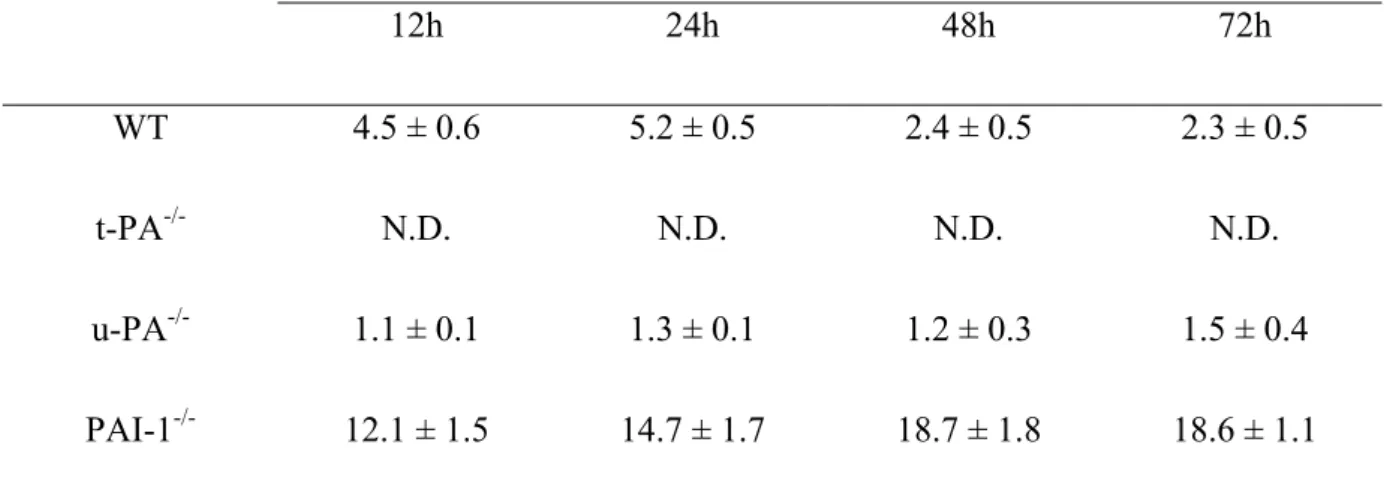
Table 1. t-PA levels in VSMC conditioned media as a function of time.
Murine t-PA levels in conditioned media (12 to 72 hours) from primary cultures of VSMC derived from WT, t-PA-/-, u-PA-/- and PAI-1-/- mice were measured by ELISA.
t-PA (ng/ml) 12h 24h 48h 72h WT 4.5 ± 0.6 5.2 ± 0.5 2.4 ± 0.5 2.3 ± 0.5 t-PA-/- N.D. N.D. N.D. N.D. u-PA-/- 1.1 ± 0.1 1.3 ± 0.1 1.2 ± 0.3 1.5 ± 0.4 PAI-1-/- 12.1 ± 1.5 14.7 ± 1.7 18.7 ± 1.8 18.6 ± 1.1
Data are expressed in ng/ml per 106 cells, and are mean ± SEM of 4 experiments. N.D., not detectable.
Table 2. PAI-1 levels in VSMC conditioned media as a function of time.
Murine PAI-1 levels in conditioned media (12 to 72 hours) from primary cultures of VSMC derived from WT, t-PA-/-, u-PA-/- and PAI-1-/- mice were measured by ELISA.
PAI-1 (ng/ml) 12h 24h 48h 72h WT 1186 ± 49 1868 ± 88 1646 ± 330 2788 ± 389 t-PA-/- 635 ± 68 998 ± 63 1770 ± 233 3190 ± 175 u-PA-/- 850 ± 68 1223 ± 184 1989 ± 123 2301± 209 PAI-1-/- N.D. N.D. N.D. N.D.
Data are expressed in ng/ml per 106 cells, and are mean ± SEM of 4 experiments. N.D., not detectable.