HAL Id: hal-02672083
https://hal.inrae.fr/hal-02672083
Submitted on 31 May 2020
HAL is a multi-disciplinary open access
archive for the deposit and dissemination of
sci-entific research documents, whether they are
pub-lished or not. The documents may come from
teaching and research institutions in France or
abroad, or from public or private research centers.
L’archive ouverte pluridisciplinaire HAL, est
destinée au dépôt et à la diffusion de documents
scientifiques de niveau recherche, publiés ou non,
émanant des établissements d’enseignement et de
recherche français ou étrangers, des laboratoires
publics ou privés.
Expanded comparative mapping between man and
rabbit and detection of a new conserved segment
between HSA22 and OCU4
Céline Chantry-Darmon, Maud Bertaud, Celine Urien, Sead Chadi, M.
Perrocheau, Claire Rogel Gaillard, Hélène Hayes
To cite this version:
Céline Chantry-Darmon, Maud Bertaud, Celine Urien, Sead Chadi, M. Perrocheau, et al.. Expanded
comparative mapping between man and rabbit and detection of a new conserved segment between
HSA22 and OCU4. Cytogenetic and Genome Research, Karger, 2005, 111, pp.134-139. �hal-02672083�
Original Article
Cytogenet Genome Res 111:134–139 (2005) DOI: 10.1159/000086382
Expanded comparative mapping between man
and rabbit and detection of a new conserved
segment between HSA22 and OCU4
C. Chantry-Darmon,
a,bM. Bertaud,
aC. Urien,
bS. Chadi-Taourit,
aM. Perrocheau,
aC. Rogel-Gaillard,
bH. Hayes
aaLaboratoire de Génétique biochimique et Cytogénétique, INRA;
bLaboratoire de Radiobiologie et Etude du Génome, INRA CEA UMR 13.314, Jouy-en-Josas (France)
Manuscript received 17 December 2004; accepted in revised form for publication by H. Hameister 19 January 2005.
C. Chantry-Darmon was supported by the Department of Animal Genetics of INRA and the SYSELAF.
Request reprints from Dr Hélène Hayes
Laboratoire de Génétique biochimique et Cytogénétique, INRA-CRJ FR–78350 Jouy-en-Josas (France)
telephone: +33 (0) 1 34 65 26 73; fax: +33 (0) 1 34 65 24 78 e-mail: [email protected]
Abstract. Rabbit, a domestic species exploited both in ani-mal production and medical research has only recently begun to be included in gene mapping projects, in particular by the French National Institute of Agronomics. By 2002, less than 60 genes had been precisely localised on rabbit chromosomes, which led us to start a large-scale project on gene mapping in rabbit with the publication of 133 gene localisations in 2003
(Chantry-Darmon et al., 2003). Here, we report the localisation of 102 new genes resulting in good coverage of the rabbit genome and an eight-fold enrichment of the gene map. In addi-tion, we have detected a new conserved segment between rabbit chromosome 4q15.3 and part of human chromosome 22 and thus improved the comparative map with the human genome.
Copyright © 2005 S. Karger AG, Basel
As previously reported (Chantry-Darmon et al., 2003), rab-bit is an interesting species both in animal production and med-ical research but its gene map was poorly developed until recently (Zijlstra et al., 2002; Chantry-Darmon et al., 2003). The production of gene-rich maps is a prerequisite both for high-resolution comparative mapping with man (or other map-rich species i.e. mouse, rat, cow, pig) and for projects on the identification of QTLs. Although bi-directional chromosome painting experiments have revealed an overall picture of the extent and distribution of conserved segments between human and rabbit chromosomes (Korstanje et al., 1999), a more detailed comparison would help to add neighbouring markers
in regions of interest. In 2003, we reported a first contribution to the large-scale mapping project in rabbit initiated by the INRA with the localisation of 133 genes by fluorescent in situ hybridisation of gene-containing BAC clones to rabbit chromo-somes (Chantry-Darmon et al., 2003). In our continuing goal to develop the comparative map between human and rabbit chro-mosomes, we have isolated and FISH-mapped rabbit BAC clones for 102 additional genes. Our aim here was to target the search of genes on chromosome bands with no mapped genes to date, based on the existing comparative mapping human/rabbit data.
Materials and methods
Choice of the genes and primer design, rabbit BAC library screening and sequencing of the PCR products
Previously described procedures (Chantry-Darmon et al., 2003) were used to design the primer sequences and to isolate gene-containing clones from our rabbit BAC library (Rogel-Gaillard et al., 2001) either by a standard PCR-based screening protocol or by hybridisation of high density filters representing the whole library. To check the identity of the sequence inserted in the BAC clones, the PCR product was sequenced and standard nucleotide-nucleotide BLASTN analyses were performed.
Cytogenet Genome Res 111:134–139 (2005) 135
Probe preparation for fluorescence in situ hybridisation
BAC DNA extracts were prepared according to standard protocols and purified with the S.N.A.P. K1900-01 Miniprep kit (Invitrogen life technolog-ies). DNA was then labelled by nick-translation with biotin-14-dATP (Bio-Nick 18247-015 labelling system, Invitrogen Life Technologies), mixed with 100× total sonicated herring sperm DNA and 100× total sonicated rabbit DNA, ethanol precipitated, slightly dried and resuspended in hybridisation buffer.
FISH on R-banded rabbit chromosomes
R-banded chromosome spreads were obtained from rabbit embryo fibro-blast cell cultures synchronized with an excess of thymidine and treated with 5-bromodeoxyuridine (BrdU) during the second half of S phase (Hayes et al., 1991). Fluorescent in situ hybridisation, signal detection and R-banding were performed as previously described (Hayes et al., 1992) with 50–100 ng biotin-14-dATP-labelled probe per slide. Before hybridisation to the chro-mosomes, probes were denatured at 100° C for 10 min and pre-hybridized at
37° C for 30–60 min. Slides were examined under a Zeiss Axioplan 2
epi-fluorescence microscope and the Applied Imaging Cytovision (version 2.7) software was used for image capturing and analysis. Chromosome and band numbering follow Hayes et al. (2002).
Results and discussion
Choice of genes and recovery of BAC clones
The 102 new genes localized in this work were chosen based on their position on the human genome i.e. on human chromo-some bands not included in our previous report (Chantry-Dar-mon et al., 2003) or to complete some human chromosome bands containing numerous genes such as 4q13.3 (SLC4A4 and
GC), 5q33.3 (ADRA1B), 6q15 (CGA), 9p13.3 (VCP), 17p13.3 (MYH3) and 18q12.1 (MAPRE2). Table 1 presents the chosen
genes with their localisation on human chromosomes. The table listing the primer pairs, the species from which the EST originated, GenBank accession numbers and expected size of PCR products is accessible online at http://dga.jouy.inra. fr/lgbc/donnees/pdf/Primer_Information_Rabbit_MS_CGR_ 2005.pdf. As explained in Chantry-Darmon et al. (2003), the availability of many rabbit ESTs and cDNAs in GenBank made it possible to design homologous primers for 93 genes. For the other nine genes, primers were designed from human cDNAs (two) and equine cDNAs (seven). At least one BAC clone was isolated from our rabbit BAC library for each of the 102 genes listed in Table 1 with 28 BAC clones recovered by PCR screening and 74 by hybridisation. In all cases, the sequence of the rabbit PCR product confirmed that the ex-pected gene was contained in the isolated BAC clone.
Localisation of 102 genes on rabbit chromosomes by fluorescent in situ hybridisation
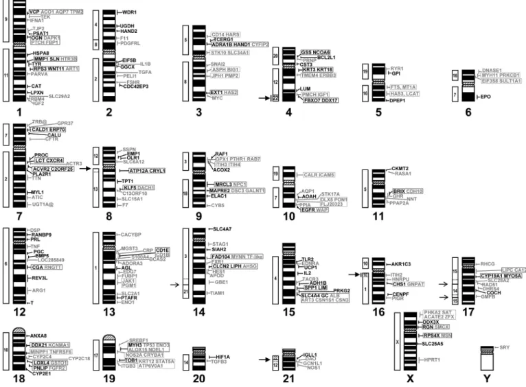
DNA extracted from each of the 102 rabbit gene-specific BAC clones was biotin-labelled and hybridised to rabbit meta-phase chromosome spreads. Clear and consistent hybridisation signals were obtained and images were captured and analysed for at least ten spreads in each case. Results are summarized in Table 1 with those in bold characters indicating localisations obtained with genes from new human chromosomal bands. Indeed, among the 102 newly localized genes on rabbit chromo-somes, 74 are positioned to chromosome bands or sub-bands devoid of any mapped gene to date. Figure 1 presents the
cur-rent status of the rabbit cytogenetic map constructed in our lab-oratory, with genes localised during this work indicated in black and those obtained in earlier reports in grey. Newly local-ized genes are distributed over all the chromosomes except chromosome Y, which still has only SRY as the single mapped gene. Figure 1 clearly shows that globally we have enriched chromosomes or chromosome arms which were either little (i.e. OCU2, OCU4, OCU7, OCU12, OCU15) or not covered (OCU6q, OCU14p, OCU16p) in our previous work. The p arm of chromosome 15 is still devoid of mapped genes as well as the very small p arms of chromosomes 20 and 21. For these partic-ular chromosomal regions, it is difficult to focus the search for genes because of the absence of any comparative mapping human/rabbit data. At present, about 43 % of the bands of the rabbit karyotype at the 347-band level (Hayes et al., 2002) carry at least one mapped gene. Most of these bands are R positive, which correlates with the fact that in mammals the R positive bands are gene-rich (Saccone et al., 1996). However, the band level used for cytogenetic positions of genes on the human genome sequence (Ensembl database http://www.ensembl.org/) is the schematic representation of chromosomes corresponding to approximately 850 bands (ISCN 1981). This is a much high-er resolution than that currently used in othhigh-er species thus more precise comparisons of gene localisations on R or G bands between man and another species are not possible. Indeed, an R positive band at the 400-band level may subdivide in two R positive and one R negative bands at the 850-band level.
Rabbit/human comparative map and detection of a new conserved segment
We have compared our results (Fig. 1) with the human/rab-bit heterologous chromosome painting data reported by Kor-stanje and colleagues (1999) and we reveal the existence of a previously undetected conserved segment between HSA22 and OCU4. Indeed the localisation of FBXO7 (HSA22q12.3) and
DDX17 (HSA22q13.1) genes on rabbit chromosome 4q15.3
shows that a small region of OCU4 band q15.3 is homologous with part of HSA22. This band is a large R positive band in rabbit and we are currently searching for genes in the distal part of HSA22 i.e. on bands 22q13.2 to 22q13.33 to determine if this conserved segment extends right to the end of OCU4q15.3. Human/rabbit heterologous chromosome painting had re-vealed that the long arm of OCU4 is entirely painted by HSA12. Together, with our results, it appears that OCU4q cor-responds in fact to a combination of ancestral chromosome forms of HSA12 and 22, an association frequently observed in other species as reviewed by Murphy and colleagues (2001). In addition, our results confirm the conservation between OCU6q and part of HSA7 with the localisation of the EPO gene, between OCU14p and part of HSA3 with the localisation of
SLC4A7 gene and between OCU16p and part of HSA10 with
the localisation of the AKR1C3 gene. These genes are the first to be mapped to these three rabbit chromosome arms and con-stitute anchors from which we can search for new genes on the corresponding human chromosomes to enrich these rabbit regions. In two cases, our results modify slightly the size of a conserved segment (i) on OCU8 the conserved segment with HSA13 extends to the centromere of OCU8 (localization of the
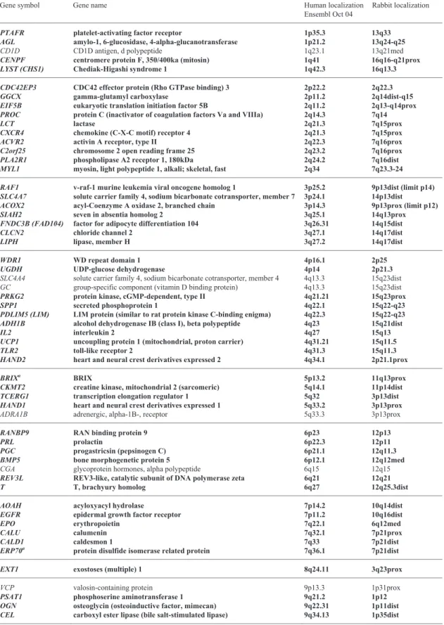
Table 1. List of genes mapped in this study, their localisation on human chromosomes, expected localisation in rabbit and FISH localisations. Results in bold characters indicate localisations obtained with genes from human chromosome bands not included in previous studies. Gene symbols follow the HUGO nomenclature (http://www.gene.ucl.ac.uk/nomenclature). For each human chromosome, genes are ordered from the p telomere to the q telomere according to the Ensembl database (http://www.en-sembl.org/).
Gene symbol Gene name Human localization Ensembl Oct 04
Rabbit localization
PTAFR platelet-activating factor receptor 1p35.3 13q33
AGL amylo-1, 6-glucosidase, 4-alpha-glucanotransferase 1p21.2 13q24-q25
CD1D CD1D antigen, d polypeptide 1q23.1 13q21med
CENPF centromere protein F, 350/400ka (mitosin) 1q41 16q16-q21prox
LYST (CHS1) Chediak-Higashi syndrome 1 1q42.3 16q13.3
CDC42EP3 CDC42 effector protein (Rho GTPase binding) 3 2p22.2 2q22.3
GGCX gamma-glutamyl carboxylase 2p11.2 2q14dist-q15
EIF5B eukaryotic translation initiation factor 5B 2q11.2 2q13-q14prox PROC protein C (inactivator of coagulation factors Va and VIIIa) 2q14.3 7q14
LCT lactase 2q21.3 7q15prox
CXCR4 chemokine (C-X-C motif) receptor 4 2q21.3 7q15prox
ACVR2 activin A receptor, type II 2q22.3 7q16prox
C2orf25 chromosome 2 open reading frame 25 2q23.2 7q16prox
PLA2R1 phospholipase A2 receptor 1, 180kDa 2q24.2 7q16dist
MYL1 myosin, light polypeptide 1, alkali; skeletal, fast 2q34 7q23.3-24 RAF1 v-raf-1 murine leukemia viral oncogene homolog 1 3p25.2 9p13dist (limit p14) SLC4A7 solute carrier family 4, sodium bicarbonate cotransporter, member 7 3p24.1 14p13dist ACOX2 acyl-Coenzyme A oxidase 2, branched chain 3p14.3 9p13prox (limit p12)
SIAH2 seven in absentia homolog 2 3q25.1 14q13prox
FNDC3B (FAD104) factor for adipocyte differentiation 104 3q26.31 14q15dist
CLCN2 chloride channel 2 3q27.1 14q17dist
LIPH lipase, member H 3q27.2 14q17dist
WDR1 WD repeat domain 1 4p16.1 2p25
UGDH UDP-glucose dehydrogenase 4p14 2p21.3
SLC4A4 solute carrier family 4, sodium bicarbonate cotransporter, member 4 4q13.3 15q23dist GC group-specific component (vitamin D binding protein) 4q13.3 15q23dist
PRKG2 protein kinase, cGMP-dependent, type II 4q21.21 15q23prox
SPP1 secreted phosphoprotein 1 4q22.1 15q22-q23
PDLIM5 (LIM) LIM protein (similar to rat protein kinase C-binding enigma) 4q22.3 15q22-q23 ADH1B alcohol dehydrogenase IB (class I), beta polypeptide 4q23 15q21dist
IL2 interleukin 2 4q27 15q13
UCP1 uncoupling protein 1 (mitochondrial, proton carrier) 4q31.21 15q11.5
TLR2 toll-like receptor 2 4q31.3 15q11.3
HAND2 heart and neural crest derivatives expressed 2 4q34.1 2p21.1prox
BRIXa BRIX 5p13.2 11q13prox
CKMT2 creatine kinase, mitochondrial 2 (sarcomeric) 5q14.1 11p14dist TCERG1 transcription elongation regulator 1 5q32 3p13dist HAND1 heart and neural crest derivatives expressed 1 5q33.2 3p13prox
ADRA1B adrenergic, alpha-1B-, receptor 5q33.3 3p13prox
RANBP9 RAN binding protein 9 6p23 12p13
PRL prolactin 6p22.3 12p11
PGC progastricsin (pepsinogen C) 6p21.1 12q11.3
BMP5 bone morphogenetic protein 5 6p12.1 12q12med
CGA glycoprotein hormones, alpha polypeptide 6q15 12q15
REV3L REV3-like, catalytic subunit of DNA polymerase zeta 6q21 12q21
T T, brachyury homolog 6q27 12q25.3dist
AOAH acyloxyacyl hydrolase 7p14.2 10q14dist
EGFR epidermal growth factor receptor 7p11.2 10q16dist
EPO erythropoietin 7q22.1 6q12med
CALU calumenin 7q32.1 7p21prox
CALD1 caldesmon 1 7q33 7p21dist
ERP70a protein disulfide isomerase related protein 7q36.1 7p21dist
EXT1 exostoses (multiple) 1 8q24.11 3q23prox
VCP valosin-containing protein 9p13.3 1p31prox
PSAT1 phosphoserine aminotransferase 1 9q21.2 1p12
OGN osteoglycin (osteoinductive factor, mimecan) 9q22.31 1p11dist CEL carboxyl ester lipase (bile salt-stimulated lipase) 9q34.13 1p35dist
Cytogenet Genome Res 111:134–139 (2005) 137
Table 1 (continued)
Gene symbol Gene name Human localization Ensembl Oct 04
Rabbit localization
AKR1C1 aldo-keto reductase family 1, member C1 10p15.1 16p12.1prox
ANXA8 annexin A8 10q11.22 18q12dist
DDX21 DEAD (Asp-Glu-Ala-Asp) box polypeptide 21 10q22.1 18q21.3dist
LOXL4 lysyl oxidase-like 4 10q24.2 18q31dist
PNLIP pancreatic lipase 10q25.3 18q33prox
CYP2E1 cytochrome P450, family 2, subfamily E, polypeptide 1 10q26.3 18q33dist
CAT catalase 11p13 1q23
LPXN leupaxin 11q12.1 1q25dist
RPS3 ribosomal protein S3 11q13.4 1q21.1
WNT11 wingless-type MMTV integration site family, member 11 11q13.5 1q21.1 TYR tyrosinase (oculocutaneous albinism IA) 11q14.3 1q14-q15 MMP1 matrix metalloproteinase 1 (interstitial collagenase) 11q22.2 1q14
SLN sarcolipin 11q22.3 1q14
HSPA8 heat shock 70kDa protein 8 11q24.1 1q12.3prox
OLR1 oxidised low density lipoprotein (lectin-like) receptor 1 12p13.2 8p12.3
EMP1 epithelial membrane protein 1 12p13.1 8p12.3dist
KRT3 keratin 3 12q13.13 4q11prox
KRT18 keratin 18 12q13.13 4q11prox
LUM lumican 12q21.33 4q15.1
CRYL1 crystallin, lambda 1 13q12.11 8q11
ATP12A ATPase, H+/K+ transporting, nongastric, alpha polypeptide 13q12.12 8q11 TPT1 tumor protein, translationally-controlled 1 13q14.13 8q13.3-q21
KLF5 Kruppel-like factor 5 13q22.1 8q22prox
COCH coagulation factor C homolog, cochlin 14q12 17q21dist HIF1A hypoxia-inducible factor 1, alpha subunit 14q23.2 20q12.1 CYP19A1 cytochrome P450, family 19, subfamily A, polypeptide 1 15q21.2 17q13prox MYO5A myosin VA (heavy polypeptide 12, myoxin) 15q21.2 17q13prox
DPEP1 dipeptidase 1 16q24.3 5q16dist
MYH3 myosin, heavy polypeptide 3, skeletal muscle, embryonic 17p13.1 19q12.3prox
TOB1 transducer of ERBB2, 1 17q21.33 19q21prox
MRCL3a myosin regulatory light chain MRCL3 18p11.31 9q13
MAPRE2 microtubule-associated protein, RP/EB family, member 2 18q12.1 9q14.2
ELAC1 elaC homolog 1 18q21.1 9q15.1
GPI glucose phosphate isomerase 19q13.11 5p12prox
CST3 cystatin C (amyloid angiopathy and cerebral hemorrhage) 20p11.21 4p11
BCL2L1 BCL2-like 1 20q11.21 4p13med
NCOA6 nuclear receptor coactivator 6 20q11.22 4p13dist
GSS glutathione synthetase 20q11.22 4p13dist
IGLL1 immunoglobulin lambda-like polypeptide 1 22q11.23 21q12prox
FBXO7 F-box protein 7 22q12.3 4q15.3prox
DDX17 DEAD (Asp-Glu-Ala-Asp) box polypeptide 17 22q13.1 4q15.3prox DDX3X DEAD (Asp-Glu-Ala-Asp) box polypeptide 3, X-linked Xp11.4 Xp13dist RGN regucalcin (senescence marker protein-30) Xp11.3 Xp11
RPS4X ribosomal protein S4, X-linked Xq13.1 Xq12
SLC25A5 solute carrier family 25, member 5 Xq24 Xq21.1
a
Symbol from NCBI Entrez Gene (http://www.ncbi.nlm.nih.gov/entrez/query).
ATP12A and CRYL1 genes) and thus covers the whole of its
long arm and (ii) on OCU16, the conservation with HSA10 includes the first band of the long arm of OCU16 while in Kor-stanje et al. (1999) it was restricted to the short arm only (local-ization of ITIH2, previous report Chantry-Darmon et al.,
2003). Finally, the comparative human/rabbit cytogenetic map confirms all the painting data obtained by Korstanje and col-leagues (1999), including the few readjustments previously dis-cussed and concerning OCU6, 10 and 11 (Zijlstra et al., 2002 and Hayes et al., 2002).
Fig. 1. Current rabbit cytogenetic map in our laboratory. The 102 newly localised genes are indicated in black characters on the right of each ideo-gram while previously mapped genes to R-banded chromosomes are given in grey (Martin-DeLeon et al., 2001; Hayes et al., 2002; Pauloin et al., 2002; Chantry-Darmon et al., 2003). Blocks on the left of each ideogram represent the chromosome correspondences with human chromosomes previously reported by Korstanje et al. (1999). Big arrows with vertical bars indicate a
newly detected conserved segment between OCU4 and HSA22 and an exten-sion of the conserved segment between OCU16 and HSA10 to the long arm of OCU16. Big arrows next to OCU8 and OCU21 denote slight modifica-tions in the extension (dotted lines correspond to the previous limits reported by Korstanje et al., 1999) of conserved segments with the human genome (see text). The small arrows next to OCU14 and OCU17 indicate previously reported modifications (Chantry-Darmon et al., 2003).
With the growing number of genes mapped to bands and sub-bands of rabbit chromosomes and although the order of genes between rabbit and human chromosomes appears to be well conserved, it is possible to detect small inversions and rearrangements. In some cases, the series of genes involved include a centromere on the human chromosome while they are situated on a single chromosome arm in rabbit. For exam-ple, CDC42EP3 (HSA2p22.2), FSHR (HSA2p16.3), PELI1 (HSA2p14), TGFA (HSA2p13.3), GGCX (HSA2p11.2), cen,
EIF5B (HSA2q11.2) and IL1B (HSA2q13) are positioned in
the following order along the long arm of rabbit chromosome 2:
EIF5B, IL1B, GGCX, TGFA, PELI1, FHSR and CDC42EP3,
which can be explained by an inversion of the segment contain-ing EIF5B and IL1B. The same situation is observed on human
chromosome segment 11p15.4 (ART1)–11q13.2 (RBM4) ver-sus rabbit chromosome segment 1q21.1→q21.2 (ART1)– 1q27med (RBM4) or human chromosome segment 9p21.3
(IFNA1)–9q22.32 (PTCH) versus rabbit chromosome segment
1p23 (IFNA1)–1p11dist (PTCH). Other single inversions are sufficient to restore the gene order between ACTR3–PROC–
LCT–CXCR4 on HSA2q and (LCT; CXCR4)–ACTR3–PROC
on OCU7q or between DAO–NOS1–GCN1L1 on HSA12q and
DAO–GCN1L1–NOS1 on OCU21q. Finally, complex
rearran-gements involving two successive or a segment transposition are necessary to explain differences between HSA7p21.3→ p11.2 and OCU10 or HSA7q31.2→q36.1 and OCU7. How-ever, it is clear that many more genes need to be mapped on rabbit chromosomes using high-resolution techniques such as
Cytogenet Genome Res 111:134–139 (2005) 139
radiation hybrid mapping or even better genome sequencing, to analyse precisely the differences in gene order and organisation between the human and rabbit genomes.
Since the beginning of the INRA project on the rabbit map in 2002, we have increased eight-fold the number of precisely localised genes. This data has enabled us to produce directly an
integrated cytogenetic and genetic map in rabbit using microsa-tellite markers isolated from anchored gene-containing BAC clones (in preparation) and to assemble a more precise compar-ative map, which in turn will speed up the construction of the genetic map.
References
Chantry-Darmon C, Rogel-Gaillard C, Bertaud M, Urien C, Perrocheau M, Chardon P, Hayes H: 133 new gene localizations on the rabbit cytogenetic map. Cytogenet Genome Res 103:192–201 (2003).
Hayes H, Petit E, Dutrillaux B: Comparison of RBG-banded karyotypes of cattle, sheep and goats. Cyto-genet Cell Genet 57:51–55 (1991).
Hayes H, Petit E, Lemieux N, Dutrillaux B: Chromo-somal localization of the ovine beta-casein gene by non-isotopic in situ hybridization and R-banding. Cytogenet Cell Genet 61:286–288 (1992). Hayes H, Rogel-Gaillard C, Zijlstra C, de Haan NA,
Urien C, Bourgeaux N, Bertaud M, Bosma AA: Establishment of an R-banded rabbit karyotype nomenclature by FISH localization of 23 chromo-some-specific genes on both G- and R-banded chromosomes. Cytogenet Genome Res 98:199– 205 (2002).
ISCN (1981): An international system for human cyto-genetic nomenclature. High resolution banding: Birth defects. Cytogenet Cell Genet 31:1–23 (1981).
Korstanje R, O’Brien PCM, Yang F, Rens W, Bosma AA, van Lith HA, van Zutphen LFM, Ferguson-Smith MA: Complete homology maps of rabbit
(Oryctolagus cuniculus) and human by reciprocal
chromosome painting. Cytogenet Cell Genet 86: 317–322 (1999).
Martin-DeLeon PA, Piumi F, Canaff L, Rogel-Gaillard C, Hendy GN: Assignment of the parathyroid hor-mone/parathyroid hormone-related peptide recep-tor (PTHR1) to rabbit chromosome band 9p14→ p13 by fluorescence in situ hybridization. Cytoge-net Cell GeCytoge-net 94:90–91 (2001).
Murphy WJ, Stanyon R, O’Brien SJ: Evolution of mammalian genome organisation inferred from comparative mapping. Genome Biology 2:1–8 (2001).
Pauloin A, Rogel-Gaillard C, Piumi F, Hayes H, Fon-taine ML, Chanat E, Chardon P, Devinoy E: Struc-ture of the rabbit alphas1- and beta-casein gene cluster, assignment to chromosome 15 and expres-sion of the alphas1-casein gene in HC11 cells. Gene 283:155–162 (2002).
Rogel-Gaillard C, Piumi F, Billault A, Bourgeaux N, Save JC, Urien C, Salmon J, Chardon P: Construc-tion of a rabbit bacterial artificial chromosome (BAC) library: application to the mapping of the major histocompatibility complex to position 12q1.1. Mamm Genome 12:253–255 (2001). Saccone S, Caccio S, Kusuda J, Andreozzi L, Bernardi
G: Identification of the gene-richest bands in hu-man chromosomes. Gene 174:85–94 (1996). Zijlstra C, de Haan NA, Korstanje R, Rogel-Gaillard C,
Piumi F, van Lith HA, van Zutphen LFM, Bosma AA: Fourteen chromosomal localisations and an update of the cytogenetic map of the rabbit. Cyto-genet Genome Res 97:191–199 (2002).