Purification and Characterization of Tomato Leaf (Lycopersicon
esculentum Mill.) Hydroperoxide Lyase
Marie-Laure Fauconnier,*,†Ana G. Perez,‡Carlos Sanz,‡and Michel Marlier† Unite´ Chimie Ge´ne´rale et Organique, Faculte´ Universitaire des Sciences Agronomiques Passage des
De´porte´s 2, B-5030 Gembloux, Belgium, and Instituto de la Grasa, CSIC, Avenida Padre Garcia Tejero 4, 41012 Sevilla, Spain
Hydroperoxide lyase (HPOOH lyase) was extracted from tomato leaves (Lycopersicon esculentum Mill.) and purified to apparent homogeneity by fractionated precipitation with polyethylene glycol 6000, ion exchange chromatography, and ultrafiltration. The enzyme is a trimer of 73 000 Da units with a molecular mass of 216 000 (determined by native-PAGE and gel filtration); its pI is around 4.9. Enzyme activity measurments realized with 9- and 13-hydroperoxides of linoleic acid (9-La OOH and 13-La OOH, respectively), R-linolenic acid (9-Ln OOH and 13-Ln OOH, respectively), and γ-linolenic acid (9-γLn OOH and γ-Ln OOH, respectively) revealed a great affinity for 13-Ln OOH. The enzyme is rapidly inhibited by its substrate (13-13-Ln OOH), but preincubation with the other five hydroperoxides, which are not degraded by the enzyme, also resulted in activity inhibition. Dialysis could not restore the activity. When 13-Ln OOH is reduced in its corresponding alcohol or converted to its methyl ester, the inhibition is reduced.
Keywords: Tomato leaves; Lycopersicon esculentum Mill.; hydroperoxide lyase; linolenic acid hydroperoxide; linoleic acid hydroperoxide
INTRODUCTION
Fatty acid hydroperoxides are key components formed by the action of lipoxygenase (EC 1.13.11.12) on 1(Z),4-(Z)-pentadiene-containing fatty acids. Depending on the origin of the lipoxygenase and on the reaction condi-tions, variable amounts of 13- and/or 9-hydroperoxides are formed. The hydroperoxides can be further de-graded in a variety of products involved in essential physiological roles in plants (jasmonic acid, traumatine) or responsible for the characteristic green notes of plants and fruits (C6 or C9 aldehydes and alcohols). Hydro-peroxide lyase is an enzyme that catalyzes the cleavage of fatty acid hydroperoxides into aldehydes and ω-oxo acids. The break takes place between the carbon that contains the hydroperoxide group and the proximate ethylenic carbon. Hydroperoxide lyase was first sus-pected in banana (Tressl and Drawert, 1973), and its presence was later demonstrated in various plant leaves and fruits (Hatanaka et al., 1986). Hydroperoxide lyases can be classified in two categories on the basis of their substrate specificity: the 13-HPOOH lyase and the 9-HPOOH lyase. The first type cleaves 13-Ln OOH into 3(Z)-hexenal and 12-oxo-9(Z)-dodecenoic acid and 13-La OOH into hexanal and 12-oxo-9(Z)-dodecenoic acid; the second type cleaves 9-Ln OOH into 3(Z),6(Z)-nonadienal and 9-oxononanoic acid and 9-La OOH into 3(Z)-nonenal and 9-oxononanoic acid. The first type has been described in watermelon seedlings (Vick and Zimmerman, 1976), alfalfa seedlings (Sekiya et al., 1979), tea leaves (Hatanaka et al., 1982), tomato fruits (Schreier and Lorenz, 1982), cultured tobacco cells (Sekiya et al., 1984), and green bell pepper fruits (Shibata et al., 1995a). Pear fruit HPOOH lyase belongs to the second type (Kim and Grosch, 1981), while that of kidney bean (Matthew and Galliard, 1978), cucumber seedlings (Sekiya et al., 1979), cucumber fruits (Galliard
and Phillips, 1976), and soybean seedlings (Gardner et
al., 1991) exhibits both activities. In cucumber
cotyle-dons (Matsui et al., 1989), it was demonstrated that the HPOOH lyase consisted of two isomers: one specific for the 9-isomer and the other specific for the 13-isomer. HPOOH lyase is difficult to study because it is a membrane-bound enzyme and because it is present in small amounts in plant tissues. It was only purified to apparent homogeneity in tea leaves (Matsui et al., 1991) and in green bell pepper fruits (Shibata et al., 1995a), in which it was shown to be a heme protein (Shibata et
al., 1995b). We have reinvestigated the extraction and
the purification of the HPOOH lyase from tomato leaves, resulting in a quite simple procedure. The pI and molecular mass of the enzyme and of its subunits were determined; particular attention was paid to the speci-ficity of the enzyme for its substrate. The inhibition of the enzyme by fatty acid hydroperoxides was studied systematically: the enzyme was preincubated with 9-and 13-OOH fromR-linolenic acid, γ-linolenic acid, and linoleic acid. The inhibitory effect of 13-Ln OOH was also checked after reduction to the corresponding alcohol and after esterification with methanol. The aldehydes, products of the reaction, and the corresponding alcohols were also used for the inhibitory assays.
This paper contribues to a better knowledge of HPOOH lyase, which has been poorly studied in spite of its physiological importance. The enzyme is essential for fruit aroma synthesis and has an application in the aroma industry for the production of natural green note compounds. The reaction products (aldehydes, oxo acids) are implicated in plant defense against pathogens and in wound healing.
MATERIALS AND METHODS
Material. HPOOH lyase was purified from tomato leaves (Lycopersicon esculentum Mill. var. Bonset) cultivated in the Institut Supe´rieur Industriel (Grand-Manil, Belgium). The potato tubers originated from the local market. EDTA, dithio-threitol, Triton X-100, PEG 6000, 2-mercaptoethanol, NADH, †Unite´ Chimie Ge´ne´rale et Organique.
‡Instituto de la Grasa.
4232 J. Agric. Food Chem. 1997, 45, 4232−4236
linoleic acid (purity by GC>99%),R-linolenic acid (purity by GC > 98%), γ-linolenic acid (purity by GC > 99%), yeast alcohol dehydrogenase, and soybean lipoxygenase (type I-S) were obtained from Sigma (St. Louis, MO). All other reagents were of analytical grade.
Methods. Enzyme Purification. All of the experiments were conducted at 4 °C. Tomato leaves (170 g) were extracted directly in a Waring blender (three times, 1 min) with 200 mL of sodium phosphate buffer (50 mM, pH 6.7) containing 3 mM EDTA, 3 mM dithiothreitol, and 0.5% (v/v) Triton X-100. The extract was centrifuged (10 min, 7250g), and the supernatant was magnetically agitated (90 min) and centrifuged again (20 min, 26000g). PEG 6000 (7%, w/v) was added slowly to the supernatant. After 30 min of agitation, the extract was centrifuged (20 min, 26000g). PEG was added to the super-natant to obtain a final concentration of 23% (w/v). The solution was again agitated for 30 min. The extract was centrifuged (15 min, 12000g), and the residue was solubilized in 15 mL of Tris-HCl buffer (20 mM, pH 9) containing 0.5% (v/v) Triton X-100. PEG was added to obtain a final concen-tration of 23% (w/v), and the extract was agitated for 30 min and centrifuged (12 min, 12000g). Tris-HCl buffer (5 mL) was used to solubilize the residue. 2-Mercaptoethanol was added to the extract to obtain a final concentration of 0.1 mM.
The extract was loaded on a DEAEE-Sepharose CL 6B (Pharmacia, Uppsala, Sweden) column (1.5× 25 cm) equili-brated with 20 mL of Tris-HCl buffer (pH 8.5) containing 0.5% Triton X-100 and 0.1 mM 2-mercaptoethanol. The column was washed with 135 mL of Tris-HCl buffer at 3 mL/min flow rate. The elution was carried out with a 135 mL linear KCl gradient (0-0.3 M) and 90 mL linear KCl gradient (0.3-1 M).
Active fractions from the anion exchange chromatography were pooled, and proteins with molecular masses<100 000 Da were removed by ultrafiltration using Centriprep-100 from Amicon (Beverly, MA). The sample was concentrated and then diluted to the initial volume with distilled water; this last operation was repeated three times.
Gel filtration was carried out with a Fast Protein liquid chromatography system (Pharmacia) on a HiLoad 16/60 Su-perdex 200 column using a sodium phosphate buffer (50 mM, pH 7) containing 1 M NaCl and Triton X-100 0.5% (v/v). The column was calibrated with the gel filtration standard kit from Bio-Rad (Hercules, CA).
Native-PAGE was performed on a Mini-Protean II cell (Bio-Rad) using a 4-15% gradient Tris-glycine gel (ready gel from Bio-Rad). The standards were the high molecular weight calibration kits from Bio-Rad and Pharmacia.
SDS-PAGE was carried out on the same kind of gel as the native-PAGE using the high-range molecular mass from Bio-Rad as standards.
The determination of the pI was realized on a gradient gel 3-9 from Pharmacia using the broad pI kit (pH 3.5-9.3) and a low pI kit (pH 2.8-6.5) from Pharmacia.
The protein content was determined according to Folin using bovine serum albumin as standard (Lowry et al., 1951).
Enzyme Activity Measurements. Three methods were used for the determination of the HPOOH lyase activity. For a rapid location of activity, hydroperoxide lyase was assayed by the decrease in absorbance at 234 nm. This method is not specific and gives the total disappearance of hydroperoxide as a function of time. A typical assay consisted of (3-x) mL of sodium phosphate buffer (0.1 M, pH 6.7), 10 µL of 13-Ln OOH in methanol (12 mM), and x mL of enzyme extract. The NADH-coupled enzyme assay (340 nm) described by Vick (1991) was used for specific determinations. A typical assay consisted of (2.9-x) mL of sodium phosphate buffer (0.1 M, pH 6.7), 50 µL of yeast alcohol dehydrogenase solution (150 units), 50 µL of NADH solution (0.01 M), 10 µL of 13-Ln OOH in methanol (12 mM), and x mL of enzyme extract. For both spectrophotometric methods, the difference in absorbance was recorded for 20 s and 1 unit of activity was expressed as the amount of enzyme consuming 1 µmol of 13-Ln OOH or 1 µmol NADH in 1 min using 25 000 or 6 220 M-1
cm-1
as extinction coefficients, respectively, for 13-Ln OOH and NADH. The NADH-coupled enzyme assay was compared to the quantita-tion of products formed during the lyase enzymatic reacquantita-tion
(aldehydes) by means of n-hexane extraction and GC deter-mination. For this purpose, (2-x) mL of sodium phosphate buffer (0.1 M, pH 6.7), 10 µL of 13-Ln OOH in methanol (12 mM), and x µL of enzyme extract were mixed and the reaction was conducted 5 min at 25 °C. The reaction medium was saturated in (NH4)2SO4 and extracted with 2 × 2 mL of
n-hexane. 1-Heptanol (internal standard) was added, and the hexanic extract was analyzed by GC (Hewlett-Packard 5880) on a Carbowax column (25 m× 0.32 µm i.d., 0.1 mm df). The
oven temperature was held at 60 °C for 1 min, increased to 90 °C at 5 °C/min, and then increased to 220 °C at 30 °C/min. Substrate Synthesis and Characterization. For 13-La OOH, 13-Ln OOH, and 13-γ-Ln OOH synthesis, respectively, 10 mg of linoleic acid,R-linolenic acid, or γ-linolenic acid and 4 mg of commercial soybean lipoxygenase were added to 100 mL of oxygenated borate buffer (0.2 M, pH 9). The reaction was carried out at 2 °C for 15 min under a constant flow of oxygen. The 9-hydroperoxides (9-La OOH, 9-Ln OOH, and 9-γ-Ln OOH) were produced using lipoxygenase extracted from potato tubers [according to the method of Galliard and Phillips (1971)]. The reaction was carried out in 100 mL of oxygenated phosphate buffer (50 mM, pH 6.7) at 25 °C during 15 min.
Hydroperoxides Extraction. The pH values of the reaction media were adjusted to 3 with 6 N HCl, and the reaction products were extracted on C18cartridges (C18Chromabond
of 500 mg from Macherey-Nagel, Du¨ ren, Germany) according to the method of Sanz et al. (1993).
Hydroperoxide Purification. 13-La OOH, 13-Ln OOH, and 13-γ-Ln OOH were used without further purification, while 9-La OOH, 9-Ln OOH, and 9-γ-Ln OOH were purified by HPLC according to the method of Sanz et al. (1993).
Hydroperoxide Analysis. The positional and geometrical isomers of hydroperoxides were analyzed by HPLC after reduction with NaBH4and methylation with diazomethane
according to the method of Sanz et al. (1993).
Enzymatic Activity Characterization. Substrate specificity was determined using the 234 nm test in triplicate with the following substrates: 13-Ln OOH, 9-Ln OOH, 13-La OOH, 9-La OOH, 13-γ-Ln OOH, and 9-γ-Ln OOH. The optimal temperature was determined using the 234 nm test in tripli-cate with 13-Ln OOH as substrate; the range of temperature tested was from 5 to 70 °C in steps of 5 °C. The optimal pH was determined in the same conditions (30 °C), using 0.1 M sodium acetate buffer for the pH range 3-4, 0.1 M sodium phosphate buffer for pH 5-8, and sodium borate buffer for pH 9-10.
Inhibition Tests. Kmand Vmaxwere determined in triplicate
in the conditions used for the test at 234 nm. The enzyme extract was used at increasing dilutions as the substrate concentration cannot be increased because of the limitations in absorbance.
Enzyme inhibition experiments were carried out after preincubation of the enzyme with various compounds. Sodium phosphate buffer (0.1 M, pH 6.7; 2.99-x mL) is mixed with x mL of enzyme extract and 10 µL of a solution of the compound to be tested at the desired concentration. After 10 min, 10 µL of 13-Ln OOH (0.12 mM) was added, and the activity was determined as previously described for the test at 234 nm.
In the same way, the enzyme was preincubated with 13-Ln OOH as described above and dialyzed for 2 days against distilled water (the absence of hydroperoxide was checked by following the absorbance at 234 nm). The activity of the enzyme was then determined using the test at 234 nm. A blank was realized exactly in the same conditions but without preincubation of the enzyme with its substrate.
RESULTS AND DISCUSSION
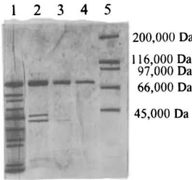
Enzyme Purification. The use of Triton X-100 was very efficient to solubilize the enzyme from the mem-branes. The concentration of 0.5% was the optimal compromise between solubilization and enzyme inhibi-tion. The fractioned precipitation with PEG 6000 between 7% and 23% (w/v) was a very simple first step of purification, allowing an increase in specific activity
of 3.7 and a recovery percentage of 47% (Table 1). The extract obtained here exhibited a great tendency to aggregate during chromatographic procedures. The conditions described under Materials and Methods are critical to avoid the elution of the enzyme in the void volume of the DEAE-Sepharose CL-6B column. Indeed, the use of Triton X-100 alone at whatever concentrations is not efficient to allow enzyme to interact with the column. The use of 2-mercaptoethanol at 0.1 mM combined with the Triton X-100 gave the best results (Figure 1). A 24-fold increase in specific activity was obtained for this purification step. The electrophoretic pattern shows that a few contaminant proteins with a molecular mass <75 000 Da were still present in the extract (Figure 2). As the HPOOH lyases are known to be high molecular mass enzymes, we decided to use an ultrafiltration membrane with a cutoff of 100 000 Da to remove the contaminants. Three cycles of ultrafil-tration were needed to obtain a single band on the electrophoretic pattern. For all purification procedures, the activity was determined by the decrease in absor-bance at 234 nm. In the DEAE step, the
spectropho-tometric activity determination was also correlated with the aldehyde synthesis by the enzyme (Figure 1). The combined test at 340 nm was used to measure the activity on the crude extract and on the final extract. In the crude extract, 77% of the hydroperoxide degrada-tion activity came from HPOOH lyase; the rest can result from hydroperoxide dehydrase activity or from another hydroperoxide metabolizing enzyme activity. In the final extract, all of the hydroperoxide metabolizing activity was attribuated to HPOOH lyase.
Enzyme Characteristics. The extract obtained previously was submitted to SDS-PAGE, revealing a single band at 73 000 Da. The native-PAGE also exhibited a single band with a molecular mass between 200 000 Da (catalase) and 232 000 Da (myosine). The accurate determination of molecular mass was difficult because native-PAGE gives quite broad bands. Gel filtration on Superdex 200, after calibration with stan-dards, gave a molecular mass for the native protein of 216 000 Da. The enzyme is then supposed to be a trimer of three 73 000 Da subunits for a total molecular mass of∼216 000 Da. The molecular mass of the enzyme has been determined to be 220 000 Da in spinach leaves (Vick and Zimmerman, 1987), between 240 000 and 260 000 Da in soybean cotyledons (Olias et al., 1990), and>200 000 Da in tomato fruits (Schreier and Lorenz, 1982). The enzyme is thought to be a tetramer of four 62 000 Da subunits in soybean cotyledons, while re-cently Shibata et al. (1995a) demonstrated that the enzyme was a trimer of 55 000 Da subunits in green bell pepper fruit. In tea leaves, subunits have also a molecular mass between 53 000 and 55 000 Da (Matsui
et al., 1991).
The pI of the enzyme determined by isoelectrofocal-ization in the presence of standards revealed that the enzyme has a pI of 4.9. A single band was present on the electrophoretic pattern, meaning that the enzyme is presumed to exist as a single isoform in tomato leaves. In tomato fruits, Schreier and Lorenz (1982) have demonstrated that the pI of HPOOH lyase was between 5.8 and 6.1. The HPOOH lyase extracted from tomato leaves is quite different from the other enzymes de-scribed in the literature, not only because of the molec-ular mass of the native form of the enzyme, which is very common, but also because of the size of the subunits (73 000 Da) and the pI value.
Enzyme Activity Characteristics. The optimal pH for tomato leaf HPOOH lyase is 7; the activity is high (90%(4% of the maximum activity) at pH between 6 and 8 but decreases to 60%(3% at pH 4 and to 70%( 5% at pH 9, meaning that the enzyme has a quite broad range of working pH. Optimal pH values described for HPOOH lyase from other origins range from 5.5 in green bell pepper fruits (Shibata et al., 1995a) to 8 in cucumber fruits (Matsui et al., 1989), while the one extracted from pear fruits (Kim and Grosch, 1981), tea leaves (Matsui et al., 1991), and soybean cotyledons (Olias et al., 1990) work at neutral pH. Optimal pH Table 1. Purification of HPOOH Lyase from Tomato Leaves
total protein (mg) total activity (units) specific activity (units/mg of protein) yield (%) purification factor (fold) crude homogenate 425 530 1.2 100 1.0
supernatant after addition of PEG 7% 150 430 2.9 81 2.4
residue after second addition of PEG 23%a 55 250 4.5 47 3.7
fraction III DEAE Sepharose CL-6B 1.20 130 108.3 25 90.2
extract after three cycles of ultrafiltration 0.55 79 143.6 15 120.0
aAfter centrifugation and solubilization.
Figure 1. DEAE-Sepharose CL 6B elution profile.
Figure 2. SDS-PAGE profile: (lane 1) residue after the second precipitation with PEG 23%; (lane 2) DEAE-Sepharose CL 6B active fraction; (lane 3) after one cycle of ultrafiltration; (lane 4) after three cycles of ultrafiltration; (lane 5) molecular mass standards.
described for HPOOH lyase from tomato fruits is 5.5 according to Schreier and Lorenz (1982). Riley et al. (1996) obseved a high enzymatic activity between pH 6 and 8 for HPOOH lyase of tomato fruits; this last result is very similar to our observations.
The optimal temperature of the enzyme is 30 °C. At 20 °C, the activity is 63%(4% of that at 30 °C, while at 40 °C it is still 90%(5% and at 50 °C, 77%(3%. The specificity of the enzyme against various peroxides was determined. The analysis of the hydro-peroxides produced as described under Materials and Methods is presented in Table 2. The 13-hydroperox-ides were used without further purification, while the 9-hydroperoxides required HPLC purification because of the lack of specificity of potato lipoxygenase. The results obtained for the study of the enzyme specificity against the six different hydroperoxides revealed that the enzyme has no activity against 9-La OOH, 9-Ln OOH, 9-γ-Ln OOH, and 13-γ-Ln OOH. The relative activities against 13-Ln OOH and 13-La OOH are, respectively, 100% and 9.8%. This specificity is also encountered in tea leaf HPOOH lyase, for which the relative activities against the same substrates are 100% and 10.9% (Matsui et al., 1991), although, in this last case, the authors also determined a small activity against 13-γ-Ln OOH (0.8%). Similar results have been described in green bell pepper fruits by Shibata et al. (1995a). In soybean seedlings, Olias et al. (1990) demonstrated that the HPOOH lyase was more specific for the 13-La OOH than for 13-Ln OOH. It seems that the specificity of the enzyme is a function of the fatty acid composition in vivo;R-linolenic acid is preponderant in leaves and in green bell pepper fruits, while linoleic acid is very abundant in seedlings. The Km values determined for tomato leaf HPOOH lyase are 60 and 34 µM for 13-Ln OOH and 13-La OOH, respectively; those Kmvalues are between those determined in green bell pepper fruits (Shibata et al., 1995a) and soybean seedlings (Olias et al., 1990). The Vmax values are 16 mM/min and 9.4 µM/min, respectively, corresponding to Kcatvalues of 154 880 mol of 13-Ln OOH/min‚mol of enzyme and 66 80 mol of 13-La OOH/min‚mol of en-zyme. The kinetic parameters of HPOOH lyase are quite ambiguous because the Kmvalues show a greater affinity for 13-La OOH, while Vmaxand Kcatindicate the contrary. This comportment can be explained by a difference in the kinetic constants: K2 (constant of dissociation of the enzyme-product complex) must be higher for 13-Ln OOH than for 13-La OOH, explaining the values of Vmax. On the other hand, the ratio K1/K-1 (constant of formation of the enzyme-substrate com-plex) must be higher for 13-La OOH, explaining the lower Kmvalue. Finally, the catalytic efficiency constant (Kcat/Km), which is frequently used to determine the affinity of an enzyme for a particular substrate, clearly demonstrates the greater affinity for 13-Ln OOH: Kcat/ Kmis 2.58× 10⁹min-1
‚mol of enzyme
-1for 13-Ln OOH and 1.85× 10⁸min-1
‚mol of enzyme
-1for 13-La OOH.
Table 3 shows the results obtained for the inhibition of HPOOH lyase after preincubation with the different hydroperoxides or hydroperoxide-derived products. The inhibitory effect obtained with Ln OOH or with 13-La OOH is less important than the one obtained with the four other hydroperoxides, which are not substrates for HPOOH lyase. A possible explanation is that part of the 13-Ln OOH or 13-La OOH is metabolized by the enzyme during the preincubation period and the enzyme is then in contact with a concentration in hydroperoxide lower than in the case of the other hydroperoxides which are not transformed. Actually, when HPOOH lyase is preincubated with 13-Ln OOH at a concentration of 40
µM, the hydroperoxides are partially degraded, and the
concentration finally stabilized at 19 µM in 5 min. When the initial concentration is 90 µM, the final concentration decreased to 54 µM. Regarding those results, all of the hydroperoxides potentially or not transformable by the enzyme seem to have the same inhibitory effect on the enzyme for a given concentra-tion. When 13-Ln OOH is transformed in the corre-sponding alcohol by reduction with NaBH4, the 13-hydroxide obtained is not a substrate for the enzyme. It inhibits the enzymatic activity but to a lesser extent than the hydroperoxides. Matsui et al, (1992) obtained also an inhibitory effect with 13-hydroxide on HPOOH lyase from tea leaf, but the residual activity was 90% of the initial one, while at the same concentration the 13-La OOH resulted in a loss of relative activity of 75%. When 13-Ln OOH was esterified using diazomethane, the inhibitory effect was very weak. The inhibitory effect of fatty acid hydroperoxides on HPOOH lyase is not specific because the same effect is obtained with hydroperoxides from linoleic acid,R-linolenic acid, or
γ-linolenic acid. The position of the function in the
molecule is not critical, as there is no difference between 9-OOH and 13-OOH. The hydroperoxide function itself is implicated, but the polarity of the molecule also influences the mechanism. When HPOOH lyase is dialyzed against distilled water for 2 days, the residual activity is 86%(4%. When the enzyme is preincubated with 13-Ln OOH at a concentration of 40 or 90 µM before the dialysis, the residual activities are, respec-tively, 63%(3% and 41%(3%. This means that the inhibition is irreversible because the elimination of the hydroperoxides of the medium could not restore the activity.
We have also checked the effect of the aldehydes issuing from the reaction and their corresponding al-cohols on the enzymatic activity. Preincubation with (E)-2-hexenol, (E)-2-hexenal, hexenal, and (Z)-3-hexenol at a concentration of 0.1 mM revealed no inhibitory effect.
Table 2. Analysis of the Hydroperoxides Synthesized Using Soybean or Potato Lipoxygenase
regioisomer proportion,
13-OOH/9-OOH stereoisomer proportion
13-La OOH 98/2 13(Z,E)/13(E, E))96/4
13-Ln OOH 99/1 13(Z,E,Z)/13(E,E,Z))97/3
13-γ-Ln OOH 98/2 13(Z,Z,E)/13(Z,E,E))96/4
9-La OOH 2/98 9(E,Z)/9(E,E))99/1
9-Ln OOH 1/99 9(E,Z,Z)/9(E,E,Z))98/2
9-γ-Ln OOH 1/99 9(Z,E,Z)/9(Z,E,E))98/2
Table 3. Residual Activity after Preincubation of HPOOH Lyase with Different Hydroperoxides
concn (µM) residual relative activity (%) none 0 100(2.5 13-Ln OOH 40 74(2.0 13-Ln OOH 90 44(2.2 13-La OOH 40 70(4.1 13-γ-Ln OOH 40 55(2.6 9-Ln OOH 40 44(3.1 9-La OOH 40 45(2.8 9-γ-Ln OOH 40 47(3.3 13-Ln OHa 40 71(2.6 13-Me-Ln OOHb 40 90 (1.8
a13-Ln OOH reduced to the corresponding alcohol by NaBH4. b13-Ln OOH esterified using diazomethane.
ABBREVIATIONS USED
HPOOH lyase, hydroperoxide lyase; 9-La OOH, 9-hy-droperoxide of linoleic acid; 9-Ln OOH, 9-hy9-hy-droperoxide of R-linolenic acid; 13-La OOH, 13-hydroperoxyde of linoleic acid; 13-Ln OOH, 13-hydroperoxide of R -lino-lenic acid; 9-γ-Ln OOH, 9-hydroperoxide of γ-lino-lino-lenic acid; 13-γ-Ln OOH, 13-hydroperoxyde of γ-linolenic acid. LITERATURE CITED
Galliard, T.; Phillips, D. R. Lipoxygenase from potato tubers. Biochem. J. 1971, 124, 431-438.
Galliard, T.; Phillips, D. R. The enzymic cleavage of linoleic acid to C-9 carbonyl fragments in extracts of cucumber (Cucumis sativus) fruit and the possible role of lipoxygenase. Biochim. Biophys. Acta 1976, 431, 278-287.
Gardner, H. W. Recent investigations into the lipoxygenase pathway of plants. Biochim. Biophys. Acta 1991, 1084, 221 -239.
Hatanaka, A.; Kajiwara, T.; Sekiya, J.; Inoue, S. Solubilization and properties of the enzyme-cleaving 13-L -hydroperoxili-nolenic acid in tea leaves. Phytochemistry 1982, 21, 13-17. Hatanaka, A.; Kajiwara, T.; Sekiya, J. Fatty acid hydroper-oxide lyase in plant tissues. In Biogeneration of Aromas; ACS Symposium Series 317; Parliment, T. H., Croteau, R., Eds.; American Chemical Society: Washington, DC, 1986; pp 167-175.
Kim, I. S.; Grosch, W. Partial purification and properties of a hydroperoxide lyase from fruits of pear. J. Agric. Food Chem. 1981, 29, 1220-1225.
Lowry, O. H.; Rosebrough, N. J.; Farr, A. L.; Randall, R. J. Protein measurement with Folin phenol reagent. J. Biol. Chem. 1951, 193, 265-275.
Matsui, K.; Shibata, Y.; Kajiwara, T.; Hatanaka, A. Separation of 13- and 9-hydroperoxide lyase activities in cotyledons of cucumber seedlings. Z. Naturforsch. 1989, 44C, 883-885. Matsui, K.; Toyota, H.; Kajiwara, T.; Kakuno, T.; Hatanaka, A. Fatty acid hydroperoxide cleaving enzyme, hydroperoxide lyase from tea leaves. Phytochemistry 1991, 30, 2109-2113. Matsui, K.; Kajiwara, T.; Hatanaka, A. Inactivation of tea leaf hydroperoxide lyase by fatty acid hydroperoxide. J. Agric. Food Chem. 1992, 40, 175-178.
Matthew, J. A.; Galliard, T. Enzymic formation of carbonyls from linoleic acid in leaves of Phaseolus vulgaris. Phy-tochemistry 1978, 17, 1043-1044.
Olias, J. M.; Rios, J. J.; Valle, M.; Zamora, R.; Sanz, L. C.; Axelrod, B. A. Fatty acid hydroperoxide lyase in germinating soybean seedlings. J. Agric. Food Chem. 1990, 38, 624-630. Riley, J. C. M.; Willemot, C.; Thompson, J. E. Lipoxygenase and hydroperoxide lyase activities in ripening tomato fruit. Postharvest Biol. Technol. 1996, 7, 97-107.
Sanz, L. C.; Perez, A. G.; Rios, J. J.; Olias, J. M. Positional specificity of ketodienes from linoleic acid aerobically formed by lipoxygenase isoenzymes from kidney bean and pea. J. Agric. Food Chem. 1993, 41 (5), 696-699.
Schreier, P.; Lorenz, G. Separation, partial purification and characterization of a fatty acid hydroperoxide cleaving enzyme from apple and tomato fruits. Z. Naturforsch. 1982, 37C, 165-173.
Sekiya, J.; Kajiwara, T.; Hatanaka, A. Volatile C-6 aldehyde formation via hydroperoxides from C-18 unsaturated fatty acids in etiolated alfalfa and cucumber seedlings. Agric. Biol. Chem. 1979, 43, 969-980.
Sekiya, J.; Tanigawa, S.; Kajiwara, T.; Hatanaka, A. Fatty acid hydroperoxide lyase in tobacco cells cultured in vitro. Phytochemistry 1984, 23, 2439-2443.
Shibata, Y.; Matsui, K.; Kajiwara, T.; Hatanaka, A. Purifica-tion and properties of fatty acid hydroperoxide lyase from green bell pepper fruits. Plant Cell Physiol. 1995a, 36 (1), 147-156.
Shibata, Y.; Matsui, K.; Kajiwara, T.; hatanaka, A. Fatty acid hydroperoxide lyase is a heme protein. Biochem. Biophys. Res. Commun. 1995b, 207 (1), 438-443.
Tressl, R.; Drawert, F. Biogenesis of banana volatiles. J. Agric. Food Chem. 1973, 21 (4), 560-565.
Vick, B. A. A spectrophotometric assay for hydroperoxide lyase. Lipids 1991, 26 (4), 315-320.
Vick, B. A.; Zimmerman, D. C. Lipoxygenase and hydroper-oxide lyase in germinating watermelon seedlings. Plant Physiol. 1976, 57, 780-788.
Vick, B. A.; Zimmerman, D. C. Pathways of fatty acid hydro-peroxide metabolism in spinach leaf chloroplasts. Plant Physiol. 1987, 85, 1073-1078.
Received for review February 3, 1997. Revised manuscript received July 10, 1997. Accepted August 13, 1997.X
JF9701042
XAbstract published in Advance ACS Abstracts, Sep-tember 15, 1997.