HAL Id: tel-01089806
https://tel.archives-ouvertes.fr/tel-01089806
Submitted on 2 Dec 2014
HAL is a multi-disciplinary open access
archive for the deposit and dissemination of sci-entific research documents, whether they are pub-lished or not. The documents may come from teaching and research institutions in France or abroad, or from public or private research centers.
L’archive ouverte pluridisciplinaire HAL, est destinée au dépôt et à la diffusion de documents scientifiques de niveau recherche, publiés ou non, émanant des établissements d’enseignement et de recherche français ou étrangers, des laboratoires publics ou privés.
Charlotte Sagne
To cite this version:
Charlotte Sagne. Polymorphisms in G-quadruplex regions of the TP53 tumour suppressor gene : Impact on cancer susceptibility and expression of p53 N-terminal isoforms. Agricultural sciences. Université Paris Sud - Paris XI, 2013. English. �NNT : 2013PA11T072�. �tel-01089806�
Université Paris-Sud
Faculté de Médecine
Ecole doctorale de Cancérologie: Biologie, Médecine, Santé
T
HÈSE DE DOCTORAT SUR TRAVAUX
LABORATOIRE: INSERM U612 - Institut Curie DISCIPLINE: Biologie
SPÉCIALITÉ: Génétique et Biologie Moléculaire
Présentée et soutenue publiquement le 27 novembre 2013 par
Charlotte SAGNE
Polymorphisms in G‐quadruplex regions of the TP53
tumour suppressor gene:
Impact on cancer susceptibility and expression of p53
I would like to thank my thesis committee: Pr Thierry Freibourg and Dr Sylvie Mazoyer for their comments on this manuscript and for their questions and discussions, Dr Joëlle Wiels, Dr Jean-Louis Mergny and Dr Pierre Hainaut for their direction and their questions/discussions.
This thesis would not have been possible without the help, support, patience and good advices of my PhD supervisor: Dr Janet Hall. Thanks for all our discussion on p53 polymorphisms and G-quadruplexes and to accept me in your team with my own subject.
I also would like to do a special thank to Dr Pierre Hainaut who help me since 2008 on my project, thank you for your help and your patience and for our scientific discussions over these 4 years. I would like also to tell you that I greatly appreciated the independency you let me during my work in your lab.
I also thank all the Scientifics that I have meet during my work, just to name them Dr Magali Olivier, Dr Maria Isabel Achatz, Dr Patricia Prolla, Dr Marie-Paule Teulade-Fichou, Dr Maria Bota, Dr David Cox. During our discussions, the writing of articles and during meeting, I have developed my reflexion and my scientific approach.
Thanks to Ghyslaine and Agnès for you help during my first experiments in the lab and for your help during my famous WB and for sequencing all the LFS patients.
Thanks also to my colleagues from MOC and U612 teams, to my friends from ESTBB and to the others who have been supportive during the past three years.
I would like to do special thanks for some persons that I met during my licence/master/PhD: Dr Virginie Marcel, Dr Doriane Gouas, Sara Chiker, thanks for our scientific discussions (or not) in or out from the lab, your help and your support during the best and the worst moments.
Thanks also to my family for their support and for their understanding of my choice when I told them that I would like to do a PhD.
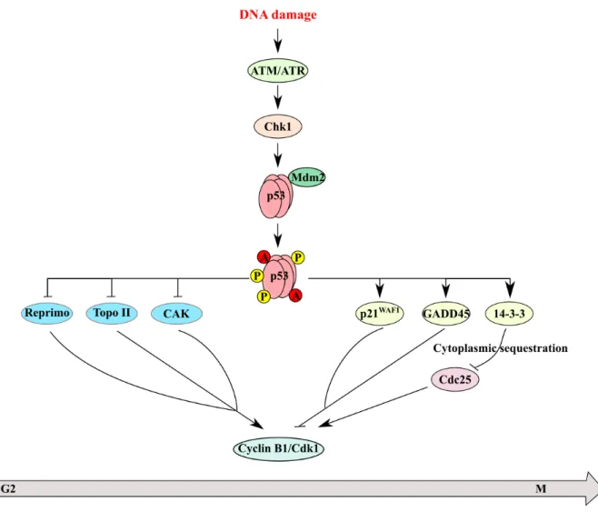
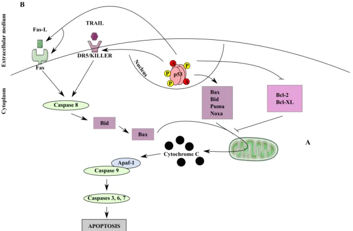
Preface 1 Introduction: The TP53 gene and its modifiers 5 Part I. The tumour suppressive protein p53 7 A. From oncogene to tumour suppressor: A short history of p53 7 B. The TP53 gene 11 I. Chromosome 17p13.1 organisation 11 II. Role and functions of each p53 domain 12 C. Regulation of p53 23 I. p53 stability 24 II. Subcellular localisation 29 III. Epigenetic silencing of TP53 promoter 30 IV. Post‐transcriptional modifications 29 V. Post‐translational modifications 33 VI. Regulation of p53 activity by its isoforms 37 D. p53 functions 38 I. Cell cycle arrest by controlling checkpoints 38 II. Apopotosis 42 III. Other p53 functions 45 IV. The p53 biological repertoire: orchestrating multiple biological functions 50 E. Genetic alterations of TP53 in cancers 53 I. Somatic mutations in TP53 gene 53 II. Germline mutations and Li‐Fraumeni syndrome 61 III. Polymorphisms in TP53 68
Part II. p53 isoforms 64
Part III. G‐quadruplex structures 80
A. Formation of G‐quadruplex structures 88
Part II. Effect of rs17878362 on age at cancer onset in germline TP53 mutation carriers
122
Part III. Role of G4 in intron 3 on p53 mRNA splicing and p53 protein isoform
expression 161 Discussion: TP53 polymorphisms, key modulators of p53 cancer susceptibility 216 Part I. Association between rs17878362 and cancer susceptibility 221 A. Meta‐analysis of rs17878362 in relation with risk of sporadic cancer 221 B. Analysis of rs17878362 in relation with risk of familial cancer 225 C. Effects of rs17878362 in sporadic or inherited contexts: an apparent paradox 228 Part II. Towards a functional hypothesis for genetic determinants of p53 regulation 231
Conclusion 236
References 240
Annexes 289
Figure 1: p53 history, from its discovery to clinical applications. 10 Figure 2: Schematic representation of the organisation of the human TP53 gene. 11 Figure 3: Schematic representation of the structural organisation of p53. 13 Figure 4: p53 trans‐activation domains functions. 15 Figure 5: Regulation of p53 stability. 24 (Figure 6: p53 regulation by miRNAs. 31 Figure 7: Post‐translational modification of p53. 34 Figure 8: Role of p53 in the G1/S transition checkpoint. 40 Figure 9: Role of p53 in G2/M transition checkpoint. 42 Figure 10: Role of p53 in apoptosis. 43 Figure 11: TP53 mutation distribution depending on cancer types. 54 Figure 12: The distribution of TP53 somatic mutation types distribution 55 Figure 13: Oncogenic effects of p53 mutants. 58 Figure 14: Codon distribution of TP53 mutation in LFS. 67 Figure 15: The most frequent polymorphisms found in the TP53 gene and in its 3'flanking region68 Figure 16: Schematic representation of human p53 isoforms. 76 Figure 17: Schematic representation of G4 formation and structures in DNA or RNA. 89 Figure 18: Schematic representation for the meta‐analysis results for the TP53 rs17878362 polymorphism 106 Figure 19: Kaplan‐Meir disease‐free probability estimates in LFS/LFL family members with or without TP53 mutations. 124 Figure 20: GFP‐reporter splicing assay to analyze the role of G4s in alternative splicing of intron 2 172 Figure 21: Effect of the G4 ligand 360A on expression of p53 transcript for A1A1 and A2A2 for rs17878362 lymphoblastoïd cell lines. 173 Figure 22: Schematic representation of the three most frequent polymorphisms between the intron 2 and the exon 4 of the TP53 gene. 219
Table 1: Summary of phenotypic observation for Knock‐in mutated mice carrying mutants at different important phosphorylation site of p53 (Adapted from (Jenkins et al 2012)). 35 Table 2: Clinical criteria for classic Li‐Fraumeni syndrome (LFS) and LFS‐like (LFL) criteria, and
Chompret criteria. 62
A Adenosine
A Alanine
aa Amino acid
ADR Adrenal cortical carcinoma
ARF Alternative reading Frame
ASSP Acid-soluble spore proteins
ATM Atexia-Telangiectasia Mutated
ATP Adenosine triphosphate
ATR ATM and Rad-3-related protein
Bax Bcl-2-associated X protein
Bcl B-cell lymphoma
BER Base excision repair
bFGF Basic fibroblast growth factor
Bid BH3-interacting domain death agonist
Bp base pair
C Cytosine
C Cysteine
CAK Cyclin-activating kinase
CBP CREB binding protein
Cdk Cyclin-dependant kinase
cDNA Complementary DNA
Chk Checkpoint kinase
CI Confidence interval
COP Constituvely photomorphic
COX-2 Cyclooxygenase-2
CTD C-terminal basic domain
D Aspartic acid
A
B
C
F Phenylalanine
FS Fully spliced
G Guanine
G Glycine
G4 G-quadruplex
GADD45 Growth arrest and DNA damage 45
H Histidine
HAUSP Herpes associated ubiquitin-specific protease
HIF Hypoxia inducible factor
hnRNP Heterogeneous nuclear ribonucleoprotein
HR Homologous recombination
JNK c-Jun N-terminal kinases
K Lysine kDA Kilodalton kb Kilo base L Leucine LFL Li-Fraumeni like LFS Li-Fraumeni Syndrome
LIF Leukaemia inhibitor factor
M Methionine
M Mitosis
MAF Minor allele frequency
MAPK Mitogen-activated protein kinase
Mdm Mouse double minute
miRNA MicroRNA
MRE Meiotic recombination
mRNA Messenger RNA
n Nucleotide
NEDD8 Neural precursor cell expressed developmentally down-regulated protein 8
NER Nucleotide excision repair
NES Nuclear export signal
NF-Y Nuclear transcription factor Y
NHEJ Non-homologous end-joining
G H J L K M N
OD Oligomerisation domain
OR Odd ratio
P Proline
p21WAF1 Cyclin-dependent kinase inhibitor 1
PGC Peroxisome proliferator-activated receptor γ co-activators
PQS Putative quadruplex sequence
PUMA p53 up-regulated modulator of apoptosis
pRB Retinoblastoma protein
PXXP Proline-rich domain
Q Glutamine
R Arginine
RE Response Element
RING Really interesting new gene
RLP Ribosomal protein
RNA Ribonucleic acid
ROS Reactive oxygen species
RPA Replication protein A
S Serine
S phase Synthesis phase
SH3 Scr homology 3-bind protein
SIRT Sirtuin
SNP Single nucleotide polymorphism
SP1 Specific protein 1
STS Soft tissue sarcoma
SUMO Small ubiquitin like modifier
SV40 Simian Vacuolating virus 40
O P
Q R
USP Ubiquitin-specific processing protease
UTR Untranslated region
UV Ultra violet
VEGF Vascular endothelial growth factor
W Tryptophan WT Wild-Type Y Tyrosine Zn Zinc U W Y Z V
human cancer. This is firstly because it has been recognised since the late eighties, that the
TP53 gene is often mutated and the presence of these mutations is associated with cancer
susceptibility and secondly that it carries out a multiplicity of biological functions that are regulated by complex and interlinked pathways. The p53 plays a role in all the functions implicated in the “Hallmarks of Cancer” a concept developed in 2000, and recently updated in 2011 (Hanahan and Weinberg 2000, Hanahan and Weinberg 2011), to understand the molecular mechanisms of carcinogenesis such as proliferation, cell death, growth, senescence, genomic and genetic stability. This multi-action capacity is perhaps the main explanation for the “success” of TP53 as cancer gene.
Given the multiplicity of its effects and functions, it is of no surprise that the TP53 gene is highly polymorphic, with significant differences in the frequency of different polymorphic alleles across populations. The TP53 gene can be expressed as multiple ribonucleic acid (RNA) transcripts, generated by using alternative splicing and alternative promoters, leading to a large diversity at the protein level. The impact of this diversity on individual and population cancer risk however is still poorly understood.
In the nineties, technological developments allowed the tri-dimensional structures of proteins to be determined and opened up the possibility of correlating deoxyribonucleic acid (DNA) and RNA sequences to their formation and their role in transcription/translation regulation. More recently, tri-dimensional structures formed in G-rich sequence in both at the DNA or RNA level called G-quadruplexes (G4) were observed. These structures are involved in different cellular processes such as gene transcription (Bochman et al 2012), genomic stability (Paeschke et al 2013, Ribeyre et al 2009), DNA replication (Cayrou et al 2012, Paeschke et al 2011), messenger RNA (mRNA) splicing (Millevoi et al 2012) and mRNA stability (Millevoi et al 2012).
In this Thesis, I have examined the impact of the complexity of the TP53 gene and in particular its polymorphic variability on cancer susceptibility in the context of both population
I
NTRODUCTION
:
T
HE
TP53
GENE
AND ITS MODIFIERS
Part I. The tumour suppressive protein p53
A. From oncogene to tumour suppressor: A short history of p53
In the past thirty years, p53 has emerged as one of the most important molecular factors in human cancer. The term the “p53” protein is used to identify an essentially nuclear phospho-protein with an apparent molecular weight of 53 kDa. The gene encoding p53 is called TP53, whereas its mouse homolog is known as Trp53. To date, mutations in TP53 are the most common genetic event in human cancer (p53.iarc.fr). Many of these mutations are missense and are identified by their codon number, preceded by the amino-acid (aa) encoded by the wild-type (WT) codon and followed by the aa encoded by the mutant codon (for example: mutation R175H: substitution of an arginine (R) by a histidine (H) at codon 175). These abbreviations will be widely used throughout this Thesis manuscript.In the mid 1950’s, small DNA tumour viruses such as Simian Vacuolating virus 40 (SV40) and polyoma virus were discovered and were shown to carry the necessary genes, termed viral oncogenes, to lead to a tumorigenic phenotype in infected cells (Eddy et al 1962). Over the following twenty years the question of how these virally encoded proteins can initiate this process and lead to the immortalisation of cells in culture was extensively studied. In 1979, several groups reported an interaction between the SV40 large T-antigen, one of the two main proteins expressed by the SV40 tumour virus, and a 53 kilodalton (kDa) protein, p53. Lane and Crawford and May and collaborators demonstrated that the SV40 large T-antigen co-precipitated with a 53 kDa protein in cells and that these cells contained an equivalent amount of both, suggesting that they two proteins were in a stoichiometric complex in the cell extracts (Lane and Crawford 1979, May et al 1979). Linzer and Levine used antisera from animals carrying SV40 induced tumours to detect both p53 and the viral
T-and to characterise its functions (Figure 1). This took about 15 years, during which the status of p53 switched from an “oncogene” to a “tumour suppressor gene”. The first Complementary DNA (cDNA) and genomic sequences of TP53 was isolated from immortalized cells. Sequences variations were reported depending on whether tumour or normal cells or tissues were the source of the DNA, leading to confusion as to which of these sequences represented the WT allele. These differences were deciphered by the seminal studies of several groups. Firstly Levine’s laboratory showed that the WT p53 protein alone could not transform cells (Reich et al 1983). Moreover, they observed that a mutant p53 protein was often found in cancer cells and could inhibit the transforming activity of oncogenes (Finlay et al 1989, Harvey and Levine 1991, Hinds et al 1989). Secondly the group of Vogelstein reported that
TP53 mutations and loss of alleles were common events in human colon carcinomas (Baker et
al 1989, Nigro et al 1989). In the early eighties, it was also shown that p53 was induced in response to DNA damage (Maltzman and Czyzyk 1984) and induced cell death by apoptosis (Yonish-Rouach et al 1991). These properties suggested that p53 was behaving as tumour suppressor protein, in a manner similar akin to the retinoblastoma gene product that had been recently identified (Geiser and Stanbridge 1989). The discovery that germline TP53 mutations were the genetic defect associated with the Li-Fraumeni syndrome (LFS), characterised by a predisposition to multiple and early onset-cancers (Malkin et al 1990, Srivastava et al 1990), led to the recognition of TP53 as the “ultimate tumour suppressor gene” (Oren 1992).
In 1992, David Lane described p53 functions as those of “the guardian of the genome” (Lane 1992), based on the role of p53 in protecting cells against carcinogenesis DNA damage and abnormal proliferation. Many studies were undertaken to understand the impact of TP53 mutations in cancers and the molecular mechanisms of p53 effects. The first important role of the p53 protein discovered was its implication in apoptotic death (Yonish-Rouach et al 1991). Next, p53 was described as a transcription factor with a sequence response element that allowed the targeting of a panel of genes (el-Deiry et al 1992, Funk et al 1992). The auto-regulatory loop of p53 stability via Mdm (Mouse Double Minute) 2 (Barak et al 1993), the crystallographic structure of the p53 core domain bound to DNA (Cho et al 1994) and the notion that TP53 mutations could be understood as “molecular fingerprints” of carcinogens (Hollstein et al 1991), all contributed to the better understanding of the relationships between DNA damage and cell proliferation and establishing the critical role of p53 in cancer.
In 1994, the discovery of the role of p53 in the response to cytotoxic therapeutic treatments suggested that its manipulation could be used to therapeutic benefit (Figure 1). For
example, it was shown that the drug PRIMA-1 can restore the suppressive functions of a mutant p53 protein (Bykov et al 2002) and that Nutlin-3 is an antagonist of the interaction between p53 and Mdm2 causing the release of p53 from this complex and by thus controlling its degradation and inducing p53 accumulation in a DNA-damage independent way (Vassilev et al 2004). Although promising, these drugs still have to find their application in cancer treatment. At about the same time, many studies showed that sequence variations in TP53 (mutations or polymorphisms) could be used as biomarkers for cancer development, treatment response, prognosis and survival (Petitjean et al 2007, Whibley et al 2009). Databases such as the IARC TP53 database (p53.iarc.fr) and the p53 Web Site (p53.free.fr) were created to compile and categorise these variations. Since the mid-nineties, studies on the p53 regulatory network have allowed a better understanding of p53 functions aided by the discovery of p73 and p63, two proteins encoded by TP53-related genes, which are implicated in development, morphogenesis and stress responses (Lane and Levine 2010). In addition, molecular epidemiological studies have more recently led to a new concept: human p53 isoforms, which can also act a large diversity at the protein level as regulators of p53 expression and function (Bourdon et al 2005, Courtois et al 2002, Marcel et al 2011).
Figure 1: p53 history, from its discovery to clinical applications. Important steps from discovery of p53 in
1979 to clinical applications in 2010. Green boxes: important p53 discoveries; Blue: important steps in p53 methods; Red: current developments from TP53 mutation studies in cancer therapy using p53 targeting drugs and TP53 mutation profiles as a prognostic biomarker. Adapted from (Hainaut and Wiman 2009) and Thesis of Virginie Marcel, University Lyon I, 2009.
B. The TP53 gene
I. Chromosome 17p13.1 organisation
The human TP53 gene is located on chromosome 17 on the short arm at locus 17p13.1 (Isobe et al 1986, McBride et al 1986). The TP53 locus is unique on this chromosome arm in having a telomere to centromere orientation, whereas flanking genes are in the reverse orientation. The gene spans about 20 kilobases (kb) and is composed of 11 exons, with a small non-coding first exon and a large conserved intron of about 10 kb (Figure 2). The gene lacks a conventional TATA box but contains several sequences with promoter activity, which may regulate p53 expression. The proximal promoter (P1) is located upstream of exon 1 (Lamb and Crawford 1986) and produces the full spliced p53 (FSp53) mRNA and p53I2 mRNA by alternative splicing. The second promoter (P1’) is located within the long intron 1 and directs the synthesis of a 1.1 kb mRNA derived from sequences from intron 1 (Hint1p53 transcript). This transcript contains several short open-reading frames but the putative proteins have not been identified so far. Its role and function are unknown (Reisman et al 1988, Reisman et al 1996). Hint1p53 is expressed in a number of human cells and is induced during the terminal differentiation of myeloid leukaemia cells. The third promoter (P2) is located between the end of exon 2 and the beginning of exon 5 and gives rise the p53I4 mRNA that lacks sequences from exon 2 to 5 (Bourdon et al 2005). The organisation of the TP53 gene is highly conserved through evolution (Soussi et al 1987).
(p53.iarc.fr) and is considered as a highly polymorphic gene. It has been demonstrated that some polymorphisms such as rs1042522 located in exon 4 at codon 72 (guanine (G) > cytosine (C); R > proline (P)) can modulate p53 functions (Siddique et al 2005, Whibley et al 2009) or cancer susceptibility (Whibley et al 2009). Some of these polymorphisms and their functions are described later in the Introduction, Section E, Part III of the Thesis manuscript.
On this part of chromosome 17p13.1, another gene, WRAP53, also called telomerase
Cajal body protein-1 (TCAB1) has also been described (Mahmoudi et al 2009). This gene is
oriented in the opposite direction from TP53 and is located immediately upstream of TP53, with an overlap between exon 1 and part of intron 1 of TP53 and exon 1α of WRAP53. The transcription level of TP53 is 100-fold higher than that of WRAP53. It was demonstrated that WRAP53 RNA could regulate TP53 RNA translation via an interaction between the sense strand region of WRAP53 and TP53 RNA. This interaction induces Wrap53 and sensitizes cells to p53-dependent apoptosis upon DNA damage (Farnebo 2009, Mahmoudi et al 2009). The interaction between Wrap53 and p53 is also required to maintain a normal level of p53 in cells (Farnebo 2009). Thus, Wrap53 is a new regulator of p53 at the post-transcriptional level.
II. Role and functions of each p53 domain
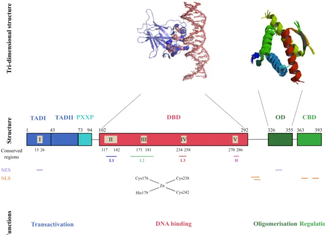
The p53 protein functions as a tetramer to bind specifically to DNA and acts as a transcription factor for target genes regulated through p53-binding DNA response elements (RE) (Figure 3). The p53 protein is divided into 5 distinct structural and functional domains: (1) the N-terminal domain located between aas 1 and 72, (2) the proline domain between aas 73 and 94, (3) the DNA binding domain into aas 102 and 292, (4) the oligomerisation domain at aas 326-355 and (5) the C-terminal basic domain between aas 363 and 393.
Figure 3: Schematic representation of the structural organisation of p53. The p53 protein has a classical
conformation with 4 important domains (“Structure” line) with particular functions (“Functions” line). The tri-dimensional conformations for the DBD and the OD, only, are known (“Tri-tri-dimensional structure” line). The conserved regions, the Nuclear Export Signal (NES) and the Nuclear Localization Signal (NLS) are also represented. The 3 loops (L1 to L3), the hydrophobic region (H) and the tertiary structure of the DBD are shown. TAD: Trans-activation Domain; PXXP; Proline Domain; DBD: DNA Binding Domain; OD: Oligomerisation Domain; CBD: C-terminal Basic Domain. Adapted from (May and May 1999).
1. N-terminal trans-activation domain (TAD, amino acids 1-72) Structure
The trans-activation domain (TAD) of p53 is located in the N-terminal part of the protein. This region contains a transcriptional activation motif ϕ-x-x-ϕ-ϕ (ϕ = hydrophobic aa and x = any other aa), which is found in many proteins regulating transcription. This region contains a few hydrophobic aas (found at positions 7-9, 16, 17, 26, 58-60), which disfavour
The TADI sub-domain contains a nuclear export signal (NES) between aas 13 and 26 (Zhang and Xiong 2001b).
Functions
Aas 13-26 of TADI represent one of the most conserved regions in the p53 protein through evolution (Soussi et al 1990). The main function of TADI is the interaction with others components of the transcription machinery such as the TATA box binding protein (TBP) and TBP-associated factors components of transcription factor TFIIB (Lu and Levine 1995, Thut et al 1995). It can also interact with viral proteins such as E1B from adenoviruses, which inhibit p53 trans-activation activity (Yew and Berk 1992).
To increase p53 trans-activation activity, cofactors and histone acetyl-transferases (HATs) interact directly with the TADI and TADII (Riley et al 2008). Recently, Attardi and collaborators showed that the TADI and TADII sub-domains, and both TAD domains together interact with different cofactors and HATs (Figure 4A) (Bieging and Attardi 2012, Brady et al 2011, Jiang et al 2011). These authors produced transgenic mice with mutations at leucine (L) at codon 25 into glutamine (Q) (L25Q) and at tryptophan (W) at codon 26 into serine (S) (W26S), resulting in inhibition of TADI function or at Phenylalalnine (F) at codon 53 into Q (F53Q) and F54S, inhibiting TADII’s role or L25Q, W26S, F53Q and F54S, inactivating both TAD domains (Figure 4B) and analysed the differences in p53 protein interactions and in expression of p53-regulated genes. They observed that TADI is involved in the activation of p53 target genes important for the response to acute DNA damage, cell-cycle arrest or apoptosis, but is not capable of tumour suppression in mice. The TADII sub-domain, in contrast to TADI, has no autonomous effects on p53 trans-activation capability or biological activity. However, collectively, the two TADs are essential for p53’s tumour suppressor activity. Overall, these results suggest that p53’s TAD mediates tumour suppressor activities via TADI-dependent trans-activation of p53- target gene and TADII contributes to tumour suppression by regulating TADI activities via unknown mechanisms. Moreover, each TAD appears to interact with specific cofactors. For example, TADI interacts with TBP, whereas TADII interacts with TFIID and with replication protein A (RPA) (Bochkareva et al 2005, Brady et al 2011). Together, both TADs interact with HATs p300 and CRE Binding protein (CBP) (Ferreon et al 2009, Teufel et al 2007).
The TAD is the most important domain for the regulation of p53 activity involving three pathways. The first is the p53-Mdm2-autoregulatory feedback loop (Barak et al 1993), the second is the post-translational modification of the serines and threonines present in p53 TAD and the third is the formation of a complex between the TAp53 and Δ40p53 isoforms (Hafsi et al 2013).
In 1992, p53 was found to trans-activate MDM2, which encodes the Mdm2 protein, a E3 ubiquitin ligase targeting p53 for ubiquitination and proteasome-dependent degradation (Barak et al 1993, Honda et al 1997, Kubbutat and Vousden 1998, Momand et al 1992, Oliner et al 1993, Wu et al 1993). Mdm2 binds to a motif at aas 18-23 (TFS acid aspartic (D) LW motif) located in the TADI sub-domain (Picksley et al 1994). This p53-Mdm2 interaction results in the inhibition of the p53 transcriptional activity by masking the p53 TADI sub-domain. It also promotes the ubiquitination of p53 and induces its ubiquitin-dependent proteosomal degradation (Haupt et al 1997, Honda et al 1997). This regulation constitutively maintains p53 at a low cellular level under normal conditions.
The p53 TAD contains several post-translational modification sites that are phosphorylated by a number of activated kinases and are critical for protein-protein interactions that either modulate the stability and subcellular localization of p53 or affect its function as a transcription factor. These regulatory sites and their upstream kinases are presented in Section I, Part C of this Thesis manuscript.
2. Proline-rich domain (PXXP, amino acids 73-94) Structure
The p53 proline-rich domain (PXXP) is located between the N-terminal and the DNA binding domain at aas 73-94 (Walker and Levine 1996). The human p53 protein contains 5 repeats of the PXXP motif. This domain contains a frequent exonic polymorphism at codon 72, rs1042522, which consists of a substitution of a guanine (G) to a cytosine (C) corresponding to the substitution of arginine to a proline (Matlashewski et al 1987). In the presence of the arginine variant, the proline-rich domain loses one of its PXXP domains.
Functions
PXXP corresponds to the Scr homology 3-binding protein (SH3) motif suggesting that it may be involved in the physical interaction with elements of signal transduction pathways that contain SH3 domains, including for example inhibitor member of the acid-soluble spore
protein (ASPP) family (iASSP) (Bergamaschi et al 2006) and c-Abelson murine leukemia viral oncogene (Abl), a kinase activated by Ataxia-Telangiectasia Mutated (ATM) (Khanna et al 1998). The deletion of the proline-rich domain does not compromise transcriptional activity or DNA binding activity (Sakamuro et al 1997, Walker and Levine 1996). In addition, in some experimental systems, it was shown that the proline-rich domain was implicated in p53-mediated apoptosis (Sakamuro et al 1997), in suppressing tumour cell growth (Walker and Levine 1996) in the degradation of p53 by the E6 protein of oncogenic human Papilloma Viruses (Li and Coffino 1996). This region of the gene encoding the proline-rich domain is rarely mutated in human cancer.
Role in p53 regulation
To date, few studies have been performed to elucidate the activity of the p53 proline-rich domain in regulating p53 tumour suppressor activity. The mutant T81A p53 failed to induce p53-mediated cell cycle arrest and apoptosis in response to ultra-violet (UV) damage, suggesting that the phosphorylation of threonine 81 by c-Jun N-terminal kinases (JNK) 2 is important for these responses (Buschmann et al 2001). Furthermore, Berger and collaborators observed that deletion of the proline-rich domain increased the sensitivity of p53 to inhibition and degradation by Mdm2, suggesting that the proline rich domain is important for the fine-tuning of p53-Mdm2 interactions (Berger et al 2001). Indeed, they observed, using mutants of the proline-rich domain, that this regulation was mediated by phosphorylation of proline 82 (Berger et al 2005). Phospho-proline 82 is important for p53-Checkpoint Kinase 2 (Chk2) interaction, which allows the phosphorylation of p53 at S20. When S20 is phosphorylated, the interaction p53-Mdm2 is inhibited, preventing p53 degradation via Mdm2 ubiquitination (Craig et al 1999).
3. DNA binding domain (DBD, amino acids 102-292) Structure
which form a direct contact with p53 RE DNA. The binding surface of p53 is made of two parts: L2 and L3 loops (two conserved region through evolution) and a short α-helix that bind within the minor groove of p53 RE DNA, with R248 as main DNA interacting aa. L2 and L3 are bridged by a zinc atom, which is linked to cysteine (C) 176, H179 on L2 and C238 and C242 on L3. L1, together with the short S2-S2’ β hairpin (aa 124-141) and a large α-helix, forms a motif that occupies the major groove of DNA, with direct aas contacting DNA (including R273, C277, R282 and Lysine (K) 120).
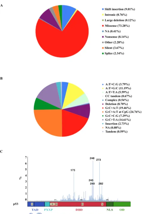
Functions
The majority of the mutations found in the human TP53 gene are located in the DNA binding domain of p53 (Petitjean et al 2007). A survey of studies using whole genome sequencing suggests that at least 70% of all cancer-related mutations are located in this domain (COSMIC database, http://cancer.sanger.ac.uk/wgs/gene/overview?ln=TP53). This high frequency of mutations is probably due to the alteration of the sequence-specificity of the p53 DNA binding activity by the presence of mutations in the DBD, which can modulate the direct contact with DNA or change the conformation of the DNA-binding structure and thus confer some selective advantage to the “mutated” protein. Indeed, the DBD can bind its target genes only by using a specific RE that allows the recognition of p53 target genes. It is constituted of a repeat of an inverted palindromic 10 base pairs (bp) element matching the consensus 5’-RRRCWW glycine (G) YYY (n=0-13) RRRCWWGYY-3’ (R is a purine, W a adenine or thymine base and Y a pyrimidine base) (el-Deiry et al 1992, Funk et al 1992). This sequence orientation increases the specificity of the binding (Funk et al 1992) and reflects the fact that p53 activity depends of the formation of a tetramer consisting of two dimers (Kitayner et al 2006, McLure and Lee 1998) with each p53 monomer in one of the dimers interacting with one half of the 10 bp site (RRRCW). Using reporter assays, Funk and collaborators showed that this palindromic sequence is necessary for p53 DNA binding activity (Funk et al 1992). This sequence is found in between 300 and 1600 potential binding sites in the human genome (Cawley et al 2004, Hoh et al 2002). To date, over 125 protein-coding genes and nonprotein-coding RNAs have been shown to be the direct transcriptional target of p53, all of which contain the p53 RE to which p53 binds activating their transcription (Poyurovsky et al 2010, Riley et al 2008). Interestingly, the p53 RE does not perfectly match the consensus sequence in the majority of these cases. The p53 protein thus may have different affinities for some p53 REs, which could explain the large number of p53 target
genes. Another activity of the p53 DBD is the trans-repression of some genes, but the mechanisms involved are less well understood (Oren 2003). For example, the B-cell
lymphoma (BCL)-2 promoter contains a p53 RE, which overlaps with the binding site of
another more potent activator (Budhram-Mahadeo et al 1999). When p53 binds to the BCL-2 promoter, the target mRNA level is decreased.
Role in p53 regulation
The p53 DBD is the domain with the least post-translational modification sites known. For instance, no phosphorylation sites have been identified (Riley et al 2008). Recently, some studies have shown that the acetylation of lysine K120 and K164 in this region could increase the trans-activation of p53 up-regulated modulator of apoptosis (PUMA), which influences p53-apoptosis (Sykes et al 2006, Tang et al 2006, Tang et al 2008). The ability to bind DNA is dependent on the DBD’s interaction with a Zinc (Zn)2+ ion via aas in two clusters around C176-H179 and C238-C242 (Hainaut and Milner 1993). The oxidation of these two particular clusters decreases the ability of the protein to bind to p53 RE and trans-activate target genes
in vivo (Parks et al 1997). The first cluster contains the aas that interact with a Zn ion and the
second is close to the aas implicated in the contact of p53 with its consensus sequence.
Two important ways to influence p53 DNA binding domain activity are well described. The first is the presence of a mutation in the DBD (Olivier et al 2002) and especially ones affecting the bases implicated in the Zn2+ ion interaction with the DBD (Kern et al 1991, Kim et al 1997). The second is the structure of the C-terminal basic domain as described later in the text (Hamard et al 2012).
4. Oligomerisation domain (OD, amino acids 326-355) Structure
The oligomerisation domain (OD) is located between aas 326-355 and is also called the tetramerisation domain (Chene 2001). The structure of this domain has been determined
and Hydrogen-bond donation between R337 and D352. Mutations at these aas prevent the formation of tetramers (Chene et al 1997, Mateu and Fersht 1998, Waterman et al 1995).
A region containing two important domains for p53 localisation is located between the p53 DBD and the OD. The first, the dominant nuclear localisation signal (NLS)-I also which is upstream of the OD at aas 315 to 325 and a weaker NLS also located upstream of the OD, in a flexible linker region at aas 305-322 (Shaulsky et al 1990b). The second domain also contains the NES (Zhang and Xiong 2001b), which is located at the end of the OD, at aas 339-352.
Functions
The p53 OD plays several roles in p53’s DNA binding activity, p53-protein interactions and cellular localisation. First, the formation of p53 oligomers by the OD increases p53 DNA binding activity. Indeed, it has been shown that p53 monomers can interact with p53’s REs in a cooperative manner and trans-activate p53 target genes but with a 10-100 fold lower affinity than a p53 tetramer (Balagurumoorthy et al 1995). Secondly, the p53 OD is important for p53’s interaction with others proteins. For example, the OD plays an indirect role in p53-Mdm2 interaction (Lomax et al 1998), or with p53-TBP interactions (Liu et al 1993).
Role in p53 regulation
Post-transcriptional modifications can modulate the dynamics of p53 oligomerisation. For example, the phosphorylation of S392 can increase the association constant for oligomerisation by 10-fold in vitro (Waterman et al 1996). This domain can interact with proteins such as REGγ, a proteasome activator, which can regulate the cellular distribution of p53 by increasing the mono-ubiquitination of p53 and its subsequent nuclear export and degradation (Liu et al 2010b). These authors also observed that REGγ inhibits p53 tetramerisation and this might enhance p53 cytoplasmic relocalisation, which decreases the amount of active p53 in the nucleus. Zhao and collaborators established mouse lines with or without the NLS-I (Zhao et al 1999). They observed that both trp53-null cells and NLS-I-null cells were deficient for p53-dependent apoptosis after exposure to γ-ionizing radiation or hydrogen peroxide treatment. These results suggest that NLS-I is necessary for p53-mediated apoptosis.
5. C-terminal basic domain (CTD, amino acids 363-393) Structure
The C-terminal basic domain of p53 (CTD) is located between aas 363-393. Its main characteristic is its natively unfolded form, which confers the capacity to interact with DNA in a sequence nonspecific fashion (Foord et al 1991, Wang et al 1993). It can interact with different DNA structures including insertion/deletion mismatches (Lee et al 1995), γ-irradiated DNA (Miyashita and Reed 1995) or supercoiled DNA (Mazur et al 1999). This domain contains two minor NLS (aas 369-375: NLSII and 379-384: NLSIII) (Dang and Lee 1989, Shaulsky et al 1990b), multiple ubiquitination sites (Michael and Oren 2003), one major site of sumoylation (K386) (Gostissa et al 1999, Rodriguez et al 1999) and several stress-inducible modification sites for phosphorylation, acetylation or glycosylation (Appella and Anderson 2001).
Functions
Early research investigating the functions of this domain was focused on its ability to regulate the sequence-specific DNA binding of p53 (Ahn and Prives 2001). Deletion of the last 30 aas of p53 and the use of the monoclonal C-terminal antibody polyclonal antibody (PAb) 421, a basic peptide which inhibits C-terminal function, resulted in an increase in p53 sequence-specific DNA binding suggesting that the CTD is important for inhibiting the high-affinity p53 DNA binding activity (Hupp et al 1992, Hupp et al 1995). These observations lead to the allosteric hypothesis namely that the C-terminal domain could control the conversion of p53 from a latent to an active form by its interaction with cellular factors resulting in enhanced sequence-specific DNA binding and the transcriptional activity of p53 (Hupp et al 1992, Hupp et al 1995). The p53 latent form was shown to arise from the interaction between the aas 80 and 93 (in the PXXP domain) with its C-terminal domain (Muller-Tiemann et al 1998). More recent studies led to the re-evaluation of this model. Anderson and collaborators found that the nonspecific DNA commonly used in standard in
repressive but the manner in which this domain causes inhibition of p53 functions remains unclear.
Role in p53 regulation
The CTD of p53 is needed for most p53 biological activities and plays an important role in p53 DNA binding and transcriptional activity (Hamard et al 2012). Hamard and collaborators suggested that the strong interaction of p53 with its targets depends on (1) the sequence of individual p53 RE and their association with p53, (2) post-translational modifications affecting the p53 protein, and more specifically the CTD, (3) p53 protein binding partners and (4) the epigenetic landscape of p53 target genes, regulating the access of p53 to relevant gene regulatory sequences (Hamard et al 2012).
The CTD contains several post-translational modification sites. Lysine aas K370, K372, K373, K381 and K382 can be each acetylated by p300/CREB-binding protein (CBP) (Knights et al 2006). The CTD seems to have a regulatory role on p53 functions by modifying the interaction on the p53 DBD with its target genes or the stability of the p53 tetramer. For example, the CTD is essential for pro-apoptotic p53 target genes such as PUMA,
Bcl-2-associated X protein (BAX) and cell cycle arrest p53 target genes such as cyclin-dependent kinase inhibitor 1 (p21WAF1) (Hamard et al 2012). For Mdm2’s interaction with p53’s CTD, it
was observed that both the CTD and TADI were involved and that Mdm2 binding to these domains was not mutually exclusively presupposing that different surfaces of the Mdm2 protein were involved (Poyurovsky et al 2010). The CTD could also interact with the signalling protein 14-3-3σ to stabilise p53 as a tetramer (Schumacher et al 2010).
Liang and collaborators have reported that K305 and K306 were essential for p53 nuclear import (Liang et al 1998, Liang and Clarke 1999a, Liang and Clarke 1999b). Using a mutagenesis approach, they observed that mutated K305A or K306A p53 proteins were deficient for nuclear import of p53. Other punctual mutations between these lysines and NLS-I did not have the same effect. However, deletion of two or more aas abolished p53 nuclear import, suggesting that a part of the basic domain is essential for NLS-I function.
C. Regulation of p53
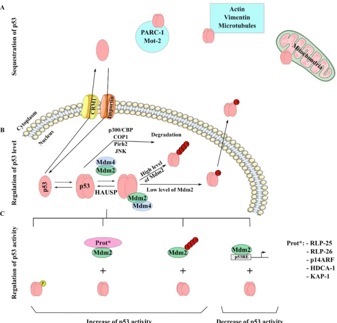
The p53 expression and activity are regulated by several pathways (Figure 5). The first is the modulation of the level of expression in the cell via its interaction with Mdm2 or other proteins interaction such as Mdm4. The second is the regulation of p53’s accumulation in the nucleus to promote the transcription of its target genes and the last level is by modulating the level of post-translational modifications (phosphorylation, acetylation), which influences p53’s activities. Transcriptional regulation of the p53 promoter does not appear to have a critical role in the induction of p53 in response to many different signals.
Figure 5: Regulation of p53 stability. (A) Sequestration of p53 in the cytoplasm by different mechanisms:
PARC-1 or Mot-1 protein-protein interaction, sequestration by actin or vimentin, microtubules or sequestration in the mitochondria. (B) Regulation of p53 level. Mdm2 and Mdm4 form a complex, which allows p53 ubiquitination. HAUSP could regulate the formation of this complex by de-ubiquitinating p53 (C). Regulation of p53 activity. p53 could be phosphorylated, the absence of Mdm4 decrease p53 degradation mediated by Mdm2, ARF could form a complex with Mdm2 or Mdm2 could poly-ubiquitinate itself and induce its degradation. Adapted from (Haupt et al 1997, Kubbutat et al 1997); Thesis of Virginie Marcel, University Lyon I, 2009.
I. p53 stability
1. The p53/Mdm2 regulatory feedback loop Mdm2: the main regulator of p53 stability
Mdm2 is a member of the really interesting new gene (RING)-finger-family protein and and an E3 ligase. p53 is one of its substrates and this modification mediates p53’s targeted degradation by the proteasome (Figure 5) (Jackson and Berberich 2000). Mdm2 can interact with the TADI of p53. Toledo and Wahls have showed that this interaction is
disrupted by phosphorylation of the N-terminal domain of p53 at S9, S15, S20 and S46 and that one or more of these phosphorylation events appeared sufficient to disrupt Mdm2 binding (Toledo and Wahl 2006). However, using mice models with an S to R mutation at S15 or S20, no effect on Mdm2 binding p53 function was noted (Chao et al 2006, Toledo and Wahl 2006). No role of S46 alone was observed on p53 stability either. Thus, threonine (T) 18 phosphorylation seems to be the essential post-translational modification for p53/Mdm2 complex formation (Sakaguchi et al 2000, Schon et al 2002). After p53/Mdm2 complex formation, Mdm2 poly-ubiquitinates p53 in the CTD, providing a signal for p53’s export to the cytoplasm and degradation by the 26S proteasome (Kubbutat et al 1999).
After DNA damage, S15 of p53 is phosphorylated, which facilitates the subsequent phosphorylation of S20, S46 and T18 resulting in the disruption of Mdm2 binding to p53 and of the degradation of p53 (Figure 5C). The stabilised p53 acts as a transcription factor for its target genes, including Mdm2 resulting in increased Mdm2 levels. This transcriptional regulation by p53 defines a negative regulatory feedback loop between p53/Mdm2 and is important for the cell to be able to restore p53 to its basal level (Figure 5B) (Chehab et al 1999, Hirao et al 2000, Stommel and Wahl 2004). The mechanisms, by which p53 escapes Mdm2 binding, depends on the type of stress but seems to be critical in most cell types. In the mouse, depletion of Mdm2 was found to be lethal at early embryonic stages whereas the double depletion of both Mdm2 and trp53rescued a normal development (Jones et al 1995, Montes de Oca Luna et al 1995). This observation suggests an important role of Mdm2 in the control of p53 levels and activity during development. Indeed the early lethality observed in Mdm2 depleted-mice may be explained by uncontrolled levels of p53 activity, inducing growth arrest and apoptosis. Moreover, over-expression of Mmd2 works as an antagonist that blocks p53-mediated cell cycle arrest and apoptosis (Kruse and Gu 2009, Vousden and Prives 2009). These results show the importance of Mdm2-mediated p53 regulation. To date, several mechanisms modulating Mdm2/p53 interaction have been described (Figure 5C). For example, the p14alternate reading frame (ARF) protein (the alternative product of the p16 locus) can
increasing its translation. Thus, the down-regulation of RLP26 by Mdm2 results in a lower level of p53 protein. Secondly, Chromatin Immuno-Precipitation assays (CHIP) have shown that Mdm2 can be found on the promoters of p53 target genes, such as p21WAF1 (Arva et al 2005, Tang et al 2008). After DNA damage, p53 is activated and Mdm2 seems to be released from such promoters. Mdm2 could thus negatively regulate some p53 target genes. In addition, Mdm2 interacts with histone modifying enzymes such as histone deacetylase (HDAC) 1 and kinase associated phosphatase (KAP) 1 that regulate the deacetylation of histones at the promoters of target genes and thus could modulate the activation of such promoters (Ito et al 2002, Wang et al 2005).
Mdm4: Regulator of the p53/Mdm2 regulator feedback loop
Mdm4 (also called MdmX) is a RING-finger homologue of Mdm2 and also has an E3 ligase activity. It presents an almost identical p53 binding domain as Mdm2 in its N-terminal region and a RING domain in its C-terminal end (Wang and Jiang 2012). It was first identified as a p53-binding domain partner in 1996 (Shvarts et al 1996). Genetic studies showed that Mdm4 is as essential as Mdm2 for the degradation and the negative regulation of p53. Indeed, Mdm4 depletion in mice is lethal at an embryonic stage due to the over-expression of p53 (Finch et al 2002, Parant et al 2001). Furthermore, embryonic lethality is also observed in mice carrying mutations in the Mdm4 RING domain due to an accumulation of p53 and an over-expression of p53 target genes such as p21WAF1, BAX and MDM2 (Huang et al 2011). Interestingly, when the level of p53 is reduced to a basal level, mice do not show embryonic lethality (Pant et al 2011). These results led to the suggestion that (1) the Mdm4 RING domain interacts with the Mdm2 RING domain and promotes Mdm2-mediated p53 poly-ubiquitination and degradation (Gu et al 2002, Kawai et al 2007, Okamoto et al 2009); (2) the level of basal p53 is important for Mdm2/Mdm4 degradation. Recently, in vitro, two main roles were described for the heterodimer of Mdm4/Mdm2: in the presence of p53, Mdm4 is an activator of the E3 ligase activity of Mdm2. In the absence of p53, Mdm4 protects Mdm2 against its self-ubiquitination (Wang et al 2011).
After DNA damage, activation of p53 requires the un-coupling of the Mdm2/Mdm4/p53 complex. Studies show that the kinases ATM/ATM and rad3-related protein (ATR) and Abelson murine leukemia viral oncogene (c-Abl) promotes the translocation of Mdm4 from the nucleus into the cytoplasm via the 14-3-3σ protein (Chen et al 2005, Jin et al 2006, LeBron et al 2006, Li et al 2002a, Okamoto et al 2005, Wang et al
2007, Waning et al 2011). The localisation of Mdm4 to the cytoplasm increases its degradation by the proteasome in an Mdm2-dependent manner (Chen et al 2005, Jin et al 2006, Okamoto et al 2005). This degradation appears to be the key for the inactivation of the Mdm2/Mdm4 E3 ligase activity during the p53 DNA damage response.
2. Others regulators of p53 stability
Other p53 stability regulators have been described in the literature such as the JNK kinase protein, other p53 E3 ligases, the de-ubiquitinating protein Herpes associated ubiquitin-specific protease (HAUSP) ubiquitin-specific protein and the p300/ CREB binding protein (CBP) co-activator proteins.
JNK
JNK appears to have a dual role on p53 stability, either increasing or decreasing p53 stability depending on cellular conditions. Under basal conditions, JNK binds p53 and acts as E3 ligase to target it for ubiquitination by the proteasome (Fuchs et al 1998b). After stress, JNK phosphorylates p53 on T81, inducing a cascade of modifications by acetylation to increase p53 activity (Fuchs et al 1998a, Prives and Hall 1999). As for Mdm2, the phosphorylation of the TAD mediated by JNK decreases p53 degradation and increases its stability.
Others p53 E3 ligases
Pirh2 is a RING domain protein, which is able to modulate p53 stability (Leng et al 2003). Like Pirh2, the human homolog of constituvely photomorphogenic 1 (COP1) has also been described as a p53 interacting RING finger protein (Dornan et al 2004a, Dornan et al 2004b). These two proteins act through a similar mechanism to Mdm2, ubiquitinating and degrading p53 through the proteasome. Interestingly, both are encoded by p53-inducible genes. These results suggest that Pirh2 and COP1 also participate in a p53 negative feedback loop, as for Mdm2. The functional impact of these proteins is similar to Mdm2:
over-2004b).
HAUSP
The HAUSP, also known as Ubiquitin-specific processing protease (USP) 7 modulates p53 activity regulation by interacting with both p53 and Mdm2 (Li et al 2002b, Lim et al 2004, Wood 2002). HAUSP directly interacts with p53, de-ubiquitinating it and activating p53 (Li et al 2002b). Studies showed that loss of HAUSP expression increases p53 stabilisation and cell cycle arrest (Cummins et al 2004, Li et al 2004).
p300/CBP: E4 ubiquitin ligases
CBP and p300 are transcription co-activators involved in p53 acetylation, which can also acts as E3/E4 ubiquitin ligases by mediating p53 degradation (Shi et al 2009a). Analysis of p300 or CBP-deficient cells revealed that both were required for endogenous p53 ubiquitination and the degradation of p53 in unstressed cells. Unexpectedly, p300/CBP ubiquitin ligase activities were absent in nuclear extracts and exclusively cytoplasmic. Consistent with the cytoplasmic localization of its E3/E4 activity, CBP deficiency specifically stabilized cytoplasmic, but not nuclear p53. This cytoplasmic localisation is associated with contrary functions of p300/CBP: cytoplasmic p300/CBP ubiquitinates and destabilizes p53, while nuclear p300/CBP activates p53 by acetylation.
p14ARF
The p14ARF protein is the product of the alternative reading frame of the p16 locus (Kamijo et al 1997, Sherr 2001). It is able to bind p53, Mdm2 or both when present in a complex (Kamijo et al 1998). Through Mdm2 binding, p14ARF inhibits Mdm2 activity (Honda and Yasuda 1999) and increases Mdm2-mediated degradation of Mdm4 (Pan and Chen 2003). The p14ARF protein is localised in the nucleolus, which co-localises with Mdm2 (Weber et al 1999). These results suggest that p14ARF could sequester Mdm2 in the nucleoplasm and indirectly activate p53. Since p14ARF is a transcriptional target of E2F transcription factors during the S (synthesis) phase of the cell cycle, its expression is increased in cells, which results in the activation of p53 as a safeguard mechanism to control cell growth (Lomazzi et al 2002).
II. Subcellular localisation
p53 is a transcriptional factor and exert its main activity in the nucleus. Export of p53 into the cytoplasm inactivates this function. During cell cycle and after genotoxic stress, p53 appears to shuttle between the nucleus and the cytoplasm (David-Pfeuty et al 1996, Moll et al 1996, Ostermeyer et al 1996, Shaulsky et al 1991a, Shaulsky et al 1991b). NLS and NES sequences are essential for the balance between the rates of import and export of p53 through the cell (Henderson and Eleftheriou 2000, Shaulsky et al 1990a, Shaulsky et al 1990b).
p53 contains two NES, one in the oligomerisation domain (aas 339-352) and the second in the N-terminal domain (aas 11-27) (Stommel et al 1999, Zhang and Xiong 2001a, Zhang and Xiong 2001b). Under basal condition, the NES are exposed when p53 presents its inactive tri-dimensional structure and p53 is exported into the cytoplasm through the export nuclear receptor CRM1 (Kudo et al 1998, Santiago et al 2013). In the cytoplasm, p53 is degraded by the proteasome in a Mdm2-dependent manner (O'Keefe et al 2003). Post-translational modifications such as phosphorylation of S15 and S392, which allows the tetramerisation of p53, interfere with nuclear export (Sakaguchi et al 1997, Zhang and Xiong 2001b). When p53 tetramers are formed, the NES in the C-terminal is not accessible and the p53 tetramers accumulate in the nucleus (Lohrum et al 2001, Stommel et al 1999).
The activity of the NLS is also under post-translational control. As explained in the Introduction, Section B, Part 5, p53 contains several NLS (NLS I: aas 315-325; NLS II: aas 369-375 and NLS III: aas 379-384), the strongest NLS is the NLS I (Shaulsky et al 1990b). This NLS includes a phosphorylation site for cyclin-dependent kinase (Cdk) 2/Cyclin A at S315 and an acetylation site for p300/CBP at K320 (Iyer et al 2004).
p53 crosses the nucleus membrane by fixation to importin α (Kim et al 2000, Liang and Clarke 1999a). Truncation of importin α results in the sequestration of p53 in the cytoplasm. Some additional proteins are implicated in p53 importation into the nucleus such as S100B and Spot-1, which bind to the NLS I motif (Elkind et al 1995, Scotto et al 1999) or
mitochondrial p53 can induce the mitochondrial intrinsic apoptotic pathway, which will be developed later in this Thesis (Introduction, Section D, part II-1).
Nuclear import/export and proteasome dependent degradation of p53 are closely related mechanisms. Controversial results have been published on p53 re-localisation into the cytoplasm and its degradation by Mdm2. Some studies showed a degradation of p53 in the cytoplasm only (Freedman and Levine 1998, Roth et al 1998), while others reported that p53 was degraded in the nucleus (Lohrum et al 2001, Stommel and Wahl 2004). Recent studies suggest that the re-localisation of p53 mediated by Mdm2 depends on its ubiquitination status. When Mdm2 levels are low, p53 is only mono-ubiquitinated and is exported to the cytoplasm; while at high level of Mdm2 or in presence of p300 and Mdm2, p53 is poly-ubiquitinated and degraded in the nucleus (Grossman et al 2003, Li et al 2003, Tao and Levine 1999).
III. Epigenetic silencing of TP53 promoter
In 1997, Schroeder and collaborators reported a correlation between hyper-methylation of the TP53 promoter and the decrease of its transcription (Schroeder and Mass 1997). In vitro studies, using reporter gene constructs, demonstrated that TP53 promoter methylation induces a 90% reduction of p53 mRNA expression (Pogribny et al 2000). Since, no reports have been published on naturally occurring DNA methylation of the TP53 promoter. A number of publications have reported that DNA hyper-methylation of the TP53 promoter was associated with low levels of mRNA expression in primary hepatocellular carcinoma (Pogribny and James 2002), acute lymphoblastic leukaemia (Agirre et al 2003), chronic lymphocytic leukaemia, breast cancer without TP53 mutation (Pharoah et al 1999) or glioma (Amatya et al 2005). Based on these results, DNA methylation could be a mechanism used to inactive p53 expression but the role of methylation remains to be fully established. However, it should be noted that there are little data on the levels of DNA methylation of the
TP53 promoter and its variation in non-cancer cells.
IV. Post-transcriptional modifications
1. miRNA
MicroRNAs (miRNAs) are non-coding RNAs of about 22 nucleotides that bind a complementary sequence in the target mRNA and can influence its post-translational regulation, usually by impairing translation or inducing target degradation. About 1,870 human miRNA sequences are currently listed in the miRBase compiled at the University of
Manchester (http://www.mirbase.org/). They are distributed on all chromosomes and miRNA sequences represent about 3-5% of all predicted genes in the human genome. Given the capacity of the same miRNA to target different mRNAs, it is considered that over 50% of all protein coding genes in the genome are regulated by miRNA. Several miRNAs could regulate p53 activity by direct modulation of p53 mRNA and indirect targeting of mRNA encoding p53 regulators. In addition, p53 controls the expression of a network of miRNA with essential functions in cell proliferation, differentiation and survival, such as miR-34, miR-145 or members of the miR-200 family (Figure 6) (Hermeking 2012).
(Figure 6: p53 regulation by miRNAs. Certain microRNAs (miR) mediate p53 down-regulation via
interaction with the 3’UTR. Others can inhibit the Mdm2-p53 interaction directly or indirectly, such as miR-122. They could also inhibit SIRT1 and YY1 regulation of p53 mRNA in the C-terminal part of the mRNA. TAD: Transcriptional Domain; PXXP: Proline Domain; DBD: DNA Binding Domain; OD: Oligomerisation Domain; CDB: C-terminal Basic Domain. Adapted from (Hermeking 2012).
miRNA-mediated direct p53 inhibition
Several publications have shown that p53 expression is regulated by the interaction between miRNAs and the 3’untranslated region (UTR) of p53 pre-mRNA (Figure 6). These miRNA include miR-125b (Le et al 2009), which contains a complementary sequence to the 3’UTR of p53 mRNA. Targeting of p53 mRNA by miR-125b results in decreased p53-mediated apoptosis. Recently, other miRNAs of the miR-125 family were shown to interact with components of the p53 effector network such as PUMA (Le et al 2011). Furthermore,
encoding miR-30d is found in about 30% of solid tumours and is associated with poor clinical survival in ovarian cancer (Li et al 2012a).
Indirect p53 inhibition by miRNAs
Some studies also observed that miRNAs could down-regulate upstream negative regulators of p53 (Figure 6). The most studied miRNAs are the miR-34 family. miR-34a can down-regulate the NAD-dependent deacetylase SIRT1, a regulator of metabolic stress responses acting as a tumour suppressor. This down-regulation increases p53 activity and p53 the expression of p53 target genes such as p21WAF1 and PUMA, which subsequently increases apoptosis (Yamakuchi et al 2008). miR-34a expression is regulated by the p53 protein, creating a positive regulatory loop between p53, miR-34a and SIRT1. miR-200a or miR-449 can increase SIRT1 expression and thus influence this p53 negative loop (Bou Kheir et al 2011, Eades et al 2011). p53 is implicated in a similar feedback loop involving miR-34, since this miRNA can inhibit YY1, a transcriptional suppressor protein, which itself inhibits p53 (Chen et al 2011).
Mdm2 is regulated by several miRNAs, which could thus indirectly modulate p53 expression and activity. miR-122 can down-regulate Cyclin G1, which inhibits the recruitment of Mdm2 by p53, resulting in an increase of p53 levels and activity (Fornari et al 2009). Mdm2 expression can be also modulated by 192, 194, 215 and miR-605, which are all induced by p53 (Braun et al 2008, Pichiorri et al 2010, Xiao et al 2011). These interactions suggest a positive feedback loop involving these miRNA, Mdm2 and p53. Of note, miR-192 appears to be down-regulated in colorectal cancer (Braun et al 2008).
These results show that a complex network of miRNAs can directly or indirectly regulate p53 mRNA levels with the indirect routes creating feedback loops and redundancies and highlight the complexity of p53 regulation.
2. WRAP53
The WRAP53 gene is located upstream of TP53, in the opposite orientation and its coding sequence overlaps with the TP53 promoter, non-coding exon 1 and the proximal part of intron 1 (Figure 2A) (Mahmoudi et al 2009), Initially identified as a natural antisense transcript that up-regulates p53 expression (Yuan et al 2011). Down-regulation of the
WRAP53 gene results in a significant decrease in p53 mRNA and in suppression of p53
induction in response DNA damage. This effect has been attributed to the capacity of WRAP53 mRNA to hybridize with and stabilize p53 mRNA. To analyse the interaction
between WRAP53 transcripts and p53, Mahmoudi and collaborators developed a siRNA against WRAP53 (Mahmoudi et al 2009). They observed, in the absence of WRAP53, a decrease of p53 mRNA level. They also observed a RNA duplex interaction in a head-head manner, which allows the p53’s transcription. However, knockdown or over-expression of p53 did not influence WRAP53 expression. These results suggest that WRAP53 is a positive regulator of p53 transcription. In addition, WRAP53 is also known as telomerase Cajal body protein-1 (TCAB1), encoding a protein that interacts with components of active telomerase dyskerin, telomerase reverse transcriptase (TERT) and telomerase RNA component (TERC) and with small Cajal body RNAs, which are involved in splicing regulation (Mahmoudi et al 2010). Depletion of TCAB1 by RNA interference prevents TERC from associating with Cajal bodies, disrupts telomerase-telomere association and prevents telomere elongation. Mutations in TCAB1 have been associated with Dyskeratosis Congenita (Venteicher and Artandi 2009).
3. G-quadruplexes in 3’UTR
G-quadruplexes (G4s) consist of four-stranded structures occurring in guanine-rich sequences with regulatory effects in DNA or RNA. These structures are widespread in the genome and have been shown to have important regulatory effects on gene transcription (Bochman et al 2012), mRNA splicing (Gomez et al 2004) and mRNA stability (Millevoi et al 2012). G4 structures are presented and discussed in detail in the Introduction, Part III of this Thesis. Decorsière and collaborators observed that after DNA damage generated by UV, the cleavage of p53 pre-mRNA and the addition of the poly(A) tail is deregulated (Decorsiere et al 2011). They showed that this cleavage of the pre-mRNA is regulated by a G4 structure located downstream of the cleavage site. Heterogenenous nuclear ribonucleoproteins (hnRNP) H and F can bind this G4 and stabilize it, enabling p53 expression and p53-induced apoptosis.
V. Post-translational modifications
cell responses, and the particular cell and tissue context.
1. Phosphorylation
Phosphorylation is located essentially in the TAD and C-terminal basic domains (Jenkins et al 2012). Overall, phosphorylation of the TAD is critical for p53 stabilisation and activation (Ohki et al 2007) whereas phosphorylation in the CBD regulates p53 oligomerisation (Figure 7).
Figure 7: Post-translational modification of p53. Known modifying enzymes (black arrow) are shown above
the modifications. Circle: Phosphorylation (yellow); Acetylation (purple); Methylation (orange); Sumoylmation (blue); Neddylation (green). Adapted from (Appella and Anderson 2001, Jenkins et al 2012))
N-terminal trans-activation domain
The p53 TAD phosphorylation network is complex, with some phosphorylations increasing p53 activity while others may decrease it. Recently, Jenkins and collaborators observed that phosphorylation sites in TADI were more important than those in TADII for activating the p53 protein (Jenkins et al 2012). The role of some phosphorylation sites is summarized in Table 1.
After DNA damage generating DNA double strand breaks (DSB), one of the first events is the auto-phosphorylation of the ATM kinase and the subsequent phosphorylation of S15 of p53 (Canman et al 1998). This phosphorylation results in the binding to p300/CBP and enables p53 to increase p53-dependent transcription (Dumaz and Meek 1999, Lambert et al 1998). The phosphorylation of S15 also enables the phosphorylation of S20 mediated by Chk1, which increases p53 stabilisation (Shieh et al 2000). These phospho-S are implicated in the p53-dependent G1 cell cycle arrest and apoptosis induction (Table 1) (Jenkins et al 2012). S4 is also phosphorylated by Chk2 after DNA damage and is associated with an increase in p53-dependent apoptosis (Shieh et al 2000). In addition, the phosphorylation of S46 by the