Abstract Argyrophilic grain disease (AgD) is a four-re-peat tauopathy that is almost exclusively restricted to allo-cortical areas. Progressive supranuclear palsy and cortico-basal degeneration also show predominant deposition of four-repeat tau filaments, and are associated with the tau H1 haplotype. We investigated a possible association be-tween AgD and the tau H1 haplotype. In AgD, no differ-ence between the prevaldiffer-ence of the tau H1 haplotype or H1/H1 genotype was observed when compared to non-de-mented control cases. These data suggest that a dysfunc-tion of the tau protein in AgD – in contrast to other four-repeat tauopathies – may arise irrespective of the genetic background regarding the tau H1 or H2 haplotypes.
Keywords Argyrophilic grain disease · Four-repeat tauopathies · Tau H1 haplotype
Introduction
Argyrophilic grain disease (AgD), first described by Braak and Braak [2, 3] is a sporadic late onset dementia that ac-counts for approximately 5% of all cases of dementia [5, 13, 17]. Among the oldest-old a recent study suggests that AgD is the second most common cause of degenerative dementia in Japan, after Alzheimer’s disease (AD) [12]. Morphologically, AgD is characterized by the presence of
neuronal argyrophilic grains (ArGs) in various limbic struc-tures, including the hippocampus, the entorhinal cortex, and the amygdala, and by coiled bodies in oligodendro-cytes [2, 3]. Both filamentous lesions consist of the mi-crotubule-associated protein tau in an abnormally hyper-phosphorylated state [16]. Recent biochemical studies fur-ther revealed that tau filamentous inclusions in AgD con-sist primarily of tau isoforms with four microtubule-bind-ing repeats (4R-tau) [14, 12, 19].
The predominant deposition of 4R-tau is also a feature of progressive supranuclear palsy (PSP) and corticobasal degeneration (CBD) [7], and both tauopathies have been shown to be associated with the tau H1 haplotype [1, 6]. A recent study also suggests a trend towards an increased tau H1 haplotype in AgD. However, no statistically signif-icant difference was found between AgD and non-tauopa-thy controls [14]. Based on these findings, we investi-gated the tau H1 haplotype in AgD, and wanted to know whether the 4R tauopathies share a common pathogenic mechanism causing dysfunction of the tau protein. Tau haplotypes H1 and H2 were analyzed in a sample of 79 sub-jects with neuropathologically confirmed AgD according to published criteria [5, 13, 17], and in a sample of 148 non-demented control subjects without ArGs.
Material and methods
AgD cases (n=79) diagnosed at the Department of Neuropathology, Basel University, Switzerland were included in the present study. There were 33 males, 46 females, with a median age of 85.9 years (66–96 years). Standard neuropathological examination was per-formed including the Gallyas silver technique, and immunohisto-chemistry using antibodies against tau (AT8; 1:1,000; Innogenet-ics, Gent, Belgium), β-amyloid (1:50; Dako, Glostrup, Denmark),
α-synuclein (1:2,000; Zymed, San Francisco, CA) and α B-crys-tallin (1:1,000; Novocastra, Newcastle, UK). As controls, 148 non-demented subjects were randomly selected from the Basel Inter-Disciplinary study on Aging (IDA), Switzerland [10]. For molecu-lar analysis, paraffin-embedded tissue samples and/or blood sam-ples from the AgD cases and blood samsam-ples from the controls were used.
Genomic DNA was extracted from tissue samples using DNeasy tissue kits from Qiagen (Hilden, Germany), and from nucleated André R. Miserez · Florence Clavaguera ·
Andreas U. Monsch · Alphonse Probst · Markus Tolnay
Argyrophilic grain disease: molecular genetic difference
to other four-repeat tauopathies
Acta Neuropathol (2003) 106 : 363–366 DOI 10.1007/s00401-003-0742-x
Received: 3 April 2003 / Revised: 16 June 2003 / Accepted: 16 June 2003 / Published online: 29 August 2003 R E G U L A R PA P E R
A. R. Miserez
Cardiovascular Genetics, Institute of Biochemistry and Genetics, Department of Clinical-Biological Sciences, University of Basel, Basel, Switzerland
F. Clavaguera · A. Probst · M. Tolnay (✉)
Department of Neuropathology, Institute of Pathology,
University of Basel, Schönbeinstrasse 40, 4003 Basel, Switzerland Tel.: +41-61-2652525, Fax: +41-61-2653194,
e-mail: [email protected] A. U. Monsch
Memory Clinics, Geriatric University, University of Basel, Basel, Switzerland
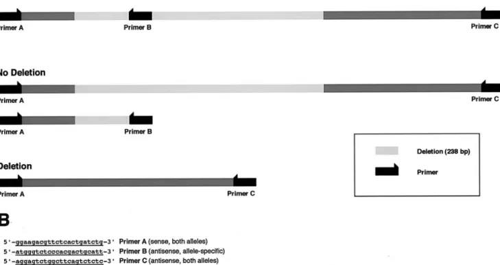
blood cells by the salting out method with modifications as de-scribed [8], or using the QIAmp DNA blood kit. Taq DNA poly-merase and deoxyribonucleotides were from Applied Biosystems (Norwalk, CT). Oligonucleotides were synthesized by Microsynth Inc. (Balgach, Switzerland); Spreadex EL 300 Wide-Mini S-100 gels were from Elchrom Inc. (Cham, Switzerland). To characterize the 238-bp deletion in intron 9, which corresponds to the H2 haplotype of the tau gene, the part of intron 9 containing the 238-bp deletion was amplified using primers as previously described [1]. The am-plicon was separated on a 2% agarose gel, dissolved, purified, and subjected to dideoxy sequencing. The presence of the intronic dele-tion was determined by allele-specific PCR. To investigate the pres-ence of the deletion in each sample, the allele-specific antisense primer tau-del9.asR, 5´-ATGGGTCTCCCACGACTGCATT-3´ (primer B), complimentary to parts of the intron sequence of the deletion, was designed. Using all three primers [del9F (A), tau-del19.asR (B), and tau-del19.R (C)] in the same reaction combined (multiplex PCR) or pairs of primers (A/B and A/C), the presence of the deletion could be determined for both alleles of the individ-ual investigated. Using the primer combination A/C, a fragment of 484 bp was obtained if the deletion was absent and a fragment of 246 bp if the intronic sequence was deleted. However, as the prob-ability of amplifying a fragment of 484 bp is theoretically lower
than the probability of amplifying a fragment of 246 bp, we also used the primer combination A/B. This PCR results in a fragment of 187 bp if the intronic fragment is not deleted (Fig. 1A). For group comparison, the Chi-square statistic was used.
Results
Histologically, all AgD cases were characterized by the presence of abundant ArGs in limbic areas, including sec-tor CA1 of the hippocampus, the ensec-torhinal cortex and the amygdala. Coiled bodies were mainly found in the white matter underneath cortical regions rich in ArGs. AT8 re-vealed various numbers of pretangle neurons in all AgD cases as previously described [16]. In all cases, α B-crys-tallin-stained ballooned neurons were predominantly found in the amygdala, corroborating earlier results [15]. Asso-ciated neurofibrillary tangles of the AD type were found in a density and distribution corresponding to early transento-rhinal or limbic Braak stages [Braak stage I, 17 (21.5%) cases; Braak stage II, 45 (57.0%) cases; Braak stage III, 17 (21.5%) cases] [4]. Some diffuse Aβ deposits were present in 40 (50.6%) cases, while the remaining cases were totally devoid of senile plaques. In three cases there were additional Lewy bodies and Lewy neurites. Vascular le-364
Fig. 1 A Length of the DNA sequence amplified by the primers A, B, and C in the case of the presence or absence of the intronic deletion of 238 bp. B Sequence alignment of the two DNA se-quences (presence and absence of the intronic 238 bp sequence)
sions (e.g., small lacunar infarcts and/or cribriform state of the basal ganglia) were found in 19 (24.1%) cases. All AgD cases were devoid of concomitant PSP and CBD pa-thology. The neuropathological findings of 24 cases have been reported in earlier studies [17]. According to the clinical records, 61 (77.2%) subjects with ArGs were re-ported to be demented.
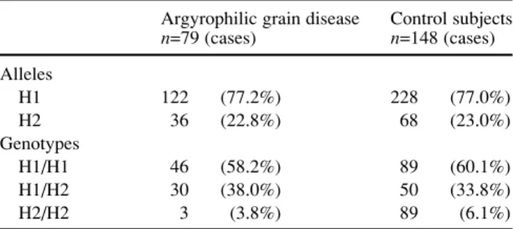
Figure 1B presents the sequence containing the 238-bp deletion as well as the localization of the deletion itself. Molecular analysis revealed the following tau genotype frequencies for the AgD cases: genotype H1/H1 46 pa-tients (58.2%), genotype H1/H2 30 papa-tients (38.0%), and genotype H2/H2 3 patients (3.8%). The tau genotype fre-quencies of the non-demented control group did not differ significantly from that of the AgD cases (P=0.672). Hence, among the AgD cases, the H1 allele was found in 77.2% and the H2 allele in 22.8%, respectively. These values did not differ significantly from those obtained in the non-de-mented control group (P=0.964) (Table 1).
Discussion
PSP and CBD are sporadic extrapyramidal syndromes characterized by predominant 4R tau deposition, and both have been shown to be associated with the tau H1 haplotype [1, 6]. The sporadic late-onset dementia AgD has recently been recognized as a 4R tauopathy [14, 18, 19]. Prelimi-nary data based on a limited number of cases opened the possibility that the tau H1 haplotype might also be over-represented in AgD [14]. To test the hypothesis whether the association of the tau H1 haplotype represents a com-mon feature acom-mong 4R tauopathies, we analyzed the tau haplotype in a cohort of 79 autopsy-confirmed AgD cases. In contrast to PSP and CBD [1, 6], no difference was found between the prevalence of the tau H1 haplotype or the H1/H1 genotype in subjects with AgD when compared to non-demented control cases.
The tau haplotype frequencies may vary when they are studied in different populations. The tau H1/H1 genotype is known to be very low in Norway (50%), while it is very high in Japan (>95%). In our study, both the AgD and the control cohorts are derived from the same urban-suburban area in a northwestern region of Switzerland. In the control cohort the frequencies of the tau H1 haplotype (77.0%)
and H1/H1 genotype (60.1%) are quite comparable with values reported in different US- and UK-based popula-tions [1, 6, 9, 14].
The present study suggests that there is no obligatory association between the tau H1 haplotype and the 4R tauo-pathies. Thus, the presence of the tau H1 haplotype seems not to be a prerequisite for the type of filamentous tau pro-tein deposited in 4R tauopathies. No association between the tau H1 or H2 haplotype has been found in Pick’s dis-ease [9, 11], a tauopathy characterized by the predominant deposition of 3R tau filaments.
PSP, CBD and AgD share pathological and biochemi-cal similarities with regard to an abnormal tau deposition. In all these disorders, predominantly 4R tau filamentous inclusions are found in neurons, astrocytes and oligoden-drocytes. A recent study further demonstrates a high preva-lence of ArGs in PSP and CBD cases [14]. In contrast, a high prevalence of PSP and CBD cases has not been ob-served in AgD cases, even in large cohorts of cases [5, 17]. Other differences, however, may confirm the separation of PSP and CBD from AgD as distinct clinicopathological entities. Clinically, PSP patients present with postural in-stability, supranuclear vertical gaze palsy, parkinsonism, pseudobulbar palsy and subcortical dementia. CBD pa-tients also exhibit motor symptoms, parkinsonism and eye movement abnormalities, and dementia has been reported as their presenting syndrome in the majority of CBD pa-tients. In contrast, although the clinical characteristics of AgD remain to be fully established, motor symptoms and parkinsonism are not characteristic features of AgD. Re-cent studies rather suggest that AgD patients present with dementia, and that personality changes and emotional im-balances may precede memory failure [17]. The different clinical presentation among 4R tauopathies is a function of the anatomical distribution of their pathology. Thus, PSP and CBD are characterized by a widespread tau pa-thology in various cortical and subcortical structures, in-cluding brain stem, while tau filamentous lesions in AgD are almost exclusively restricted to allocortical areas. Whether the tau haplotypes may affect the different topol-ogy of neurodegeneration among 4R tauopathies remains to be elucidated.
In conclusion, the present study further establishes AgD as a rather distinct pathological entity within the heteroge-neous group of tauopathies. Hence, the clinicopathologi-cal differences between the 4R tauopathies PSP and CBD on one hand and AgD on the other might be explained by genetic or non-genetic differences that are involved in the development of the tau protein dysfunction.
Acknowledgements This work was supported by the Basel Foundation for Dementia Research (to A.R.M. and M.T.) and by the Swiss National Science Foundation (SNSF) (A.R.M.; grant number 3200–049125.96/1). A.R.M. is supported by the Swiss Clinicians Opting for Research (SCORE), a grant number 3231-048896.96 of the SNSF. F.C is supported by the SNSF (grant num-ber 3100-068328). We thank Michelle Pfeifer for help with the manuscript.
365 Table 1 Tau haplotype and genotype frequency in
neuropatho-logically confirmed cases of argyrophilic grain disease and healthy control subjects
Argyrophilic grain disease Control subjects
n=79 (cases) n=148 (cases) Alleles H1 122 (77.2%) 228 (77.0%) H2 36 (22.8%) 68 (23.0%) Genotypes H1/H1 46 (58.2%) 89 (60.1%) H1/H2 30 (38.0%) 50 (33.8%) H2/H2 3 (3.8%) 89 (6.1%)
References
1. Baker M, Litvan I, Houlden H, Adamson J, Dickson D, Perez-Tur J, Hardy J, Lynch T, Bigio E, Hutton M (1999) Association of an extended haplotype in the tau gene with progressive supranuclear palsy. Hum Mol Genet 8:711–715
2. Braak H, Braak E (1987) Argyrophilic grains: characteristic pathology of cerebral cortex in cases of adult onset dementia. Neurosci Lett 76:124–127
3. Braak H, Braak E (1989) Cortical and subcortical argyrophilic grains characterize a disease associated with adult onset de-mentia. Neuropathol Appl Neurobiol 15:13–26
4. Braak H, Braak E (1991) Neuropathological stageing of Alz-heimer-related changes. Acta Neuropathol 82:239–259 5. Braak H, Braak E (1998) Argyrophilic grain disease: frequency
of occurrence in different age categories and neuropathological diagnostic criteria. J Neural Transm 105:801–819
6. Houlden H, Baker M, Morris HR, et al (2001) Corticobasal de-generation and progressive supranuclear palsy share a common tau haplotype. Neurology 56:1702–1706
7. Lee VM-Y, Goedert M, Trojanowski JQ (2001) Neurodegener-ative tauopathies. Annu Rev Neurosci 24:1121–1159
8. Miserez AR, Laager R, Chiodetti N, Keller U (1994) High prevalence of familial defective apolipoprotein B-100 in Switzer-land. J Lipid Res 35:574–583
9. Morris HR, Baker M, Yasojima K, et al (2002) Analysis of tau haplotypes in Pick’s disease. Neurology 59:443–445
10. Perrig-Chiello P, Perrig WJ, Stähelin HB, Krebs-Roubicek E, Ehrsam R (1996) Wellbeing, health and autonomy in old age: the Basel IDA Study (Interdisciplinary Aging Study). Z Geron-tol Geriatr 29:95–109
11. Russ C, Lovestone S, Baker M, Pickering-Brown SM, Ander-sen PM, Furlong R, Mann D, Powell JF (2001) The extended haplotype of the microtubule associated protein tau gene is not associated with Pick’s disease. Neurosci Lett 299:156–158
12. Saito Y, Nakahara K, Yamanouchi H, Murayama S (2002) Se-vere involvement of ambient gyrus in dementia with grains. J Neuropathol Exp Neurol 61:789–796
13. Togo T, Cookson N, Dickson DW (2002) Argyrophilic grain disease: neuropathology, frequency in a dementia brain bank and lack of relationship with apolipoprotein E. Brain Pathol 12:45–52
14. Togo T, Sahara N, Shu-Hui Y, Cookson N, Ishizawa T, Hutton M, De Silva R, Lees A, Dickson DW (2002) Argyrophilic grain disease is a sporadic 4-repeat tauopathy. J Neuropathol Exp Neurol 61:547–556
15. Tolnay M, Probst A (1999) Ballooned neurons expressing al-phaB-crystallin as a constant feature of the amygdala in argyro-philic grain disease. Neurosci Lett 246:165–168
16. Tolnay M, Spillantini MG, Goedert M, Ulrich J, Langui D, Probst A (1997) Argyrophilic grain disease: widespread hyper-phosphorylation of tau protein in limbic neurons. Acta Neuro-pathol 93:477–484
17. Tolnay M, Monsch AU, Probst A (2001) Argyrophilic grain disease: a frequent dementing disorder in aged patients. Adv Exp Med Biol 487:39–58
18. Tolnay M, Sergeant N, Ghestem A, Chalbot S, Vos RAI de, Jansen Steur ENH, Probst A, Delacourte A (2002) Argyro-philic grain disease and Alzheimer’s disease are distinguished by their different distribution of tau protein isoforms. Acta Neuropathol 104:425–434
19. Zhukareva V, Shah K, Uryu K, Braak H, Del Tredici K, Sun-darraj S, Clark C, Trojanowski JQ, Lee VM (2002) Biochemi-cal analysis of tau proteins in argyrophilic grain disease, Alz-heimer’s disease, and Pick’s disease. A comparative study. Am J Pathol 161:1135–1141