A look ahead: PET/MR versus PET/CT
Gustav K. von Schulthess&Heinz-Peter W. SchlemmerPublished online: 23 December 2008
# Springer-Verlag 2008 Abstract
Introduction Integration of positron emission tomography (PET) and magnetic resonance (MR) has become a topic of increasing interest to the imaging community over the past two years.
Objectives In this text, the authors attempt to distinguish facts from fiction concerning such integrated systems. Analysis of existing information of combined imaging on existing brain PET/MR systems and imaging experience with PET-computed tomography (CT) is reviewed. Various types of system integration of PET and MR are discussed with completely independent systems on one hand and completely integrated systems with the possibility of simul-taneous data acquisition on the other hand. Furthermore, it is discussed, what simultaneous data acquisition with nuclear imaging systems combined with MR or CT really means, as technical simultaneity may not be relevant in light of the pharmacokinetics of the nuclear tracers used.
Discussion The authors conclude that combining PET/MR is an interesting research endeavor with uncertain outcome. They argue that, while completely simultaneous brain applications are of research interest immediately, clinical applications do not currently warrant the construction of fully integrated systems. Systems adjacent to each other, where imaging tables are linked with a patient “shuttle”
thereby requiring only patient translation but no reposition-ing, may be a good start to assess the value of integrated PET/MR.
Keywords Integrated imaging . Dual modality imaging . PET/CT . MRI
Introduction
Discussion on integration of positron emission tomography (PET) and magnetic resonance (MR) has become fashion-able in the last two years, and there is much speculation with relatively few hard facts available. Combining PET and MRI is an interesting undertaking with uncertain outcome: a true research project encompassing basic science, technologic and clinical questions. The aim of this paper is to summarize existing knowledge on this topic and with a specific focus on human applications.
Proof of concept of integrated imaging with single-photon emission computed tomography (SPECT)-computed tomography (CT) [1,2] was provided in the late 1980s and with PET/CT [3] in 1998. In both cases, the more functional imaging methods of SPECT and PET are com-bined with the more anatomical CT imaging information within a single examination. With the advent of clinical PET/CT in early 2001, it was quickly shown that integration of PET and CT into a single system is advantageous and synergistic, as it provides anatomic referencing and at the same time permits transmission correction in a much shorter time [4,5], thus making PET/CT a more accurate [6] and faster [7] imaging modality than PET alone, CT alone, and even PET and CT read side-by-side [8–10]. Since its clinical introduction in early 2001, it has become evident that imaging with PET/CT will have an impact on
DOI 10.1007/s00259-008-0940-9
G. K. von Schulthess (*)
Nuclear Medicine, Department of Medical Radiology, University Hospital,
Raemistr. 100,
CH-8091 Zurich, Switzerland e-mail: [email protected] H.-P. W. Schlemmer
Department of Radiology, University Hospital, Tuebingen, Germany
therapeutic management in 20–50% of the examined oncology cases [11]. Thus, this decade in imaging is clearly the age of PET/CT and, to a lesser extent, SPECT-CT.
As MR has some advantages over CT, it is obvious to evaluate, whether a combined PET/MR system may have certain advantages over PET/CT. In order to design proper PET/MR systems, it makes sense to use the experience of PET/CT and try to look at the specifics of MR technology interacting with PET technology and at their respective clinical applications. The conjectures will be mostly clini-cally oriented, as the authors are both clinicians and they look to physicists, engineers, and the equipment manufac-turers to provide technological solutions.
Facts
Image integration of two modalities like PET and MR or PET and CT can be achieved in different ways as will be analyzed in more detail in the next section. Essentially integration can be achieved by software-based fusion or by integration of the hardware of the two devices. Other issues relevant to the ensuing discussion are:
1. MRI has become the clinical imaging modality of choice in most neuro- and musculoskeletal applications. 2. MRI imaging from the head and neck to the pelvic floor has its clinical role mainly in focal imaging of the head and neck, the liver and other upper abdominal organs as well as the pelvis.
3. Despite MR, CT has remained the method of choice in staging patients from the head and neck to the pelvic floor region; reasons include speed, robustness, and availabili-ty of CT data acquisition and higher cost of MR. 4. CT has remained the method of choice in most
cross-sectional chest imaging.
5. MR offers functional imaging aspects not available through CT.
6. Depending on how CT is used, it can contribute a sub-stantial radiation burden to an integrated modality exam. A low-dose CT scan in PET/CT yields a radiation expo-sure of 2–3 mSv, while some CT protocols can yield effective doses of up to 20 mSv [12]. This is mainly the case in CT perfusion imaging because repetitive imaging of the body region evaluated is needed to obtain the bolus tracking data. FDG-PET imaging, for example, causes effective dose levels on the order of 7–15 mSv, depending on the injected dose.
What have we learned from PET/CT?
In clinical practice, we have learned several important things. 1. Inline PET/CT systems are quite adequate for most
body applications.
2. The most important clinical applications cover areas, in which MR has not replaced CT, i.e., body oncology applications, parts of infection imaging, and cardiac imaging.
3. In areas, where MR excels clinically, i.e., neuro- and musculoskeletal applications as well as some focal body applications in the upper abdomen, pelvis, and head and neck, both MR and PET/CT are frequently performed both.
4. Comparison of separate PET/CT and MR data using “mental” rather than software fusion is readily possible because, with the CT of PET/CT, the PET data are anatomically “framed”. Thus, there are enough ana-tomic landmarks on both examinations, that computer assisted image fusion is not necessary, when comparing PET/CT with MR data.
5. Consecutive imaging on tightly coupled integrated imaging systems may be inefficient from a workflow perspective as either imaging system idles during parts of the examination. Therefore, integrated imaging proce-dures tend to be costly.
6. On the other hand, multimodality imaging performed with separate imaging devices requires patient reposi-tioning, which results in a greater risk of voluntary or involuntary patient movements between procedures. If images are misaligned, time-consuming side-by-side image interpretation or software-based image fusion may be required. Furthermore, patients need to be positioned and repositioned twice rather than once between scans, which is time consuming.
7. There are some physiologic processes such as tissue perfusion, which may change so rapidly over time that consecutive data acquisition of PET and MR data may yield inaccurate results and fully integrated imaging is necessary. However, this is of less importance for current clinical applications.
Integrated PET/MR imaging: systems design issues Recently, the feasibility of simultaneous PET/MR imaging of the human head has been demonstrated by using a novel avalanche photodiode-based PET detector technology, which is insensitive even to high magnetic fields [13]. For simultaneous PET/MR imaging of the human brain, a detector ring was constructed, which fits in a standard clinical 3-Tesla MRI scanner (TRIO, Siemens Medical Solutions) and contains a standard bird cage transmit/ receive head coil [14]. All PET detector components (amplifiers, resistors, shielding material, and housing) were selected and dimensioned in order to exhibit minimal interference with the static and gradient magnetic fields as well as the radiofrequency irradiation of the MRI system.
Initial human examinations have proven that the perfor-mance neither of the PET nor the MRI scanner are qualitatively compromised by each other compared to separate systems [15]. Animal MR-PET systems have also been described which allow quantitative assessment of tracer uptake [16, 17]. With these experimental systems, integrated PET/MR systems appear technically feasible. An evaluation whether they might be of clinical or research use is warranted. To better define the potential uses of a PET/ MR system, we have to first look at the various technical options of how PET and MR can be integrated and second, at some pharmacokinetic issues, relevant in PET imaging with radiotracers.
First, there are three major ways to technically integrate PET and MR systems:
A. Separate imaging in devices placed far from each other. The patient has to get off one and onto the other imaging system during the examination process. This approach requires integration through software-based image fusion and leaves full flexibility regarding the placement of the two imaging systems and temporal flexibility in the examination sequence of the patient. The systems can also be used independently as single imaging devices without interfering in their respective operation.
B. Sequential imaging by systems which are linked by a patient “shuttle”. The patient does not have to get off the examination table of one device to get onto the table of the second device as the transfer is accomplished by the“shuttle”. Such devices provide “hardware-fused” data. Sequential imaging designs (B) can be further divided into 3 categories:
B1 Systems distant from each other, patients shuttled, e.g., in restraining mattresses; B2 Systems are in adjacent rooms and “shuttle”
links systems through a door;
B3 Systems in one room with patient “shuttle” between both devices.
C. Fully integrated systems with technically simultaneous data acquisition. Neither patient nor table motion is required when imaging single fields of view
It is of note that all currently available PET/ and SPECT-CT systems are of type B3. In these devices, the examination table is the patient shuttle, and the distance between the detection fields of view of the two imaging systems is in the range of 50–100 cm. Similar solutions to B2 and B3 exist in operating theaters where the operating room table is linked to imaging devices such as MR scanners for intraoperative imaging. The recently described experimen-tal head PET/MR system is of type C [15]. In PET/CT and SPECT-CT, such systems are also conceivable, but would
require development of detectors which can simultaneously detect gamma rays and CT X-rays and discriminate between them.
Second, we want to look at pharmacokinetics, as this identifies whether and when simultaneity of imaging is required. What does “simultaneous” really mean when dealing with a combined PET/CT or PET/MR imaging procedure. In Nuclear Medicine procedures, a radioactive tracer is injected and—depending on its pharmacologic properties—it distributes to the sites of interest. Hence, pharmacokinetic behavior is critical, when we talk about simultaneity. There are PET-tracers like O-15 water, which enter and exit the volume of interest very quickly (within minutes), while there are other tracers, such as ammonia (N-13H3), which enter the cells quickly (also within a few minutes) and then remain there in a pseudo-steady-state (PSS) in proportion to local perfusion. Finally, there are tracers, which build up in the structures of interest over an extended period of time. In the case of fluorodeoxyglucose (18F-FDG), a PSS distribution is reached after around 45 min and lasts for 30–60 min. Obviously, in a setting, where no such PSS is reached, physically simultaneous imaging may be relevant. However, when the distribution of a radiopharmaceutical reaches a PSS, the imaging protocol, i.e., the injection of radiotracer and possibly also the MR contrast agent, as well as the subsequent imaging steps in MR and PET have to be driven by considering the pharmacokinetics of the tracer and contrast agents used. For example, in a perfusion experiment of the heart or brain with ammonia, it is important that the injection is done while imaging the patient in the MR scanner because the tracer distribution will reflect the state of perfusion at the time of the MR examination. This is particularly important, if a pharmacologic stress test, i.e., with Dipyridamole or Adenosine is done. PET scanning can be done later, as the perfusion state of the patient during the “adenosine stress” MR scan was “frozen” by the pharmacokinetic properties of the radiotracer ammonia. With a study using FDG, the situation is yet different, and the tracer may have to be injected even prior to the MR scan. Once, the tracer distribution reaches a PSS, MR and PET imaging can be acquired in sequence during the PSS phase, or assuming that the patient state is not changing during the initial uptake phase, MR can be done during the FDG uptake phase assuming the patient state is not changing.
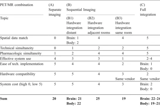
With this insight, we can discuss options A, B, and C defined above, and for this, refer to Table 1. The options B1, B2, and B3 are considered separately. Note that the sum total given in this table in the last row is of very limited value as there is no way to attach appropriate relative weights to the different categories. Furthermore, the categories are not independent. Some comments are in
order regarding the relative assessment of the five solutions defined above:
1 Spatial data match: the shorter the time difference between the data acquisitions on the two systems the better the spatial match of the data. Here, full integration is ideal. It is assumed that the patient“shuttling” times between scanners in adjacent rooms are similar to those between scanners within the same room. This makes sense because, in one case, the distance between the detection devices is 2–3 m, in the other, 5–8 m, which is hardly relevant. Obviously, this is only true if shuttling devices are efficient.
2 Technical simultaneity can only be achieved in systems of type C, but transport times between systems are much shorter, when they are adjacent (B2 and B3). Thus, these options are advantageous compared to options A and B1.
3 Pharmacological simultaneity sometimes mandates si-multaneous data acquisition in PET and MR (thus five points), but—in light of pharmacokinetics as discussed above—in many current clinical applications, it is rather contraindicated (thus two points). Therefore, in such applications, flexibility in designing the proper proto-cols driven by pharmacological considerations make adjacent linked systems preferable.
4 Effective system use is a somewhat complex issue. Separate systems have the advantage of full flexibility, but the disadvantage, that for all patients, who get both exams, placing and removing the patient from the examination table occurs twice, and thus, patient
positioning in separate imaging requires twice as long as for integrated imaging. The sum of these two times per system is on the order of 15 min. During this time, the systems are idling. If the imaging time in the single device is less than this time, the changing time becomes the rate-limiting step. For imaging times longer than the times for re-/positioning and removing the patient, and for imaging times, which are very different for each of the integrated devices, independent systems are advan-tageous. Thus, with typical effective imaging times for PET of 15 min, CT of 5 min, and MR of 30 min or more, PET/MR systems in independent rooms could frequently be used more effectively than when in a single room. The situation for PET/CT is different, as particularly the CT examination is much faster. This is not an in-depth analysis but rather states the relevant issues, when considering workflow.
5 Ease of technical integration is related to the additional engineering costs incurred for system integration. For options A and B1 this is minimal (software and posi-tioning devices); B2 requires a patient shuttle and B3 a patient shuttle plus additional shielding measures of the PET scanner from the main magnetic field of the MR scanner. Option C requires complete re-engineering of the detector systems for brain imaging and additional re-engineering of the MR system. These measures are all very costly, and personal experience of one of the authors (GvS) with an interventional magnet with central surgical access suggests that for magnet redesign for a body system, development cost may be over EUR 7 M or€ 7 M.
Table 1 Comparison of differ-ent options (A–C) of integrat-ing PET and MR usintegrat-ing grades from 0 to 5 (0 worst, 5 best)
PET/MR combination (A) Separate imaging (B) Sequential Imaging (C) Full integration Topic (B1) Hardware integration distant (B2) Hardware integration adjacent rooms (B3) Hardware integration same room
Spatial data match 0 Brain: 1 4 4 5 Body: 2
Technical simultaneity 0 1 2 2 5 Pharmacologic simultaneity 1 2 4 4 5 Effective system use 4 3 3 1 2–4 Ease of tech. implementation 5 4 4 2 Brain: 1 Body: 0 Hardware compatibility 5 5 4 3 2
Same vendor Same vendor System cost (high 0, low 5) 5 5 4 3 Brain: 2
Body: 0 Sum 20 Brain: 21 25 19 Brain: 22–24
6 Hardware compatibility is again related to the degree of integration. While options A and B1 do not require any compatibility except for DICOM image format, B2, B3, and, in particular, option C will likely have to be provided by a single vendor, at least for a body-imaging system. The overall assessment suggests that integrating a system, where the two components are in adjacent but independent rooms, is preferable when technical issues and costs are taken into consideration. If engineering funds are not an important restraint, then the fully integrated system has its attractions, and research into this option is highly desirable.
7 Overall system cost is directly related to points 5 and 6. In summary, a deployment of a PET/MR system has to take into account various technical, physiological, and design aspects. While full integration even of body systems may be the final goal, at this point, the fully integrated brain imaging systems allow experience to be gained with this new technology, while experience with non-neuro applica-tions of PET/MR body imaging (but even including some brain applications) may be gained with systems integrated to a lesser degree. Table1suggests that the preferred design for body applications is a“shuttle” linked PET/MR scanner consisting of two independent scanners in adjacent rooms, as it combines the advantage of a closely linked system with maximum flexibility of use.
Integrated PET/MR: attenuation correction
Variable photon attenuation in tissue is a cause of image degradation in all Nuclear Medicine emission imaging procedures including PET. Accordingly, measurement of the spatial distribution of attenuation is a prerequisite to reconstruct PET images rendering true tracer distributions, thereby permitting semiquantitative evaluation of images by measuring standard uptake values (SUV). In PET/MR systems, two potential sources of photon attenuation have to be considered:
(1) PET photon attenuation caused by the MR gradient and RF coils with their electronic components: This problem can be solved by acquiring a CT image of the head coil once and integrating the data into the image reconstruction algorithm. The problem only occurs in option C with fixed coils, but may be a problem with surface coils in all types of integrated systems, since removing of surface coils likely requires patient re-positioning, which is undesirable.
(2) Attenuation correction is a caused by patient tissue is relatively straightforward in PET/CT examinations, as non-enhanced whole-body CT scans are an integral part of the examination. CT scans (around 100 kVp data) can be easily converted into an attenuation
mu-map at 511 kVp for PET using a bilinear function [5], which can then be incorporated into the reconstruction algorithm for PET thereby correcting for patient attenuation. MRI, on the other hand, yields no information about photon attenuation but rather tissue proton densities and magnetic relaxation times. Par-ticularly, bone and air appear with similar low signal intensity on MR images despite having the highest and lowest photon attenuation coefficients, respectively. Attenuation correction of PET/MR must therefore be based on an indirect voxel-by-voxel assignment of MR signal intensities to empirical values of photon attenuation coefficients [18]. Alternative methodologi-cal approaches are currently under development for calculating so called “pseudo-CT images”. One ap-proach uses image segmentation [19]; another a combination of local pattern recognition with atlas registration for additionally capturing global variations of anatomy [20]. The latter approach has been shown to be successful for brain MR/PET images by suc-cessfully calculating artefact-free images and quanti-tative SUV values. However, the brain is a relatively homogeneous structure and using the same procedure in body PET/MR may be more difficult. Despite these initial successes, an integrated PET/MR for body imaging would probably be based on sequential partly integrated PET/CT and an MR systems for the time being.
Advantages of PET/CT and PET/MR
PET enables quantitative and semiquantitative evaluation of tracer uptake kinetics of any small molecule which can be labeled with standard positron emitters. Clinically, by far, the most frequently used radiopharmaceutical is FDG, and FDG-PET/CT has been proven to be an important tool for diagnosis, staging, and therapy monitoring. CT is a fast and standardized method which performs well even in uncoop-erative patients, reliably yielding highly standardized image data even in examinations repeated on different days [21]. In recent years, PET/CT has established itself as an imaging method that works robustly in clinical routine. Long-standing clinical experience exists for both modalities PET and CT. In a PET/CT examination, PET and CT are performed in rapid sequence, and as CT is very fast and also provides the data for PET attenuation correction, a PET/CT examination is typically 25–30% less time con-suming than the acquisition of an attenuation-corrected PET scan.
PET/MR on the other hand is still in its early stages of development and several technological and methodical issues have to be addressed before PET/MR can establish itself as a clinical routine tool. As discussed, the technical
issues are related to the incompatibility of current MR and PET systems mandating new developments in MR and PET systems mandating technology as well as related to the currently unresolved issue of PET attenuation correction with MR image data. The latter is critical particularly for therapy monitoring. Consequently, fully integrated PET/ MR at the present stage of development basically represents a research tool and so far is only available as a dedicated brain scanner.
From a scientific and clinical point of view, a fully integrated PET/MR may have some advantages over PET/ CT:
1. The superior soft tissue contrast of MRI allows better anatomical visualization of soft tissue structures and bone marrow than CT.
2. Simultaneous image acquisition enables temporal co-registration of dynamic PET data acquisition and morphologic/functional MR data. A variety of func-tional information can be acquired by MR, e.g., perfusion (microvessel density, vessel leakage, etc.), diffusion (cell density, microstructure, etc.), and me-tabolism (cell death, proliferation, etc.). Moreover, brain activation can be studied by functional MRI (fMRI). A vast array of applications mostly relevant in research can be conceived with such a system [14,15], but even PET/MR systems of type B2 and B3 will be quite useful in view of the discussion on the relevance of tracer pharmacokinetics given above.
3. Some studies comparing whole-body MR with PET/CT have shown potential advantages of MR particularly regarding the early detection of brain-, liver- and bone marrow metastases [22–24]. If these data are con-firmed, PET/MR can be assumed to have an advantage over PET/CT in some oncology applications. To benefit from these potential advantages, full PET/MR system integration is not required.
4. In fully integrated systems, MRI could also be used to provide a gating signal in addition to imaging. This is however only advantageous, when MR data acquisi-tion, MR gating data, and PET data are acquired simultaneously and for similar amounts of time, as otherwise MR is just an expensive device to provide gating information for PET.
5. MR does not expose the patient to ionizing radiation. Thus, replacing CT by MR in such an integrated system is expected to result in radiation dose reductions depending on the imaging protocol. This is mainly relevant in pediatric applications.
6. Fully integrated PET and MR imaging reduces the scan time for the patient compared to sequential scanning. Summarizing and hypothesizing on the limited data available, PET/MR in its various deployment options will
be a valuable tool, which in some applications is likely to be superior to PET/CT. Clinically, these will be most likely in brain- and musculoskeletal imaging. However, in the brain, current clinical applications will be equally as effective using software rather than hardware image integration. In musculoskeletal imaging, PET/MR may be of major clinical interest, as MR is excellent and PET has two routine tracers available for musculoskeletal imaging, which have been shown to provide clinically relevant data: FDG and 18F-flouride for PET-bone imaging. Integrated PET/MR for brain imaging is likely to be a formidable research tool for the foreseeable time.
Expertise and training:
As an increasing number of PET/CT and in the future possibly also PET/MR scanners are installed, there will accordingly be an increasing demand for imaging special-ists in reading the imaging data from such combined systems. Appropriate training in Radiology and Nuclear Medicine is a prerequisite for successful clinical interpre-tation and also for performing research on these systems. The most effective way of reading the images is by doubly board certified experts, but currently, in many sites, a nuclear physician and a radiologist read the images separately with ensuing discussion of the results. It is not acceptable to read CT or MR only for anatomic correlation of PET findings, as important findings can be missed, which have to be an integral part of the diagnosis. Efforts by the US and European [25] societies are under way for optimized training, and there are also fellowship training programs available, which enable the relevant expertise to be obtained [
http://www.nuk.usz.ch/german/LehreUndFor-schung/Fellowship+Program/TrainingProgram/].
Conclusions
At the current stage of technological development, PET/MR systems based on two imaging systems in close proximity and connected by some kind of a patient shuttle together with software tools for image integration including methods for MR-based attenuation correction appear optimal for evaluating the clinical value of whole-body PET/MR. Likely, the PET part of such systems is actually a PET/ CT, which solves the yet unresolved PET attenuation correction issues with MR data. Fully integrated systems have several scientific and possibly also clinical advan-tages. Two types of integrated PET/MR for head imaging research studies are already in use. Fully integrated whole-body PET/MR systems will however need considerable more technological and methodological analysis and devel-opment before it can be decided whether such systems are
useful in settings beyond those of research-oriented academic centers. Prime clinical applications for PET/MR systems are likely in the brain and the musculoskeletal system, but interesting applications in other body areas can also be envisioned.
Conflict of interest GvS is a conlsultant to GE Healthcare and a Board member of Timaq Inc.. HS declares no conflict of interest.
References
1. Seo Y, Mari C, Hasegawa BH. Technological development and advances in single-photon emission computed tomography/com-puted tomography. Semin Nucl Med. 2008;38(3):177–98. May. 2. Hasegawa BH, Gingold EL, Reilly SM, Liew SC, Cann CE.
Description of a simultaneous emission-transmission CT system. Proc SPIE. 1990;1231:50–60.
3. Beyer T, Townsend DW, Brun T, Kinahan PE, Charron M, Roddy R, et al. A combined PET/CT scanner for clinical oncology. J Nuc Med. 2000;41:1369–79.
4. Kinahan PE, et al. Attenuation correction for a combined 3D PET/ CT scanner. Med Phys. 1998;25:2046–53.
5. Burger C, Goerres GW, Schoenes S, Buck A, Lonn AHR, von Schulthess GK. PET attenuation coefficients from CT images: experimental evaluation of the transformation of CT- into PET 511 keV attenuation coefficients. Europ J Nucl Med. 2002;29(7): 922–7.
6. Hany TF, Steinert HC, Goerres GW, Buck A, von Schulthess GK. PET diagnostic accuracy: improvement with in-line PET/CT System: initial results. Radiology. 2002;225:575–81.
7. von Schulthess GK. Cost considerations regarding an integrated CT-PET system. Eur Radiol. 2000;Suppl 3:377–80.
8. Lardinois D, Weder W, Hany TF, Kamel EM, Korom S, Seifert B, et al. Integrated PET/CT imaging improves staging of non-small-cell lung cancer. N Engl J Med. 2003;348(25):2500–7.
9. Czernin J, Allen-Auerbach M, Schelbert HR. Improvements in cancer staging with PET/CT: literature-based evidence as of September 2006. J Nucl Med. 2007;48(Suppl 1):78S–88S. Jan. 10. Hillner BE, Siegel BA, Liu D, Shields AF, Gareen IF, Hanna L, et al.
Impact of positron emission tomography/computed tomography and positron emission tomography (PET) alone on expected management of patients with cancer: initial results from the National Oncologic PET Registry. Clin Oncol. 2008;26(13):2155–61. May 1.
11. Gambhir SS, Czernin J, Schwimmer J, Silverman DH, Coleman RE, Phelps ME. A tabulated summary of the FDG PET literature. J Nucl Med. 2001;42(5 Suppl):1S–93S. May.
12. Brix G, Lechel U, Glatting G, Ziegler SI, Münzing W, Müller SP, et al. Radiation exposure of patients undergoing whole-body dual-modality 18F-FDG PET/CT examinations. J Nucl Med. 2005;46 (4):608–13. Apr.
13. Pichler BJ, Judenhofer MS, Catana C, Walton JH, Kneilling M, Nutt RE, et al. Performance test of an LSO-APD detector in a 7-T MRI scanner for simultaneous PET/MRI. J Nucl Med. 2006;47 (4):639–47.
14. Schlemmer HP, Pichler B, Wienhard K, et al. Simultaneous MR/ PET for Brain Imaging: first Patient Scans. J Nucl Med. 2007;48 (Suppl):152.
15. Schlemmer HP, Pichler BJ, Schmand M, Burbar Z, Michel C, Ladebeck R, et al. Simultaneous MR/PET imaging of the human brain: a feasibility study. Radiology. 2008;248:1028–35. 16. Judenhofer MS, Catana C, Swann BK, Siegel SB, Jung WI, Nutt
RF, et al. PET/MR images acquired with a compact MR-compatible PET detector in a 7-T magnet. Radiology. 2007;244 (3):807–14.
17. Judenhofer MS, Wehrl HF, Newport DF, Catana C, Siegel SB, Becker M, et al. Simultaneous PET-MRI: a new approach for functional and morphological imaging. Nat Med. 2008;14(4):459– 65. Apr.
18. Beyer T, Weigert M, Quick HH, Pietrzyk U, Vogt F, Palm C, et al. MR-based attenuation correction for torso-PET/MR imaging: pitfalls in mapping MR to CT data. Eur J Nucl Med Mol Imaging. 2008;35(6):1142–6.
19. Zaidi H, Montandon ML, Slosman DO. Attenuation compensation in cerebral 3D PET: effect of the attenuation map on absolute and relative quantitation. Eur J Nucl Med Mol Imaging. 2004;31 (1):52–63.
20. Hofmann M, Steinke F, Scheel V, Charpiat G, Farquhar J, Aschoff P, et al. MR-based attenuation correction for PET/MR: a novel approach combining pattern recognition and atlas registration. J Nucl Med. 2008; (in press).
21. Carroll TJ, Teneggi V, Jobin M, Squassante L, Treyer V, Hany TF, et al. Absolute quantification of cerebral blood flow with magnetic resonance, reproducibility of the method, and comparison with H2 (15)O positron emission tomography. J Cereb Blood Flow Metab. 2002;22(9):1149–56. Sep.
22. Antoch G, Vogt FM, Freudenberg LS, Nazaradeh F, Goehde SC, Barkhausen J, et al. Whole-body dual-modality PET/CT and whole-body MRI for tumor staging in oncology. JAMA. 2003;290 (24):3199–206. Dec 24.
23. Muller-Horvat C, Radny P, Eigentler TK, Schafer J, Pfannenberg C, Horger M, et al. Prospective comparison of the impact on treatment decisions of whole-body magnetic resonance imaging and computed tomography in patients with metastatic malignant melanoma. Eur J Cancer. 2006;42(3):342–50.
24. Pfannenberg AC, Aschoff P, Eschmann SM, Plathow C, Eigentler TK, Garbe C, et al. Prospective comparison of 18F-fluorodeoxy-glucose positron emission tomography/computed tomography and whole-body magnetic resonance imaging in staging of advanced malignant melanoma. Eur J Cancer. 2007;43(3):557–64. 25. Bischof Delaloye A, Carrió I, Cuocolo A, Knapp W, Gourtsoyiannis
N, McCall I, et al. White paper of the European Association of Nuclear Medicine (EANM) and the European Society of Radiology (ESR) on multimodality imaging. Eur J Nucl Med Mol Imaging. 2007;34(8):1147–51. Aug.