HAL Id: hal-02713265
https://hal.inrae.fr/hal-02713265
Submitted on 1 Jun 2020
HAL is a multi-disciplinary open access
archive for the deposit and dissemination of
sci-entific research documents, whether they are
pub-lished or not. The documents may come from
teaching and research institutions in France or
abroad, or from public or private research centers.
L’archive ouverte pluridisciplinaire HAL, est
destinée au dépôt et à la diffusion de documents
scientifiques de niveau recherche, publiés ou non,
émanant des établissements d’enseignement et de
recherche français ou étrangers, des laboratoires
publics ou privés.
protein specific to the mouse epididymis
Norbert B. Ghyselinck, Isabelle Dufaure, Jean-Jacques Lareyre,
Marie-Geneviève Mattei, Jean-Pierre Dufaure
To cite this version:
Norbert B. Ghyselinck, Isabelle Dufaure, Jean-Jacques Lareyre, Marie-Geneviève Mattei, Jean-Pierre
Dufaure. Structural organization and regulation of the gene for the androgen-dependent glutathione
peroxidase-like protein specific to the mouse epididymis. Molecular Endocrinology -Baltimore-,
En-docrine Society, 1993, 7 (2), pp.258-272. �10.1210/mend.7.2.8469239�. �hal-02713265�
Structural
Organization
and
Regulation
of the Gene for the
Androgen-Dependent
Glutathione
Peroxidase-Like
Protein Specific to
the Mouse Epididymis
Norbert
B. Ghyselinck,
lsabelle
Dufaure,
Jean-Jacques
Lareyre,
Nicole
Rigaudibre,
Marie-Genevieve
Mattei,
and
Jean-Pierre
Dufaure
Laboratoire de Biologic Cellulaire
Centre National de la Recherche Scientifique URA 360
Universitb Blaise Pascal
63177 Aubikre Cedex, France
Centre de Gkktique
Mkdicale (M.-G.M.)
lnstitut National de la Santa! et de la Recherche Medicale U-242
H6pital d’Enfants de la Timone
18385 Marseille Cedex 5, France
Genomic
clones containing
the gene for the gluta-
thione
peroxidase-like
androgen-regulated
murine
epididymal
protein of 24 kilodaltons
(arMEP24)
were
isolated.
A 9-kilobase
DNA fragment
was sequenced
and found to contain
the entire coding
region
of the
gene, which
is divided
into five exons. The exact
sizes and boundaries
of the exon blocks were de-
duced by comparison
with the cDNA sequence.
One
major and four weak transcription
initiation
sites in
the epididymis
were localized
by primer
extension.
The promoter
of the gene does not contain
a con-
ventional
TATA
box immediatly
up-stream
of the
start site; rather,
the sequence
TATCA
occurs
at
residue
-35. Two CAAT boxes in opposite
orienta-
tion and two putative
binding
sites for the transcrip-
tion factor
Spl
were
identified
up-stream
of the
TATA-like
box. To localize
the cis-acting
sequences
responsible
for androgen
regulation
of expression,
fragments
of the arMEP24
gene
promoter
region
were cloned
in front of the luciferase
(LUC) reporter
gene and cotransfected
with an androgen
receptor
expression
vector into CV-1 cells in a transient
as-
say. LUC activities
of CV-1 cells grown
in the pres-
ence of various concentrations
of Sa-dihydrotestos-
terone were compared
to LUC activities
of untreated
controls.
The DNA fragment
containing
up to 200
nucleotides
up-stream
from the major transcription
start site was sufficient
for the full promoter
activity,
but not for the responsiveness
to androgen
induc-
tion. Depending
on the Sa-dihydrotestosterone
con-
centration,
a 2- to 4-fold
induction
of LUC activity
0e&3-8809/93/0258-0272$03.00/0 Molecular Endocrinology
Copyright 0 1993 by The Endocrine Society
was found if a -1797
to -167
arMEP24
gene frag-
ment was used linked to the reporter
gene driven by
either the homologous
promoter
or the heterologous
thymidine
kinase
promoter.
Two or three copies of
the imperfect
palindromic
sequence
TGlTGAgag-
AGAACA,
found
at position
-896
to -882
in the
gene
and ressembling
the consensus
steroid
hor-
mone-responsive
element,
are able to confer andro-
gen
regulation
to the thymidine
kinase
promoter
independently
of their
orientation.
These
findings
support
evidence
that transcriptional
regulation
of
the
arMEP24
gene
occurs
via
the
sequence
TGTTGAgagAGAACA.
Homologies
found in the se-
quence
up-stream
of the promoter
with several pu-
tative binding
sites for erythroid-specific
trans-act-
ing regulatory
proteins
are discussed.
Finally, the
arMEP24
gene is located
by in situ hybridization
in
the [A2-A41
region
of mouse chromosome
13. (Mo-
lecular
Endocrinology
7: 258-272,
1993)
INTRODUCTION
We previously described
an androgen-dependent
pro-
tein (arMEP24)
specific to the caput epididymidis
of the
mouse (1, 2). It constitutes
the major synthesized
pro-
tein of this organ, where it is secreted
into the epididy-
mal fluid and binds to the spermatozoa
(3, 4). The
amino acid sequence
of the polypeptide
has been de-
termined
(5). It shows high homology with the glutathi-
one peroxidase
(GSHPx) (6, 7) which is an enzyme of
considerable
importance
for several reasons.
First, it
has a protective
role in removing
lipid hydroperoxides
258
and H202 formed during normal oxidative
metabolism,
principally in the red blood ceils (8) but also in other
tissues. Secondly, in certain tissues (e.g. platelets, leu-
kocytes,
and lungs), GSHPx
reduces,
and thereby
cleaves, the hydroperoxi-fatty
acids formed by the ac-
tion of lipoxygenases
on lipid fatty acids; this generates
the immediate precursors
of the leukotrienes,
which are
involved in inflammatory
responses
(9). Finally, mouse
spermatozoa
and epididymal fluid have GHSPx activity,
which is the major protective
system against oxidative
damage (10). This could maintain
membrane
flexibility
and prevent the premature
acrosome
reaction
that
would otherwise
occur (6).
In previous studies we have shown
that arMEP24
mRNA expression
is androgen
responsive
(1, 2, 11).
Androgen-induced
expression
of genes is generally me-
diated through
the androgen
receptor
(AR), which has
been recently cloned (12-14).
The AR is a member of
the superfamily of ligand-inducible
transcription
factors,
which includes steroid, thyroid hormone,
and retinoic
acid receptors
(15-l 7). These
ligand-inducible
tran-
scription
factors can bind to specific DNA sequences
and regulate
the transcriptional
activity of their target
genes (17, 18). The DNA structures
to which steroid
receptors
bind are imperfect
palindromic
sequences.
The consensus
sequence of the responsive
element for
glucocorticoid
and
progesterone
receptors
is
GGTACAnnnTGTTCT
(17). Although
the expression
of
many genes is known to be regulated
by dihydrotes-
tosterone
(DHT), well defined androgen-responsive
ele-
ments (ARE) interacting
with the DHT-AR complex have
not yet been clearly identified
(19). The best known
example
is the mouse mammary tumor virus (MMTV)
promoter, which is able to confer androgen-responsive-
ness to a reporter gene (20-22).
In the C3(1) gene (23,
24) the first intron is also able to bind the AR (19, 25)
and confer weak androgen
responsiveness
to the thy-
midine kinase (tk) promoter
(25, 26). More recently, a
new functional ARE has been identified in the promoter
of the prostate-specific
antigen (PSA) gene (27). In the
epididymis,
several genes have been described
as an-
drogen
dependent
(28). Nevertheless,
there is no evi-
dence that their transcription
is directely controlled
by
androgens
via DNA &-acting
elements.
In this study, we report on the molecular
cloning of
the gene encoding
arMEP24.
Its organization,
se-
quence, and chromosomal
localization
are described.
The promoter
and the 5’-flanking
region contain some
noteworthy
structural
elements,
which are discussed.
We also provide evidence that the imperfect palindromic
sequence TGTTGAgagAGAACA,
located about 900 nu-
cleotides (nt) up-stream of the major transcription
start
site, is involved in androgen-regulated
transcription
of
the gene.
RESULTS
Isolation
and Structure
of arMEP24
Gene
To isolate genomic clones for arMEP24,
we used the
cDNA clone M53 as a probe (5). About 1.5 x 1 O6 phage
plaques
of a mouse
BALB/c
genomic
library were
screened,
and two plaques were identified. This is in
agreement
with a single copy gene (1). These
two
overlapping
clones were
partially restriction
mapped
(Fig. 1, top). Southern
hybridization
of DNAs from
X91arMEP24
and
X151arMEP24
showed
that the
cDNA clone hybridized
within a 9-kilobase
(kb) HindIll
fragment and that clone Xl 51 was positioned
3’ relative
to X91. To attest that the phage sequences
are normally
present in this orientation
in the murine genome, mouse
genomic DNA was cut with appropriate
restriction
en-
zymes, and the fragments
were analyzed
by hybridi-
zation of Southern
blots to probes derived from X91
and Xl 51 subclones.
The genomic DNA yielded bands
of the expected
sizes (not illustrated),
confirming
the
gene structure.
A Hindlll fragment of about 9 kb con-
taining the whole
gene was further
mapped
(Fig. 1,
bottom)
and then sequenced
(Fig. 2). Comparison
of
the genomic DNA sequence
with the cDNA sequence
(6) indicates that the mRNA is encoded
by five exons
interrupted
by four introns
(Figs. 1 and 2). The se-
quences at the exon/intron
boundaries
are consistent
with the splice junction
sequences
observed
for other
genes (29). In the first exon, an in-frame ATG codon is
found 63 nt up-stream
from the first codon ACC of the
mature protein (4, 7) and is likely to be the translation
initiation
codon.
This ATG
is in an environment,
GTCATGG,
ressembling
the optimal sequence
for initi-
ation of translation
by eukaryotic
ribosomes
(30). The
deduced
21 first amino acids (underlined
in Fig. 2) form
a sequence
rich in hydrophobic
residues, as expected
for a signal peptide (31). The cleavage site between
the
two threonine
residues
is in agreement
with the rule
previously defined (32). According
with this, arMEP24
is demonstrated
to be a secretory
protein
(1, 3, 4)
whose amino-terminal
sequence
was determined
(4, 7)
by direct Edman
degradation
on the mature
protein
(boxed in Fig. 2). Additionally,
two Bl Alu-like repeated
elements are observed
in the sequence
(dotted-under-
lined). The first one, which
is flanked by 8 nt direct
repeats (AAAGAGCT)
starts at position 299 and ends
at nt 510. The second element
in the last exon has
been described
previously (6). Together
they contain
sequences
that are reminiscent
of the two intragenic
RNA polymerase-Ill
control regions (33).
Analysis
of the Promoter
Region
The 5’-terminus
of the gene was determined
by primer
extension
experiments
in which the primer consisted of
a 5’-end-labeled
synthetic
oligonucleotide
20 nt in
length located in the first exon (Fig. 3). A sequencing
ladder of a known
DNA fragment run side by side with
the primer-extended
fragment positioned
a major tran-
scription start site 109 nt up-stream from the 5’-end of
the primer. Four additional
weaker
initiation sites were
present at 117, 129, 133, and 134 nt. Other faint bands
smaller than 109 nt probably resulted from premature
termination
of reverse transcription
(not shown).
The
MOL END0.1993
260
Vol7 No. 2
Fig. 1. Restriction Map of the arMEP24 Gene and Sequencing Strategy
Top, Partial Hindlll map of phage clones X91arMEP24 and X151arMEP24. Middle, Enlarged map of the gene on which exon boxes are numbered El-E5. The protein-coding region is indicated by the black boxes, while the 5’- and 3’-untranslated regions of the mRNA are indicated by white boxes. Initiation and stop codons are shown below. Restriction endonucleases sites are indicated. Bottom, Sequencing strategy. Fragments were subcloned into pGem7Zf(-) vector and sequenced on both strands by the dideoxy chain termination procedure. Arrows indicate the direction and extent of sequencing.
A residue preceded
by a C (Fig. 2) the generally pre-
ferred cap site (29). The genomic DNA 5’ to the major
transcription
start point was analyzed for potential cis-
acting elements that may control initiation of transcrip-
tion. Several structural
elements
indicative
of a pro-
moter were found (Fig. 2). An unconventional
TATA
box (TATCA)
was present at -40 to -35 in the sense
strand relative to the major start site. The TATA box is
believed to aid in fixing the start site of transcription,
and most genes that lack the authentic TATA element
have multiple start sites (34-36).
Such gene promoters
contain one or more putative binding sites for transcrip-
tion factor Spl (GC box) or its complement
(35, 36).
This motif was found at -150 to -142
and -63 to -54.
Three other GC boxes were found up-stream
of the
promoter
region at -1023
to -1014,
-1163
to -1154,
and -1193
to -1184.
Two CAAT boxes were present
in the promoter. One motif occurred in the coding strand
at -122 to -118 (CAAT), and the other was located at
-78
to -74
(ATTGG)
in the complementary
strand.
CAAT elements in this orientation
have been found to
be functional in other genes (37).
A series of arMEP24
putative
promoter
fragments
was cloned in front of a reporter gene. Since the major
transcription
start site was found 19 nt up-stream
from
a Spel site, we ligated the Hindlll-Spel (-2000
nt), Kpnl-
Spel (-600
nt) and EcoRl-Spel
(-200
nt) fragments
containing
this transcription
start point to the .firefly
luciferase gene (LUC) as a transcriptional
reporter
(38)
to obtain
pMEPl-LUC
to pMEP6-LUC
vectors
(see
Materials and Methods or Fig. 8 for details). Two types
of constructs were made, which contained
the promoter
sequence
and flanking regions in either the sense or
antisense orientation
with respect to the LUC gene. A
schematic
representation
of the different constructs
is
shown
in Fig. 4. To study androgen
regulation
of the
arMEP24
promoter, CV-1 cells were cotransfected
with
each of the pMEP-LUC
constructs
and with pSVARo,
an AR expression
vector (39). After transfection,
the
stimulatory effect of the different 5’-genomic
fragments
on the rate of LUC gene transcription
in the absence
and the presence
of lo-’
MDHT was determined
by
performing
LUC assays (Fig. 4). The luciferase activities
were normalized
for efficiency of transfection
by co-
transfection
with a plasmid containing
the P-galactosid-
ase gene under the control of simian virus-40
early
promoter
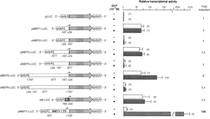
(40). In the absence
of hormone
(Fig. 4,
q
),
the construct
pMEP1 -LUC containing
the minimal pro-
moter
region
(EcoRISpel,
-167/+24)
as well
as
pMEP3-LUC
and pMEP5-LUC
containing
larger DNA
fragments
(KpnlSpel,
-577/24;
Hindlll-Spel,
-17971
24) stimulated
transcription
of the LUC gene several-
fold compared
to the background
activity of the parent
vector pLUC. The control plasmid ptk-LUC, which con-
tains the herpes virus tk gene promoter,
showed
a
transcriptional
activity similar to those of pMEPl-LUC,
pMEP3-LUC,
and pMEP5-LUC.
In contrast, none of the
other pMEP-LUC
vectors, containing
the same genomic
DNA fragments
in the antisense
orientation,
showed
significant
stimulation
of LUC activity. To investigate
whether
transcription
of the promoter/LUG
reporter
gene constructs
started at the same sites as transcrip-
tion of the wild-type
arMEP24
promoter,
we performed
primer extension
experiments
with total RNA extracted
from the transfected
CV-1 cells, using a primer derived
from the published
luciferase
sequence
(38). The 5’-
start sites in RNA from transfected
cells mapped at the
same positions
as those observed
using RNA from
The mouse genomic Hindlll fragment of 9 kb was cloned and sequenced, as described in Materials and Methods. The nt are numbered according to the major transcription initiation site, as determined by primer extension. The major transcription start site is indicated by a large arrowhead, while minor sites are indicated with small arrowheads. The exon sequences, which were determined by comparison with the cDNA insert of clone M53 (5) are in boldface. Each exon is translated into the amino acid sequence below the nt sequence. The signal peptide is underlined, and the boxed residues are confirmed by direct peptide sequencing of purified 24-kDa protein. The consensus splice signal GTRAG/CAG flanking the introns are overlined. The proposed TATA-like box, CAAT boxes, GC-rich boxes, the two polyadenylation signals AATAAA, and the homopurine-homopyrimidine stretch are underlined. The 16-core putative ARE TGTYCT are boxed. Repeat and dyad structures are indicated by arrows, while the Bl Alu-like elements are shown by dotted lines.
mouse epididymis
(Fig. 5). It can be concluded,
there-
fore, that the region
-167/24
indeed represents thearMEP24
gene promoter.
Evidence
for an ARE
In the presence of 1 O-7
MDHT (Fig. 4,
q
), the constructs
pMEPl-LUC
and pMEP3-LUC
were
not induced.
In
contrast,
pMEP5-LUC
was responsive
to androgens,
with an average induction
factor of 2, compared
to 1 or
1 .I for all other constructs.
The plasmid ptk-LUC was
used as a negative control of stimulation.
As a positive
control
of induction
efficiency, we used a construct
MOL END0.1993
262
Vol7 No. 2A.
B.
a
-x
ladder
8
TCGA
start sites
5’
UH
1
exon 1
3
p
primer
109 nt
Fig.
3. Primer Extension Analysis of the 5’-End of arMEP24 mRNAEpididymal poly(A)+ RNA was hybridized with a 3ZP-labeled primer located in the first exon and extended with AMV reverse transcriptase. The products were analyzed by polyacrylamide gel electrophoresis and autoradiography, as described in Ma- terials and Methods. The sizes of the extented products, as determined from the known fragments sizes of the DNA se- quence ladder, are indicated on the left. A, Primer extension products from yeast carrier transfer RNA (lane tRNA) and from epididymal poly(A)’ RNA [lane poly(A)+]. Lanes T, C, G, and A are 35S-labeled DNA sequencing reaction. B, Diagram of the 5’-portion of the gene. The El exon sequence is indicated by an open box. The thick line and the wavy line stand for the synthetic primer and the extended cDNA transcripts, respec- tively. Transcription start points are represented by vertical arrows.
repeat (LTR) in front of the LUC reporter gene (pMMTV-
LUC). Such MMTV-LTR-reporter
gene constructs
were
previously described
as androgen
responsive
in trans-
fected cells (20-22).
An induction
factor of 188 was
observed with pMMTV-LUC.
Omission of pSVARo from
the transfection
mix abolished
hormonal
induction
of
expression
(not shown).
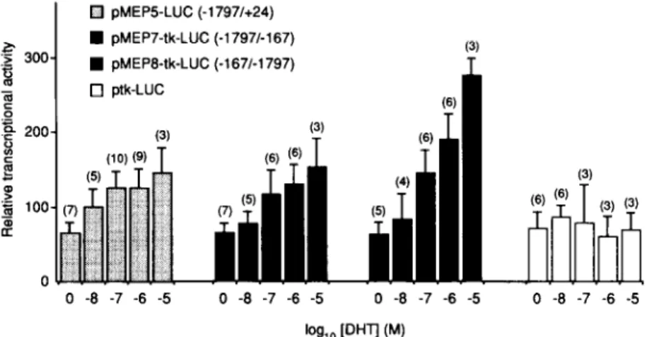
To verify whether the fragment HindIll-EcoRI
(-1797/
-167) contains a functional ARE responsible
for andro-
gen responsiveness
of the pMEPS-LUC
construct,
it
was cloned in both sense and antisense orientations
in
front of the heterologous
tk promoter-LUC
construct.
Cotransfections
with the AR expression
vector in the
presence of DHT ranging from 1 O-‘-l Ow5
Mevidenced
an average 2- to 4-fold increase
in LUC activity inde-
pendently of the orientation
of the Hindlll-EcoRI
frag-
ment (Fig. 6). This effect was not observed
with the
control ptk-LUC vector. From the sequence
analysis of
the genomic 5’-flanking
region, we found the imperfect
palindromic
motif TGTTGAgagAGAACA
at position
-896
to -882,
which ressembles
the core consensus
ARE (19). To obtain evidence that this sequence
can
act as a functional ARE, it was taken out of the context
of the arMEP24
promoter,
and two or three copies
were linked to the ptk-LUC construct
(see Materials
and Methods).
The results of the cotransfections
with
pSVARo in the presence of various DHT concentrations
are shown
in Fig. 7. The stimulation
of transcription
increased with the amount of available DHT in a dose-
dependent
manner, to reach a maximal efficiency at
10m5
M.A relative induction
factor of 3 was measured
with pARE3-tk-LUC
containing
three copies of the pu-
tative ARE in the sense orientation
(Fig. 7, m. The
construct pARE2-tk-LUC,
harboring
two copies of the
ARE in the antisense
orientation,
induced a 4-fold in-
crease in transcription
(0). Expression
of the ptk-LUC
control construct
without
any ARE was not inducible
by DHT (Fig. 7,0), while pMMTV-LUC
expression
was
stimulated
with an average induction
factor of about
200 (0).
Other Potential
Regulatory
Elements
In the region up-stream of the promoter, dyad symetry
structures
were found at -610
to -597
(DSl), -595
to -586 (DS2), and -600 to -590 (DS3). DSl and DS2
or DS2 and DS3 overlap (Fig. 2). Thus, only one of
them can form a stem-loop
structure
in a single mole-
cule. They are preceded
by a (TG)r4 stretch (Fig. 2). A
similar structure is present about 1500 nt down-stream
of the 3’-end of the gene (not shown). These elements,
which have the potential to adopt the Z-DNA confor-
mation (see Discussion),
are common
(lo5 copies) in
eukaryotic
DNA (41). They are believed to regulate the
expression
of the adjacent genes (42). We also ana-
lyzed the 5’-flanking
region for homology with potential
regulatory elements
by comparison
with a catalog of
consensus
sequences
(43). Several interesting
motifs
were found (Fig. 8), including three copies of the AP-2-
binding protein (44), three motifs for the CFI transcrip-
tional activator
(45) and one recognition
sequence
for
Ott-4 transcription
factor (46, 47). The putative PEA3-
responsive
element for Ets oncogenes,
serum growth
factor, and phorbol ester is found three times in the up-
stream 5’-region
(48-51).
Most notable is the presence
of six consensus
sites for the GATA-1 -binding protein,
previously called GF-1, NF-E2, and Eryfl (52-54)
an
erythroid-specific
zinc finger protein
that has been
shown
to regulate
the transcription
of a number
of
erythroid-specific
genes, including the a, 0, and r-globin
genes (55). An AP-1 -like motif for the second erythroid-
specific factor NF-E2 (56-58)
is also present associated
with several GT/CAC boxes similar to those observed
up-stream
of many erythroid promoters
and regulatory
elements (59, 60).
Chromosomal
Localization
of arMEP24
Gene
The high degree of amino acid conservation
and simi-
larity in the overall structures
of arMEP24
and GSHPx
Relative transcriptional activdy DHT (WM) : 1 50 1 1 100 1 1 150 1 2104 Fold I. ? 295c tnductlon - pLUC (7) 5 3’ + I (4) 1 ptvtEP1 -LUC 5 pMEPP-LUC 5 pMEP3.LUC 5 pMEP4.LUC 5 pMEP5.LUC 5 1.1 2 pMEPG-LUC 5’ pMMTV-LUC 5
Fig. 4. Analysis of arMEP24 Gene Promoter Activity in CV-1 Cells
Different lengths of arMEP24 gene sequences up-stream of the transcription start sites were inserted into the plasmid containing a promoterless LUC reporter gene to generate pMEP-LUC vectors. Nucleotide numbering is according to Fig. 2 and previously reported sequences.
Broken
arrows represent the transcription start sites. Details of constructs are described inMaterials and
Methods.
Cotransfections of pMEP-LUC constructs with pSVARo were performed in CV-1 cells in the absence @) and presence (Cl) of 1 O-’ M DHT. Values were standardized relatively to P-galactosidase activities. Each bar is the mean of several independent assays (indicated inparentheses),
and the errorbars
indicate the SD. Induction factors, indicated on theright,
were calculated by dividing the relative LUC activities obtained in extracts of cells grown in medium with hormone by that obtained in extracts of cells grown in the absence of hormone.(6, 7) strongly suggest that they evolved from a single
ancestral gene by duplication. We found it of interest
to determine the chromosomal localization of the ar-
MEP24 gene.
In situ
hybridization experiments were
carried out using mouse metaphases
spreads. A recom-
binant pGEM7Zf(-) plasmid containing a 1.4-kb ar-
MEP24 cDNA (5) was used as a probe. In the 100
metaphase cells examined after
in situ
hybridization,
there were 303 silver grains associated with chromo-
somes, and 44 of these (15%) were located on chro-
mosome 13. The distribution of grains on this chromo-
some was not random; 75% of them mapped to the
[A2-A41 region of chromosome 13 (Fig. 9). These re-
sults demonstrate that the localization of the arMEP24
gene is to the [A2-A41 region of chromosome 13 in the
murine genome.
DISCUSSION
We previously described the homology of arMEP24
with GSPHx (6,7). Here we report on the structure and
hormonal regulation at the molecular level of the gene
encoding for arMEP24. Primer elongation of mRNA
indicated the existence of a major and four weak start
sites of transcription that map within a stretch of about
25 nt. The region up-stream of the multiple initiation
sites contains an unusual TATCA box located -35 nt
from the major one. Since
in
vitro transcription studies
have shown the authentic TATA box serves to fix the
site at which transcription will start (61) the fact that
the arMEP24 gene has numerous transcriptional initia-
tion sites is not surprising. Several sequences ressem-
bling the consensus
CAAT box (62) and the GC box for
the Spl-binding site (63) are present. It has been hy-
pothesized that the presence of the latter facilitates the
recognition of a weak TATA box (64). Similar GC boxes
were first found in the simian virus-40 early promoter
region (61) the tk gene of herpes simplex virus (65)
and many so-called GC-rich housekeeping genes
whose activity is required in all cells and are not subject
to environmental control (66-69). Nevertheless, the
arMEP24 gene promoter cannot be considered a GC-
rich one. Rather, it belongs to another class of genes
that have no consensus
TATA element and are not GC
rich. Unlike GC-rich promoters, many of these pro-
moters are not constitutively active, but are regulated
during differentiation or development and initiate tran-
scription at only one or a few tightly clustered start
sites (70). In these promoters, an initiator element may
be essential for both core promoter strength and deter-
MOL END0.1993
264
A.
Vol7 No. 2
98,
99 nt
94nt=
82nt--,
74nt+
B.
-
primer
74 nt
Fig. 5. Primer Extension Analysis of the 5’-End of Hybrid arMEP24/LUC mRNA in Transfected CV-1 Cells
Total RNA from transfected CV-1 cells was hybridized with a 32P-labeled primer located in the coding region for LUC and
extended with AMV reverse transcriptase. The products were analyzed by polyacrylamide gel electrophoresis and autoradiography,
as described in
Materials and Methods.The sizes of the extented products, as determined from the known fragments sizes of the
DNA sequence ladder, are indicated on the left. A, Primer extension products from total RNA of untransfected CV-1 cells (lane CV-
1) and from total RNA of CV-1 cells transfected with pMEP8LUC plasmid DNA (lane pMEP5LUC). Lanes T, C, G, and A are 35S-
labeled DNA-sequencing reaction. B: Bottom, Diagram of the promoter region of the hybrid reporter gene. The LUC-coding sequence
is indicated by an
open arrowbox.The
shadedbox represents the Smal linker. The
thickline and the
wavyline stand for the
synthetic primer and the extended cDNA transcripts, respectively. Transcription start points are represented by verfical
arrows.Top, An enlargement shows the nt sequence of the hybrid gene with the primer position. Untranscribed sequences are in lowercase
letters, arMEP24/LUC hybrid mRNA sequences are in uppercase
letters,and LUC sequences are in
italics.The LUC translation
start codon ATG is
boxed.The Spel/Smal junction is in
boldfaceand
underlined.Major (+l) and weak transcription start points
are indicated by
large and small arrowheads,respectively.
mining the actual initiation
site
(71).
interestingly,
be-
tween position
-21 to IO, the arMEP24 gene promoter
shows 65% homology
with the transcription
start site
region
of the terminal
deoxynucleotidyl
transferase
gene, in which the initiator element was first described
(70). Furthermore, residues that are essential for the
initiator activity are perfectly conserved (70). It has been
recently hypothesized that the transcription initiation
complex assembly might be nucleated by binding of
transcription factors on either TATA-like or initiator
elements, depending upon the relative concentrations
and activities, or on both elements
in concert (71). This
would result in more diversified responses to distal
regulatory elements. In the 5’-flanking region, most
notable is a run of (TG)14.
TG repeats have been found
in genomes of yeast, mouse, and man (72, 73). These
sequences often show hypersensitivity toward single
strand-specific nucleases (74). This might reflect the
presence of conformational alterations in the DNA,
which could act as general recognition signals for the
nearby presence of a transcription-initiation
or promoter
region. By virtue of their ability to form left-handed DNA
helices of Z-DNA (75) the conformational changes
might facilitate or direct the binding of transcription or
regulatory factors to more specific sequences on the
promoter. Several DS structures are present near the
(TG)r4 stretch. The rat prostatic binding protein C2 and
C3(1) genes (24, 76), the rat seminal vesicle F and S
Q pMEPB-LUC (-1797/+24) 4%
n
pMEP7-tk-LUC (.1797/-167) :z 300 2 W pMEP6-tk-LUC (-167/-1797) 3 s ‘g 200 P 5 z J f 100 $ 0 0 -8 -7 -6 -5 0 -8 -7 -6 -5 0 -8 -7 -6 -50 -a
-7 -6 -5log,, VT] P4
Fig.
6. Transcription-Stimulating Activity of the Hindlll-EcoRI Fragment (-1797/-167) Transfected into CV-1 Cells in the Presence of AndrogensCV-1 cells were cotransfected with pMEP5-LUC (containing the genomic fragment -1797/24) or pMEP7-tk-LUC or pMEP8-tk- LUC (containing the genomic fragment -1797/-167 linked to the heterologous tk promoter in either the sense or antisense orientation, respectively) and pSVARo. LUC activity was measured in the presence of various DHT concentrations and normalized to @-galactosidase activity. Each bar represents the mean of several independent assays (indicated in parentheses), and the error bars indicate the SD
::;;] /L-: . pMMTV-LUC 2000 i 500 - 400- 300 - 200 - 0 pA=-tk-LUC . pARE_3-tk-LUC
./-P---P---
---*...--
p
o ptk-Luc
OI
0 -a -7 -6 -5 log ,o PHTI CM)Fig. 7. Transcription-Stimulating Activity of the -896 to -882 ARE Motif Transfected into CV-1 Cells in the Presence of Androgens
CV-1 cells were cotransfected with pSVARo and PARES-tk- LUC (m) or pARE2-tk-LUC (0) containing three or two copies of the putative ARE sequence TGTTGAgagAGAACA (-896/ -882) cloned in front of the tk-promoter LUC reporter gene construct (ptk-LUC) in the sense or antisense orientation, as indicated by arrows. Transcriptional stimulation was measured in the absence and presence of various DHT concentrations. Constructs ptk-LUC (0) and pMMTV-LUC (0) were used as negative and positive controls, respectively. Values were standardized relative to @-galactosidase activities. Experimen- tal details are described in Materials and Methods. Each point represents the mean of at least three independent assays, and the error bars indicate the SD.
genes (77, 78) the mouse renin 2 gene (79) and the
rat androgen-binding
protein gene (80) all androgen-
controlled
genes, have short inverted repeats in their
promoter
regions. Therefore,
it is likely that the repeats
of DS structures
in androgen-controlled
genes could be
involved in the regulation
of expression.
In this study, we have shown that expression
of the
arMEP24
gene can be up-regulated
on the level of
transcription
by DHT. We have evidenced
a functional
ARE
located
at
position
-896
to
-882
(TGTTGAgagAGAACA)
that can function
both in the
context
of the arMEP24
promoter
or as a separate
element when cloned in front of the tk promoter.
The
sequence
of this imperfect palindromic
ARE ressembles
the
reverse
complement
consensus
sequence
AGAACAnnnTGTACC
for binding of glucocorticoid
and
progesterone
receptors
(17) in which the order of the
two half-sites is inverted. The homology is less marked
with the other ARES characterized
so far (21, 26, 27).
The level of induction
is rather low in our experiments
(-2-fold).
However,
it is comparable
to the inductions
observed
in a study of the effect of androgen
on the
transcription
rate of the arMEP24
gene in run-on ex-
periments
performed
on isolated
nuclei from the epidi-
dymides of castrated
and normal mice (Lareyre, J. J.,
unpublished
results). Similarly, transcription
of the pros-
tate-specific
kallikrein-like
gene is only 2-fold induced
by androgens
(81). This low induction
will represent
at
least part of the total androgen-responsive
regulation
mechanisms,
which determines
the final mRNA concen-
tration. Obviously, the change in the rate of arMEP24
mRNA transcription
is not sufficient to account for the
very dramatic in vivo changes observed
in the steady
state level after castration
and testosterone
replace-
ment (1, 2). Then, the role of arMEP24
mRNA turnover
in gene control is also likely to be important.
Indeed,
MOL ENDO. 1993 Vol7” - 266
About 2000 residues of the sequence in the promoter region extending from nt -1831 to 93 are shown. The major initiation site of transcription, numbered 1, is indicated with a large arrowhead. Other weak start sites are indicated by small arrowheads. The transcribed sequence is in boldface, and the translated amino acid sequence is indicated above with the three-letter code. Restriction sites used to construct pMEP-LUC vectors are underlined. The motifs found to be homologous to &-acting elements in other promoters and/or enhancers are boxed, and the arrow indicate their orientation. Reference numbers are given in the text.
Fig. 9. Localization of the arMEP24 Gene to Mouse Chromosome 13 by in Situ Hybridization
A, Two partial WMP mouse metaphases, showing the specific site of hybridization to chromosome 13. Top, Arrowheads indicate silver grains on Giemsa-stained chromosomes after autoradiography. Bottom, Chromosomes with silver grains were subsequently identified by R-banding. B, Diagram of WMP mouse Rb (13, 15) chromosome, indicating the distribution of labeled sites.
when the changes in the steady state level of a partic-
ular mRNA
under
two different
conditions
are not
matched by a comparable
change in de nova transcrip-
tion (as measured
by nuclear run-on assays), changes
in mRNA turnover or stability are often presumed
(82).
There are two limitations of this approach.
First, there
is no numerical
estimation
of half-life, only a relative
comparison.
Second, no account is made for changes
in mRNA maturation
events or nuclear transport
that
could influence steady state mRNA levels.
Increased
attention
has been paid in the last few
years to the regulation
of mRNA stability as an impor-
tant control point of gene expression
(reviewed in Refs.
82-84). Steroid hormones represent
some of the earli-
est agents shown to control the degradation
of specific
mRNAs and have been shown to regulate the stability
of a substantial
number
of mRNAs (83). Vitellogenin
mRNA stabilization
by estrogen
is a clear example of
specific regulation
of gene expression
by control of
mRNA stability (83, 85). In mouse kidney, androgens
have little or no effect on the synthesis of three different
mRNAs species whose steady state concentrations
are
increased
lo- to 20-fold during testosterone
treatment,
suggesting
that mRNA stabilization
is a major factor in
the inductions
(86). Similarly, androgenic
stimulation
of
mRNA concentrations
in the prostate
appears to occur
predominantly
via transcript
stabilization
(81, 87). In
different reports, both 5’- and 3’-terminal
sequences
have been claimed to play crucial roles in mRNA stability
(82, 83). Although
stretches of AT-rich sequences
were
found in the 3’-untranslated
portion of arMEP24
mRNA,
a carefull analysis of the sequence
failed to exhibit a
conserved
sequence
motif with that mediating
changes
in mRNA turnover (84). The rate of mRNA turnover was
also demonstrated
to be highly dependent
on the pres-
ence of the 3’-terminal
poly(A) sequences,
and the
deadenylated
mRNA apparently had a much faster rate
of turnover
(88). Estrogen
stabilization
of very low
density apolipoprotein-II
mRNA (89) and glucocorticoid
stabilization
of GH mRNA (90) result in an increase in
the length of their poly(A) tails. Interestingly,
expression
of the cytoplasmic
poly(A) polymerase
is positively con-
trolled by androgens
(91). Nevertheless,
modulation
of
the poly(A) tail length
as a possible
mechanism
of
regulation
of mRNA stability does not apply for ar-
MEP24, since no change in mRNA size was seen after
induction
by androgens
(1). Finally, the control of mRNA
degradation
could also be coupled to ribosome
loading
and translational
engagement
of the mature RNA (92).
For instance,
the maintenance
of a high density of
ribosomes
on vitellogenin
mRNA increases its stability
(93). Implication
of the translational
machinery
in the
regulation
of arMEP24
expression
was recently sus-
pected (94).
In summary, it seems evident that posttranscriptional
events, such as regulation
of the mRNA turnover
rate,
have an important
place in the overall scheme
of ar-
MEP24 gene expression.
Their contribution
to the final
cellular mRNA concentration
remains to be determined
by other
experiments.
The last important
question
would be to determine
whether
the posttranscriptional
effects of steroid hormones are direct. The effect of the
hormone-receptor
complexes
may be to induce tran-
scription of gene(s) coding for a protein(s)
required
for
stabilization,
as previously shown (83, 95).
In synthetic promoter
constructs,
synergistic activity
has been described
for the steroid
receptors,
with
factors binding
to the Spl,
Ott-1,
or CACCC
box
consensus
sequences
(96-98).
In the arMEP24
gene,
several motifs for cell-specific
or ubiquitous
transcrip-
tion factors are found. Their implication
in the full cell-
specific responsiveness
of transcription
to androgens
is suspected.
In this respect, the PEA3 motifs are of
great interest, since PEA3 protein, which
belongs
to
the Ets oncogene
family, is specifically expressed
in the
epididymis
and brain, but not in erythroid
cell lines or
hematopoietic
tissues, where all of the other Ets onco-
genes are synthesized
(49). In contrast, GATA-l-
and
NF-E2-binding
proteins
are specifically
restricted
in
these latest tissues (52, 53, 56, 57). The presence
of
several consensus
sequences
for erythroid-specific
transcription
factors is quiet surprising.
Furthermore,
the general organization
in the region up-stream
of the
arMEP24
gene promoter
(Fig. 10) is similar to that
observed
in the GATA-l-binding
protein gene (54), the
porphobilinogen
deaminase
gene (56, 59) the erythro-
poietin
receptor
gene (99) the LY- and @globin
locus
control regions (58, 60, 100, lOl), and the erythrocyte
GSHPx gene (102). Whether these motifs are functional
in the epididymal
gene is not known.
They could be
related to the regulatory environment
of the ancestral
erythroid
gene from which the arMEP24
gene is prob-
ably derived.
Although
the two proteins
show
high
homology
(6, 7)
the conserved
regions
between
GSHPx (103) and arMEP24
are not encoded
by identi-
cal exons. This may indicate
a diverging
evolution
of
the genes that can descend
from a single progenitor
gene. The red cell GSHPx is not secreted, and the gene
does not contain any exon for a signal peptide
(103).
The exon encoding
the N-terminal
region of arMEP24,
which may be critical for secretion
of the peroxidase-
like protein in the epididymis,
could have been added
to a member of the GSHPx gene family after duplication
and evolution
of an ancestral
unit. To support
this
hypothesis,
the arMEP24
gene is located
in the [A2-
A41 region of the mouse chromosome
13 in a region
homologous
to human chromosome
7 (104) while the
human
GSHPx gene is located
on chromosome
3 in
region 3qll-13.1
(105).
The identification
of epididymal-specific
regulatory
elements
from the arMEP24
gene will be extremely
important
for further
analysis
of hormone-regulated
gene expression
of GSHPx and related proteins in the
epididymis.
MATERIALS
AND METHODS
Isolation of Clones
The mouse BALB/c genomic library was purchased from Clontech (Palo Alto, CA). It was screened by using DNA frag- ments from the M53 cDNA clone (5) as probe in plaque hybridization, as described previously (1). DNA was prepared from purified clones by the liquid-lysis method (106) and was mapped with restriction endonucleases under conditions rec- ommended by the suppliers (Boehringer Mannheim, Maylan, France: Bethesda Research Laboratories, Cergy Pontoise, France). Suitable restriction fragments were subcloned into pGem7Zf(-) vector (Promega Biotec, Madison, WI), using standard protocols (107).
Nucleotide Sequence Analysis
All of the nt sequences were determined on NaOH-denatured double stranded plasmid DNA by the dideoxy chain termination method (108) using a Pharmacia sequencing kit (St-Quentin en Yvelines, France). For gene sequence determination, each clone was sequenced at least twice on each strand. Se- quences were assembled and analyzed using the BISANCE program (109) at C.I.T.l.2 (Paris, France).
Primer Extension Analysis
Conditions for the extraction of total RNA and poly(A)’ RNA purification have been described previously (1, 2). Ten pico-
MOL END0.1993 268
Vol7 No. 2
al 5 3
GATA GT GATA NF-EZ GT GTGATA GATA
9) 5’Pf-ll I H -3
Fig. 10. Comparison of arMEP24 Promoter Region with Erythroid-Specific Regulatory Regions
Schematic representation of promoters or enhancers from arMEP24 gene (a), erythrocyte GSHPx gene (b), GATA-l-binding protein gene (c), porphobilinogen deaminase gene (d), erythropoietin receptor gene (e), @-globin (f), and n-globin (g). Control locus regions are shown (not to scale). The black boxes are GATA-1 consensus sequences; the dotted boxes are NF-E2-binding sites, and the open boxes named GT indicate the presence of GGTGG motifs. The broken arrows localize the transcription start sites. Reference numbers are given in the text
moles of synthetic primer specific for arMEP24 mRNA (5’- CATCTTTTCCGGCCTCCCCC-3’) or specific for luciferase mRNA (5’-GTTTTTGGCGTCTTCC-3’; Eurogentec SA, Liege, Belgium) were radiolabeled using T4 kinase (Promega Biotec) in the presence of 50 &i [?P]deoxy-ATP (Amersham, Les Ulis, France). One picomole was then hybridized to 10 pg poly(A)+ RNA from-caput epididymidis, i0 pg carrier yeast transfer RNA. or 10 ua total RNA from transfected CV-1 cells for 12 h at 35.C in 1 d 2 0.04 M 1,4-piperazine diethanesulfonic acid (PIPES), pH 6.4, buffer containing 0.4 M NaCI, 1 FM EDTA, and 60% (vol/vol) formamide. Reverse transcription was per- formed in 20 ctl 50 PM Tris-HCI (pH 6.3) 30 PM KCI, 6 PM MgC12, 6 PM dithiotreitol, 0.5 FM of each deoxy-NTP, and 50 U AMV reverse transcriptase (Promega Biotec). The samples were incubated at 42 C for 30 min, boosted by the addition of 50 U enzyme, and incubated for 1 h more. The products were analyzed on 6% (wt/vol) polyacrylamide urea gels used for sequence determination.
Construction of Plasmids
The promoterless basis plasmid pLUC, which was used for cloning promoter region fragments from the arMEP24 gene in front of the LUC gene, was derived from pSVOA (36) by inserting a Smal adaptor in the unique HindIll-cloning site. Constructs pMEPl-LUC and pMEP2-LUC (EcoRI-Spel, -167/ 24) pMEP3-LUC and pMEP4-LUC (KpnlSpel, -577/24), and pMEP5-LUC and pMEP6-LUC (/iindlll-Spel, -1797/24) were generated by ligation of the appropriate blunt-ended restriction fragments into the Smal site of pLUC. Insertion of the correct fragment and the orientation were checked by restriction en- zyme mapping and sequencing. The tk promoter (fragment from -193 to 50) and MMTV-LTR (from -631 to 125) were obtained by polymerase chain amplification, using oligonucle- otides orimers 5’-CCAAGCTTCATCCCCGTGG-3’ and 5’- CCAAGCTTCGGCACGCTGTTGACGCTGT-3’ or 5’-GCA- AGCTTGGCCTAGAAGTAAAAAAGGG-3’ and 5’-GCAAGCT TGGCCGTCCTGAGGGTGACCG-3’. which were directed against previously reported sequences (110, 111). DNA was amplified in lOO-~1 reactions containing 50 PM KCI; 1.5 PM
MgCl*; 10 PM Tris-HCI (pH 6.3); 0.01% gelatin (wt/vol); 0.2 PM each of dATP. dGTP. dCTP. and dTTP: 5 U Taa DNA oolvm- erase; 0.5 ~~‘of the appropriate oligonucleotide primers; and 10 ng pMMTV-chloramphenicol acetyltransferase (CAT) (20) or pBLCAT2 (112) plasmid DNA. Reactions were amplified for 25 cycles, each consisting of 1 min at 94 C, 2 min at 60 C, and 3 min at 72 C. Amplified fragments were purified on 2% (wt/vol) agarose gels, restricted with Hindlll, and ligated into the Hindlll site of pSVOA to generate pMMTV-LUC and ptk- LUC. Constructs were verified by DNA sequencing. Constructs pMEP7-tk-LUC and pMEP&tk-LUC (HindIll-EcoRI, -1797/ -167) were generated by ligation of the appropriate blunt- ended restriction fragment into the up-stream filled Hindlll site of ptk-LUC. Two (pARE2-tk-LUC) or three (pARE3-tk-LUC) ARES were cloned in front of the tk promoter in the ptk-LUC vector. Oligonucleotides (positioned -696 to -662 in the arMEP24 gene) 5’-AGCTATATTGTTGAGAGAG AACATGTA- 3’ and 5’-AGCTTACATGTTCTCTCTCAACAATAT-3’ were ki- nased, annealed, and subsequently cloned into the up-stream HindIll site of ptk-LUC. Copy number and orientation of ARES were checked by sequencing.
Cell Culture
African green monkey kidney (CV-1) cell were maintained in Dulbecco’s Modified Essential Medium (DMEM) containing 5% (vol/vol) dextran-charcoal-treated fetal calf serum (Gibco-Be- thesda Research Laboratories, Cergy Pontoise, France), 15 PM HEPES (pH 7.2) 2 PM glutamine, 100 U/ml penicillin, and 50 @g/ml streptomycin. They were harvested in 0.25% (wt/ vol) trypsin. Cells were grown in a tissue culture incubator at 37 C, with an atmosphere of 5% CO* (vol/vol).
Transient DNA Transfection
Supercoiled plasmid DNA used for transfection experiments was purified by two successive CsCl density gradient centrif- ugations (107). For each vector, two different isolates were transfected at least twice in independent experiments. This approach minimized uncertainties in the interpretation of the
results that might stem from vanabillty between plasmid prep- aratlons. In all cases, plasmid pCHll0 containing the jj-galac- tosidase gene under the control of the simian virus-40 early promoter was used as an internal control of the efficiency of transfection (40). Typically, 2 gg reporter plasmid, 1 pg pCH110, and 1 pg pSVARo (39), the human AR expression plasmid, were used. The total amount of DNA was increased to 15 pg by adding pGem7Zf(-). CV-1 cells were transiently transfected by the calcium phosphate precipitation method (113) without glycerol shock, using lo6 cells in each lo-cm plate. The precipitates were left for 12 h on the cells, which were then washed with 5 ml DMEM. Cells were Incubated for 24 h In 10 ml fresh medium [DMEM supplemented with glu- tamine, antibiotics, and 5% (vol/vol) steroid-depleted fetal calf serum] with either vehicle alone or DHT (Theramex Laborato- rles, Monaco) at flnal concentrations of 10 nM to 10 @M.
Luciferase Assays
Each 1 O-cm plate of transfected CV-1 cells was washed once with PBS without Ca’+ or Mg’+, and cells were harvested in 1 ml extraction buffer (100 PM potassium phosphate, pH 7.8, and 1 PM dithiothreitol) by scraping. The cells (-5 x 1 06) from a single dish were pelleted by centrifugation and resuspended in 100 ~1 extraction buffer. Cells were lysed by three cycles of freezing in liquid nitrogen and thawing at 37 C. Cell debris was pelleted by centrifugation for 5 min. A 3O-j~l aliquot of each extract was tested for i-l-galactosidase (40). The remaining 60- ~1 samples were added to 350 ~1 25 /IM glycylglycine, pH 7.8, containing 5 PM ATP and 15 PM MgSO+ The samples were placed in an LKB luminometer (LKB, Rockville, MD), and the reaction was initiated by the injection of 100 ~1 1 FM luciferin (Sigma, St. Louis, MO). The peak light emission was recorded. Luciferase activities were then standardized according to the internal levels of ij-galactosidase. In each experiment, ptk-LUC was transfected three times, and the mean tk-luciferase value was determined. Luciferase data from separate experiments, in which the mean tk-luciferase values were similar, were pooled. Three to 10 independent values were considered for calculation of the mean f SEM.
Chromosome Preparation
In situ hybridization experiments were carried out using met- aphase spreads from a WMP male mouse, in which all auto- somes, except 19, were In the form of metacentric robertson- ian translocations. Concanavalin-A-stimulated lymphocytes were cultured at 37 C for 72 h, with 5-bromodeoxyundine added for the final 6 h of culture (60 pg/ml medium), to ensure a chromosomal R-banding of good quality. The 1.4-kb ar- MEP24 cDNA (5) in pGem7Zf(-) was tritium labeled by nick translation (107) to a specific activity of 1.8 x 10’ dpm/pg. The radiolabeled probe was hybridized to metaphase spreads at a flnal concentration of 25 rig/ml hybridization solution, as previously described (1 14). After coating with nuclear track emulsion (NTB2, Eastman Kodak, Rochester, NY), the slides were exposed for 13 days at 4 C, then developped. To avoid any slipping of silver grains during the banding procedure, chromosomes spreads were first stained with buffered Glemsa solution, and metaphases were photographed. R-Banding was then performed by the fluorochrome-protolysis-Giemsa method, and metaphases were rephotographed before analy-
SIS.
Acknowledgments
The authors thank Dr. E. Pailhoux for many helpful discus- sions, and S Guenn for her skilled technical assistance. N G is also gratefull to Dr. M. T. Bocquel for providing excellent advice concerning transfections and LUC assays. Plasmid pSVARo was a generous gift from Dr. A. 0. Brinkmann (Rot- terdam, The Netherlands), pMMTV-CAT was obtained from
Prof. P. Chambon (Strasbourg, France), and pBL.CAT2 from Dr. J. L. Coudert (Clermont-Ferrand, France).
Received September 17, 1992. Revision received Novem- ber 30. 1992. Accepted November 30, 1992,
Address requests for reprints to: Jean-Pierre Dufaure, La- boratoire de Blologle Cellulaire, CNRS lJRA360. 24 avenue des Landaus, 63177 Aubi&re Cedex. France.
Present address: Dr. Norbert B Ghyselinck, Laboratoire de G&-&lque Moleculaire des Eucaryot& du CNRS. INSERM U184, lnstitut de Chimie Biologique, FacultB de Medecine, 11 rue Humann, 67085 Strasbourq Cedex, France.
This work was supported by-a grant from CNRS (URA 360. Expression et Rbgulatlon du GBnome) and flnanclal assistance from the Fondation pour la Recherche Medlcale FranCaise and INSERM (Contrat 89 4004).
The nt sequence reported in this paper has been submitted to the GenbankTM/EMBL data Bank with accession no. M68896.
REFERENCES
1 2. 3 4 5. 6. 7. 8. 9. 10. 11. 12Ghyselinck NB. Jlmenez C, Lefranqols AM, Dufaure JP 1990 Molecular cloning of a cDNA for androgen-regu- lated proteins secreted by the mouse epldidymis. Mol Endocrinol 4:4-l 5
Faure J, Ghyselinck NB, Jimenez C. Dufaure JP 1991 Specific distribution of mRNA for 24 kDa proteins In the mouse epididymis as revealed by in situ hybridization: developmental expression and regulation in the adult. Biol Reprod 44:13-22
Jimenez C, Ghysellnck NB, Depeiges A, Dufaure JP 1990 lmmunochemical localization and association with spermatozoa of androgen-regulated proteins of Mr 24000 secreted by the mouse epididymis Biol Cell 6:171-174
Jimenez C. LefranCols AM, Ghyselrnck NB. Dufaure JP 1992 Charactenzatlon and hormonal regulation of 24kDa proteins synthesis by the murine adult epididymis J Endocnnol 133: 197-203
Ghyselinck NB. Dufaure JP 1990 A rnouse cDNA se- quence for epididymal androgerl-regulated proteins re- lated to glutathione peroxidase. Nucleic Acids Res 18:7144
Ghyselinck NB, Jimenez C, Dufaure JP 1991 Sequence homology of androgen regulated epididymal proteins with glutathione peroxidase. J Reprod Fertll 93.461-466 Ghyselinck NB, Rlgaudiere N, Dufaure JP 1991 Une prot&ne androgeno dependante s&r&e par la tite de I’epldidyme de souris presente des homologies &ev&es avec differentes glutathion peroxydases. C R Acad SCI [D] (Paris) 313:l -6
Flohe L 1982 Glutathlone peroxldase brought Into focus. In: Pryor WA (ed) Free Radicals in Biology Academic Press, New York. voi 5:223--253
Samuelson B 1983 Leukotrienes: mediators of immedi- ate hypersensltivlty reactions and inflammation Science 220:568-575
Alvarez JG, Storey BT 1984 LIpid peroxidation and the reactions of superoxlde and hydrogen peroxide in mouse spermatozoa. Biol Raprod 30 833.-841
Ghyselinck NB, Jlrnenez C, Courty Y, Dufaute JP 1989 Androgen dependent messenger RNA(s) related to se- cretory proteins In the mouse epididymls. J Reprod Fertll 85:631-639
Chang C, Kokontis J, Llao S 1988 Structural analysis of complementary DNA and amtno acid sequence of human and rat androgen receptors Proc Nat1 Acad SCI USA 85:721 l-721 5