HAL Id: hal-01826158
https://hal.archives-ouvertes.fr/hal-01826158
Submitted on 7 Jan 2020
HAL is a multi-disciplinary open access
archive for the deposit and dissemination of
sci-entific research documents, whether they are
pub-lished or not. The documents may come from
teaching and research institutions in France or
abroad, or from public or private research centers.
L’archive ouverte pluridisciplinaire HAL, est
destinée au dépôt et à la diffusion de documents
scientifiques de niveau recherche, publiés ou non,
émanant des établissements d’enseignement et de
recherche français ou étrangers, des laboratoires
publics ou privés.
Distributed under a Creative Commons Attribution - NonCommercial - ShareAlike| 4.0
International License
in a mouse model of fragile X syndrome
Kwang-Mook Jung, Marja Sepers, Christopher Henstridge, Olivier Lassalle,
Daniela Neuhofer, Henry Martin, Melanie Ginger, Andreas Frick, Nicholas
Dipatrizio, Ken Mackie, et al.
To cite this version:
Kwang-Mook Jung, Marja Sepers, Christopher Henstridge, Olivier Lassalle, Daniela Neuhofer, et al..
Uncoupling of the endocannabinoid signalling complex in a mouse model of fragile X syndrome. Nature
Communications, Nature Publishing Group, 2012, 3 (1), �10.1038/ncomms2045�. �hal-01826158�
Received 10 Apr 2012 | Accepted 1 Aug 2012 | Published 25 sep 2012 DOI: 10.1038/ncomms2045
Fragile X syndrome, the most commonly known genetic cause of autism, is due to loss of the fragile X mental retardation protein, which regulates signal transduction at metabotropic glutamate receptor-5 in the brain. Fragile X mental retardation protein deletion in mice enhances metabotropic glutamate receptor-5-dependent long-term depression in the hippocampus and cerebellum. Here we show that a distinct type of metabotropic glutamate receptor-5-dependent long-term depression at excitatory synapses of the ventral striatum and prefrontal cortex, which is mediated by the endocannabinoid 2-arachidonoyl-sn-glycerol, is absent in fragile X mental retardation protein-null mice. In these mutants, the macromolecular complex that links metabotropic glutamate receptor-5 to the 2-arachidonoyl-sn-glycerol-producing enzyme, diacylglycerol lipase-α (endocannabinoid signalosome), is disrupted and metabotropic glutamate receptor-5-dependent 2-arachidonoyl-sn-glycerol formation is compromised. These changes are accompanied by impaired endocannabinoid-dependent long-term depression. Pharmacological enhancement of 2-arachidonoyl-sn-glycerol signalling normalizes this synaptic defect and corrects behavioural abnormalities in fragile X mental retardation protein-deficient mice. The results identify the endocannabinoid signalosome as a molecular substrate for fragile X syndrome, which might be targeted by therapy.
1 Department of Anatomy and Neurobiology, University of California, Irvine, California 92697, USA. 2 INSERM U862, Circuit and Dendritic Mechanisms
Underlying Cortical Plasticity Group, Neurocentre Magendie, 146 Rue Léo-Saignat, F 33077 Bordeaux Cedex, France. 3 University of Bordeaux, Bordeaux
F 33077, France. 4 Institute of Experimental Medicine, Hungarian Academy of Sciences, H-1083 Budapest, Hungary. 5 INSERM U901, Marseille 13009,
France. 6 Université de la Méditerranée UMR S901, Aix-Marseille 2, France. 7 INMED, Marseille 13009, France. 8 Department of Psychological and Brain
Sciences, Gill Center for Biomolecular Science, Indiana University, Bloomington, Indiana 47405, USA. 9 Department of Biological Chemistry, University
of California, Irvine, California 92697, USA. 10 Unit of Drug Discovery and Development, Italian Institute of Technology, Genova 16163, Italy. *These
authors contributed equally to this study. Correspondence and requests for materials should be addressed to I.K. (email: katona@koki.hu) or to D.P. (email: piomelli@uci.edu) or to O.J.M. (email: olivier.manzoni@inserm.fr).
uncoupling of the endocannabinoid signalling
complex in a mouse model of fragile X syndrome
Kwang-mook Jung
1,*, marja sepers
2,3,*, Christopher m. Henstridge
4,*, olivier Lassalle
5,6,7,
Daniela neuhofer
5,6,7, Henry martin
5,6,7, melanie Ginger
2,3, Andreas Frick
2,3, nicholas V. DiPatrizio
1,
Ken mackie
8, Istvan Katona
4, Daniele Piomelli
1,9,10& olivier J. manzoni
2,3,5,6,7F
ragile X syndrome (FRAX), the most frequently known genetic cause of autism, is characterized by a series of physical, cogni-tive and emotional symptoms that include social deficits and diminished ability to learn1. FRAX is due to the mutation of a single gene, called FMR1 (ref. 2), which encodes for the fragile X mental retardation protein (FMRP)3. FMRP regulates the translation and transport of messenger RNAs in dendritic spines of brain neurons, and is involved in modulating signal transduction through type-I metabotropic glutamate (mGlu1 and mGlu5) receptors at glutama-tergic synapses throughout the brain. Loss of FMRP causes pro-found changes in the structure of neuronal dendrites and enhances, in the hippocampus and cerebellum of FMRP-deficient (fmr1 − / − )mice, a form of long-term depression (LTD) that requires mGlu5 receptor-mediated protein synthesis3–7.
In medium spiny neurons of the ventral striatum and pyramidal neurons of the prefrontal cortex (PFC), activation of mGlu5 receptors initiates a distinct form of LTD, which does not depend on protein synthesis and is affected by the retrograde endocannabinoid (eCB) transmitter, 2-arachidonoyl-sn-glycerol (2-AG)8,9. Stimulation of mGlu5 receptors releases 2-AG through the sequential recruitment of two enzymes: phospholipase C-β, which produces 1,2-diacyl-sn- glycerol, and diacylglycerol lipase-α (DGL-α), which converts 1,2-diacyl-sn-glycerol into 2-AG10–12. Morphological studies have shown that both phospholipase C-β and DGL-α are localized to the perisynapse, a subdivision of the dendritic spine that forms a 100– 200-nm-thick border around the postsynaptic density13. mGlu5 receptors are concentrated in this region14 where they may be physi-cally linked to DGL-α through the adaptor protein Homer10,15. This supramolecular complex is likely to have an important role in eCB-mediated transmission at excitatory synapses, by providing a focal point for the generation of a 2-AG pool that is specifically commit-ted to retrograde transmission. The 2-AG produced by this complex can readily cross the synaptic cleft to access CB1 cannabinoid recep-tors on presynaptic terminals, where the 2-AG-deactivating enzyme monoacylglycerol lipase (MGL) is also found16,17. We propose that this complex, which we refer to as ‘eCB signalosome’, allows for the separation of the 2-AG pool responsible for eCB signalling from other cellular pools of 2-AG, such as those involved in eicosanoid production and phospholipid remodelling.
FMRP deletion is known to be associated with greater eCB-mediated responses at GABAergic synapses of the dorsal striatum and CA1 region of the hippocampus18,19. Here, we show that loss of FMRP is accompanied by marked deficits in mGlu5-dependent 2-AG release and eCB-mediated LTD at excitatory synapses of the forebrain, as well as by a structural disorganization in the eCB signa-losome. We further show that inhibition of presynaptic MGL activ-ity normalizes eCB-mediated LTD and corrects key behavioural abnormalities in fmr1 − / − mice, suggesting that pharmacological
enhancement of 2-AG signalling might offer a new therapeutic strategy for FRAX.
Results
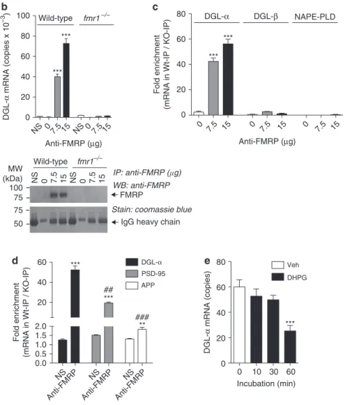
FMRP binds DGL- mRNA and controls 2-AG signalling. FMRP
regulates protein translation in postsynaptic spines by interacting with mRNAs that often, albeit not always, contain a G-quartet FMRP-binding motif 20. A bioinformatics search showed that an FMRP-binding sequence is present in DGL-α mRNA (Fig. 1a), but not in mRNAs encoding for structurally or functionally related enzymes such as DGL-β, MGL, N-acylphosphatidylethanolamine-selective phospholipase D (NAPE-PLD) and α/β hydrolase domain-6 (ABHD6) (data not shown). To test whether FMRP binds the DGL-α message, we immunoprecipitated FMRP from mouse forebrain homogenates and used quantitative PCR to measure DGL-α mRNA in the pellets. The pellets contained DGL-α mRNA (Fig. 1b), but no detectable levels of mRNAs encoding for DGL-β or NAPE-PLD (Fig. 1c). As expected, the precipitates
also contained mRNAs encoding for PSD-95 and amyloid precursor protein, which are known to bind FMRP21,22 (Fig. 1d). Moreover, experiments with rat cortical neurons in primary cultures showed that exposure to the mixed mGlu1/mGlu5 receptor agonist (S)-3,5-dihydroxyphenylglycine (DHPG) (100 µM) caused the dissociation between FMRP and DGL-α mRNA (Fig. 1e), as previously shown for other FMRP-regulated messages22.
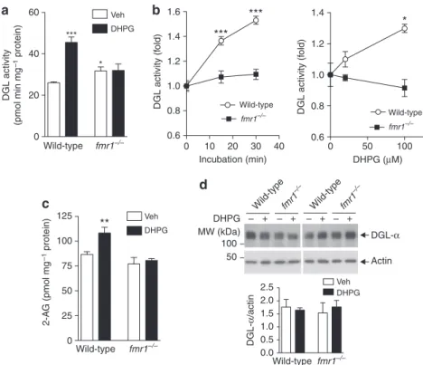
To examine whether FMRP deletion influences mGlu5-dependent 2-AG signalling, we used mouse brain synaptoneurosomes, a subcellular preparation enriched in resealed postsynaptic spines attached to their corresponding axon terminals23. As expected for previous results10,11, incubation of wild-type synaptone-urosomes with DHPG resulted in a rapid increase in DGL activ-ity, which was dependent on both incubation time and DHPG concentration (Fig. 2a,b), and was accompanied by an elevation in 2-AG levels (Fig. 2c). These effects were not associated with changes in DGL-α content (Fig. 2d) and were not prevented by the translation inhibitor cycloheximide (Supplementary Fig. S1A), indicating that they did not require the synthesis of new DGL-α protein. In synaptoneurosomes prepared from fmr1 − / − mice, DHPG failed to stimulate DGL activity and 2-AG production (Fig. 2a–c), even though baseline levels of 2-AG were similar to those measured in wild-type controls (Supplementary Fig. S1B). The results suggest that genetic loss of FMRP disrupts mGlu5-dependent 2-AG signalling at excitatory synapses.
FMRP targets DGL- to the perisynapse. One mechanism
through which FMRP deletion might affect receptor-operated 2-AG production is by altering the structure of the supramolecular com-plex that couples mGlu5 receptors to DGL-α (which we refer to here as ‘eCB signalosome’). To test this possibility, we immunostained for mGlu5 or DGL-α brain sections from wild-type and fmr1 − / − mice. We first focused on the core of the ventral striatum, a brain region in which the eCB system mediates excitatory LTD8. Immu-noperoxidase labelling for mGlu5 or DGL-α revealed a dense punctuate distribution throughout the neuropil, which appeared to be similar between wild-type and fmr1 − / − mice (Fig. 3a,b,g,h).
Further analyses by immunogold electron microscopy revealed a widespread presence of mGlu5 and DGL-α in postsynaptic profiles—predominantly in dendritic spine heads that received asymmetric, putatively glutamatergic inputs from DGL-α or mGlu5 -immunonegative axon terminals (Fig. 3c,d,i,j). To assess whether FMRP deletion alters the subcellular distribution of mGlu5 and DGL-α, in three separate experiments we measured the distance along the plasma membrane between the gold particles attached to DGL-α or mGlu5 and the closest edge of the postsynaptic density. There was no statistically detectable difference in the subcellular distribution of mGlu5 between wild-type and fmr1 − / − mice (n = 294 and 285 synapses for wild-type and fmr1 − / − mice,
respectively; Kolmogorov–Smirnov test, P = 0.917) (Fig. 3k). By contrast, the cumulative distribution functions of the subcellular localization of DGL-α were significantly different between the two genotypes (n = 259 and 185 synapses for wild-type and fmr1 − / −
mice, respectively; P = 0.001) (Fig. 3e). This suggests that FMRP deletion selectively impairs the subcellular targeting of DGL-α in postsynaptic spine heads, without affecting the targeting of mGlu5.
To investigate this phenomenon further, we divided the spine-head plasma membrane into 60-nm bins and calculated the fre-quency of gold particles within each bin. As previously shown for the hippocampus13, particles associated with DGL-α were con-centrated in the perisynaptic zone, with a gradient that decreased towards the spine neck, and were nearly absent from the intrasynap-tic domain (Fig. 3f). This skewed distribution was clearly detectable in the ventral striatum core of wild-type mice, but was totally absent in fmr1 − / − mutants (Fig. 3f). Swapping all data points in the first
60-nm bin of wild-type mice to the same data set of fmr1 − / −
ani-mals and vice versa eliminated statistical significance (Kolmogorov– Smirnov test, P = 0.187 and P = 0.332 for wild-type to fmr1 − / − swap
and fmr1 − / − to wild-type swap, respectively). As swapping data sets
in other bins did not reveal differences, the disappearance of DGL-α labelling from the perisynaptic domain may underlie the difference in the cumulative distribution functions. Furthermore, the same
analytical approach revealed a biased perisynaptic localization of mGlu5 in both wild-type and fmr1 − / − mice (Fig. 3l).
To determine whether FMRP deletion influences the subcellu-lar targeting of DGL-α in other forebrain structures, we examined the localization of mGlu5 and DGL-α in the CA1 subfield of the hippocampal formation,9,13,24,25. The perisynaptic localization of mGlu5 at hippocampal excitatory synapses was identical between
IP: anti-FMRP (�g) WB: anti-FMRP Stain: coomassie blue
100
a
b
d
e
c
h 2630A A A A C G G G G G G G G C C C C C C C C C G A A A G G G A G G G G G G G T G G G G G G A G A G G G G G G C C C C C C C C C G G G T T T T T T T G G G G G G G G G G G G G G G G G G G G G G G G G G T T T T T T G G G G G G G G G G A A A A A A A – – – A G G G A A A C m 2633 r 2633 Wild-type *** *** DGL-α mRNA (copies x 10 –3 ) DGL-α mRNA (copies) fmr1–/– 80 60 40 20 0 80 DGL-α DGL-α APP NS NS NS Anti-FMRP Anti-FMRP Anti-FMRP PSD-95 DGL-β NAPE-PLD 60 60 *** *** *** *** ** ### ## 40 40 Fold enrichmen t(mRNA in Wt-IP / KO-IP)
Fold enrichment
(mRNA in Wt-IP / KO-IP)
20 20 2.0 1.5 1.0 0.5 0.0 0 80 DHPG Veh *** 0 10 60 60 30 Incubation (min) 40 20 0 Anti-FMRP (µg) Wild-type FMRP
IgG heavy chain 100 75 75 50 MW (kDa) NS 0 7.5 15 NS 0 7.5 15 fmr1–/– Anti-FMRP (µg) NS 07.5 15 NS 07.5 15 0 7.5 15 0 7.5 15 0 7.5 15
Figure 1 | DGL- mRNA binds to FMRP. (a) Alignment of the coding region in human (h), mouse (m) and rat (r). DGL-α mRnAs reveal a putative
G-quartet sequence within a G-rich region containing several DWGG repeats. The canonical G-quartet motif is DWGG-n(0–5)-DWGG-n(0–3) -DWGG-n(0–2)-DWGG, where D is any nucleotide except C, and W is A or u. The DWGG repeats are boxed in red. (b) Co-immunoprecipitation of FmRP with DGL-α mRnA. Brains from wild-type or fmr1 − / − mice were homogenized and centrifuged at 70,000 g for 30 min. The supernatant (1 mg protein) was
incubated with the indicated amounts of anti-FmRP antibody or normal serum (ns), and the immunocomplex was precipitated using protein G-sepharose beads. Top, levels of DGL-α mRnA in the immunoprecipitates were quantified by real-time quantitative PCR (n = 3, ***P < 0.001). Bottom, a portion of the immunoprecipitate was subjected to sDs–PAGE and western blot analyses to confirm the presence of FmRP. (c,d) Analyses of mRnAs encoding for DGL-α, DGL-β and nAPE-PLD (c) or positive control PsD-95 and amyloid precursor protein (d) in anti-FmRP-immunoprecipitates (n = 3, **P < 0.01 and ***P < 0.001 compared with ns, ##P < 0.01 and ###P < 0.001 compared with DGL-α-anti-FmRP). similar results were obtained using fmr1 − / − mice bred
on either a FVB.129 background (b,c) or a C57BL/6J background (d). (e) DHPG-induced dissociation of DGL-α mRnA from FmRP in cultured cortical neurons. Rat primary neurons were prepared from embryonic day 18 cortex, as described55. Cells were treated with DHPG (100 µm) in culture medium
and harvested at the indicated time. Levels of FmRP-bound DGL-α mRnA were determined by anti-FmRP immunoprecipitation and quantitative PCR, as described in methods (n = 5, ***P < 0.001). Results are representative of at least two independent experiments. significance was determined using two-tailed student’s t-test. Error bars represent s.e.m.
fmr1 − / − and wild-type mice, whereas the localization of DGL-α
was markedly impaired in fmr1 − / − mutants (Supplementary
Fig. S2). An economical interpretation of these findings, which is consistent with our biochemical data, is that FMRP enables the correct targeting of DGL-α and its assembly with mGlu5 into a functional eCB signalosome at excitatory synapses throughout the forebrain.
FMRP is a core component of the translational machinery in dendritic polyribosomes and has been implicated in the transport and localization of mRNAs to dendrites and synapses3.
We reasoned therefore that, in the absence of FMRP, DGL-α mRNA might be translated ectopically. Consistent with this prediction, an analysis of 300 synapses per genotype revealed an increased aver-age density of intracellular DGL-α labelling in the ventral striatum of fmr1 − / − mice, compared with wild-type animals (0.91 (range:
0.6–1.43) gold particles per µm2 and 3.02 (range: 2.19–3.63) gold particles per µm2 in wild-type and fmr1 − / − animals, respectively,
normalized to background levels) (Fig. 3m). This increase in nor-malized average density was also reflected in the ratio of intracel-lularly localized DGL-α (14% (range: 10–17%) and 36% (range:
60 1.6 DGL activity (f old) DGL activity (f old) 1.4 1.2 1.0 0.8 0.6 1.4 1.2 1.0 0.8 0.6 Veh DHPG Veh DHPG DGL-α Actin MW (kDa) Wild-t ype Wild-t ype – + – + – + – + fmr1 –/– fmr1 –/– 100 50 DHPG Veh DHPG 2.5 2.0 DGL-α /acti n 1.5 1.0 0.5 0.0 40 40 50 100 30 Incubation (min) DHPG (µM) 10 Wild-type Wild-type fmr1–/– fmr1–/– *** *** * *** * 20 20 Wild-type Wild-type DGL activity (pmol min mg –1 protein) fmr1–/– Wild-type fmr1–/– fmr1–/– 0 0 25 50 75 100 ** 2-A G (pmol mg –1 protein) 125 0 0
Figure 2 | mGlu5 receptor-stimulated DGL activity and 2-AG release is disrupted in fmr1 − / − mice. (a) synaptoneurosome (sn) fractions prepared
from wild-type or fmr1 − / − mice (1 mg ml − 1) were incubated at 37 °C for 30 min in the presence of 100 µm DHPG, and DGL activity was measured in vitro
using 10 µm diheptadecanoylglycerol as a substrate (n = 4). (b) DGL activity was measured in sn fractions after incubation with various concentrations of DHPG (n = 3). (c) 2-AG levels in sn fractions were measured after 45-min incubation with DHPG (100 µm) or vehicle (Veh, PBs) (n = 5–9). (d). After treatment of synaptoneurosomes with DHPG, as described above, levels of DGL-α protein were measured by western blot. Representative images for DGL-α and loading control actin are shown (n = 6 each). Experiments were conducted on fmr1 − / − mice on C57BL/6J (a,d) or FVB.129 background (b,c).
Results are representative of at least two independent experiments. significance was determined using two-tailed student’s t-test. *P < 0.05, **P < 0.01 and ***P < 0.001. Error bars represent s.e.m.
Figure 3 | Impaired targeting of DGL- to the perisynaptic domain of excitatory synapses in fmr1 − / − mice. DGL-α immunoperoxidase labelling in
wild-type (a) and fmr1 − / − mice (b) reveals a similar granular staining pattern in the neuropil of the accumbens/ventral striatum (arrowheads). medium spiny
neuron somata are devoid of labelling (asterisks). aco, anterior commissure. Electron micrographs of asymmetric synapses reveal that DGL-α-positive gold particles (arrowheads) are predominantly found perisynaptically, close to the PsD (red arrows) in wild-type animals (c), but not in fmr1 − / − synapses
(d) s, spine head; b, bouton. (e) Cumulative distribution plot of DGL-α localization in wild-type (solid line, n = 259) and fmr1 − / − spines (dashed line,
n = 185). (f) Histogram summarizing the normalized distribution of DGL-α-immunogold on the spine head membrane of wild-type mice (black arrow and black bars, n = 259) and fmr1 − / − mice (grey arrow and grey bars, n = 185). Large arrow = edge of PsD. mGlu
5 immunoperoxidase labelling in wild-type
(g) and fmr1 − / − animals (h) depicts a similar granular labelling pattern (arrowheads) as DGL-α. Electron micrographs of asymmetric synapses containing
mGlu5-immunogold labelling (arrowhead) revealed that mGlu5 was predominantly found perisynaptically (red arrows) in both wild-type (i) and fmr1 − / −
spines (j). (k) Cumulative distribution plot of mGlu5-immunogold localization in wild-type (solid line, n = 294) and fmr1 − / − mice (dashed line, n = 285).
(l) Histogram representing the normalized distribution of mGlu5-immunogold on spine heads of wild-type (black arrow and black bars, n = 294) and fmr1 − / − spines (grey arrow and grey bars, n = 285). The number of DGL-α-positive spines was higher in fmr1 − / − mice; however, the density of spine head
membrane labelling was the same in both strains. Instead, a significantly higher density of DGL-α labelling was found within the spine head and neck cytoplasm (arrowheads; m1–m2) and along the spine neck membrane (black arrowheads; n). symmetrical synapses are considered inhibitory GABAergic inputs and frequently synapse onto spine necks (red arrowhead; n) and dendritic shafts Importantly, the number of DGL-α positive symmetrical synapses were similar in wild-type (o) and fmr1 − / − mice (p). scale bars: 15 µm in a,b,g and h and 100 nm in c,d,i,j,m–p.
33–46%) of all gold particles found in dendritic spine heads in wild-type and fmr1 − / − animals, respectively). In another experiment, we
assessed the presence of DGL-α labelling in a second data set of 75 random intact spines per animal. We found that fmr1 − / − spines
had a higher probability of DGL-α labelling than did wild-type spines (χ2 test, P < 0.001).
In which subcellular structure was DGL-α labelling concentrated? To address this question, we divided each spine into three distinct Wild-type
DGL-α mGlu5
fmr1–/– aco fmr1–/– fmr1–/– fmr1–/–
Wild-type Wild-type Wild-type
aco aco aco b b 100 80 30 25 20 Immunopar ticle % Immunopar ticle % 15 10 5 0 30 25 20 15 10 5 0 Wild-type fmr1–/– Wild-type fmr1–/– fmr1–/– 60 % of gold par ticles % of gold par ticles 40 20 0 100 80 60 40 20 0 0 –200 –120 –60 0 60120 180 240 300 360 420 480 540 600 660 –100 100 200 300 Distance from PSD (nm) Distance from PSD (nm) –120 –60 0 60120 180 240 300 360 420 480 540 600 660 Distance from PSD (nm) Wild-type fmr1–/– Wild-type DGL-α fmr1–/– DGL-α fmr1–/– fmr1–/– Wild-type DGL-α fmr1–/– b b b b b s s s d d 400 P = 0.001 P = 0.917 500 700 800 b b s s s s * * * * * * * * 600 –200 –100 0 100 200 300 Distance from PSD (nm) 400 500 600 700 800
compartments: spine-head membrane, spine-neck membrane, and head and neck cytoplasm. The number of DGL-α gold particles on the spine head membrane was very similar between wild-type and
fmr1 − / − mice (χ2 test, P = 0.696). By contrast, we found a signifi-cantly higher number of DGL-α gold particles in spine neck mem-brane (χ2 test, P < 0.001) (Fig. 3n) and cytoplasm (χ2 test, P = 0.003) of fmr1 − / − mice. In agreement with previous studies26,27, we found that dendrites in fmr1 − / − mice exhibited longer spine necks than
wild-type (852 ± 19 and 1030 ± 24 nm in wild-type and fmr1 − / −
respectively; Kolmogrov–Smirnov test, P < 0.001), which might account for the higher number of DGL-α gold particles observed in these animals. However, when we calculated the density of DGL-α labelling for each spine compartment, this was also signifi-cantly higher in the fmr1 − / − necks than wild-type mice (2.29 (range:
1.63–3.01) gold particles per µm2 and 4.67 (range: 3.63–5.85) gold particles per µm2 in wild-type and fmr1 − / − animals, respectively).
By contrast, the density of DGL-α labelling in the spine-head membrane was similar between the two strains (1.26 (range: 0.43–1.89) gold particles per µm2 and 1.42 (range: 1.06–1.86) gold particles per µm2 in wild-type and fmr1 − / − animals, respectively),
but higher intracellular labelling was observed in fmr1 − / − mice
(0.4 (range: 0.2–0.8) gold particles per µm2 and 0.91 (range: 0.78– 0.92) gold particles per µm2 in wild-type and fmr1 − / − animals,
respectively).
Finally, we examined whether FMRP deletion might affect DGL-α localization at GABAergic synapses, which are also dys-functional in fmr1 − / − mice18,19. DGL-α gold particles were found at the postsynaptic plasma membrane of both wild-type (Fig. 3o) and fmr1 − / − mice (Fig. 3p), but no statistically detectable difference
was noted in their distribution (χ2 test, P = 0.6, n = 150). The results suggest that genetic FMRP deletion alters the normal localization of DGL-α at excitatory, but not inhibitory, synapses of the forebrain, causing this enzyme to accumulate ectopically in the neck and cytoplasm of dendritic spines.
FMRP enables eCB-LTD in ventral striatum and PFC. The
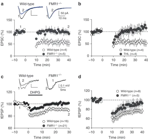
bio-chemical and ultrastructural results reported above suggest that FMRP deletion reduces 2-AG-dependent LTD at excitatory synapses of the forebrain. In acutely dissected brain slices from wild-type mice, low-frequency electrical stimulations (10 min, 10 Hz) elic-ited robust LTD of excitatory postsynaptic currents (EPSCs, Fig. 4a, white symbols) or extracellular field potentials (fEPSP, see Fig. 5a) in medium spiny neurons of the ventral striatum8. The induction of LTD was prevented by the DGL-α inhibitor tetrahydrolipstatin (10 µM) (Fig. 4b), as expected from previous work9,28, and was not affected by pharmacological blockade of protein synthesis (Supplementary Fig. S3). Importantly, the same low-frequency stimulations that caused LTD in wild-type mice failed to do so in fmr1 − / − mutants (Figs 4a
and 5a, black symbols). Moreover, bath applications of the mixed mGlu1/mGlu5 agonist, DHPG, which consistently elicited LTD in brain slices prepared from wild-type mice, were ineffective in slices obtained from fmr1 − / − mutants (Fig. 4c). To evaluate whether the
deficits in eCB-LTD observed in fmr1 − / − mutants extended beyond
the ventral striatum, we took advantage of the fact that this brain region receives major excitatory projections from the medial PFC, a structure where mGlu5 activation also underlies 2-AG-dependent eCB-LTD at synapses of pyramidal neuron layers 2/3 and 5/6 (refs 9,29). Similar to the ventral striatum, eCB-LTD was absent in PFC slices obtained from fmr1 − / − mutants (Fig. 4d). Together, these
results support the idea that FMRP deletion causes a widespread impairment of mGlu5-dependent LTD at excitatory synapses.
To test the alternative possibility that the LTD deficit observed in
fmr1 − / − mice might result from impaired CB1 receptor function,
we assessed the ability of the cannabinoid agonist CP55940 (0.01– 10 µM) to inhibit synaptic transmission in slices of ventral striatum prepared from wild-type or fmr1 − / − mice. The drug’s potency and
efficacy were identical in the two strains (Supplementary Fig. S4A), indicating that CB1 receptors function normally in fmr1 − / − mice. Moreover, tonic eCB signalling did not appear to be affected by FMRP deletion, because the selective CB1 inverse agonist AM251 (4 µM, applied 30 min before testing) did not alter baseline excita-tory transmission in the ventral striatum of wild-type or fmr1 − / −
mice (Supplementary Fig. S4B). Thus, the lack of eCB-dependent plasticity observed in fmr1 − / − mice cannot be accounted for by
alterations in CB1 function or tonic eCB activity. We also compared the intrinsic properties of ventral striatum medium spiny neurons in wild-type and fmr1 − / − mice, and found no difference in medium
spiny neurons excitability (Supplementary Fig. S5). Deletion of FMRP may negatively impact the basic properties of ventral stria-tum excitatory synapses and, to test for this possibility, we recorded AMPA receptor-mediated spontaneous EPSCs (sEPSCs) in medium spiny neurons of both genotypes. The amplitude and frequency of AMPAR sEPSCs were similar in wild-type and fmr1 − / − mice
(Supplementary Fig. S6). The results rule out the possibility that modifications of excitability or basal synaptic properties of ventral striatum medium spiny neurons may be responsible for impaired eCB-LTD in fmr1 − / − mice.
To test whether FMRP might influence intracellular signal trans-duction pathways mediating other forms of LTD, we also analysed the LTD evoked by presynaptic mGlu2/3 receptors, which utilizes the same transduction mechanisms that are engaged by presyn-aptic CB1 receptors30,31. Bath application of the selective mGlu2/3 agonist LY379268 (100 nM) produced comparable levels of LTD in wild-type and fmr1 − / − mice (Supplementary Fig. S7). Together,
our experiments support the conclusion that FMRP enables mGlu5 to initiate retrograde 2-AG signalling and elicit LTD at excitatory synapses of the forebrain.
Enhancement of 2-AG signalling normalizes LTD in fmr1 − / −
mice. The results reported above suggest that, in fmr1 − / − mice, the
failure of DGL-α to associate with mGlu5 into a functional eCB sig-nalosome results in a loss of eCB-LTD. We have previously shown that pharmacological blockade of intracellular 2-AG hydrolysis, which is predominantly catalysed by presynaptic MGL16,32, lowers the threshold for LTD induction at excitatory synapses of the ven-tral striatum9,29. We hypothesized therefore that inhibitors of 2-AG degradation might normalize LTD in fmr1 − / − mice by
reinforc-ing 2-AG activity at residual intact synapses or, possibly, extendreinforc-ing the reach of ectopically produced 2-AG. To test this possibility, we blocked 2-AG hydrolysis in slices using the potent and irreversible MGL inhibitor, JZL18433. Brain slices were incubated with JZL184 (1 µM) for 45–90 min before LTD induction. This treatment was sufficient to restore both synaptic and pharmacologically induced LTD in slices prepared from fmr1 − / − mutants (Fig. 5a,b), but had
no effect in slices from wild-type controls (Supplementary Fig. S8). In addition, inhibition of the postsynaptic 2-AG-hydrolysing enzyme ABHD629 with WWL70 (10 µM, 45–90-min incubation) also restored LTD in slices from ventral striatum of fmr1 − / − mice
(Fig. 5a,b). The effects of JZL184 and WWL70 indicate that phar-macological blockade of either presynaptic or postsynaptic 2-AG degradation rescues eCB-dependent LTD in fmr1 − / − mice.
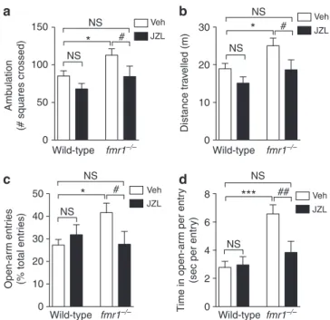
Enhancing 2-AG corrects behavioural changes in fmr1 − / − mice. When bred on a C57BL/6 background, fmr1 − / − mice display a
behavioural phenotype that is characterized by elevated motor activity in an open field (Fig. 6a,b) and decreased aversion to the open arms of an elevated plus maze34 (Fig. 6c,d). The MGL inhibi-tor JZL184 (16 mg kg − 1, intraperitoneal) increased brain 2-AG levels (Supplementary Fig. S9A) and corrected these behavioural abnormalities within 6 h of administration (Fig. 6a–d). In fmr1 − / −
mice, JZL184 reduced the number of squares crossed (Fig. 6a) and the total distance travelled (Fig. 6b) in the open field test.
The compound also reduced the number of entries and the time spent in the open arm in the elevated plus maze test (Fig. 6c,d and Supplementary Fig. S9C, D). By contrast, JZL184 did not signifi-cantly affect open field or elevated plus maze behaviours in wild-type mice (Fig. 6a–d and Supplementary Fig. S9B–D). These experiments suggest that pharmacological enhancement of 2-AG signalling normalizes two key behavioural changes observed in fmr1 − / − mice.
Discussion
In the present study, we provide three lines of evidence indicating that FMRP exerts a tight regulatory control over 2-AG-dependent retrograde signalling at excitatory synapses of the mouse forebrain. First, we show that an FMRP antibody selectively immunoprecipi-tates DGL-α mRNA in forebrain synaptoneurosome extracts. This is suggestive of a direct association between the two molecules, pos-sibly through an interaction of FMRP with the G-quartet motif of DGL-α mRNA35. Second, we report that the normal perisynaptic colocalization of DGL-α and mGlu5 is severely impaired in fmr1 − / − mice, indicating that FMRP is necessary for the correct targeting of DGL-α to dendritic spines. Finally, we find that FMRP deletion disrupts both mGlu5-stimulated 2-AG production in synaptoneu-rosomes and LTD at excitatory synapses of the ventral striatum and PFC. These findings suggest that FMRP may direct the assembling
of DGL-α and mGlu5 into a functional signalosome, and is essential for retrograde 2-AG transmission at glutamatergic synapses (Fig. 7). Underscoring the functional significance of these results, we show that pharmacological blockade of 2-AG degradation rein-states eCB-dependent LTD and corrects key behavioural changes in
fmr1 − / − mice.
The eCB 2-AG is considered to be a primary effector of mGlu5 -dependent plasticity in the brain9,12,28, but the dynamics of 2-AG signalling at central synapses are still unclear. One important ques-tion pertains to the mechanism through which mGlu5 activation stimulates production of the 2-AG pool responsible for retrograde eCB transmission. Based on the present results, we hypothesize that the stable association of mGlu5 and DGL-α into a multiprotein complex at the perisynaptic annulus of the dendritic spine provides a structural scaffold that allows for the localized formation of a sig-nalling-competent pool of 2-AG, which may be distinct from the intracellular 2-AG pools involved in phospholipid remodelling and eicosanoid biosynthesis36,37. We propose to call this complex ‘eCB signalosome’, to signify its pivotal role in eCB-mediated retrograde transmission (Fig. 7).
How does the lack of FMRP expression alter the structure of the eCB signalosome? The available data suggest two possibilities. The first is that, in the absence of FMRP, the DGL-α message may
EPSC (% ) 0 10 20 30 40 0 1 2 1 2 1 2 50 pA 10 ms 60 100 100 80 1 2 1 2 1 2 DHPG 5ms 0.1 mV 40 60 80 150
a
b
c
d
100 50 0 150 100 50 EPSC (% ) Time (min) –10 0 10 20 30 Time (min) –10 0 10 20 30 40 Time (min) –10 0 10 20 30 40 Time (min) –10 Wild-type (n=4) FMR1–/– (n=5) 120 120 fEPSP (% ) fEPSP (% ) Wild-type (n=16) FMR1–/– (n=21) Wild-type (n=8) FMR1–/– (n=5) Wild-type (n=4) THL (n=4) Wild-type FMR1–/– Wild-type FMR1–/–Figure 4 | LTD is abolished in ventral striatum and PFC of fmr1 − / − mice. (a) summary graph showing whole-cell evoked EPsC amplitudes. Values are
normalized to baseline before the induction of LTD and averaged per minute. Tetanic stimulation (10 min at 10 Hz starting at time 0) of PFC afferent fibres to medium spiny neurons in the ventral striatum induces a robust LTD in wild-type littermates (open symbols, n = 4) but not fmr1 − / − mice (filled
symbols, n = 5). Here as in all physiology figures, n equals the number of animals. (b) summary graph showing averaged time courses of the experiments in which the 10 min at 10-Hz protocol was given in control ACsF (open symbols, n = 4) and after pre-treatment with tetrahydrolipstatin (THL, 10 µm, filled symbols, n = 4), an inhibitor of the DGL-α. Graphs show EPsC amplitudes normalized to baseline before the induction of LTD and averaged per minute. (c) Direct pharmacological activation of mGlu1/5 with 50 µm (S)-DHPG induces LTD in the ventral striatum of wild-type littermates (open symbols, n = 16) but not fmr1 − / − mice (filled symbols, n = 21). summary graphs show excitatory postsynaptic field potentials (fEPsP) amplitudes. (d) summary
graph showing fEPsP amplitudes recorded in pyramidal neurons of the PFC. Values are normalized to baseline before the induction of LTD and averaged per minute. Tetanic stimulation (10 min at 10 Hz starting at time 0) of layers 2/3 to layers 5/6 pyramidal synapses induces a robust LTD in wild-type littermates (open symbols, n = 8) but not fmr1 − / − mice (filled symbols, n = 5). statistical significance was determined using mann–Whitney U-test.
be incorrectly targeted within the dendritic spines, leading to errors in the timing of DGL-α translation or the localization of the trans-lated protein. An error in spatial mRNA delivery caused by FMRP deletion was recently demonstrated for another key postsynaptic protein, calmodulin-activated protein kinase II38. A second possi-bility is that FMRP deletion may produce an alteration in the func-tion of Homer proteins. These scaffolding proteins bind both mGlu5 and DGL-α10, and single-point mutations in the Homer-2-binding region of DGL-α result in an enzyme that retains functional activ-ity, but is found in intracellular membrane compartments rather than the plasma membrane10. Notably, uncoupling of mGlu5 from the postsynaptic Homer scaffold results in the absence of mGlu- activated retrograde 2-AG signalling at excitatory synapses39. Previ-ous studies suggest a similar functional uncoupling between mGlu5 and Homer-2 in fmr1 − / − mice, despite comparable protein levels in synaptoneurosome preparations40. In support of this idea, an altered balance in mGlu5 scaffolding between short-form Homer 1a and long-form Homer proteins was found in fmr1 − / − mice41. The hypotheses outlined above will be tested in future experiments.
The mGlu receptor theory of FRAX is based on the seminal observation that fmr1 − / − mice display a marked enhancement
of a form of hippocampal LTD that depends on mGlu receptor-mediated protein synthesis3,5–7. Consistent with this theory, the eCB-mediated LTD induced at CA1 GABAergic synapses by phar-macological activation of mGlu5 is similarly enhanced in fmr1 − / − mice18. Likewise, in the dorsal striatum of these mutants mGlu5 -mediated eCB activity at GABAergic synapses is increased19. The present results, showing that eCB-LTD is abolished in the PFC and
ventral striatum of fmr1 − / − mice, are only apparently in opposition
with those data. Indeed, the notion that different neuron and syn-apse types adapt differently to the lack of FMRP is supported by a recent study showing that mGlu receptor-dependent LTP is reduced in the lateral amygdala of fmr1 − / − mice42. Although we found no changes in DGL-α density at GABAergic synapses, 2-AG signal-ling was suggested to be heterosynaptic between glutamatergic and GABAergic synapses43 (that is, 2-AG released from dendritic spines might activate CB1 receptors on more distant GABAergic terminals). Thus, it is possible that the shifted DGL-α localization gradient from the perisynaptic annulus to the spine neck compart-ment, where GABAergic synapses are often present (for example see red arrowhead in Fig. 3n), may underlie the opposite altera-tions in CB1-dependent regulation of synaptic plasticity between these synapses (refs 18,19 and present study). Our findings are in line, therefore, with the core prediction of the mGlu receptor the-ory—namely, the idea that loss of FMRP alters most consequences of mGlu5 activation44. On the other hand, the present results reveal molecular disparities between excitatory and inhibitory synapses of the same brain region, and highlight that distinct forms of LTD may also behave differentially in FRAX. While protein synthesis-dependent LTD is enhanced, presynaptic mGlu2/3-dependent LTD is unaffected, and finally the protein synthesis-independent, but eCB-mediated, LTD is absent. This palette of synaptic changes might underlie the complex neurobehavioural abnormalities observed in FRAX patients.
Pharmacological or genetic modulation of mGlu5 signalling nor-malizes various functional and structural aberrations occurring in 60 70 80 90 100 0 10 20 30 40 40 60 80 100
a
b
fEPSP (% ) Time (min) –10 0 10 20 30 Time (min) –10 fEPSP (% ) 110 DHPG Wild type (n=16) Control FMR1–/– (n=21) JZL184 FMR1–/– (n=5) WWL70 FMR–/– (n=8) Wild type (n=19) Control FMR1–/– (n=20) JZL184 FMR1–/– (n=6) WWL70 FMR–/– (n=8)Figure 5 | Inhibitors of 2-AG degradation rescues eCB-dependent LTD in
fmr1 − / − mice. (a) normalized fEPsP amplitude in fmr1 − / − mice without
(n = 20; black symbols) or with JZL184 (1 µm; blue symbols, n = 6) or WWL70 (10 µm; green symbols, n = 8) pretreatment (starting 45–90 min before LTD induction) followed by the LTD induction protocol at time zero. (b) normalized fEPsP amplitude in fmr1 − / − mice without (n = 20;
black symbols) or with JZL184 (1 µm; blue symbols, n = 5) or WWL70 (10 µm; green symbols, n = 8) pretreatment (starting 45–90 min before LTD induction) followed by 10-min bath perfusion with 50 µm (S)-DHPG at time zero. statistical significance was determined using mann–Whitney U-test. Error bars represent s.e.m.
150
a
b
c
d
NS NS * # *** ## NS 50 8 6 4 2 0 40 30 20 10 0 NS Veh Veh JZL JZL Veh JZL Veh JZL NS NS NS NS * # * # Wild-type fmr1–/– Wild-type fmr1–/– Wild-type fmr1–/– Wild-type fmr1–/– 100 50 0 0 10 20 Distance tr av elled (m ) Amb ulatio n (# squares crossed) Open-a rm entr ies (% total ent ries) Time in open-ar m per entr y(sec per ent
ry
)
30
Figure 6 | MGL inhibition normalizes behavioural alterations in fmr1 − / −
mice. Effects of the mGL inhibitor JZL184 on open field (a,b) and elevated
plus maze (c,d) behaviours. Wild-type and fmr1 − / − mice received JZL184
(JZL, 16 mg kg − 1, intraperitoneal) or vehicle (Veh), and were tested 6 h
after injections. Activity in the open field was measured by counting the number of squares crossed (a) and total distance travelled (b). In the elevated plus maze test, we measured percentage of open arm entries (c) and average time in open per each entry (d). significance was determined using two-way analysis of variance with post-hoc student– newman–Keuls test. ns, not significant; *P < 0.05, and ***P < 0.001 compared with wild-type-Veh; #P < 0.05, and ##P < 0.01 compared with
fmr1 − / − mice—including hippocampal LTD, immature dendritic
arbourization and behavioural deficits44. However, this approach is unlikely to restore LTD at synapses of the ventral striatum and the PFC, where we find mGlu5-dependent eCB production to be down-regulated. We reasoned therefore that one possible alternative could be to restore eCB-mediated plasticity by reducing the degradation of its main effector, 2-AG. We used JZL184, an irreversible inhibitor of the 2-AG-degrading enzyme MGL33,45,46. This compound proved to be effective at restoring eCB-LTD in ventral striatum slices as well as correcting two key behavioural changes observed in fmr1 − / −
mice—elevated motor activity in an open field and decreased aver-sion to the open arms of an elevated plus maze34. Despite these promising findings, the therapeutic potential of JZL184 might be limited by its ability to induce CB1 receptor desensitization46. Future experimentation will be required to assess whether other classes of MGL inhibitors (for example, reversible inhibitors)47 or agents that block additional 2-AG-hydrolysing enzymes, such as ABHD629, might normalize behaviour in fmr1 − / − mice without inducing CB1
desensitization. Supporting the latter possibility, we observed here that acute blockade of ABHD6 re-establishes eCB-LTD in ventral striatum slices from fmr1 − / − mice.
In conclusion, our results indicate that FMRP expression is nec-essary for the correct assembling of the eCB signalosome at gluta-matergic synapses of the frontal cortex and ventral striatum core, and identify this macromolecular complex as a previously unknown substrate for FRAX, which might be targeted by therapy.
Methods
Physiology. Animals were treated in compliance with the European Communities Council Directive (86/609/EEC) and the United States National Institutes of Health Guide for the Care and Use of Laboratory Animals. All animals were housed, grouped and acclimated to laboratory conditions for 4 days before experiments with 12-h light/dark cycles, and access to food and water ad libitum.
Male fmr1 − / − mice on a C57BL/6J genetic background48 aged 4 to 12 weeks
(fmr1 − / − ) were used, with wild-type littermates used as control group. They were
anesthetized with isoflurane and decapitated according to institutional regulations.
The brain was sliced (300 µm) in the coronal plane with a vibratome (Integraslice, Campden Instruments, Loughborough, UK) in a sucrose-based solution at 4 °C (in mM: 87 NaCl, 75 sucrose, 25 glucose, 5 KCl, 21 MgCl2, 0.5 CaCl2 and 1.25
NaH2PO4). Immediately after cutting, slices were stored at room temperature
in a low-calcium artificial cerebrospinal fluid (low Ca–ACSF) that contained (in mM): 130 NaCl, 11 glucose, 2.5 KCl, 2.4 MgCl2, 1.2 CaCl2, 23 NaHCO3
and 1.2 NaH2PO4, and was equilibrated with 95% O2/5% CO2 until the time
of recording.
Whole-cell patch-clamp of visualized medium spiny neurons, PFC pyramidal neurons and field potential recordings were made in coronal slices containing the ventral striatum or the PFC as previously described8,9,49,50. For more details,
see Supplementary Methods.
All values are given as mean ± s.e.m. For all experiments, n corresponds to the number of animals tested for each condition. Data were analysed using Clampfit 10 (Molecular Devices, Sunnyvale, USA). Unless otherwise stated, the statistical test used was a Mann–Whitney U-test performed with GraphPad Prism (GraphPad Software, La Jolla, CA) and significance determined by P < 0.05. The magnitude of LTD was calculated by comparing average responses (30–40 min) after induction to baseline-averaged responses before induction protocol (increased stimulation for 10 min at 10 Hz).
Picrotoxin, THL and anisomycin were from Sigma (St Quentin Fallavier, France). 6,7-dinitroquinoxaline-2,3-dione (DNQX), (1R,3R,4R)-3-[2-hydroxy- 4-(1,1-dimethylheptyl)-phenyl]-4-(3-hydroxypropyl)-cyclohexan-1-ol (CP55940), DHPG, AM251, WWL70 and JZL184 were from Tocris (Bristol, UK). Other chemicals were of the highest commercial grade available.
Biochemistry. Male fmr1 − / − mice (FVB.129P2-Fmr1tm1Cgr/J) and wild-type
control mice (FVB.129P2-Pde6b + Tyrc − ch/AntJ) from Jackson Laboratories
(Bar Harbor, ME), aged between 6 to 10 weeks, or adult male fmr1 − / − mice on
C57BL/6J background (Fmr1 KO2) were used for the biochemical studies. All procedures met the National Institutes of Health guidelines for the care and use of laboratory animals and were approved by the University of California, Irvine, Institutional Animal Care and Use Committee.
All manipulations of brain homogenates were performed at 4 °C51.
Synaptone-urosomes were prepared from whole brains of wild-type or fmr1 − / − mice. Briefly,
mice were quickly decapitated, brains were removed and homogenized by 20 strokes with a glass-Teflon homogenizer in 10 ml of homogenizing buffer (118 mM NaCl, 4.7 mM KCl, 1.2 mM MgSO4, 2.5 mM CaCl2, 1.53 mM KH2PO4, 212.7 mM
glucose, 1 mM DTT, pH7.4, and protease inhibitor) and filtered through a series of polypropylene filters (twice through 100 µm and once through 10 µm (BD Filcon). Filtrates were spun briefly (1,000 g, 15 min) and the pellets were reconstituted in 3 ml of homogenizing buffer at a protein concentration of 1 mg ml − 1.
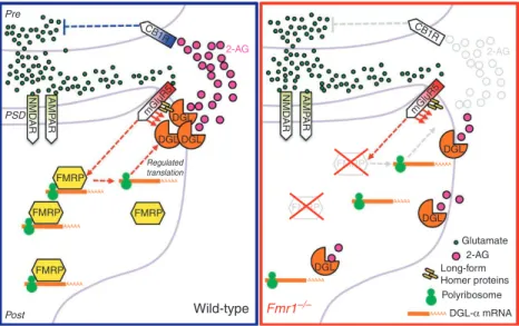
FMRP FMRP Wild-type Glutamate 2-AG 2-AG Long-form Homer proteins Polyribosome DGL-α mRNA Post Fmr1–/– FMRP FMRP PSD Pre CB1R CB1R mGluR5 mGluR5 NM D A R AMP AR AMP AR NMD AR FMRP FMRP DGL DGL DGL DGL DGL Regulated translation DGL 2-AG
Figure 7 | Hypothetical role of FMRP in eCB-mediated retrograde transmission. At glutamatergic synapses of wild-type mice (left), mGlu5 receptors
and DGL-α are linked together in a postsynaptic signalling complex (eCB signalosome), which includes the scaffolding protein Homer. stimulation of mGlu5 receptors (mGluR5) by glutamate (green circles) increases DGL-α activity (red arrows), causing a localized rise in 2-AG levels at the perisynapse.
2-AG (red circles) travels across the synaptic cleft and inhibits glutamate release by stimulating CB1 receptors (CB1R) on nerve endings. We hypothesize that FmRP helps target DGL-α to the eCB signalosome, possibly by positioning the DGL-α or Homer message(s) in specific subcellular nanodomain(s). In fmr1 − / − mice (right), the lack of FmRP expression leads to disruption of this spatial organization, loss of localized 2-AG production and, consequently,
impaired retrograde 2-AG-mediated signalling. AmPAR, AmPA receptors; nmDAR, nmDA receptors; PsD, postsynaptic density. Red arrows indicate intracellular signalling pathways triggered by the activation of mGlu5 receptors. molecular pathways that are impaired in fmr1 − / − mice are shown
Dissected brain regions were homogenized in methanol (1 ml) containing [2H8]-2-AG and N-heptadecanoylethanolamide (Cayman Chemicals, Ann Arbor,
MI, USA) as internal standards. Protein concentration was determined in the homogenate to normalize samples using the bicinchinonic acid protein assay (Pierce, Rockford, IL, USA). Lipids were extracted with chloroform (2 ml) and water (1 ml), and further fractionated by open-bed silica gel column chromato-graphy, as previously described52. Lipids were reconstituted in chloroform,
loaded onto small glass columns packed with Silica Gel G (60-Å 230–400 Mesh ASTM; Whatman, Clifton, NJ, USA) and washed with 2 ml of chloroform. 2-AG were eluted with 1 ml of chloroform/methanol (9:1, vol/vol). Eluates were dried under N2 and reconstituted in chloroform/methanol (1:3, 50 µl)
for liquid chromatography/mass spectrometry (LC/MS) analyses.
An 1100-LC system coupled to a 1946A-MS detector (Agilent Technologies, Inc., Palo Alto, CA, USA) equipped with an electrospray ionization interface was used to measure anandamide and 2-AG levels in lipid extracts. Lipids were sepa-rated using a XDB Eclipse C18 column (50×4.6 mm i.d., 1.8 µm, Zorbax), eluted with a gradient of methanol in water (from 75 to 85% in 2.5 min, to 90% in 7.5 min, to 100% in 14 min and to 75% in 20 min) at a flow rate of 1.0 ml min − 1. Column
temperature was kept at 40 °C. MS detection was in the positive ionization mode, capillary voltage was at 3 kV and fragmentor voltage varied from 120 V. N2
was used as drying gas at a flow rate of 13 l min − 1 and temperature of 350 °C.
Nebulizer pressure was set at 60 PSI. Quantifications were conducted using an isotope-dilution method52 by monitoring Na + adducts of the molecular ions
([M + Na] + ) in the selected ion-monitoring mode. Quantification limits for 2-AG
was 0.4 pmol. For reverse transcriptase-PCR and quantitative PCR and immuno-precipitation, detailed procedures are provided in Supplementary Methods.
Results are expressed as the mean ± s.e.m. Statistical significance was evaluated using the Student’s t-test.
Animal behaviour. Adult male fmr1 − / − C57BL/6J mice were used for the animal
behaviour studies. For open field and elevated plus maze, detailed procedures provided in Supplementary Methods53,54.
Anatomy. Experiments were performed according to the guidelines of the insti-tutional ethical code and the Hungarian Act of Animal Care and Experimentation (1998; XXVIII. Section 243/1998). Adult male C57BL/6J mice (three wild-type and three fmr1 − / − mice (8 weeks old) were deeply anesthetized with a mixture of
keta-mine–xylazine (25 mg ml − 1 ketamine, 5 mg ml − 1 xylazine, 0.1% w/w pipolphen in
H2O; 1 ml per 100 g, intraperitoneal). Animals were then perfused transcardially
with 0.9% saline for 2 min, followed by 100 ml of fixative containing 4% parafor-maldehyde and 0.1% glutaraldehyde in 0.1 M phosphate buffer (PB), pH 7.4, for 20 min. After perfusion, the brain was removed from the skull, cut into blocks, post-fixed for 2 h and washed in PB. The blocks containing the ventral striatum were sliced into 50-µm-thick coronal sections of the brain with a Leica VTS-1000 vibratome (Vibratome, St Louis, MO, USA).
After slicing and extensive washing in 0.1 M PB, the sections were incubated in 10% sucrose for 15 min and 30% sucrose overnight, followed by freeze thawing over liquid nitrogen four times. Subsequently, all washing steps and dilutions of the antibodies were performed in 0.05 M Tris-buffered saline (TBS), pH 7.4. After extensive washing in TBS, the sections were blocked in 5% normal goat serum for 45 min and then incubated in rabbit anti-DGL-α (1:3,000, ‘INT’;13) or rabbit
anti-mGlu5 (1:1,000, AB5675, Millipore, Billerica, MA, USA) for a minimum of 48 h at
4 °C. In the immunoperoxidase staining procedure, after primary antibody incuba-tions, the sections were treated with biotinylated goat anti-rabbit IgG (1:400, Vector Laboratories) for 2 h and after three washes in TBS, with avidin biotinylated– horseradish peroxidase complex (1:500; Elite ABC; Vector Laboratories) for 1.5 h. After extensive washing, the immunoperoxidase reaction was developed using 3,3′-diaminobenzidine as the chromogen. In the immunogold staining procedure, after the primary antibody incubation, the sections were washed extensively in TBS before incubation in 0.8 nm gold-conjugated goat anti-rabbit secondary antibody (1:50; AURION, Wageningen, The Netherlands), overnight at 4 °C. Then sections were silver intensified using the silver enhancement system R-GENT SE-EM according to the kit protocol (AURION). After development of both types of immunolabelling, the sections were treated with osmium tetroxide (0.5% for immunogold labelling, 1% for immunoperoxidase reaction) in 0.1M PB for 20 min (at 4 °C for immunogold labelling) and dehydrated in an ascending series of ethanol and acetonitrile, before being embedded in Durcupan (ACM, Fluka, Buchs, Switzerland). During dehydration, sections were treated with 1% uranyl acetate in 70% ethanol for 15 min (at 4 °C for immunogold labelling). Immunoperoxidase-labelled sections were imaged with a Nikon Eclipse 80i microscope (Nikon Instru-ments Europe B.V., Amsterdam, The Netherlands) and light micrographs were captured with a DS-Fi1 digital camera (Nikon Instruments Europe B.V., Amsterdam, The Netherlands). For electron microscopy analysis, areas of interest in the ventral striatum core were removed from Durcupan-embedded sections, then re-embedded and re-sectioned. Ultrathin (60 nm) sections were collected on Formvar-coated single-slot grids and stained with lead citrate. Electron micrographs were taken at ×40,000 magnifications with a Hitachi 7100 electron microscope (Tokyo, Japan).
Quantitative analysis of the distribution of DGL- and mGlu5 in dendritic spines:
see Supplementary Methods.
References
1. Penagarikano, O., Mulle, J. G. & Warren, S. T. The pathophysiology of fragile X syndrome. Annu. Rev. Genomics. Hum. Genet. 8, 109–129 (2007). 2. Verkerk, A. J. et al. Identification of a gene (FMR-1) containing a CGG repeat
coincident with a breakpoint cluster region exhibiting length variation in fragile X syndrome. Cell 65, 905–914 (1991).
3. Ronesi, J. A. & Huber, K. M. Metabotropic glutamate receptors and fragile × mental retardation protein: partners in translational regulation at the synapse.
Sci. Signal. 1, pe6 (2008).
4. Kelleher, R. J. 3rd. & Bear, M. F. The autistic neuron: troubled translation?
Cell 135, 401–406 (2008).
5. Bear, M. F., Dolen, G., Osterweil, E. & Nagarajan, N. Fragile X: translation in action. Neuropsychopharmacology 33, 84–87 (2008).
6. Dolen, G. et al. Correction of fragile X syndrome in mice. Neuron. 56, 955–962 (2007).
7. Huber, K. M., Gallagher, S. M., Warren, S. T. & Bear, M. F. Altered synaptic plasticity in a mouse model of fragile X mental retardation. Proc. Natl Acad.
Sci. USA 99, 7746–7750 (2002).
8. Robbe, D., Kopf, M., Remaury, A., Bockaert, J. & Manzoni, O. J. Endogenous cannabinoids mediate long-term synaptic depression in the nucleus accumbens. Proc. Natl Acad. Sci. USA 99, 8384–8388 (2002). 9. Lafourcade, M. et al. Molecular components and functions of the
endocannabinoid system in mouse prefrontal cortex. PLoS One. 2, e709 (2007).
10. Jung, K. M. et al. A key role for diacylglycerol lipase-alpha in metabotropic glutamate receptor-dependent endocannabinoid mobilization. Mol. Pharmacol. 72, 612–621 (2007).
11. Jung, K. M. et al. Stimulation of endocannabinoid formation in brain slice cultures through activation of group I metabotropic glutamate receptors.
Mol. Pharmacol. 68, 1196–1202 (2005).
12. Tanimura, A. et al. The endocannabinoid 2-arachidonoylglycerol produced by diacylglycerol lipase alpha mediates retrograde suppression of synaptic transmission. Neuron 65, 320–327 (2010).
13. Katona, I. et al. Molecular composition of the endocannabinoid system at glutamatergic synapses. J. Neurosci. 26, 5628–5637 (2006).
14. Lujan, R., Nusser, Z., Roberts, J. D., Shigemoto, R. & Somogyi, P. Perisynaptic location of metabotropic glutamate receptors mGluR1 and mGluR5 on dendrites and dendritic spines in the rat hippocampus. Eur. J. Neurosci. 8, 1488–1500 (1996).
15. Won, Y. J., Puhl, H. L. 3rd. & Ikeda, S. R. Molecular reconstruction of mGluR5a-mediated endocannabinoid signaling cascade in single rat sympathetic neurons. J. Neurosci. 29, 13603–13612 (2009).
16. Dinh, T. P. et al. Brain monoglyceride lipase participating in endocannabinoid inactivation. Proc. Natl Acad. Sci. USA 99, 10819–10824 (2002).
17. Katona, I. & Freund, T. F. Endocannabinoid signaling as a synaptic circuit breaker in neurological disease. Nat. Med. 14, 923–930 (2008).
18. Zhang, L. & Alger, B. E. Enhanced endocannabinoid signaling elevates neuronal excitability in fragile X syndrome. J. Neurosci. 30, 5724–5729 (2010).
19. Maccarrone, M. et al. Abnormal mGlu 5 receptor/endocannabinoid coupling in mice lacking FMRP and BC1 RNA. Neuropsychopharmacology 35, 1500–1509 (2010).
20. Brown, V. et al. Microarray identification of FMRP-associated brain mRNAs and altered mRNA translational profiles in fragile X syndrome. Cell 107, 477–487 (2001).
21. Todd, P. K., Mack, K. J. & Malter, J. S. The fragile X mental retardation protein is required for type-I metabotropic glutamate receptor-dependent translation of PSD-95. Proc. Natl Acad. Sci. USA 100, 14374–14378 (2003).
22. Westmark, C. J. & Malter, J. S. FMRP mediates mGluR5-dependent translation of amyloid precursor protein. PLoS Biol. 5, e52 (2007).
23. Raiteri, L. & Raiteri, M. Synaptosomes still viable after 25 years of superfusion.
Neurochem. Res. 25, 1265–1274 (2000).
24. Yoshida, T. et al. Localization of diacylglycerol lipase-alpha around postsynaptic spine suggests close proximity between production site of an endocannabinoid, 2-arachidonoyl-glycerol, and presynaptic cannabinoid CB1 receptor. J. Neurosci. 26, 4740–4751 (2006).
25. Lujan, R., Roberts, J. D., Shigemoto, R., Ohishi, H. & Somogyi, P. Differential plasma membrane distribution of metabotropic glutamate receptors mGluR1 alpha, mGluR2 and mGluR5, relative to neurotransmitter release sites. J. Chem.
Neuroanat. 13, 219–241 (1997).
26. Bagni, C. & Greenough, W. T. From mRNP trafficking to spine dysmorphogenesis: the roots of fragile X syndrome. Nat. Rev. Neurosci. 6, 376–387 (2005). 27. Irwin, S. A., Galvez, R. & Greenough, W. T. Dendritic spine structural anomalies in fragile-X mental retardation syndrome. Cereb. Cortex 10, 1038–1044 (2000).
28. Heifets, B. D. & Castillo, P. E. Endocannabinoid signaling and long-term synaptic plasticity. Annu. Rev. Physiol. 71, 283–306 (2009).
29. Marrs, W. R. et al. The serine hydrolase ABHD6 controls the accumulation and efficacy of 2-AG at cannabinoid receptors. Nat. Neurosci. 13, 951–957 (2010).