HAL Id: hal-01822221
https://hal.umontpellier.fr/hal-01822221
Submitted on 14 Apr 2020
HAL is a multi-disciplinary open access
archive for the deposit and dissemination of
sci-entific research documents, whether they are
pub-lished or not. The documents may come from
teaching and research institutions in France or
abroad, or from public or private research centers.
L’archive ouverte pluridisciplinaire HAL, est
destinée au dépôt et à la diffusion de documents
scientifiques de niveau recherche, publiés ou non,
émanant des établissements d’enseignement et de
recherche français ou étrangers, des laboratoires
publics ou privés.
New drugs vs. old concepts: A fresh look at
antiarrhythmics
J Thireau, Jean-Luc Pasquie, Eric Martel, Jean-Yves Le Guennec, Sylvain
Richard
To cite this version:
J Thireau, Jean-Luc Pasquie, Eric Martel, Jean-Yves Le Guennec, Sylvain Richard. New drugs vs.
old concepts: A fresh look at antiarrhythmics. Pharmacology and Therapeutics, Elsevier, 2011, 132
(2), pp.125 - 145. �10.1016/j.pharmthera.2011.03.003�. �hal-01822221�
Associate Editor: P. Madeddu
New drugs vs. old concepts: A fresh look at antiarrhythmics
Jérôme Thireau
a, Jean-Luc Pasquié
a, Eric Martel
b, Jean-Yves Le Guennec
a, Sylvain Richard
a,⁎
aInserm U1046 Physiologie & Médecine Expérimentale du Cœur et des Muscles, Université Montpellier-1, Université Montpellier-2, 371 avenue du doyen Gaston Giraud, 34295 Montpellier Cedex 5, FrancebCentre de Recherches Biologiques (CERB), chemin de Montifault, 18800 Baugy, France
a b s t r a c t
a r t i c l e i n f o
Keywords: Drug therapy Reentry Atrial fibrillation Ventricular tachycardia Calcium homeostasis Cardiac remodelingCommon arrhythmias, particularly atrial fibrillation (AF) and ventricular tachycardia/fibrillation (VT/VF) are a major public health concern. Classic antiarrhythmic (AA) drugs for AF are of limited effectiveness, and pose the risk of life-threatening VT/VF. For VT/VF, implantable cardiac defibrillators appear to be the unique, yet unsatisfactory, solution. Very few AA drugs have been successful in the last few decades, due to safety concerns or limited benefits in comparison to existing therapy. The Vaughan-Williams classification (one drug for one molecular target) appears too restrictive in light of current knowledge of molecular and cellular mechanisms. New AA drugs such as atrial-specific and/or multichannel blockers, upstream therapy and anti-remodeling drugs, are emerging. We focus on the cellular mechanisms related to abnormal Na+and Ca2+
handling in AF, heart failure, and inherited arrhythmias, and on novel strategies aimed at normalizing ionic homeostasis. Drugs that prevent excessive Na+entry (ranolazine) and aberrant diastolic Ca2+release via the
ryanodine receptor RyR2 (rycals, dantrolene, and flecainide) exhibit very interesting antiarrhythmic properties. These drugs act by normalizing, rather than blocking, channel activity. Ranolazine preferentially blocks abnormal persistent (vs. normal peak) Na+currents, with minimal effects on normal channel function
(cell excitability, and conduction). A similar “normalization” concept also applies to RyR2 stabilizers, which only prevent aberrant opening and diastolic Ca2+leakage in diseased tissues, with no effect on normal
function during systole. The different mechanisms of action of AA drugs may increase the therapeutic options available for the safe treatment of arrhythmias in a wide variety of pathophysiological situations.
© 2011 Elsevier Inc. All rights reserved.
Contents
1. Introduction . . . 126
2. Origins of arrhythmias. . . 126
3. Antiarrhythmic drugs . . . 126
4. Current and future strategies. . . 126
5. Conclusion . . . 126
Financial support . . . 128
Conflict of interest . . . 128
Acknowledgments . . . 129
References . . . 129
Abbreviations:AA, antiarrhythmic; ACE, angiotensin-converting enzyme; AF, atrial fibrillation; ANS, autonomic nervous system; AP, action potential; AT-1, angiotensin-1; CAMKII, Ca2+/calmodulin kinase type II; CatB, cysteine cathepsin B; CatL, cysteine cathepsin L; CatS, cysteine cathepsin S; CAVB, chronic atrio-ventricular block; CPVT, catecholaminergic polymorphic ventricular tachycardia; DAD, delayed afterdepolarization; DHA, docosahexaenoic acid; EAD, early afterdepolarization; ECG, electrocardiogram; EPA, eicosapentaenoic acid; hERG, human ether-a-go-go-related gene; ICD, implantable cardioverter defibrillator; HF, heart failure; HRV, heart rate variability; ICaL, L-type calcium current; If, funny current (hyperpolarization-activated current); IKAch, potassium current activated by acetylcholine; IKR, rapid component of potassium current; IKS, slow component of potassium current; IKUR, ultra-rapid potassium current; INa, sodium current; INaP, persistent sodium current; ITO, transient outward current; LQT, long QT syndrome; LV, left ventricle; MAPK, mitogen-activated protein kinase; MI, myocardial infarction; MMP, matrix metalloproteinase; mPTP, mitochondrial permeability transition pore; n-3 LC-PUFA, n-3 long-chain polyunsaturated fatty acid; NADPH oxidase, nicotinamide adenine dinucleotide phosphate oxidase; NCX, sodium–calcium exchanger; NOS, nitric oxide synthase; PF, Purkinje fiber; PKA, protein kinase A; PKC, protein kinase C; PLN, phospholamban; QTc, Q-T interval corrected for heart rate; ROS, reactive oxygen species; RP, refractory period; RyR1, ryanodine receptor type 1; RyR2, ryanodine receptor type 2; SCD, sudden cardiac death; SERCA2, sarco/endoplasmic reticulum Ca2+ATPase type 2; SR, sarcoplasmic reticulum; TdP, Torsades de pointes; TGF, transforming growth factor; TNF, tumor necrosis factor; TRPM4, transient receptor potential type M4; TTX, tetrodotoxin; VA, ventricular arrhythmia; VF, ventricular fibrillation; VT, ventricular tachycardia; VW, Vaughan Williams.
⁎Corresponding author at: Inserm U1046 Physiologie & Médecine Expérimentale du Cœur et des Muscles, CHU Arnaud de Villeneuve, 371, Rue du doyen G. Giraud, 34295 Montpellier Cedex 5, France. Tel.: +33 4 67 41 52 40; fax: +33 4 67 41 52 42.
E-mail address:sylvain.richard@inserm.fr(S. Richard).
130 131 139 140 140 140 140
0163-7258/$ – see front matter © 2011 Elsevier Inc. All rights reserved. doi:10.1016/j.pharmthera.2011.03.003
Contents lists available atScienceDirect
Pharmacology & Therapeutics
1. Introduction
Efficient cardiac contraction depends on the sinus rhythm and atrioventricular as well as inter- and intraventricular synchronization, given the integrity of the cardiac conduction pathway and well-organized excitation–contraction coupling. The term ‘arrhythmia’ refers to any change in the normal sequence and/or shape of electrical impulses during the cardiac cycle. Arrhythmias are a major public health concern and represent a significant and increasing economic burden for healthcare systems. The most common forms of arrhyth-mia leading to a high risk of cardiac morbidity and mortality are atrial fibrillation (AF) and ventricular tachycardia/fibrillation (VT/VF). Other rhythm disorders result mostly from unique mechanisms such as intranodal reentry, accessory pathways or focal abnormal automaticity.
Over the last decade, no novel antiarrhythmic (AA) drug has come onto the market. Are there still prospective drugs in the wings? The approaches used for drug development are many and varied. There are a number of excellent recent reviews on arrhythmias and pharmacological approaches to them (Rubart & Zipes, 2005; Nattel & Carlsson, 2006; Nattel et al., 2008; Das & Zipes, 2010; Dobrev & Nattel, 2010; Ravens, 2010). Until now, AAs have been considered modulators of ion channels, and several classifications, mostly based on the targeted protein, have been developed. With this logic in mind, a key question to answer is: which ion channels are the best candidates for new AA drugs? One logical strategy to treat AF is to develop atrial-specific ion channel blockers that are devoid of proarrhythmogenic risks at the ventricular level. Treating ventricular arrhythmia (VA) is more problematic, despite the development of the implantable cardioverter defibrillator (ICD). Novel concepts regarding the origin of arrhythmias are leading to new therapeutic approaches based on recent advances in our understanding of the cellular mechanisms underlying arrhythmias and the normalization of Ca2+
cycling, with clinical potential for heart failure (HF), inherited arrhythmias, catecholaminergic polymorphic ventricular tachycardia (CPVT) and AF. Ranolazine and blockers or stabilizers of the ryanodine receptor (RyR2) are emerging as novel drugs or prototypes (Antzelevitch et al., 2004; Wehrens & Marks, 2004; Wehrens et al., 2005; Blayney & Lai, 2009; Kaneko et al., 2009) aimed at preventing Ca2+-dependent
arrhythmias and, eventually, structural remodeling as well. Here, after a brief review of established and emerging therapeutic strategies, we will focus on ion-channel blockers, with particular emphasis on concepts that integrate the role of intracellular Ca2+as a key element in arrhythmia
generation.
2. Origins of arrhythmias
2.1. Diverse mechanisms: from the tissue to the cell and gene
Arrhythmias are most often caused by an underlying cardiac disease, and can arise from the adverse remodeling associated with morphological and structural alterations of cardiac tissues (Michael et al., 2009). Schematically, the mechanisms underlying arrhythmias can be greatly simplified by saying that there is a substrate and a trigger (e.g., at the cellular level, a disturbance of Ca2+homeostasis
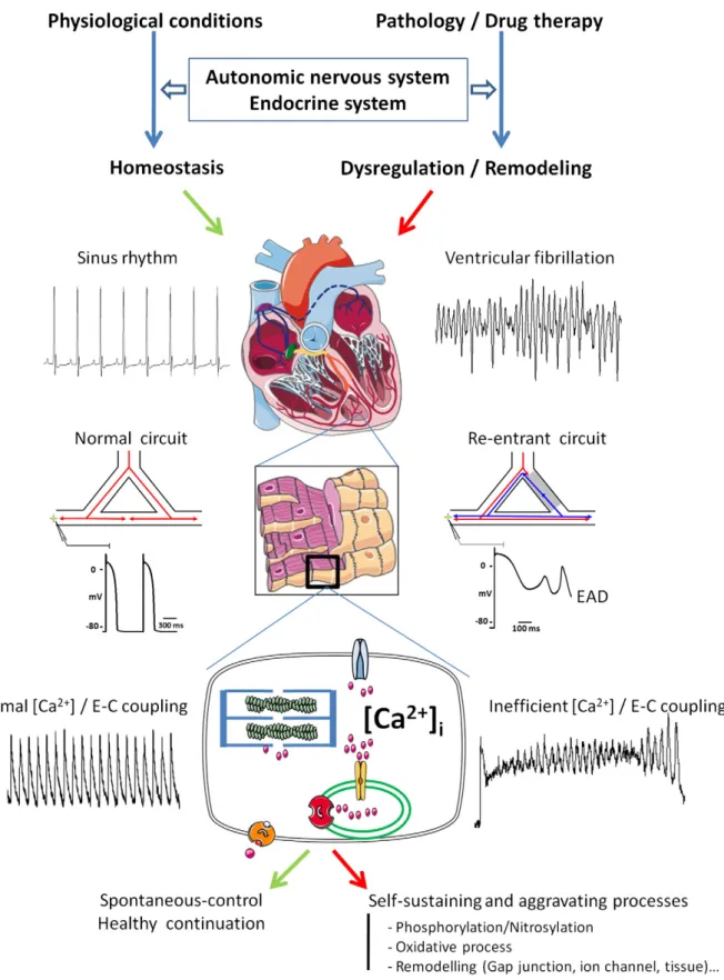
leading to delayed afterdepolarization, DAD). In addition, environ-mental considerations such as the autonomic nervous system (ANS), neurohormones and/or metabolism that regulate the cardiac rhythm to a great extent, are involved in the triggering of arrhythmias as well as in their maintenance, both at the atrial and ventricular levels (Fig. 1) (Corr et al., 1986; Coumel & Maison-Blanche, 1991; Zipes & Wellens, 1998; Zipes & Rubart, 2006; Workman, 2010).
Arrhythmias may also depend on conduction defects, such as reentry, that could lie at the origin of Torsades de pointes (TdP) and VF (Napolitano et al., 1994). Besides enhanced or abnormal impulse formation, reentry occurs when a propagating impulse persists after
sinus activation of the heart and re-excites cardiac tissue after the expiration of its refractory period (RP) (Antzelevitch, 2001). Reentry can have two different origins: electrophysiological or structural. Circus movement reentry involves the circuitous propagation of an impulse around an anatomical or functional obstacle, leading to re-excitation of the heart. The first model described for this phenomenon was reentry due to the presence of an anatomical obstacle (Mines, 1913). In this model, there is a circular propagation of the depolarization that is mainly determined by the size of the obstacle (i.e. fibrosis) and the RP (Allessie et al., 1977). In this case, the type of reentry observed is called a macro-reentry. In the presence of slow-conducting tissue (e.g. depolarized cells, and fibrosis), the action potential (AP) can propagate rapidly in normal tissue and return through the area of slow conduction (Fig. 1). However, at least in the atrium, there could be micro-reentry due to a difference as low as 16 ms in the effective RP (Antzelevitch, 2001). This small difference in the RP is enough to maintain the type of circus movement exemplified by the “leading circle model”. The same kind of process is thought to occur in the ventricle. Phase 2 reentry occurs due to the electrical heterogeneity of the ventricular myocardium between the endocar-dial, epicardial and mid-myocardial regions, and in response to pharmacological agents and pathophysiological states (Antzelevitch, 2001, 2007). As a consequence, the right ventricular epicardium displays a much more prominent AP notch than the left ventricular (LV) epicardium. Transmural voltage gradients, generated by differ-ences in the time-course of repolarization of the three cell types, could increase the QT interval and the risk of VA.
At the cellular level, complex mechanisms take part in the genesis of arrhythmias. They depend broadly on aberrant electrical sig-naling with a multifaceted interplay between different types of ion channels and/or disordered Ca2+signaling. In the normal heart, both
mutations in a variety of ion channel genes – like SCN5A or its associated regulating protein (i.e. SCN4B gene) – and drugs that affect cellular AP repolarization may increase the risk of severe arrhythmia (Wilde & van den Berg, 2005; Medeiros-Domingo, 2007; Zhu & Clancy, 2007; Saenen & Vrints, 2008). In addition to contributing to AP duration, Ca2+plays an important role in the triggering of both
early afterdepolarizations (EADs; Ca2+ entry through L-type Ca2+
channels) and DADs. For example during HF, intracellular diastolic Ca2+ overload can trigger ectopic activity (Wit & Rosen, 1986)
(Fig. 1). The key role played by a panel of Ca2+-handling proteins
(Ca2+ATPase SERCA2a, the Na+–Ca2+exchanger NCX, RyR2 channel,
and proteolytic enzymes) is now well-established (Shannon & Bers, 2004; Antoons & Sipido, 2008). Overall, some of the mechanisms listed here are involved in both arrhythmias related to remodeling in chronic pathology and inherited arrhythmias (e.g. RyR2), providing relevant avenues of research for new AA drugs and novel concepts, which we will discuss later. Indirect strategies targeting cellular remodeling on a pathological substrate very early in the process are undoubtedly highly relevant to many diseases with structural modifications and deleterious remodeling.
2.2. Tissue sources of arrhythmias 2.2.1. Atrial level
For cardiologists, arrhythmias are mostly characterized by their tissue of origin. AF is a disorder defined by a rapid and irregular rhythm leading to uncoordinated atrial contraction.
AF is initially thought to originate from the ectopic activity of venous structures, mostly the pulmonary veins but also the coronary sinus, the ligament of Marshall or the superior vena cava. It is the most complex arrhythmia, frequently associated with an underlying structural and evolutive heart disease. As the AF lasts longer and longer, the mechanisms become more and more complex, involving fibrosis through multiple reentry circuits, rotors considered as “mother” waves, and parasympathetic cardiac innervation (Loomis
Fig. 1. Schematics of the main mechanisms involved in arrhythmia. The cardiac rhythm is regulated by hormones and the autonomic nervous system. Efficient myocardial contraction depends on a normal sinus rhythm, synchronization among the different cardiac tissues, conduction pathway integrity and well-organized excitation-contraction coupling. Pathological conditions and/or medications can alter or interfere with these regulatory systems at different levels and can lead to cardiac remodeling and rhythm disorders. Complex mechanisms take part in the genesis of arrhythmias. Reentry can lead to ion accumulation (in particular Na+and Ca2+) (Munteanu et al., 2008) and, conversely, high intracellular Ca2+can initiate cardiac remodeling that creates a substrate for reentry. AP prolongation can initiate early afterdepolarisations (EADs), and aberrant spontaneous diastolic Ca2+release via leaky RyR2 activates Iti(through NCX) to trigger DAD and, subsequently, VA (typically in HF and CPVT). Environmental conditions (redox status, phosphorylation or nitrosylation) and pathological remodeling can play a pivotal role in susceptibility to arrhythmia.
& Krop, 1955; Yue et al., 2011). Symptoms of AF include palpitations and sometimes weakness, dyspnea and presyncope, and in more severe forms, AF may lead to death.
Clinically, the type of AF is determined based on its duration and its resistance to therapy. As such, AF is classified as paroxysmal (self-terminating, generally N48 h and lasting up to 7 days), persistent (episodes lasting more than 7 days and requiring pharmacological or direct-current cardioversion), long-standing persistent (lasting more than 1 year) and permanent, when the AF is considered as being definitive and accepted by the patient (Camm et al., 2010). In addition to its complex etiology, there is a lack of satisfactory models for sustained AF in connection with specific ion channels. This is especially true for small animals (rodents), and the only valid models for the reproduction of sustained human AF are large animal models (goats, sheep) due to the critical size of their atria (Schotten et al., 2010). This contributes to the difficulty in developing new anti-AF drugs.
The prevalence of AF increases with age (1/25 after 60 years and 1/10 after 80 years in the USA) and is expected to increase by a factor of 2.5 until 2050 (Go et al., 2001). More strikingly, the number of hospitalizations related to AF has increased by 66% in 20 years (Fuster et al., 2006). A study from Olmsted County reports that, from 1980 to 2000, the risk of death related to cardiovascular disease decreased significantly but that the risk of death related to AF remained unchanged (Miyasaka et al., 2007; Miyasaka et al., 2008). The latest guidelines of the European Society of Cardiology clearly highlight that AF is an independent risk factor for mortality (Camm et al., 2010). The critical issue is appropriate antithrombotic therapy, but until recently, existing AA drugs were associated with increased mortality. Besides drug therapy to maintain the sinus rhythm, non-drug treatments such as catheter ablation show promising results (Cappato et al., 2005; Ellis et al., 2009), despite a 6–8% global complication rate with a mortality rate as high as 0.5%. Following the recent AFFIRM study (Wyse et al., 2002), it is now commonly accepted that the problem for patients presenting with AF is not rhythm or rate control. A more global approach is desirable in order to decrease the morbidity–mortality linked to AF. Indeed, available AA drugs are associated with an increase in mortality, suggesting that we need better drugs that allow sinus rhythm to be maintained while avoiding iatrogenicity (Wyse et al., 2002).
2.2.2. Ventricular level
At the ventricular level, US vital statistics mortality data for 1989– 1998 showed that of 719,456 cardiac deaths among adults aged 35 years in 1998, 456,076 (63%) were defined as sudden cardiac death (SCD) (Zheng et al., 2001). A recently completed large study of 121,701 women (Nurses' Health Study) over a 20-year period estimated that 88% of SCDs were due to arrhythmias (Albert et al., 2003). Deaths from SCD each year outnumber deaths from all cancers (MMWR, 1999; Anderson, 2001). SCD can have multiple origins. Coronary artery disease is present in 80–85% of patients who undergo SCD (Cobb et al., 1975; Myerburg, 2001). A reduced LV ejection fraction remains the single most important risk factor for overall mortality and SCD (Priori et al., 2001b). In HF, QT-prolonging drugs are a major cause for concern. Polytherapy also increases the risk of lethal arrhythmias, estimated to cause 100,000 deaths/year (De Bruin et al., 2007), a figure that is even higher in the presence of underlying heart disease. In a recent study with over 8000 patients, a QTc of greater than 450–470 ms was associated with a tripling of the risk of SCD after adjustment for age, sex, body mass index, hypertension, cholesterol, diabetes, myocardial infarction (MI), HF and heart rate (Straus et al., 2006). Thus, clinical studies have revealed that SCD resulting from arrhythmias is a major and growing public health problem worldwide.
VA is relatively unpredictable, and remains a major source of avoidable SCD. VF is a chaotic VA leading to the disorganized pumping
of blood and possibly leading to death. VF occurs consecutive to reentry circuits, but the underlying mechanisms are still not fully understood. It occurs due to reentries that are the result of an increase in transmural dispersion (Antzelevitch & Fish, 2001). It was originally hypothesized that the transmural dispersion was due to an electrical gradient with, notably, the existence of an intermediate cell layer “M” (for mid-myocardial) between the epicardium and the endocardium (Antzelevitch et al., 1996). This gradient was based on differences in the density of various ion currents, including the IKS. Any
environ-mental change that reduced repolarizing currents, such as the IKR,
would accentuate the dispersion of repolarization (viewable on an ECG by measuring the Ve–Vp of the T wave) and could result in reentry and thus VF. However, M cells exist in myocardial clusters that vary in spatial location and extent across the heart, and are not present uniformly at a given depth of myocardium (Akar et al., 2002). This heterogeneous distribution of M cells as islands of M cells, coupled with electrophysiological differences between epi- and endo-cardiomyocytes, reinforces the notion of a heterogeneous repolariza-tion sufficient to cause a funcrepolariza-tional block and reentry in response to a premature stimulus, and contributing to arrhythmia.
Two different strategies have been adopted in recent decades to reduce the risk of developing lethal arrhythmias, and are used alone or in combination according to ACC/AHA/ESC guidelines (Epstein et al., 2008). These are AA drugs and the implantation of ICD, eventually combined with surgical or percutaneous catheter ablation (Reddy et al., 2007). Currently, no drug is effective enough to obvi-ate the need for ICD implantation, which is not only a palliative but clearly a life-saving treatment. However, although the effective-ness of this therapeutic protocol/strategy is known, it raises a number of problems: 1) it is only a symptomatic treatment, 2) shocks are stressful and may be associated with a deterioration of LV pump function, and 3) it does not prevent disease progression or the development of remodeling. Although their effectiveness in saving lives is high, even for prophylactic indications, ICDs treat but do not prevent SCD (Coats, 2002). Moreover, both appropriate and inappro-priate shocks have deleterious effects on the quality of life (Das & Zipes, 2010), suggesting that the development of novel pharmaco-logical therapies is essential.
2.2.3. Purkinje tissue
The role of the Purkinje fibers (PFs) in the triggering and main-tenance of VF has been described before (Berenfeld & Jalife, 1998; Haissaguerre et al., 2002a, 2002b; Lindsay, 2009). However, their contribution to the generation of arrhythmias through reentry has probably been underestimated (Sasyniuk & Mendez, 1971; Boyden et al., 2010). The QRS complex of the ECG, which reflects the conduction of electrical activity by PF, has been described as the best predictive factor for VT in patients suffering from perturbations of systolic function (Fazelifar et al., 2009). This suggests that conduction defects are very important in the generation of arrhythmias.
Conduction abnormalities are involved in the genesis of arrhyth-mias and SCD (Marriott, 1964; Titus, 1973; Herron et al., 2010). This has been shown in ischemic cardiac disease and LV dysfunction after MI, where slowed conduction has been localized to the zone bordering the infarct, as well as in non-ischemic dilated cardiomy-opathy, as confirmed by genetic models that exhibit a more global slowing of conduction (Akar & Tomaselli, 2005). A unidirectional conduction block in a branch of a PF can trigger sustained reentry (Gilmour & Watanabe, 1994; Antzelevitch, 2008). Structural heart disease, electrical instability, and increased sympathetic activity provide substrates for ECG changes and arrhythmias in patients with congestive HF (Hombach, 2006). HF patients commonly exhibit prolonged QRS duration and/or ventricular late potentials, predictive of life-threatening VT, aberrant heart rate variability (HRV, an index of MI), and repolarization abnormalities detected by studies of QT dispersion, QT/QTc fluctuation or T-wave alternans (Hombach, 2002).
The impact of conduction delays has been supported by clinical studies, with late potentials on abnormal signal-averaged ECG being an independent predictor of all-cause cardiac death and of great interest for the risk stratification of arrhythmias (Mancini et al., 1993; Lander et al., 1997; Fauchier et al., 2000).
In addition to anisotropic reentry, notably arising in HF after MI-induced injury, inappropriate impulse propagation leading to ar-rhythmia generation is determined by numerous structural, cellular and molecular changes in fibrosis, extracellular matrix, cell–cell coupling, the expression and function of gap junction proteins (Cx43), and the combination, availability and gating properties of ion channels (Akar & Tomaselli, 2005). Ca2+handling proteins are
also involved, in particular the RyR2 Ca2+release channel. It was
recently reported that focally activated arrhythmias originate in the specialized electrical conducting cells of the His-Purkinje system in the RyR2(R4496C) mouse model of CPVT (Herron et al., 2010). In this model, PFs have a higher propensity to develop abnormalities in intracellular Ca2+ handling than ventricular myocytes, in relation
with aberrant Ca2+release events due to the spontaneous opening of
RyR2 in diastole, which can be suppressed with flecainide (Kang et al., 2010).
PFs have electrophysiological particularities that are not shared by ventricular cells, such as a longer plateau involving a persistent Na+
current reflecting a “window” current, considered to be the steady-state component of the fast sodium current (INaf) resulting from the
crossover of the activation and inactivation curves of the Na+channel
(Attwell et al., 1979; Coraboeuf et al., 1979), rather than slow inactivation due to modified gating properties. Recently, it has also been shown that PFs express channels that are not expressed in ventricular cells, at least under normal conditions. For example, the Ca2+-activated nonselective cation channel TRPM4 is highly
expressed in cardiac PFs. Also, recently, a mutation in the TRPM4 channel has been shown to induce a gain-of-function mechanism related to elevated TRPM4 channel density at the cell surface in progressive familial heart block type I, a progressive cardiac bundle
branch disease of the His-Purkinje system (Kruse et al., 2009; Liu et al., 2010).
PFs are also prone to developing EADs, which can be prevented by ryanodine (Boyden et al., 2004). EADs occur against a background in which the AP duration is increased, and are among the first events leading to VF. In addition, it has been shown that in a post-MI animal model, arrhythmias begin at the level of the PFs following the dysregulation of intracellular Ca2+homeostasis (Hirose et al., 2008).
These results are of great interest because the origins of CPVT are currently being investigated at the ventricular level, and PFs may also be involved in this process (Hirose et al., 2008). Recent experiments using a mutant mouse model of CPVT have demonstrated that the His-Purkinje system is an important source of focal arrhythmias in CPVT (Cerrone et al., 2007). These findings underline a role for PFs in the generation and maintenance of VA that has been too long ignored, and that could be specifically targeted.
2.3. Pharmacological origin of arrhythmias
2.3.1. Antiarrhythmic therapy and proarrhythmogenicity
The pharmacologic treatment of AF can generate conditions favoring the triggering of VA. There have been dramatic examples in the past of a deleterious effect of AAs on clinical criteria. A classic example of the dangers of the use of intermediate criteria to judge drug efficacy is that of class I AA drugs in post-MI. In the Cardiac Arrhythmia Suppression Trial (CAST), the effects of encainide and flecainide were evaluated in patients with asymptomatic or mildly symptomatic VA after MI (1989). The conclusion was that neither encainide nor flecainide was advisable in the treatment of patients with asymptomatic or minimally symptomatic VA after MI, even though these drugs were initially effective in suppressing VA. Overall, the toxicity associated with class I AA drugs is very common and can be life-threatening (Denaro & Benowitz, 1989; Kim & Benowitz, 1990; Koppel et al., 1990). Examples of poisoning with β-blockers (class III) and Ca2+antagonists (class IV), mostly due to high doses, can also be
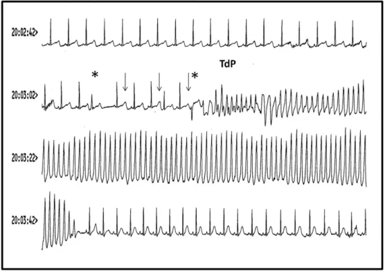
Fig. 2. Example of VT and Torsade de Pointes in a 32-year old woman treated for paroxysmal AF and after attempted suicide using sotalol. In the upper part of the monitor, the trace shows a dramatic prolongation of the QT interval. After an atrial ectopy (first asterisk), there is a pause followed by electrical QT alternans (arrows). A ventricular ectopy (second asterisk) occurs at a vulnerable point during prolonged QT and is responsible for a Torsade de Pointes onset that turns into very fast monomorphic VT and stops after about 30 s (syncope of the patient). After cessation of tachycardia, the sinus rhythm is notably accelerated with a much shorter QT interval.
found (Pearigen & Benowitz, 1991; Reith et al., 1996). This is illustrated inFig. 2, which shows VT and TdP induced in a 32-year old woman treated for paroxysmal AF after an attempted suicide using sotalol.
Apart from adverse effects related to proarrhythmogenicity, clinical trials have sometimes been discontinued because of other adverse effects as well, suggesting that proarrhythmia is not the only potential safety concern with AA drugs. For instance, amiodarone, in spite of its clearly positive AA effects, was stopped because it was found to induce pulmonary, neurological or liver toxicity as well as thyroid dysfunction in patients under specific circumstances (Kamath & Mittal, 2008). In patients with severe HF and LV systolic dysfunction, treatment with a noniodinated amiodarone derivative, dronedarone, was associated with increased early mortality related to the aggravation of HF (Kober et al., 2008). Since dronedarone was found to block the ICaL current (Gautier et al., 2003), it was
hypothesized that a negative inotropic effect may have promoted the worsening of chronic HF in these patients. This propensity of a candidate drug to adversely affect physiological systems other than the cardiovascular system is the second aspect addressed by regulatory authorities in the context of non-clinical safety documen-tation (Guidance for Industry, S7A, 2001).
2.3.2. Safety concerns
The need to evaluate the proarrhythmic potential of AA drugs was stimulated by the outcomes of theCAST (1989)and SWORD clinical trials (Waldo et al., 1996). It was obvious that AA drugs could also be proarrhythmogenic. Drugs that prolong cellular AP repolarization could increase the risk of severe arrhythmia. After the mid-1990s and the withdrawal from the market of the antihistamine terfenadine because of increasing clinical evidence that the drug induced life-threatening arrhythmias (June & Nasr, 1997), regulatory authorities rapidly addressed the need for improved pre-clinical experiments for the documentation of such safety issues. Therefore, at the end of 1997, the Committee for Proprietary Medicinal Products (CPMP) of the European Medicines Agency first raised a point to be considered in the assessment of the potential for QT-interval prolongation of non-cardiovascular medicinal products (CPMP/SWP/986/96), followed in 2005 by guidelines from the International conference on Harmoniza-tion (Guidance for industry, 2005) defining the regulatory require-ments for the non-clinical evaluation of this cardiovascular risk. These regulatory guidelines recommend the use of two main approaches: in vitro patch clamp studies to evaluate effects on the hERG current, and in vivo studies dealing with effects on the duration of ventricular repolarization in appropriate species, thereby providing an estimation of the risk of TdP induction. In order to interpret the results and draw appropriate conclusions regarding the proarrhythmic risk, the integration of all these results and others arising from primary pharmacological studies or other safety studies is recommended. Depending on the results of these initial experiments, integrated risk assessment may require additional follow-up experiments, including an assessment of effects on APs in isolated tissues and specific in vivo proarrhythmia models.
Animal models have been developed in order to detect the potential of drugs to induce a rare VA, TdP. The common feature of these models is the achievement of enhanced susceptibility to arrhythmia, to mimic the situation encountered in vulnerable patients. Two models are currently used: the methoxamine-sensitized rabbit model of TdP (Carlsson et al., 1990) and the chronic atrio-ventricular block (CAVB) dog model (Vos et al., 1995). The methoxamine-sensitized rabbit model is considered to be mainly effective in detecting pro-arrhythmic drugs with class III AA features. Likewise, the CAVB dog model is capable of displaying TdP after exposure to class III drugs, but also in response to others such as dronedarone or setindole (Gintant, 2008). Moreover, this model has also helped to highlight the fact that the QT interval duration, the
most common biomarker used as a surrogate for drug-induced TdP to estimate pro-arrhythmic potential, could in some ways be misleading. Indeed, drugs like amiodarone and moxifloxacin prolong QT interval duration to a similar extent but fail to induce TdP as expected based on clinical data. From this finding, a new surrogate, the beat-to-beat variability of left ventricular repolarization estimated from short term variability measurements, has been proposed for the evaluation of pro-arrhythmic potential (Gintant, 2008).
Apart from actual regulatory requirements, the patch clamp study on hERG was initially considered a go/no go test in the industrial development process because all drugs inducing TdP were identified as hERG channel blockers. The need for the documentation of the electrophysiological effects of all new chemical entities, whatever their therapeutic targets, on the hERG current led to the development of high-throughput methods like automated patch clamp assays to manage the large number of chemical entities, especially for early safety assessment. New methodologies have had a positive impact on the development of new AA drugs because they provide, at an early stage, helpful information for the adjustment of drug design. A variety of relevant cell-based screening assays and systems for arrhythmia assessment have been reviewed recently (Nattel et al., 2008). 3. Antiarrhythmic drugs
3.1. Ion channels: The traditional targets of antiarrhythmic drugs The electrical activity of the heart is primarily driven by cellular ion currents. Experimental electrophysiology really gave birth to the concept of the “voltage-gated ion channel” more than a half-century ago in a series of remarkable papers (Hodgkin & Huxley, 1952a, 1952b; Hodgkin et al., 1952) and established a theoretical model paving the way for our understanding of how voltage-gated channels work and how drugs could interact with them and modify their function. The “voltage-clamp” technique, and later, the concomitant development of patch clamping methods (Hamill et al., 1981; Sakmann & Neher, 1983; Hille, 1992) and the enzymatic isolation of single cardiomyocytes fostered the emergence of new knowledge and concepts that greatly contributed to mechanistic investigations and the development of AA drugs. Consequently, it became possible to measure ion currents at the level of a single cell or even of a single channel protein, making most tissues (including human tissue samples obtained during surgery) amenable to powerful electrophys-iological investigation. These advances made possible the determina-tion of the molecular effects of drugs on the gating of channels for an improved prediction of drug effects on function.
In the meantime, the determination of the protein nature of ion channels, the identification of drug binding sites, and the sequencing of genes encoding these proteins have laid the foundations of “molecular medicine”that address the complex problem of cardiac arrhythmias. It has become apparent that numerous mutations in a variety of genes coding for different types of ion channels (channe-lopathies) are responsible for prolonging the QT interval, which can precipitate TdP and irreversible VF (Shah et al., 2005; Pasquie & Richard, 2009). Thirteen genotypes characterize the long QT syn-drome. Hundreds of mutations have been identified for at least eight different ion channel complexes, the structural anchoring protein Ankyrin, and a caveolin protein (Antzelevitch, 2007).
3.2. What is a classic antiarrhythmic drug?
Most voltage-gated ion channels possess druggable binding sites for pharmacological therapy in general and AA drugs in particular (Clarkson & Hondeghem, 1984). These drugs usually prevent channel opening, resulting in a decrease in a given ion current. Interestingly, the biophysical properties of ion channels are important determinants of current amplitude and kinetics, and can strongly modulate/govern
pharmacological effects. In particular, the affinity of a ligand for a channel can be strongly modulated by the conformational state of the protein, which in turn is highly dependent on the membrane potential. In this regard, the modulated receptor hypothesis has been useful in understanding the kinetics of Na+channels and the changes
produced by various AA drugs (Hondeghem & Katzung, 1984). 3.3. Classification of antiarrhythmic drugs
Based on initial knowledge regarding the origin of arrhythmias (ectopic activity, conduction disorders leading to reentry, and over-excitability), strategies to stop arrhythmias have led to the classifi-cation of AA agents since the 70s. The first classificlassifi-cation was established byVaughan Williams (1970)(VW), and was successively completed bySingh and Hauswirth (1974),Harrison et al. (1980)and Hondeghem (1992)(seeTable 1). Although other types of classifica-tion exist, such as Touboul's classificaclassifica-tion, which takes into account data from in vivo electrophysiological explorations (Touboul et al., 1979), the autonomic classification ofGoldberger and Curtis (1982)or theGambit (1991), the VW classification has remained robust over time, being by far the most used despite its obvious limitations. It continues to be used for the development of new drugs and the design of approaches to clinical therapy. Its main advantage is that it explains complex effects simply (as a teaching tool), in terms of pharmaco-logical targets. However, it does not address the etiology of the disorder. This classification also does not integrate the concept that AA drugs can be effective in various ways on multiple targets. Indeed, the VW classification was originally based on four classes of drugs that interfered with, respectively, the fast Na+current, ß-adrenergic
receptor, K+channel and Ca2+channel (Vaughan Williams, 1984,
1992). In other words, the VW system enabled the classification of AA drugs based on their effects on the three major types of ion currents identified in the seventies on the one hand, and on sympathetic activity on the other. In all cases, the concept is based on the notion that arrhythmias arise due to abnormal electrical activity and somehow reflect the inappropriate functioning of ion channel proteins. However, the fact that drug action can change during pathology due to channel or receptor modification or a sympathova-gal imbalance is not taken into account.
Class I includes Na+channel blockers. Blockade of the Na+current
slows conduction (Fozzard & Hanck, 1992), which could help prevent arrhythmias by transforming a unidirectional block into a bidirec-tional one. However, slowing conduction can also promote reentry by decreasing wavelength. This could be the reason for the failure of Class Ic drugs in theCAST (1989). An important aspect of the generation of ectopic beats is linked to the RP. Depending on the type of arrhythmia, ectopic beats can be prevented by increasing the RP following blockade of K+ currents. Sympathetic innervation also plays an
important role in the regulation of cardiac excitation–contraction coupling and in the generation of arrhythmias (Anderson, 2003; Verrier & Antzelevitch, 2004; Vaseghi & Shivkumar, 2008). In the VW classification, this role is undertaken mainly through a regulation of ion currents such as ICaL. Presently, beta-blockers (class II according to
the classification) are among the most widely used AA drugs, but the precise reason for their properties is not fully understood. As mentioned above, an important aspect in the generation of arrhyth-mias is the decrease in the RP. The inhibition of some K+channels
generally leads to an increase in the AP duration and thus of the RP (Kim & Benowitz, 1990; Ravens & Cerbai, 2008). This is why the VW classification created a particular group, class III, which includes K+
channel blockers. However, no member of this group is exclusively a K+channel blocker. Sotalol, which is the leading molecule in this
group, is also a powerful ß-blocker, while amiodarone, which is a powerful AA drug, also inhibits many other channels.
Class IV comprises Ca2+channel blockers including verapamil and
diltiazem. These drugs block the ICaLcurrent in all cardiomyocytes but
have few effects on the AP waveform. Their effects resemble those of class II AA drugs, with a slowing of the late spontaneous diastolic depolarization of sinus node tissue and an increase of the atrionodal conduction time and RP. With Class IV compounds, the heart rate is not systematically attenuated but the PR interval is often prolonged. Class IV drugs are the reference treatment for reentrant supraven-tricular tachycardia (Colucci et al., 2010), and the potential decrease of the heart rate could be sufficient to prevent AF (Tsuneda et al., 2006). A major side effect of these drugs is a decrease in contraction, which could be undesirable in HF patients. Finally, a fifth class, whose agents work through other or unknown mechanisms, has been added (Vaughan Williams, 1992). Class V agents include digoxin and adenosine. Digoxin increases vagal activity via its action on the central nervous system, thus decreasing the conduction of electrical impulses through the AV node (van Veldhuisen et al., 1996). It should be noted that Class V agents are not part of the original VW classification. Moreover, new molecules with potential AA properties but that are not compatible with the original VW classification, have emerged: some can be placed in several classes (vernakalant) while others cannot be listed under any of the four original VW classes (ivabradine). This suggests that the VW classification may no longer be best suited to the realities of new AA drug design.
4. Current and future strategies 4.1. Where do we stand?
AA drugs are designed to maintain a normal sinus rhythm and prevent rapid and irregular heartbeats. They are expected to relieve symptoms related to arrhythmias such as palpitations, faintness or HF. Theoretically, the final and ideal goal of AA therapy is to reduce mortality directly related to arrhythmia. So far, this goal has been purely utopian. Currently available pharmacological agents have limited efficacy and/or carry a risk of relevant side effects, such as drug toxicity or proarrhythmic potential (Gramley et al., 2009). As we have all been aware for a very long time, studies of the effectiveness of AA drugs at the atrial (e.g. AFFIRM) and ventricular (e.g. CAST I) levels have shown an increase in mortality directly related to drug side-effects. The only therapies with a mortality-reducing effect are anticoagulants in the case of AF (Hylek et al., 2003; Boden et al., 2007; Connolly & Poston, 2009) and ICDs in VA (Moss, 2003; Cappato et al., 2005; Budde, 2006). In other words, we can reduce mortality by treating the complications of arrhythmias, but we still do not know how to effectively prevent arrhythmias in the long run using drugs or non-drug treatments such as surgery (Klein et al., 1986) or catheter ablation (Wilber, et al., 2010; Pappone et al., 2011).
In complex pathological conditions such as HF, the diversity of the systems that contribute to cardiac remodeling and arrhythmia (beta-adrenergic receptors, renin–angiotensin–aldosterone and endothelin systems or NO synthases) and their polymorphisms necessitates extensive gene cartography in order to understand the different outcomes in patients (Cascorbi et al., 2004). The pharmacogenomic approach has established that individual responses could result from specific gene variants that modify how a drug is absorbed or eliminated. These genetically based differences in drug efficacy, which have long been recognized (Kalow, 2006), are only now on the verge of clinical application in drug development and the design of clinical trials (Shah, 2004; Roden, 2005; Perez et al., 2008; Winkelmann and Herrington, 2010). Gene variants can also hold the key to under-standing why individuals who are asymptomatic for cardiac disease are at risk for long QT syndrome, VF, syncope or SCD when under medication or during exercise (Hedley et al., 2009).
A major obstacle in the development of novel AA drugs is related to the complexity of electrical signaling, which in turn introduces safety concerns. The regulation of criteria defining the safety of a drug has been greatly tightened following several cases of malignant
Table 1 Historical and mechanistic classi fication. 1970 Singh and Vaughan Williams Class I Class II Class III Class IV Class V (1979) Na + Channel Blocker β -adrenergic receptor Blocker K + channel Blocker Ca 2+ Channel blocker Adenosine 1974 Singh and Hauswirth Ia ↑ repolarization β 1 or/and β 2 receptor blocker ↑ repolarization Slower conduction velocity Digoxin (↑ vagal activity) Ib ↓repolarization Sympatholytic drugs ↑ refractory periods ↑ PR, ↑ QTc, ↓ HR Ivabradine 1981 Harrison Ic No effect on repolarization ↑ QTc negative inotrope (speci fic sinus node action Slower conduction velocity AV Blocks If blocker 1992 Hondeghem ↑ QRS, (± ↑ PR) ↑ PR, ↓ HR Class IIIA verapamil, diltiazem ↓ diastolic depolarization negative inotrope negative inotrope acting during HR acceleration ↓ HR, (± ↑ PR)) reentry sustaining AV blocks disopyramide (a) propranol, atenolol … Class IIIB lidocaine (b) acting during bradycardia fl ecainide (c) sotalol Class IIIAB acting independently of heart rate amiodarone Positive inotrope Torsades de pointe Touboul et al., 1979 Class I Class IIA Class IIB Class III ↓ AV conduction velocity ↑ His-Purkinje ERP ↓His-Purkinje RP without ↑ HV ↑AH conduction velocity and ↑HV ↑ AH and ↑ ERP ↑ HV and ↑ atrial RP phenotoine, lidocaine ↑ AV RP Beta blockers, verapamil, digitalic quinidine, disopyramide ↑His-Purkinje RP without ↑ HV aprindine bretylium ↑ Atrial RP ↑ His-Purkinje ERP Amiodarone 1982 Autonomic classi fi cation of Goldberger and Curtis Class I Class II Class III Class IV Local anesthetics digitalis glycoside with vagotonic effect antiadrenergic agents calcium channel blockers 1A quinidine-like agents with cholinergic 1blocking Class IIIA 1B agents without autonomic action beta-blockers lidocaine propranolol Class IIIB norepinephrine-release inhibitors bretylium Class IIIC non speci fic adrenergic blockers amiodarone 1991 Taormina “Sicilian gambit ” Channels Receptors Pumps Na + α − adrenergic Na +/K + ATPase Ca 2+ β− adrenergic K + M2 -muscarinic If Purinergic
arrhythmias and SCD related to medication used in cardiology and non-cardiological fields. Although a drug may prove effective in most patients, rare cases of drug-related SCD support the view that positive results on major safety screening tests (mortality, QT interval, hERG assay …) lead to the exclusion of many molecules of clear therapeutic interest. One immediate consequence is the slowing down of AA drug development, due to the difficulty in equating an hERG block with the actual risk of induction of TdP in humans (Roden, 2008), and the discarding of candidate compounds of possible therapeutic interest (Gintant, 2008). In an attempt to define well-balanced criteria for proarrhythmic risk, it has been proposed that setting an IC50 value for an hERG block at 30 times the maximum calculated unbound effective therapeutic plasma concentration would provide a good safety margin (Redfern et al., 2003). Interestingly, there are examples of drugs with multiple effects on different ion channels that minimize torsadogenic risks, suggesting that combinations of drugs should be taken into account on a more systematic basis.
4.2. Experimental tools available
The starting point in AA development is the availability of relevant experimental models of arrhythmias on the one hand, and for each specific step of efficacy and safety testing on the other hand. Different models are required for different purposes, including the improve-ment of our understanding of the mechanisms underlying arrhythmia, the identification of “druggable” targets, and the screening and design of molecules (i.e. lead optimization). Very early, extensive consider-ation has been paid to experimental investigconsider-ation of arrhythmia in vivo (rat, guinea-pig), particularly at the ventricular level during ischemia, infarction and reperfusion, to provide the Lambeth Con-vention guidelines covering many practical aspects (definition, classification, quantification, and analysis) (Walker et al., 1988). In vivo models in a number of species allow the induction of AF or VA and aim to reproduce human pathophysiology. However, animal models did not deliver their full potential in this field. In particular, no model covers all aspects of the clinical situation in patients. Therefore, the choice of model used needs to be better defined depending on the goal pursued: identification of mechanisms or development of a therapeutic product.
AF models were developed mainly in large animals (i.e. dogs, pigs, sheep and goat) in order to induce AF associated with electrical (paroxysmal or persistent/permanent models) or structural (persis-tent/permanent models) remodeling (Nishida et al., 2010; Schotten et al., 2010). These also included the influence of the ANS on the induction of AF. There are now several transgenic mouse models available that exhibit pronounced atrial enlargement and spontane-ous or inducible AF, but their comparison with human AF is inherently problematic (for review:Schotten et al., 2010). In different models of VT/VF (iatrogenic, in vivo models of ischemia-induced arrhythmia, Langendorff models and naturally occurring models) the mechanisms involved have been widely documented (for review,Hamlin, 2007). However, due to significant differences in cardiac electrophysiological profiles and heart structure that influence the spatial component of arrhythmia, the translation of these findings to humans also needs to be carried out with caution (Nerbonne et al., 2001). Since the topic of this review is not to describe extensively the limits of all experimental models available for mechanistic investigation of arrhythmia, we invite the reader to consider recent reviews (e.g.Hamlin, 2007for VF andSchotten et al., 2010for AF).
Apart from in vivo models for mechanistic studies of arrhythmias, a number of cell-based in vitro assays were developed especially for drug screening and design both for efficacy and safety of new drugs. Ion channel assays based on electrophysiological technologies are essential in drug discovery and evaluation of their potential side effects. Other assays include non-functional tests with ligand-receptor binding techniques targeting ion channels, fluorometric imaging for intracellular
Ca2+ assessment, and direct ion-flux, which provide more or less
functional information. Proceeding beyond the set-up of these new cell-based methods, the achievement of automated assays offers increased drug screening capacity (Nattel et al., 2008). Finally, in combination with the experimental data provided by the models described above, in silico modeling is another emerging technology specifically proposed for the characterization of the mechanisms responsible for efficacy (Vigmond et al., 2009) and adverse effects (Corrias et al., 2010).
The absence of effective drugs in human patients suggests that the relevance of available pre-clinical models is questionable. On the other hand, the absence of reference drugs in the clinical field for VA makes the validation of novel AA molecules difficult to achieve. 4.3. Emerging concepts regarding novel antiarrhythmic drugs
AA drug discovery has been problematic for a long time and development of new AA drugs, notably to treat VA, has failed. The prevailing therapy for VA is the ICD, but this remains a symptomatic treatment which does not prevent disease progression. Interestingly, newly acquired knowledge through genetics of ion channels, functional genomics and use of miniaturized devices (echocardiogra-phy, ECG by telemetry, intracardiac electrophysiological exploration), developed in particular for the phenotyping of transgenic mouse models, has advanced our understanding of not only the role of ion channels but also of complex intracellular mechanisms involved critically in the genesis of arrhythmias. Clearly genetic models are easier to study than conventional models of chronic diseases and, although they do not reproduce the complex phenotype of such chronic diseases, they can provide comprehensive mechanistic information. These new approaches are undoubtedly helpful for the development of new AA drugs and for preclinical studies. For example, drugs normalizing altered Na+and Ca2+signaling and/or
homeostasis were shown to prevent arrhythmias not only in inherited diseases related to ion channels mutations (e.g., LQT3, CPVT) but also in chronic disease. Although they are not always specific, some emerging AA drugs might pose a dual therapeutic benefit at both atrial and ventricular levels where intracellular Na+and Ca2+overload has
been shown to trigger arrhythmias. Other emerging pharmacological treatments include anti-remodeling drugs targeting primarily factors that are extrinsic to the cardiomyocyte, like mechanistic pathways involved in disease progression (e.g. neurohormonal systems, extracellular matrix remodeling, fibrosis), but involved in its electrical remodeling (ion channels, Ca2+signaling).
4.3.1. Sino-atrial level
4.3.1.1. Monotask antiarrhythmic drugs: Do they really exist? Although the cause of AF does not primarily involve ion channels, but is more likely due to a remodeling of atrial tissue, the treatment of choice is aimed at ion channels. There are several ways to envisage the development of novel AA drugs that are effective and safe. An ideal approach is to target a specific ion channel involved exclusively at the cardiac stage that exhibits the arrhythmia in question. A good example of this is ivabradine, a novel specific heart-rate lowering drug that acts selectively at the level of the sinoatrial node by inhibiting the pacemaker If current involved in setting the sinus
rhythm (Thollon et al., 1994; DiFrancesco & Camm, 2004). Recent studies have demonstrated a powerful effect of ivabradine in ischemic patients, in whom it reduces the sinus rhythm to below 70 beats/min (‘SHIFT’ study) (Cleland et al., 2010; Swedberg et al., 2010). Its place in the treatment of angina pectoris is now established. Due to its own specific effect, we should expect a beneficial effect of this drug on abnormal automaticity of the sinus node. As a matter of fact, ivabradine has recently been shown to be effective, and a safe alternative to Ca2+channel blockers and ß-blockers for the treatment
In the same vein of specific inhibition of ion channels, a promising AA strategy targeting AF currently under development, is the use of atrial-tissue specific ion channel blockers that do not fit within the classic VW classification. A new family of drugs called ARDAs, for atrial repolari-zation delaying agents, designed to block the ultra-rapid delayed rectifier current (IKur) and the acetylcholine-regulated K+ current
(IKACh) that are not expressed at the ventricular stage, is emerging. A
more exhaustive treatment of this subject can be found in several recent reviews (Ehrlich & Nattel, 2009; Kozlowski et al., 2009; Ravens, 2010). An example of these new drugs is vernakalant, which has been presented as an atrial-selective compound, with a reduced risk of TdP, and expected to be of use in the acute chemical cardioversion of AF. Its capacity to rapidly reduce AF episodes by 75%, 45 min after IV injection (compared to IV amiodarone; AVRO study) has led to its inclusion in the new ESC 2010 guidelines (Camm et al., 2010). Although vernakalant primarily targets the Kv1.5 ion channel, it does, however, have other effects (on INa, IKAch, IToand IKS), and can be classified as a multichannel
blocker, in particular with mixed effects on K+and Na+channels.
4.3.1.2. Multitask antiarrhythmic drugs. Other AA candidates are multichannel blockers or, may be more appropriately, “multitask” drugs, as some of them have far more complex effects than the modification of AP duration (e.g. effects on Na+and Ca2+homeostasis).
A good example of a multichannel blocker is amiodarone, which is the oldest and most effective drug used for the maintenance of the sinus rhythm, despite its poor objective efficacy (Lafuente-Lafuente et al., 2007). It is also the AA drug associated with the greatest number and variety of side effects, although its QT-prolonging effect, due to a complex mechanism of action, is unlikely to promote VA. Nowadays, however, new pharmacological agents for AF have to demonstrate a reduction of mortality and a low rate of side effects, in addition to their effect on sinus rhythm maintenance or frequency control. Dronedarone is a derivative of amiodarone that is expected to be devoid of most of its adverse side-effects, particularly on thyroid function (Boden et al., 2007). A benefit of dronedarone treatment on hospitalization rate and cardiovascular mortality was reported in high-risk patients with AF and normal LV function in the ATHENA study (Kober et al., 2008). The EURIDIS and ADONIS studies showed a modest benefit of this drug in reducing the recurrence of AF by, respectively, 22 and 27%, with a clear reduction in the ventricular rate during fibrillation (Singh et al., 2007). Improved rate control during persistent AF was confirmed by the ERATO study (Davy et al., 2008). However, no benefit in terms of mortality was associated with dronedarone in HF patients (Kathofer et al., 2005). Dronedarone is also currently suspected of being hepatotoxic by the EMA (http://www.ema.europa.eu/ema/index.jsp? curl=pages/medicines/human/medicines/001043/human_med_ 001207.jsp&murl=menus/medicines/medicines.jsp&jsenabled=true) and the FDA (http://www.fda.gov/Safety/MedWatch/SafetyInformation/ SafetyAlertsforHumanMedicalProducts/ucm240110.htm). In conclusion, while the benefit/risk ratio of dronedarone has not been well evaluated, amiodarone is still, despite its side effects, the best AA available for AF.
4.3.2. Ventricular level
4.3.2.1. Drugs targeting Na+and/or Ca2+homeostasis. Among other
emerging AA drugs, ranolazine seems to have great potential despite the fact that it has multiple effects on several ion channels. However, ranolazine has a very interesting panel of effects on intracellular Na+
and Ca2+homeostasis. Similarly, drugs inhibiting or stabilizing RyR2
activity in diastole define a novel therapeutic concept that applies to diverse conditions favoring arrhythmias.
4.3.2.2. Rationale for the normalization of Na+and Ca2+homeostasis
by antiarrhythmic drugs.VA can be triggered by erratic intracellular Ca2+movements that promote abnormal Ca2+-dependent electrical
activity. This can occur in a variety of proarrhythmogenic conditions including chronic diseases, such as congestive HF and ischemic heart disease, and inherited arrhythmogenic diseases such as CPVT. Numerous channels and exchangers regulate Ca2+influx from the
extracellular space, Ca2+exchange between intracellular stores and
the cytoplasm, as well as Ca2+exit, in order to maintain intracellular
Ca2+homeostasis. During each heart beat, fast depolarization of the
cardiomyocyte membrane by the AP generates a small Ca2+influx via
voltage-gated transmembrane L-type Ca2+channels localized in the
transverse tubules. This Ca2+ current releases Ca2+stored in the
sarcoplasmic reticulum (SR) by triggering the opening of the SR Ca2+
-release channel (the cardiac isoform of which is currently referred to as the ryanodine receptor; RyR2) via the “Ca2+-induced Ca2+release”
mechanism (Fabiato & Fabiato, 1979; Bers, 2002). This release rapidly increases cytoplasmic Ca2+, thus enabling the activation of contractile
proteins. Relaxation occurs with the concerted closing of L-type Ca2+
channels, and Ca2+ re-uptake into the SR via SERCA2a and/or
extrusion via NCX (Bers, 2002; Richard et al., 2006). The rapid return of cytoplasmic Ca2+to its low resting levels determines both the
appropriate conditions for full diastolic relaxation as well as its effectiveness.
There is a very close relationship between myocardial contractile dysfunction and the occurrence of VA, most often in chronic cardiac diseases. Patients affected by LV contractile dysfunction are at particular risk for SCD. Ca2+undeniably constitutes a common key
factor involved in these two abnormalities, for instance, in HF. Both weaker contraction and the increased propensity for disordered cellular electrical activity result from uncontrolled spontaneous Ca2+
release (Pogwizd & Bers, 2004; Tomaselli & Zipes, 2004). In addition to abnormal repolarization, HF is in fact associated with major changes in Ca2+ handling, both in experimental and human cell models
(Beuckelmann et al., 1992; Bers et al., 2003; Yano et al., 2006). Ca2+
homeostasis can also be influenced by changes in Ca2+binding to
contractile proteins and/or in Ca2+buffering capacity. The reduced
affinity of the troponine C protein (TnC) for Ca2+, which leads to an
increase in cytoplasmic Ca2+ (Allen & Kentish, 1988), can initiate
DADs (Miura et al., 2010). Spatial non-uniformity of excitation– contraction coupling within the border zone between ischemic and healthy cardiac regions promotes DADs (Miura et al., 2008). Myofilament sensitization of contractile proteins, which occurs in pathological situations like hypertrophic cardiomyopathy, has been proposed to cause susceptibility to arrhythmia (Baudenbacher et al., 2008) but the underlying molecular mechanisms are unclear (Huke & Knollmann, 2010). Ca2+ buffering within the SR may also be
important, as shown by the occurrence of CPVT-2 consecutive to mutations of calsequestrin (CSQ), and the associated reduction in Ca2+buffering capacity (Rizzi et al., 2008; Venetucci & Eisner, 2008).
The electrical abnormalities seen in CPVT are very similar to those associated with cardiotonic steroid (ouabain, digoxin, and digitonin) toxicity (Priori et al., 1988; Priori et al., 2001a). The striking similarity between ECG abnormalities (bidirectional VT) observed with RyR 2 mutations (CPVT) or induced by cardiotonic steroid toxicity is related to aberrant Ca2+overload and RyR2-mediated Ca2+release.
Cardio-tonic steroids inhibit Na+/K+ATPase leading to the accumulation of
diastolic intracellular Na+, which increases Ca2+influx mediated by
NCX during the AP and limits diastolic Ca2+extrusion, which in turn
results in SR Ca2+store overfilling, the subsequent activation of a
transient inward Na+current mediated by NCX (I
ti) and the triggering
of DADs (Ferrier et al., 1973; Rosen et al., 1973; Lederer & Tsien, 1976; Levi et al., 1997). Recently, Na+-dependent SR Ca2+overload was
shown to induce arrhythmogenic events both in a mouse model with a human CPVT mutation (Sedej et al., 2010) and with oleandrin, a cardiotonic steroid obtained from oleander extract (Poindexter et al., 2007). It should be noted that ouabain-induced abnormalities in Ca2+
handling can be partially rescued by the RyR2 stabilizer JTV-519 (Sedej et al., 2010).
4.3.2.3. The ranolazine example
4.3.2.3.1. Ranolazine and cardioprotection.Ranolazine (Ranexa) is a novel anti-ischemic and antianginal drug (Chaitman et al., 2004; Chaitman, 2006) with therapeutic benefits during the early events following MI. Ranolazine, was patented in 1986 and approved in 2006 for patients resistant to standard antianginal therapy (Hale et al., 2008; Keating, 2008; Nash & Nash, 2008). This drug reduces electrical and mechanical dysfunction during myocardial ischemia presumably via the inhibition of persistent Na+currents (I
NaP). The
electrophys-iological consequences on AP duration and intracellular Na+and Ca2+
homeostasis are probably critical for its therapeutic effects. Ranola-zine has a structure similar to that of lidocaine (Fredj et al., 2006) and is a class IB AA, according to the VW classification. It blocks a number of ion currents at concentrations approaching the therapeutic plasma level, including both inward depolarizing currents, such as the L-type Ca2+channel current (I
CaL) and the persistent INaP, as well as different
outward repolarizing K+currents such as I
Krand IKs(Hale et al., 2008;
Saint, 2008). Although ranolazine was not originally proposed as a Na+ channel inhibitor, its antianginal effect may involve the
inhibition of INaP. The rationale for this mechanism of action can be
intuited from several converging observations. First, INaP is more
sensitive to ranolazine than other ion currents (Antzelevitch et al., 2004), which is indeed consistent with its therapeutic effects. Second, ranolazine reduces the intracellular Na+and Ca2+overload resulting
from any condition that promotes INaP(Fredj et al., 2006). Finally, like
other agents that block INaP, ranolazine has a protective effect during
ischemia–reperfusion. The inhibition of INaP is recognized as a
cardioprotective approach of choice (Hale et al., 2008; Saint, 2008). 4.3.2.3.2. Pathophysiological role and molecular origin of INaP.Our
understanding of the role of INaPin cardiac pathophysiology today has
been influenced by the landmark work of Professor E. Coraboeuf, showing the participation of a Na+current in the plateau phase of the
AP in PF, although its origin was unclear at the time (‘window’ current of INaF) (Coraboeuf et al., 1979). The importance of this finding for the
mechanism of action of AA drugs was already evident. Many studies have subsequently demonstrated the existence of INaPin
cardiomyo-cytes of both humans and animals using the patch-clamp technique (Saint, 2008). The physiological role of INaP in cardiomyocytes is,
however, poorly understood. INaPis clearly associated with
patholog-ical situations, both acquired and genetic. For example, INaPis induced
by hypoxia, present in HF and implicated in the cellular damage that results (Hammarstrom & Gage, 2002). INaPis also involved in serious
genetic arrhythmias (e.g. congenital long QT syndrome type 3 or LQT3, LQT10, and Brugada syndrome) associated with mutations in the cardiac Na+channel (Na
v1.5 isoform in LQT3; genes encoding a
regulatory subunit, such as Navβ4 in LQT10; and the gene encoding
caveolin in LQT9) (Tan et al., 2003; Vatta et al., 2006; Medeiros-Domingo et al., 2007; Saint, 2008). Finally, INaP can be induced
artificially by the binding of different toxins to the Nav1.5 channel
(Quignard et al., 1997; Boccara et al., 1999; Wasserstrom et al., 2009). In cardiomyocytes, the fast inactivating Na+current (referred to as
INaFfor simplicity) is responsible for the rapid depolarization of the AP
and closely determines cell excitability. The opening of Na+channels
is initiated by slight membrane depolarizations. Under normal conditions, the Na+channel opens quickly and briefly (for a few
milliseconds) after membrane depolarization, which accounts for the transitory properties of INaF. When depolarization is maintained,
typically during the plateau of the AP, all Na+channels inactivate by
rapidly entering a non-conductive and absorbing state (non-activa-table). Compared to these “normal” gating properties, INaP hardly
inactivates (it can last for seconds), even for large depolarizations far above the voltages that determine the so-called ‘window’ current (Boccara et al., 1999). At the single-channel level, longer openings and/or re-openings during depolarization have been demonstrated in different experimental models, including in human cardiomyocytes (Maltsev & Undrovinas, 2006). This biophysical behavior of the Na+
channel most probably determines the pharmacological efficacy of ranolazine.
The molecular origin of INaPis still undefined. Although INaFand
INaP could be generated by distinct Na+ channel isoforms (splice
variants), a single isoform (Nav1.5) has been shown to generate both
fast- and slow-inactivating components (Maltsev & Undrovinas, 2006), which is the case for mutations of the SCN5A gene (LQT3) (Fredj et al., 2006). Although the mechanistic details are unclear, the promotion of INaP under various pathological situations induces
similar changes in the biophysical properties (gating) of the Nav1.5
Na+channel.
4.3.2.3.3. Ranolazine normalizes Na+ channel gating and inhibits
INaP.What is the molecular mechanism of action of ranolazine on INaP?
Ranolazine inhibits INaPwith a much higher affinity than INaF(Fredj
et al., 2006), by a factor of 40 to 60, in terms of EC50, according to some
reports (Undrovinas et al., 2006; Keating, 2008). This suggests that ranolazine is capable of inhibiting INaPwith no effect on INaF(Fig. 3), a
mechanism that is fundamentally different from the effect of tetrodotoxin (TTX) which is expected to block both INaFand INaP
(Quignard et al., 1997; Boccara et al., 1999). In other words, the strategic effect of ranolazine may be associated with a normalization of the kinetics of global INa, then turned into a fast inactivating current
INaF, and ideally with little or no effect on the peak current as a whole.
The observed effects of the molecule correspond to the underlying mechanistic concept: at concentrations that selectively block INaP,
ranolazine does not affect global current amplitude (indicating that all channels open normally), as shown with channels carrying a mutation implicated in the LQT3 syndrome (Fredj et al., 2006), but the time spent in the open state is limited by the drug. At the physiological level, ranolazine is expected not to affect the participation of the Na+
current in cell excitability or conduction, in stark contrast to the effect of an inhibitor such as TTX, which blocks the channel and prevents its opening (Fig. 3). Consistent with this supposition, ranolazine shortens the AP in the presence of an INaPcomponent, as in the case of a LQT3
mutation (Fredj et al., 2006; Moss et al., 2008).
4.3.2.3.4. Ranolazine as a novel antiarrhythmic drug? Despite a selective effect on INaP, ranolazine blocks different types of ion
channels. In particular, ranolazine inhibits IKr, a major
proarrhythmo-genic ion current, thereby causing a modest but significant dose-dependent prolongation of the cellular AP and of the QT interval, events regarded as predictive of VA and TdP (Singh & Wadhani, 2004). In fact, several studies suggest that proarrhythmic risk, usually associated with IKrblockers, is limited in the case of ranolazine, an
effect that could, among other things, arise from its concomitant effect on INaP. Blocking the inward depolarizing INaP is expected to
counterbalance the inhibitory effect of ranolazine on the repolarizing IKrcurrent, thereby resulting in a ‘neutral’ or minor effect on the AP
plateau. In fact, ranolazine has remarkable AA properties and, in addition, may prevent the deleterious effects of various IKrblockers
(Antzelevitch et al., 2004; Keating, 2008; Moss et al., 2008; Wang et al., 2008; Antoons et al., 2010). Ranolazine is a promising new treatment option for patients with atrial rhythm disturbances (AF-terminating drug) and diastolic dysfunction (Nattel & Carlsson, 2006; Dobrev & Nattel, 2010; Sossalla et al., 2010). It has also been shown to exhibit a marked AA effect in the setting of acute ischemia/reperfusion at low doses, consistent with its inhibition of INaP(Kloner et al., 2010).
At the cellular level, a normalization of the ventricular AP duration, in association with an INaPblockade, is expected to decrease the risk of
developing EADs during the plateau phase (Fig. 3). More importantly, an overload of intracellular diastolic Na+(linked to a ‘window’ I
NaPor
after a rise in heart rate) plays a role in inducing a Ca2+overload and
the occurrence of arrhythmias via Ca2+-dependent DADs. These
abnormal depolarizations are associated with the existence of spontaneous Ca2+ waves (not triggered by an AP) (Wasserstrom
et al., 2009), resulting from intracellular Ca2+overload and increased