HAL Id: hal-02951618
https://hal-univ-paris.archives-ouvertes.fr/hal-02951618
Submitted on 28 Sep 2020HAL is a multi-disciplinary open access
archive for the deposit and dissemination of sci-entific research documents, whether they are pub-lished or not. The documents may come from teaching and research institutions in France or abroad, or from public or private research centers.
L’archive ouverte pluridisciplinaire HAL, est destinée au dépôt et à la diffusion de documents scientifiques de niveau recherche, publiés ou non, émanant des établissements d’enseignement et de recherche français ou étrangers, des laboratoires publics ou privés.
Electrocatalytic O 2 Activation by Fe
Tetrakis(pentafluorophenyl)porphyrin in Acidic Organic
Media. Evidence of High-Valent Fe Oxo Species
Nikolaos Kostopoulos, Ceĺia Achaibou, Jean-Marc Noel, Fredeŕic Kanoufi,
Marc Robert, Claire Fave, Elodie Anxolabéhère-Mallart
To cite this version:
Nikolaos Kostopoulos, Ceĺia Achaibou, Jean-Marc Noel, Fredeŕic Kanoufi, Marc Robert, et al.. Elec-trocatalytic O 2 Activation by Fe Tetrakis(pentafluorophenyl)porphyrin in Acidic Organic Media. Evidence of High-Valent Fe Oxo Species. Inorganic Chemistry, American Chemical Society, 2020, 59 (16), pp.11577-11583. �10.1021/acs.inorgchem.0c01379�. �hal-02951618�
Electrocatalytic O
2
Activation by Fe
Tetrakis(penta
fluorophenyl)porphyrin in Acidic Organic Media.
Evidence of High-Valent Fe Oxo Species
Nikolaos Kostopoulos, Célia Achaibou, Jean-Marc Noël, Frédéric Kanoufi, Marc Robert, Claire Fave,
and Elodie Anxolabéhère-Mallart
*
ABSTRACT: O2 activation under mild conditions remains a weighty challenge for chemists. Herein we report a study of
electrochemical O2 reductive activation catalyzed by FeIII(F
20TPP)Cl, by means of cyclic voltammetry and UV−vis
spectroelectrochemistry in acidic solutions of N,N-dimethylformamide. Two parallel catalytic pathways have been evidenced occurring at different overpotentials. At high overpotential a classical electron−proton (EPT) pathway where protonation of Fe peroxo ultimately leads to the formation of high-valent Fe oxo species dominates. At low overpotential a proton−electron (PET) pathway involving a hydrosuperoxo species has been identified.
1. INTRODUCTION
Oxidation reactions are of fundamental importance not only in Nature but also in the chemical industry for the production of quantities of organic molecules. In order to successfully perform such processes, stoichiometric harmful oxidants, noble-metal catalysts, and/or high temperatures and pressures are generally required and large quantities of waste are generated.1The current economic, environmental, and climatic context demonstrates the need to urgently develop greener
processes. In this framework, the ideal “green” oxidant is
molecular oxygen (O2) because it is abundant and
environ-mentally benign (with H2O as a byproduct).2 However, the
kinetic inertia of O2 (triplet ground state) necessitates
reductive activation steps. Highly efficient and selective
oxidation reactions are achieved in Nature by metalloenzymes
such as heme-containing CytP450, which unravel the O2
potent oxidizing power under mild conditions through the so-called reductive activation.3−6 This corresponds to partial (2e−) and controlled reduction of O2bound at the Fe active site via sequential e− and H+ transfers to realize O−O bond cleavage. The 4e−and 4H+reduction of O2to H2O (ORR), a
key reaction in fuel cell technology, also occurs through
activation of O2 and breaking of O−O bonds at
heme-containing active site of cytochrome c oxidase.7,8
Taking inspiration from the structure and efficient reactivity of these natural systems, researchers have carried out many chemical synthetic efforts over the past decades.6,9−11In order to achieve better control over proton and electron delivery, mechanistic studies and identification of reactive intermediates are necessary. Recently, the Mayer group obtained important insights into the kinetics of the ORR catalyzed by Fe porphyrins.12,13 In a parallel effort, our own approach relies on controlling the production of some key postulated intermediate species such as FeIIIOO− (peroxo), FeIIIOOH
(hydroperoxo), and FeIVO (oxo) (Scheme 1) and thus
reductive activation of O2by Fe porphyrins, not only the ORR reaction.6−11,14
In this context, we recently reported the electrochemical
generation of FeIIIOO− peroxo and FeIIIOOH hydroperoxo
species from organic solutions of the commercially available
[FeIII(F20TPP)Cl] (1; F20TPP =
5,10,15,20-tetrakis-(pentafluorophenyl)porphyrinate) and O2 in an organic
medium.15 Using complementary techniques, i.e. cyclic
voltammetry and low-temperature electronic absorption and EPR spectroscopy, we demonstrated that reductive activation
of O2 could be achieved using FeIII(F20TPP)Cl through
electrochemical reduction of the FeIIIOO• superoxo complex
(at an electrolysis potential of−0.60 V vs SCE), leading to the formation of the FeIIIOO−peroxo complex (Scheme 1). In this
paper, we report the effect of protons and of potential values on the generation and reactivity of such intermediates. This approach allowed us to identify and probe, in addition to the classical electron−proton transfer (EPT), a new and less
demanding (occurring at more positive potential) proton−
electron transfer (PET) pathway.
2. CYCLIC VOLTAMMETRY UNDER VARIOUS ACIDIC CONDITIONS
Figure 1shows cyclic voltammograms (CVs) obtained for the
iron porphyrin 1 under argon and O2and in the presence of
acid. Under an argon atmosphere, CVs display three reversible monoelectronic waves corresponding to the successive FeIII/
FeII (E
1/2 = +0.02 V vs SCE), FeII/FeI (E1/2 = −0.80 V vs
SCE), and FeI/Fe0 (E1/2 = −1.31 V vs SCE) reduction
processes (Figure 1, black trace).16Upon O2saturation, a new
monoelectronic wave appears at ca. Epc = −0.60 V vs SCE
(Figure 1, red trace) attributed to the reduction of FeIIIO 2•
into the FeIIIOO−complex (Scheme 1).15 The large peak at −0.85 V vs SCE in the red trace corresponds to the direct reduction of the excess O2 in the diffusion layer that is not
bound to the Fe. When a strong acid (HClO4) is added, the
signal at −0.60 V sharply increases, indicative of a catalytic process17(Figure 1, blue trace) attributed to the O2catalytic
reduction18 triggered by the protonation of the FeIIIOO−
intermediate (Scheme 1). To describe this catalytic process, the direct reduction of O2on a glassy-carbon electrode in the presence of protons has to be subtracted. For this purpose, CVs of air-saturated DMF solutions in the absence of
porphyrin with various concentrations of HClO4 were
recorded (Figure S1, left) and then subtracted from the traces shown in Figure 1. Figure 2A displays the current responses thus obtained at each concentration of HClO4. Figure 2B,C
shows the dependence of the peak potential of the FeIIIOO•/
FeIIIOO−couple (E
p,FeIIIOO•/FeIIIOO−) and of the normalized peak
current (ip/i0) with the concentration of acid. With less than 2
equiv of HClO4the peak potential of the FeIIIOO•/FeIIIOO−
couple (Ep,FeIIIOO•/FeIIIOO−), initially centered at−0.60 V, slightly
Scheme 1. Proposed Catalytic Cycle of O2Reductive
Activation by FeIII(F20TPP)Cla
aTotal charges of intermediates and axial ligands are omitted for
clarity.
Figure 1.(left) Molecular structure of FeIII(F20TPP)Cl. (right) CVs
at a glassy-carbon-disk electrode of a 1 mM solution of 1 in DMF + TBAPF6(0.1 M), with v = 0.1 V s−1and T = 293 K: under argon
(black trace); under O2(air saturated, 1 mM) (red trace); under O2
with 10 mM HClO4(blue trace).
Figure 2.(A) CVs of 1 (0.5 mM) in DMF + TBAPF6(0.1 M) with v = 0.1 V s−1and T = 293 K at a glassy-carbon electrode, under O2(air
saturated, 1 mM) (black line) and in the presence of increasing concentrations of HClO4(0.025 mM/light blue, 0.5 mM/orange, 1 mM/blue, 2
mM/violet, 3 mM/green, and 4 mM/red), after subtraction of the current recorded in the absence of porphyrin (seeFigure S1). For clarity only the forward scan is shown. Variation of (B) ip/i0and (C) the peak potential for the FeIIIOO•/FeIIIOO−redox couple with increasing concentrations
shifts to a less negative potential, indicative of an electron transfer followed by a fast proton transfer (EC) process. Upon an increase in the acid concentration, the peak potential shifts toward more negative values and the increase of ip/i0indicates
a catalytic process. These experimental observations suggest
that, in a CytP450-like behavior,3 the protonation of the
FeIIIOO−peroxo intermediate involves more than one proton
before the O−O bond breaking occurs and the catalytic
process starts. Full kinetic analysis of this catalytic process is beyond the scope of the present study.
In addition to this strong catalytic current, a slight increase in the current at the level of the FeIII/FeIIwave is also observed at +0.02 V vs SCE along with a modest anodic shift.Figure 3
shows the half-wave potential evolution of this redox couple
upon addition of increasing amounts of HClO4 (up to 20
mM). In the absence of acid under argon (black trace) the CV exhibits a classical cathodic wave corresponding to the reduction of FeIII(F20TPP)Cl, with the anodic wave being a
composite involving two overlapping waves (see the
Supporting Information for details). Upon addition of HClO4in the presence of oxygen, an increase in the cathodic current occurs with a concomitant decrease in the anodic component until the wave becomes fully irreversible and plateaus, indicative of a kinetically controlled catalytic process (Figure 3).
Meanwhile, the addition of HClO4to a porphyrin solution
in the absence of O2 does not lead to any current variation (Figure S5). These observations indicate that a catalytic process involving both O2and protons is triggered at the FeIII/
FeIIreduction wave. In this process, the Fe superoxo complex FeIIIO
2• generated at the first reduction wave is further
protonated in the presence of the strong acid (HClO4) to give
the hydrosuperoxo FeIIIO
2•H+ species and then the reduced
FeIIIOOH hydroperoxo intermediate upon one-electron trans-fer (Figure 4, PET green path). This PET pathway results in
the overall O−O bond breaking at a remarkably weakly
negative potential of only +0.02 V vs SCE. At this potential, the EPT pathway (Figure 4, blue path) is not thermodynami-cally accessible, since the FeIIIOO−peroxo species could not be
formed.15The PET pathway lead to a smaller catalytic current yet occurs at a much lower cathodic potential in comparison to EPT, being energetically more favorable.
Addition of a weaker acid,fluoro-tert-butyl alcohol (pKa (C-(CF3)3OH) in DMF = 11.819), has no effect on the FeIII/FeII
wave, while catalytic activity is observed at the level of the peroxo wave (Figure S7), indicating that the superoxo FeIIIO2•
is less basic than the peroxo FeIIIOO−. The protonation of the
Fe superoxo complex by a strong acid is also supported by the linear variation of the half-wave potential with log[HClO4] (Figure 3). Protonation of a superoxo complex has been previously proposed in water with the iron tetrakis(N-methyl-4-pyridyl)porphyrin.20Recently Mayer et al. also put forward the formation of the hydrosuperoxo intermediate in organic solvents for the Fe tetrakis phenyl porphyrin.13
The present study provides new experimental proof and thermodynamic arguments for the accessibility of the PET pathway, the role of which has been recently taken into consideration for mechanistic analysis catalyzed by the ORR with Fe13and Co21porphyrins.Figure 4summarizes the two
different pathways that lead to the formation of FeIIIOOH
species and to the subsequent O−O bond cleavage. The PET
process occurs at a low overpotential (green path), while the EPT occurs at a higher overpotential (blue path). A change of
the catalytic pathway upon modification of the medium has
been evidenced in the case of the ORR catalyzed by
ferrocene-decorated Fe porphyrins.22 More recently, thermodynamic
aspects of reduction and protonation pathways of a ferric superoxide have been also studied in detail.23
It may be noted that the breaking of the O−O bond is not
the only possible reaction from FeIIIOOH; Fe−O bond
breaking is also possible, leading to the formation of H2O2. Nevertheless, it has been shown that the production of H2O2is
unfavorable in the case of Fe porphyrins in organic solvents and it may be neglected in mechanistic analyses.12
Figure 3.(A) CVs sof 1 (0.5 mM) in DMF + TBAPF6(0.1 M) with v
= 0.1 V s−1and T = 293 K, at a glassy-carbon electrode under O2(air
saturated) in the presence of increasing concentrations of HClO4(1
mM/orange, 2 mM/blue, 5 mM/violet, 10 mM/green, and 20 mM/ red). (B) Variation of the half-wave potential of the FeIII/FeIIcouple
with increasing concentrations of acid. The color codes are identical in (A) and (B).
Figure 4.CV of 1 (1 mM) in DMF + TBAPF6(0.1 M) with v = 0.1 V
s−1 and T = 293 K, at a glassy-carbon electrode under O2 (air
saturated) in the presence of 10 mM HClO4(after subtraction of the
current due to the direct reduction of O2) and parallel catalytic
pathways occurring at Eelec=−0.10 V vs SCE (green) and at Eelec=
The observed slow catalytic activity along the PET route offers favorable conditions for detecting intermediate species.
To do so, we recorded low-temperature UV−vis spectra upon
controlled applied potential (Eelec) under various acidic
conditions.
3. UV−VIS SPECTROELECTROCHEMISTRY AND DETECTION OF INTERMEDIATES
Thin-layer UV−vis spectroelectrochemistry can provide useful information about the intermediate species that are formed in the reaction layer surrounding the electrode surface. Upon reduction of an air-saturated solution of 1 at Eelec=−0.20 V vs
SCE in the presence of 1 equiv of HClO4, the initial FeIII
spectrum (Soret band,λ = 414 nm and Q bands, λ = 500, 558,
and 610 nm) evolves to a new spectrum showing a
bathochromic shift of the Soret band a (λ = 420 nm) and
two new Q bands centered at 530 and 550 nm, respectively (Figure 5A, blue trace). This spectrum is attributed to FeIIOH− on the basis of a comparison with the spectrum of
an electrochemically prepared FeIIOH− in our own
exper-imental configuration in an argon-saturated DMF solution in
the presence of OH−(seeFigure S8). Hydroxo species of the
porphyrin under study have been previously generated
chemically and characterized by UV−vis spectroscopy in
acetonitrile solution.24As partial reduction of O2to H2O2has
been reported as a minor pathway in organic media,12 we
propose that the formation of the OH−ligand results from O−
O bond cleavage (Scheme 2A) rather than an H2O2
disproportionation reaction.
Upon addition of 10 equiv of protons at the same electrode potential (Eelec = −0.20 V vs SCE), the spectrum evolves to new features at 410 (Soret band) and 490, 555, and 605 nm (Figure 5B, orange trace) with isosbestic points. This spectrum is close to that of a chemically prepared ferric aquo species (see
Figure S9). This new FeIIIspecies is less easily reducible than
the initial FeIIICl, and its accumulation stops the catalysis. Once protons in the probed optical part of the spectroelec-trochemical cell are consumed, the FeIIOH−signature appears again (Figure S12).
Upon reduction of an air-saturated solution of 1 at more negative potential (−0.60 V vs SCE) in the presence of 1 equiv of acid, a characteristic Q band at 547 nm with a shoulder at
570 nm appears after ∼5 min (Figure 6, red trace). This
spectral signature corresponds to the generation of the high-valent FeIVO species. Indeed, the spectral signature of this
electrochemically produced species matches well to that of a species prepared chemically using an excess of m-chloroper-benzoic acid (m-CPBA, seeFigure S10) and is also in perfect accordance with data previously published.25−27
We propose that the FeIVOπ radical cation, resulting from the heterolytic breaking of the O−O bond (Scheme 2) in the
FeIIIOOH complex, can either react with the solvent or be
further reduced to FeIVO (Figure 4) (by the electrode or in
solution by the electrogenerated FeII). We propose a
heterolytic cleavage rather than a homolytic cleavage that could result directly in FeIVO on the basis of recent studies by Fujii and co-workers, where it is shown that heterolytic cleavage prevails under acidic conditions.28
That FeIVO accumulates under reductive conditions is
supported by the disproportionation reaction that takes place between FeIIIOH and FeIVOπ radical cation (Scheme 2B) and
is known to favor FeIVO formation in organic media.23Under Figure 5.(A) UV−vis spectra of 1 (0.05 mM) in DMF + TBAPF6
(0.2 M) at T = 258 K, under O2(air saturated) + HClO40.05 mM at
open circuit (black trace) and at Eelec=−0.20 V vs SCE after 5 min
(blue trace). (B) Spectral evolution of the same solution upon addition of HClO4 0.5 mM at Eelec= −0.20 V vs SCE after 5 min
(orange trace).
Scheme 2. (A) Reactions Taking Place in the Catalytic Cycle Involving the FeIIOH−and FeIVO Species and (B)
our conditions, the electrochemically generated FeIIIOH species reacts with the FeIVO π radical cation and leads to
FeIVO. The formation of the FeIVO is further favored by the electron-withdrawing substituents of the porphyrin ring.25
The possible involvement of H2O2(obtained from direct O2
reduction) in the generation of these high-valent Fe species was ruled out by control experiments (seeFigure S11).
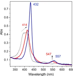
As shown in Figure 6 (blue trace), when a reductive
potential (Eelec= −0.60 V vs SCE) is maintained, after about
20 min the FeIVO signature fades and the characteristic spectral
signature of the FeIIIOO−species appears (Soret band at 432 nm and Q band at 557 nm).15Such an evolution is attributed to the consumption of protons in the optical part of the spectroelectrochemical cell before diffusion of protons from the bulk solution compensates it, thus stopping the catalysis. Interestingly, the spectral evolution of FeIIIOO−at open circuit
as a function of time (minutes scale) displays a signature corresponding to FeIVO species (seeFigure S13), as protons of
the bulk solution diffuse to the electrode, supporting the
proposition that this latter species originates from the O−O bond cleavage.
The signature of the FeIVO species has been previously
observed by resonance Raman spectroscopy among signatures of other species under electrocatalytic conditions, in the case of Fe porphyrins immobilized onto gold electrodes.29,30However, to the best of our knowledge, the present observation of accumulated high-valent species under reductive conditions has never been reported. This high-valent species can react with organic molecules such as cyclohexene and toluene. Preparative-scale electrolysis and analysis of the oxidation products are currently being performed and will be the subject of a future publication.
4. CONCLUSIONS AND PERSPECTIVES
Using the porphyrin FeIIIF20TPP as a catalyst for the
electrochemical reduction of O2 and a combination of CV
and UV−vis spectroelectrochemistry, we have experimentally
evidenced two catalytic pathways depending on the applied potential in acidic solutions of N,N-dimethylformamide. For
the low-overpotential pathway (−0.20 V vs SCE) a proton−
electron (PET) pathway involving a hydrosuperoxo species has
been identified. At large overpotential (−0.60 V vs SCE) the electron−proton (EPT) pathway prevails where protonation of FeIIIperoxo ultimately leading to the formation of high-valent
FeIVoxo species dominates. The observation that two different species are accumulated in each catalytic pathway suggests that the kinetics in the O−O bond cleavage differ. We thus propose that in the EPT case the more efficient catalysis likely favors the production of a larger amount of high-valent species. The unexpected observation of a high-valent Fe species under reductive conditions is quite remarkable and reminiscent of the
O2 reductive activation process observed in the Cyt P450
cycle. It opens the route to electrochemically triggered activity for oxidation processes using O2reductive activation.31−33
5. EXPERIMENTAL SECTION
5.1. Chemicals. All reagents and solvents were obtained commercially (Acros Organics and Aldrich). [FeIII(F
20TPP)Cl] (1),
tetrabutylammonium hexafluorophosphate (TBAPF6) supporting
electrolyte, tetrabutylammonium chloride (TBACl), N,N-dimethyl-formamide (DMF, anhydrous, 99.8%), silver perchlorate (AgClO4),
m-chloroperbenzoic acid 77% (m-CPBA), and perchloric acid 70% (HClO4) were used without further purification.
5.2. Cyclic Voltammetry. Electrochemical experiments were run under an argon or O2atmosphere. A dry O2atmosphere was obtained
by purging the solution with compressed air via a glass tubefilled with CaCl2. Cyclic voltammograms were recorded on a Metrohm
potentiostat (AUTOLAB Model PGSTAT302N). For cyclic voltammetry, the counter electrode used was a Pt wire and the working electrode a glassy-carbon disk (3 mm diameter) carefully polished before each voltammogram with a 1 μm diamond paste, sonicated in an ethanol bath, and then washed with ethanol. The reference electrode used was an SCE (saturated calomel electrode), isolated from the rest of the solution with a fritted bridge. The supporting electrolyte had a concentration of 0.1 M (293 K) or 0.2 M (258 K). Low-temperature regulation was ensured by a Julabo circulation cryostat.
5.3. Low-Temperature UV−Visible Spectroelectrochemis-try. Thin-cell spectroelectrochemical data were obtained using a combination of three electrodes in a thin cell (optical length 0.2 cm) mounted on a UV/vis Varian Cary 60 spectrophotometer, equipped with a transparent Dewar.34It consists of a 0.1 cm quartz UV−vis− NIR cell surmounted by a glass compartment. A homemade carbon-paper electrode was used as the working electrode, Ag/AgNO3as the
reference electrode, and a platinum grid in a frit as the counter electrode. The entire solution was saturated with air (1 mM O2), and
the cell was cooled to 258 K by a Julabo circulation cryostat.
ASSOCIATED CONTENT
CV analysis and simulation, including the CVs of an air-saturated DMF solution with increasing amounts of protons in the presence and absence of [FeIII(F
20
TPP)-Cl], CVs of [FeI I I(F2 0TPP)]+ (1-DMF) and
[FeIII(F
20TPP)Cl] (1-Cl) in DMF under argon, and
[FeIII(F20TPP)Cl] (1) in DMF under argon in the
presence of HClO4, the variation of the ip/i0ratio of the
FeIII/FeIIwave potential of 1-Cl with acid concentration under catalytic conditions, the effects of C(CF3)3OH, and CVs of chemically prepared FeIIIand FeIVO species of Fe(F20TPP), UV−vis absorption spectra of chemically prepared FeIIFeIIIand FeIVO species of the Fe(F20TPP),
Addition of H2O2 to an acidic [FeIII(F
20TPP)Cl]
Figure 6.UV−vis spectra of 1 (0.05 mM) in DMF + TBAPF6(0.2
M), under O2 (air saturated) + HClO4 (0.05 mM), at T = 258 K
(black trace), with Eelec=−0.60 V vs SCE after 5 min electrolysis (red
solution, and evolution of FeIIIOH
2+ and FeIIIOO−
species of Fe(F20TPP) (PDF) AUTHOR INFORMATION
Corresponding Author
Elodie Anxolabéhère-Mallart − Université de Paris,
Laboratoire d’Electrochimie Moléculaire, CNRS, F-75013 Paris,
France; orcid.org/0000-0002-8708-802X;
Email:elodie.anxolabehere u-paris.fr
Authors
Nikolaos Kostopoulos − Université de Paris, Laboratoire d’Electrochimie Moléculaire, CNRS, F-75013 Paris, France Célia Achaibou − Université de Paris, Laboratoire
d’Electrochimie Moléculaire, CNRS, F-75013 Paris, France Jean-Marc Noël − Université de Paris, ITODYS, CNRS,
F-75013 Paris, France
Frédéric Kanoufi − Université de Paris, ITODYS, CNRS,
F-75013 Paris, France; orcid.org/0000-0002-9784-2380
Marc Robert − Université de Paris, Laboratoire d’Electrochimie Moléculaire, CNRS, F-75013 Paris, France; Institut
Universitaire de France (IUF), F-75005 Paris, France;
orcid.org/0000-0001-7042-4106
Claire Fave − Université de Paris, Laboratoire d’Electrochimie Moléculaire, CNRS, F-75013 Paris, France; orcid.org/ 0000-0001-8146-8702
Complete contact information is available at:
https://pubs.acs.org/10.1021/acs.inorgchem.0c01379
Notes
The authors declare no competingfinancial interest.
ACKNOWLEDGMENTS
We thank the LabEx MiChem part of French state funds
managed by the ANR within the Investissements d’Avenir
programme under reference ANR-11-IDEX-0004-02 (E.A.-M., C.A., C.F., F.K., and J.-M.N.). N.K. acknowledges funding from the French government for his Ph.D. Partialfinancial support to M.R. from the Institut Universitaire de France (IUF) is also gratefully acknowledged. E.A.-M. thanks Pr. Frédéric Banse (ICMMO, Université Paris Saclay) for fruitful discussions.
ABBREVIATIONS
PET proton electron transfer EPT electron proton transfer DMF N,N-dimethylformamide ORR oxygen reduction reaction CytP450 cytochrome P450 oxidase SCE standard calomel electrode
CV cyclic voltammetry or cyclic voltammograms
REFERENCES
(1) Cavani, F. J. H.; Teles, J. H. Sustainability in catalytic oxidation: an alternative approach or a structural evolution? ChemSusChem 2009, 2, 508−534.
(2) Sheldon, R. A.; Arends, I. W. C. E.; Hanefeld, U. In Green Chemistry and Catalysis; Wiley-VCH: 2007; pp 133−221.
(3) Meunier, B.; De Visser, S. P.; Shaik, S. Mechanism of Oxidation Reactions Catalyzed by Cytochrome P450 Enzymes. Chem. Rev. 2004, 104, 3947−3980.
(4) Denisov, I. G.; Makris, T. M.; Sligar, S. G.; Schlichting, I. Structure and Chemistry of Cytochrome P450. Chem. Rev. 2005, 105, 2253−2278.
(5) Ortiz de Montellano, P. R. Hydrocarbon Hydroxylation by Cytochrome P450 Enzymes. Chem. Rev. 2010, 110, 932−948.
(6) Huang, X.; Groves, J. T. Oxygen Activation and Radical Transformations in Heme Proteins and Metalloporphyrins. Chem. Rev. 2018, 118, 2491−2553.
(7) Kaila, V. R. I.; Verkhovsky, M. I.; Wikström, M. Proton-Coupled Electron Transfer in Cytochrome Oxidase. Chem. Rev. 2010, 110, 7062−7081.
(8) Yoshikawa, S.; Shimada, A. Reaction mechanism of cytochrome c oxidase. Chem. Rev. 2015, 115, 1936−1989.
(9) Groves, J. T.; Han, Y.-Z. Models and mechanisms of Cytochrome P450 Action; Ortiz de Montellano, P. R., Ed.; Springer: Boston, MA, 1995.
(10) Tani, F.; Matsu-Ura, M.; Nakayama, S.; Naruta, Y. Synthetic models for the active site of cytochrome P450. Coord. Chem. Rev. 2002, 226, 219−226.
(11) Collman, J. P.; Boulatov, R.; Sunderland, C.; Fu, L. Functional Analogues of Cytochrome c Oxidase, Myoglobin, and Hemoglobin. Chem. Rev. 2004, 104, 561−588.
(12) Pegis, M. L.; McKeown, B. A.; Kumar, N.; Lang, K.; Wasylenko, D. J.; Zhang, X. P.; Raugei, S.; Mayer, J. M. Homogenous Electrocatalytic Oxygen Reduction Rates Correlate with Reaction Overpotential in Acidic Organic Solutions. ACS Cent. Sci. 2016, 2, 850−856.
(13) Pegis, M. L.; Martin, D. J.; Wise, C. F.; Brezny, A. C.; Johnson, S. I.; Johnson, L. E.; Kumar, N.; Raugei, S.; Mayer, J. M. Mechanism of Catalytic O2 Reduction by Iron Tetraphenylporphyrin. J. Am.
Chem. Soc. 2019, 141, 8315−8326.
(14) Anxolabéhère-Mallart, E.; Bonin, J.; Fave, C.; Robert, M. Small-molecule activation with iron porphyrins using electrons, photons and protons: some recent advances and future strategies. Dalton Trans. 2019, 48, 5869−5878.
(15) Oliveira, R.; Zouari, W.; Herrero, C.; Banse, F.; Schöllhorn, B.; Fave, C.; Anxolabéhère-Mallart, E. Characterization and Subsequent Reactivity of an Fe-Peroxo Porphyrin Generated by Electrochemical Reductive Activation of O2. Inorg. Chem. 2016, 55, 12204−12210. In
this study, the spectrum with λmax = 420, 530, 550 nm has been
mistakenly attributed to FeIIIOO•; according to our current knowledge this spectrum corresponds to the FeIIOH complex.
(16) Gueutin, C.; Lexa, D.; Savéant, J. M.; Wang, D. L. σ-Alkyl Iron Porphyrins from Sterically Encumbered Alkyl Halides and lron (“0”)Porphyrins. Organometallics 1989, 8, 1607−1613.
(17) Savéant, J.-M. In Elements of Molecular and Biomolecular Electrochemistry; Wiley-Interscience: New York, 2006.
(18) Wasylenko, D. J.; Rodríguez, C.; Pegis, M. L.; Mayer, J. M. Direct comparison of electrochemical and spectrochemical kinetics for catalytic oxygen reduction. J. Am. Chem. Soc. 2014, 136, 12544− 12547.
(19) Izutsu, K., Acid-Base Dissociation Constants in Dipolar Aprotic Solvents, Blackwell Scientific Publications, Brookline Village, 1990, Vol. 35, p 166.
(20) Costentin, C.; Dridi, H.; Savéant, J. M. Molecular Catalysis of O2 Reduction by Iron Porphyrins in Water: Heterogeneous versus
Homogeneous Pathways. J. Am. Chem. Soc. 2015, 137, 13535−13544. (21) Wang, Y. H.; Schneider, P. E.; Goldsmith, Z. K.; Mondal, B.; Hammes-Schiffer, S.; Stahl, S. S. Bronsted Acid Scaling Relationships Enable Control Over Product Selectivity from O2 Reduction with a
Mononuclear Cobalt Porphyrin Catalyst. ACS Cent. Sci. 2019, 5, 1024−1034.
(22) Mittra, K.; Chatterjee, S.; Samanta, S.; Dey, A. Selective 4e-/4H + O2reduction by an iron(tetraferrocenyl)porphyrin complex: from
proton transfer followed by electron transfer in organic solvent to proton coupled electron transfer in aqueous medium. Inorg. Chem. 2013, 52 (24), 14317−25.
(23) Kim, H.; Rogler, P. J.; Sharma, S. K.; Schaefer, A. W.; Solomon, E. I.; Karlin, K. D. Heme-Fe(III) Superoxide, Peroxide and Hydroperoxide Thermodynamic Relationships: FeIII-O2•− Complex
H-Atom Abstraction Reactivity. J. Am. Chem. Soc. 2020, 142 (6), 3104−3116.
(24) Franke, A.; Fertinger, C.; van Eldik, R. Axial Ligand and Spin-State Influence on the Formation and Reactivity of Hydroperoxo− Iron(III) Porphyrin Complexes. Chem. - Eur. J. 2012, 18, 6935−6949. (25) Pan, Z.; Newcomb, M. Kinetics and Mechanism of Oxidation Reactions of Porphyrin-Iron(IV)-Oxo Intermediates. Inorg. Chem. 2007, 46, 6767−6774.
(26) Cong, Z.; Yanagisawa, S.; Kurahashi, T.; Ogura, T.; Nakashima, S.; Fujii, H. Synthesis, characterization, and reactivity of hypochloritoiron(III) porphyrin complexes. J. Am. Chem. Soc. 2012, 134, 20617−20620.
(27) Cong, Z.; Kurahashi, T.; Fujii, H. Oxidation of chloride and subsequent chlorination of organic compounds by oxoiron(IV) porphyrin pi-cation radicals. Angew. Chem., Int. Ed. 2011, 50, 9935− 9939.
(28) Yokota, S.; Fujii, H. Critical Factors in Determining the Heterolytic versus Homolytic Bond Cleavage of Terminal Oxidants by Iron(III) Porphyrin Complexes. J. Am. Chem. Soc. 2018, 140 (15), 5127−5137.
(29) Sengupta, K.; Chatterjee, S.; Samanta, S.; Dey, A. Direct observation of intermediates formed during steady-state electro-catalytic O2reduction by iron porphyrins. Proc. Natl. Acad. Sci. U. S.
A. 2013, 110, 8431−8436.
(30) Sengupta, K.; Chatterjee, S.; Dey, A. In Situ Mechanistic Investigation of O2 Reduction by Iron Porphyrin Electrocatalysts
Using Surface-Enhanced Resonance Raman Spectroscopy Coupled to Rotating Disk Electrode (SERRS-RDE) Setup. ACS Catal. 2016, 6 (10), 6838−6852.
(31) Minteer, S.; Baran, P. Electrifying Synthesis: Recent Advances in the Methods, Materials, and Techniques for Organic Electrosyn-thesis. Acc. Chem. Res. 2020, 53 (3), 545−546.
(32) Anxolabéhère-Mallart, E.; Banse, F. Bioinspired molecular catalysts for homogenous electrochemical activation of dioxygen. Current Opinion in Electrochemistry 2019, 15, 118−124.
(33) Sengupta, K.; Chatterjee, S.; Samanta, S.; Bandyopadhyay, S.; Dey, A. Resonance Raman and electrocatalytic behavior of thiolate and imidazole bound iron porphyrin complexes on self assembled monolayers: functional modeling of cytochrome P450. Inorg. Chem. 2013, 52 (4), 2000−14.
(34) Gueutin, C.; Lexa, D. Low temperature spectroelectrochemistry for the characterization of highly reduced σ-alkyl iron halogenated porphyrins. Electroanalysis 1996, 8, 1029−1033.