Protoplasma (2000) 212:1-7
F o c u s o n
Cellular Biochemistry
PROTOPLASMA
9 Springer-Verlag 2000 Printed in AustriaRegulation of raft architecture
T. Harder 1'* and P. Scheiffele 2
1 Basel Institute for Immunology, Basel and 2 Department of Molecular and Cell Biology, University of California at Berkeley, California Received April 19, 1999
Accepted May 11, 1999
Summary. Raft membrane domains are envisioned as lateral assem-
blies of cholesterol and sphingolipids which adopt a liquid-ordered membrane phase. Our understanding of the raft architecture in cell membranes is developing rapidly. The current view describes raft domains as small and highly dynamic subdomains of cell mem- branes. The size and stability of raft domains are essential parame- ters which determine the function of raft domains in cells. Here we discuss how the architecture and stability of raft domains is regu- lated by oligomerisation of raft components and by modulation of their molecular composition.
Keywords: Membrane raft; Signal transduction; Sphingolipid;
Cholesterol; Oligomerisation.
Abbreviations: DIGs detergent-insoluble glycolipid-enriched membranes; GPI glycosylphosphatidylinositol; PDZ PSD95/discs- large/ZO-1; PI phosphoinositide; PIPz phosphatidylinositol 4,5- bisphosphate; TCR T-cell receptor.
Introduction
According to the raft hypothesis lateral assemblies of sphingolipids and cholesterol f o r m lipid-based domains (rafts) within biological m e m b r a n e s floating in an e n v i r o n m e n t of glycerophospholipids. R a f t domains c o n c e n t r a t e specific proteins while excluding others. Following this principle rafts are thought to serve as platforms for the assembly of signalling com- plexes, the generation of t r a n s p o r t vesicles in polarised m e m b r a n e transport, and the f o r m a t i o n of cell-cell or c e l l - s u b s t r a t u m adhesion sites (Brown and L o n d o n 1998, Simons and I k o n e n 1997). In this review we discuss the m e c h a n i s m s which m a y regulate raft size and c o m p o s i t i o n and why these features of rafts m a y be central to their function.
* Correspondence and reprints: Basel Institute for Immunology, Grenzacherstrasse 287, CH-4005 Basel, Switzerland.
Large detergent-insoluble membrane domains, but small raft domains
The biophysical nature of raft domains has b e e n studied in artificially reconstituted lipid bilayers. These studies led B r o w n and colleagues to p r o p o s e that raft m e m b r a n e domains exist in a liquid-ordered (1o) phase, whereas the surrounding nonraft m e m b r a n e forms a liquid-disordered (ld) p h a s e (Brown and L o n d o n 1997). In lipid vesicles which mimic the com- position of the p l a s m a m e m b r a n e , coexistence of these two liquid m e m b r a n e phases was d e m o n s t r a t e d . Phase separation in these m e m b r a n e s depends on choles- terol; and, importantly, m o d e l m e m b r a n e s which a d o p t an 1o phase as well as glycosylphosphatidylinositol ( G P I ) - a n c h o r e d proteins reconstituted into such m e m b r a n e s are insoluble in mild detergents ( A h m e d et al. 1997, Schroeder et al. 1994).
Making use of this p r o p e r t y a detergent-insoluble m e m b r a n e fraction ( D I G ) isolated f r o m cell m e m - branes is used to analyse the m o l e c u l a r composition of raft domains. D I G s are highly enriched in sphin- golipids and cholesterol and contain a specific subset of m e m b r a n e proteins (Simons and I k o n e n 1997). A p r o m i n e n t protein class in the D I G fraction are G P I - a n c h o r e d proteins. D I G association of t r a n s m e m b r a n e proteins is d e t e r m i n e d by specific amino acids in their m e m b r a n e - s p a n n i n g domains (Scheiffele et al. 1997). M o r e o v e r , palmitoylations which anchor cytoplasmic domains of t r a n s m e m b r a n e proteins to the inner leaflet of the lipid bilayer support D I G association (Melkonian et al. 1999). Several cytoplasmic periph- eral m e m b r a n e proteins, for e x a m p l e Src-kinase family m e m b e r s , are targeted to the D I G fraction via multi- ple palmitoylation and are thus thought to interact
2 T. Harder and R Scheiffele: Raft architecture with the cytoplasmic face of raft membranes (Rodgers
et al. 1994, Shenoy-Scaria et al. 1994).
Even though detergent insolubility has proven a valuable tool and technically easy approach to assay both protein and lipid requirements for raft associa- tion, it has its problems. Detergent extraction causes coaggregation of raft markers on the surface of mam- malian cells, and the morphology of the DIG mem- branes does not reflect the organisation of rafts in vivo (Mayor et al. 1994). In order to understand the bio- logical function of rafts, however, their organisation in cell membranes, i.e., their size and composition, needs to be unravelled.
Several recent studies using novel biochemical and biophysical methods analysed raft morphology in the plasma membrane of living cells. Varma and Major (1998) introduced a noninvasive measurement of the distance between GPI-anchored folate receptor mole- cules. Energy transfer between GPI-anchored folate receptor was monitored by anisotropy measurement of the emitted light. Importantly, energy transfer was independent of the concentration of the folate recep- tor in the plasma membrane, indicating that folate receptors are clustered in lipid microdomains. These studies, moreover, demonstrated that clustering was cholesterol dependent in line with the role of choles- terol in formation of an lo phase in model membranes. These domains, however, were not resolvable by conventional light microscopy, indicating their small size. Similarily, analysis of GPI-anchored green- fluorescent-protein mutants on the cell surface indi- cated some degree of clustering (De Angelis et al. 1998). Furthermore, it was demonstrated biochemi- cally that crosslinking efficiencies between GPI- anchored proteins were independent of their concentration on the cell surface (Friedrichson and Kurzchalia 1998). Altogether these papers strongly indicate that GPI-anchored proteins on the surface of resting cells are clustered in small domains (<70 nm diameter) with dimensions below the resolution limit of conventional light microscopy.
Fission and fusion of raft domains - a functional switch?
What is the functional significance of these raft micro- clusters? Such small raft domains can harbour only relatively few protein molecules. Moreover, it appears improbable that membrane domains of this size can be shaped into transport vesicles or function as platforms
for signal transduction. However, assuming a regu- lated equilibrium of fusion and fission of raft domains in the cell surface the localisation of a molecule in rafts would have important functional consequences.
In the resting state there is a large number of domains which are dispersed over the cell surface. The molecules in the raft domain would only have limited contact with molecules in the surrounding "nonraft" membrane. For signal transduction molecules this could mean that unwanted cross talk is prevented. As the domains are small, the local representation of signalling compounds will not be sufficient to recruit downstream binding partners.
When rafts coalesce, molecules previously separated by dispersion of the raft domains are combined in a common large patch (for detailed discussion, see Vaz and Almeida 1993). Furthermore, now large domains with high local concentrations of raft components are formed. These concentrations would now be above the threshold to tightly bind downstream molecules (Fig. 1).
Different mechanisms should allow to shift the equilibrium of raft size. The availability of raft lipids or the lipid composition in a membrane as well as extracellular or intracellular cross-linking events and their influence on raft architecture will be discussed below.
Regulation of rafts by changes in lipid composition
The amount of raft lipids in a membrane should be an important parameter for the size of the domains. According to the proposed equilibrium of raft fusion and fission, increase of a raft lipid should shift this equilibrium towards the fusion and decrease of raft lipids towards the fission of the domains. The endo- plasmic reticulum membrane might represent an extreme case for fission as it contains cholesterol but only minute amounts of sphingolipids. No DIG asso- ciation of proteins is observed in the endoplasmic reticulum membrane, suggesting that the sphingolipid levels are below the threshold to significantly assem- ble rafts (Brown and Rose 1992).
Relatively little is known how physiological changes in sphingolipid composition change the properties of raft domains. Such changes in glycolipid expression have been described for cell differentiation processes. For example, maturation of neurons to form a fully polarised phenotype is accompanied by an upregula- tion of specific glycolipids (Futerman et al. 1998).
T. Harder and E Scheiffele: Raft architecture 3
i
. . . ~ r
T MIt
U
Fig. 1. Recruitment of cytosolic proteins to stabilised raft domains triggered by clustering. Clustering of a raft-associated membrane protein
creates large patches with a high local concentration of this membrane protein. This allows efficient recruitment of a cytosolic ligand which binds only weakly to this membrane protein under steady-state conditions. Multiple binding sites or lateral interaction between the ligand molecules may lead to a high cooperativity in the association
Interestingly, this upregulation correlates with the sta- bility of detergent-insoluble complexes and sorting of membrane proteins to the axon (Ledesma et al. 1999). Similarly, the amount of ganglioside GM1 in resting T lymphocytes strongly increases after priming by engagement of T-cell receptor (TCR) and costimula- tion. This priming induces differentiation of the resting T lymphocyte towards an effector T cell. Upregulation of the glycolipid GM1 may drive raft fusion and thereby increase responsiveness of the cells (A. Viola and T. Harder unpubl, results). It will be most inter- esting to see which other lipid components are up- regulated in such maturation processes, as one would expect that for example also the cholesterol pool in the plasma membrane increases.
Besides sphingolipids and cholesterol, phospho- inositides (PI) such as phosphatidylinositol-4,5-bis- phosphate (PIP2) are highly enriched in the D I G fraction (Hope and Pike 1996). PI are rapidly turned over by the activity of PI kinases and their respective counteracting phosphatases (Liu et al. 1998) and may be subject to short-term changes in rafts. PI kinases and phosphatases m a y therefore play an essential role in regulating raft composition. Downstream targets which bind PIP2, such as cytoskeletal proteins or sig- nalling molecules, could then aid the assembly of the raft patches and stabilise these domains (Fukami et al. 1994). Furthermore PIP2 is rapidly hydrolysed in acti- vated cells by the activity of phospholipase C, yielding second messengers inositol 1,4,5-trisphosphate and
diacylglycerol. This hydrolysis may be a mechanism of local raft destabilisation and downregulation of its activity (Pike and Casey 1996, Stauffer et al. 1998).
Clustering rafts from within the membrane: caveolin
Members of the caveolin family were initially described as major constituent of the caveolar invagi- nations in the plasma membrane and the D I G frac- tion. A number of features make caveolin an ideal candidate for sequestering raft domains. The protein tightly binds to cholesterol, approximately in equimo- lar stoichiometry. Furthermore, each caveolin mole- cule is modified by three palmitic acid chains in the C-terminal cytoplasmic domain. Caveolins form large oligomers of 15-20 molecules, which might allow cre- ation of large stabilised rafts within the membrane. Consistently, the ganglioside GM1 is found concen- trated in cell surface caveolae (for a review, see Parton 1996). A multitude of biological functions have been assigned to caveolae. The common theme in all these processes might be the organisation of a specific mem- brane subcompartment. A m o n g these functions are cholesterol exchange, endocytosis and transcytosis of compounds in endothelia, formation of signalling cen- tres, etc. (Okamoto et al. 1998).
However, caveolins were also observed in noncave- olar regions of the cell surface and in intracellular compartments (Dupree et al. 1993). Different com- plexes of caveolins are involved in polarised mere-
4 T. Harder and E Scheiffele: Raft architecture brane sorting from the trans-Golgi network in MDCK
cells. Apical transport appears to involve caveolin-1 homo-oligomers, whereas basolateral transport vesi- cles contain complexes of caveolin-1 and -2 (Scheiffele et al. 1998). The apical complexes show a significant size shift upon leaving the Golgi complex, possibly reflecting the specific concentration of apical protein and lipid cargo. Interestingely caveolin was shown to be required for the formation of active signalling complexes of integrins, Fyn kinase, and associating signalling molecules (Wary et al. 1998). Future studies will show to which extent caveolins function in these complexes by coordinating protein-protein or pro- tein-lipid interactions.
Extracellular cross-linking: oligomeric ligands
of raft-associated receptors
Many ligands induce clustering of their receptors upon binding. Additionally, repetitive antigens and other oligomeric molecules can cluster their cognate cell surface receptors, and this may induce formation of stabilised raft domains. One of the first examples described is the cross-linking of the immunoglobulin E receptor by repetitive antigens via receptor-bound immunoglobulin E molecules (Holowka and Baird 1996). These domains are preferentially stained with specific lipid dyes and moreover transiently accumu- late the glycosphingolipid GM1 (Stauffer and Meyer 1997, Thomas et al. 1994). Extracellular cross-linking of membrane proteins is a frequently recurring theme in the interaction of cells with their environment. For example, membrane protein clusters are formed when cell receptors interact with extracellular matrix or when cell adhesion molecules line up at cell-cell contact sites. It is tempting to speculate that some of these clusters have raft properties which are recog- nised by an intracellular machinery.
Intracellular cross-linking: adaptor cross-linking
Also cytoplasmic molecules can confer clustering of transmembrane receptors. For example P D Z (PSD-95, discs-large, ZO-1) domain-containing proteins are found at different cellular junctions like synapses and adherens junctions in the epithelium. Via the P D Z domain they bind to a recognition sequence located at the C terminus of their ligands. One of the most prominent members of this family, PSD-95, was
recently reported to be recovered in DIGs (Perez and Bredt 1998). Surprisingly, D I G association in heterol- ogous cells did not require the two N-terminal palmi- toylations of PSD-95 but the P D Z domain-containing region. However, targeting of PSD-95 to synaptic clus- ters in primary neurons was dependent on the palmi- toylation (Craven et al. 1999). Possibly, in heterologous cells binding to a raft-associated transmembrane protein mediates recruitment into DIGs as it was observed for another P D Z domain-containing protein, GRIP2. Via one of its seven P D Z domains GRIP2 interacts with the C terminus of EphrinB1, a trans- membrane ligand of Eph-tyrosine kinase receptors, and is thereby recruited into rafts (Br~ickner et al. 1999). Binding of transmitter receptors or channels to PSD-95 results in clustering in the membrane which is driven by oligomerisation of PSD-95 (Hsueh et al. 1997). It will be interesting to know whether these patches formed by PSD-95 at the synapse represent raft domains.
A series of recent reports suggest that many pro- teins involved in TCR-mediated signalling are asso- ciated to rafts. These include the phosphorylated ~-subunit of the CD3-TCR complex itself, the Src kinase family members Fyn and Lck, the adaptor protein LAT (linker for activation of T lymphocytes), and a large number of other molecules involved in T-lymphocyte activation (Montixi et al. 1998, Xavier et al. 1998, Zhang et al. 1998). For LAT and Lck a link between their DIG association and their capability to transduce a signal via TCR was described (Kabouridis et al. 1997, Rodgers et al. 1994, Shenoy-Scaria et al. 1994, Zhang et al. 1998).
Under physiological conditions antigen-presenting cells display only few peptide-major histocompatibil- ity complexes specific for a TCR of a particular T cell. Thus, triggering of T lymphocytes most probably does not involve the formation of large TCR clusters. Rather it appears that cognate major histocompatibil- ity molecules can serially trigger multiple rounds of TCRs until a threshold for activation is reached (Vali- tutti et al. 1995b, Viola and Lanzavecchia 1996). A series of recent reports underline the importance of raft domain sequesteration and stabilisation in the T lymphocyte's plasma membrane region contacting the antigen-presenting cell. Firstly raft-associated glycosphingolipid GM1 accumulates at contact sites between a naive T lymphocyte and a TCR-activating bead in a manner depending on costimulation by
T. Harder and R Scheiffele: Raft architecture 5
CD28 engagement (Viola et al. 1999). Moreover co- engagement of TCR and the GPI-anchored surface protein CD48 enhanced T-cell activation and Triton X- 100 insolubility of TCR subunits (Moran and Miceli 1998). A multitude of additional interactions between accessory molecules and ligands support the formation of a contact zone between an antigen-presenting cell and a T cell. This zone adopts a well defined structure which may represent a highly differentiated scaffold for raft patches involved in the activation of T cell by antigen-presenting cells (Dustin and Shaw 1999, Monks et al. 1998, Moran and Miceli 1998).
It appears probable that intracellular proteins are responsible for accumulating and stabilising membane domains at the site of TCR engagement. Cross-linking of raft-associated molecules could occur through mul- tiple interactions of adaptor molecules which generate a complex network of signalling molecules sufficient to stabilise raft domains. Most probably the actin cytoskeleton plays an important role in this assembly process. Signalling via TCR requires an intact actin cytoskeleton (Valitutti et al. 1995a). Moreover, it was shown that artificially induced clusters of the gly- cosphingolipid GM1 accumulate phosphotyrosine as well as actin filaments (Harder and Simons 1999). The actin cytoskeleton may help organise the T-cell contact site via an active actomyosin-mediated transport of molecules and/or by forming a scaffold for raft domains (Dustin and Shaw 1999, Kupfer et al. 1987, Wtilfing and Davis 1998).
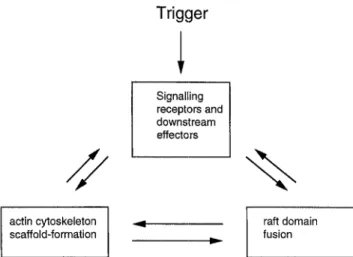
Rafts, actin cytoskeleton or other scaffolding pro- teins, and signalling molecules are likely to be tied up in an interplay as presented in Fig. 2. Triggering of receptors may induce the recruitment of proteins which aid raft sequestration. Raft-associated PIP2 may induce actin polymerisation and recruit a set of cytoskeletal molecules to the rafts, which could form a scaffold for further stabilisation of the domains (Fukami et al. 1994). Moreover, signalling may induce actin polymerisation for example via activation of GTPases of the Rho family (Reif and Cantrell 1998). Large raft patches could then serve as a platform for assembly of activated signalling complexes. This posi- tive feedback circuit may ensure that a threshold for a cellular response is reached even if few signalling receptors are triggered. It is the challenge for the future to elucidate how a starting point for raft stabil- isation is seeded and which mechanisms mediate and regulate the ongoing stabilisation.
Trigger
Signalling receptors and downstream effectors/ /
%
actin cytoskeleton scaffold-formation 4 IP raft domain fusionFig. 2. Possible interplay of signalling molecules, raft domains, and cytoskeletal elements. Membrane receptors are triggered by binding of ligands. This may induce fusion and accumulation of raft domains either by cross-linking of receptors via the ligands or by recruitment of intracellular signalling molecules to the membrane. Further recruitment of raft-associated signalling molecules and actin accu- mulation allows stabilisation of the raft patches. Once overcoming the initial threshold for the formation of a large raft patch, a posi- tive feedback cycle may serve to amplify signalling via the raft- associated receptors
Acknowledgments
We thank Christian Wimmer and Antonella Viola for critical reading of the manuscript. The Basel Institute for Immunology was founded and is supported by Hoffman La Roche Ltd. RS. is supported by an EMBO long-term fellowship.
References
Ahmed SN, Brown DA, London E (1997) On the origin of sphingolipid/cholesterol-rich detergent-insoluble cell membranes: physiological concentrations of cholesterol and sphingolipid induce formation of a detergent-insoluble, liquid-ordered lipid phase in model membranes. Biochemistry 36: 10944- 10953
Brown DA, London E (1997) Structure of detergent-resistant mem- brane domains: does phase sepaiation occur in biological mem- branes? Biochem Biophys Res Commun 240:1-7
- - (1998) Functions of lipid rafts in biological membranes. Annu Rev Cell Dev Biol 14:111-136
- Rose JK (1992) Sorting of GPI-anchored proteins to glycolipid- enriched membrane subdomains during transport to the apical cell surface. Cell 68:533-544
BriJckner K, Labrador JR Scheiffele R Herbst A, Seeburg PH, Klein R (1999) EphrinB ligands recruit GRIP family P D Z adaptor proteins into raft membrane microdomains. Neuron 22: 511-524
Craven SE, E1-Husseini AE, Bredt DS (1999) Synaptic targeting of the postsynaptic density protein PSD-95 mediated by lipid and protein motifs. Neuron 22:497-509
6 T. Harder and R Scheiffele: Raft architecture De Angelis DA, Miesenbock G, Zemelman BV, Rothman JE (1998)
PRIM: proximity imaging of green fluorescent protein-tagged polypeptides. Proc Natl Acad Sci USA 95:12312-12316 Dupree R Parton RG, Raposo G, Kurzchalia TV, Simons K (1993)
Caveolae and sorting in the trans-Golgi network of epithelial cells. EMBO J 1993 12:1597-1605
Dustin ML, Shaw AS (1999) Costimulation: building an immuno- logical synapse. Science 283:649-650
Friedrichson T, Kurzchalia TV (1998) Microdomains of GPI- anchored proteins in living cells revealed by crosslinking. Nature 394:802-805
Fukami K, Endo T, Imamura M, Takenawa T (1994) Alpha-actinin and vinculin are PIP2-binding proteins involved in signaling by tyrosine kinase. J Biol Chem 269:1518-1522
Futerman AH, Boldin S, Brann AB, Schwarz A, Zisling R (1998) Regulatory roles for sphingolipids in the growth of polarized neurons. Ann N Y Acad Sci 845:176-187
Harder T, Simons K (1999) Clusters of glycolipid and glycosylphos- phatidylinositol-anchored proteins in lymphoid cells: accumula- tion of actin regulated by local tyrosine phosphorylation. Eur J Immunol 29:556-562
Holowka D, Baird B (1996) Antigen-mediated IGE receptor aggre- gation and signaling: a window on cell surface structure and dynamics. A n n u Rev Biophys Biomol Struct 25:79-112
Hope HR, Pike LJ (1996) Phosphoinositides and phosphoinositide- utilizing enzymes in detergent-insoluble lipid domains. Mol Biol Cell 7:843-851
Hsueh YP, Kim E, Sheng M (1997) Disulfide-linked head-to-head multimerization in the mechanism of ion channel clustering by PSD-95. Neuron 18:803-814
Kabouridis PS, Magee AI, Ley SC (1997) S-acylation of LCK protein tyrosine kinase is essential for its signalling function in T lym- phocytes. EMBO J 16:4983-4998
Kupfer A, Swain SL, Singer SJ (1987) The specific direct interaction of helper T ceils and antigen-presenting B cells II: reorientation of the microtubule organizing center and reorganization of the membrane-associated cytoskeleton inside the bound helper T cells. J Exp Med 165:1565-1580
Ledesma MD, Brugger B, Btinning C, Wieland FT, Dotti CG (1999) Maturation of the axonal plasma membrane requires upregula- tion of sphingomyelin synthesis and formation of protein-lipid complexes. EMBO J 18:1761-1771
Liu Y, Casey L, Pike LJ (1998) Compartmentalization of phos- phatidylinositol 4,5-bisphosphate in low-density membrane domains in the absence of caveolin. Biochem Biophys Res Commun 245:684-690
Mayor S, Rothberg KG, Maxfield FR (1994) Sequestration of GPI- anchored proteins in caveolae triggered by cross-linking. Science 264:1948-1951
Melkonian KA, Ostermeyer AG, Chen JZ, Roth MG, Brown DA (1999) Role of lipid modifications in targeting proteins to detergent-resistant membrane rafts: many raft proteins are acylated, while few are prenylated. J Biol Chem 274:3910-3917 Monks CR, Freiberg BA, Kupfer H, Sciaky N, Kupfer A (1998)
Three-dimensional segregation of supramolecular activation clus- ters in T celts. Nature 395:82-86
Montixi C, Langlet C, Bernard AM, Thimonier J, Dubois C, Wurbel MA, Chauvin JR Pierres M, He HT (1998) Engagement of T cell receptor triggers its recruitment to low-density detergent- insoluble membrane domains. EMBO J 17:5334-5348
Moran M, Miceli MC (1998) Engagement of GPI-linked CD48 con- tributes to TCR signals and cytoskeletal reorganization: a role for lipid rafts in T cell activation. Immunity 9:787-796
Okamoto T, Schlegel A, Scherer PE, Lisanti MP (1998) Caveolins, a family of scaffolding proteins for organizing "preassembled sig-
naling complexes" at the plasma membrane. J Biol Chem 273: 5419-5422
Parton R G (1996) Caveolae and caveolins. Curr Opin Cell Biol 8: 542-548
Perez AS, Bredt DS (1998) The N-terminal PDZ-containing region of postsynaptic density-95 mediates association with caveolar-like lipid domains. Neurosci Lett 258:121-123
Pike LJ, Casey L (1996) Localization and turnover of phos- phatidylinositol 4,5-bisphosphate in caveolin-enriched membrane domains. J Biol Chem 271:26453-26456
Reif K, Cantrell DA (1998) Networking Rho family GTPases in lym- phocytes, Immunity 8:395401
Rodgers W, Crise B, Rose JK (1994) Signals determining protein tyrosine kinase and glycosyI-phosphatidylinositol-anchored pro- tein targeting to a glycolipid-enriched membrane fraction. Mol Cell Biol 14:5384-5391
Scheiffele R Roth MG, Simons K (1997) Interaction of influenza virus haemagglutinin with sphingolipid-cholesterol membrane domains via its transmembrane domain. EMBO J 16: 5501- 5508
- Verkade R Fra AM, Virta H, Simons K, Ikonen E (1998) Caveolin-1 and -2 in the exocytic pathway of MDCK cells. J Cell Biol 140:795-806
Schroeder R, London E, Brown D (1994) Interactions between saturated acyl chains confer detergent resistance on Iipids and glycosylphosphatidylinositol (GPI)-anchored proteins: GPI- anchored proteins in liposomes and cells show similar behavior. Proc Natl Acad Sci USA 91:12130-12134
Shenoy-Searia AM, Dietzen D J, Kwong J, Link DC, Lublin DM (1994) Cysteine3 of Src family protein tyrosine kinase determines palmitoylation and localization in caveolae. J Cell Biol 126: 353-363
Simons K, Ikonen E (1997) Functional rafts in cell membranes. Nature 387:569-572
Stauffer TP, Meyer T (1997) Compartmentalized IgE receptor- mediated signal transduction in living cells. J Cell Biol 139: 1447- 1454
Ahn S, Meyer T (1998) Receptor-induced transient reduction in plasma membrane PtdIns(4,5)P2 concentration monitored in living cells. Curr Biol 8:343-346
Thomas JL, Holowka D, Baird B, Webb WW (1994) Large-scale co- aggregation of fluorescent lipid probes with cell surface proteins. J Cell Biol 125:795-802
Valitutti S, Dessing M, Aktories K, Gallati H, Lanzavecchia A (1995a) Sustained signaling leading to T cell activation results from prolonged T cell receptor occupancy: role of T ceil actin cytoskeleton. J Exp Med 181:577-584
- Muller S, Cella M, Padovan E, Lanzavecchia A (1995b) Serial trig- gering of many T-cell receptors by a few peptide-MHC complexes. Nature 375:148-151
Varma R, Mayor S (1998) GPI-anchored proteins are organized in submicron domains at the cell surface. Nature 394:798-801 Vaz WL, Almeida PF (1993) Phase topology and percolation in
multi-phase bilayers: is the biological membrane a domain mosaic2 Curt Opin Struct Biol 3:482-488
Viola A, Lanzavecchia A (1996) T cell activation determined by T cell receptor number and tunable thresholds. Science 273: 104- 106
- Schroeder S, Sakakibara Y, Lanzavecchia A (1999) T lymphocyte costimulation mediated by reorganization of membrane micro- domains. Science 283:680-682
Wary KK, Mariotti A, Zurzolo C, Giancotti FG (1998) A re- quirement for caveolin-1 and associated kinase Fyn in integrin signaling and anchorage-dependent cell growth. Cell 94: 625- 634
T. Harder and R Scheiffele: Raft architecture 7 W~ilfing C, Davis MM (1998) A receptor/cytoskeletal movement
triggered by costimulation during T cell activation. Science 282: 2266-2269
Xavier R, Brennan T, Li Q, McCormack C, Seed B (1998) Membrane compartmentation is required for efficient T cell activation. Immunity 8:723-732
Zhang W, Trible RE Samelson LE (1998) LAT palmitoylation: its essential role in membrane microdomain targeting and tyro- sine phosphorylation during T cell activation. Immunity 9: 239- 246