HAL Id: hal-01511994
https://hal.archives-ouvertes.fr/hal-01511994
Submitted on 21 Apr 2017HAL is a multi-disciplinary open access archive for the deposit and dissemination of sci-entific research documents, whether they are pub-lished or not. The documents may come from teaching and research institutions in France or abroad, or from public or private research centers.
L’archive ouverte pluridisciplinaire HAL, est destinée au dépôt et à la diffusion de documents scientifiques de niveau recherche, publiés ou non, émanant des établissements d’enseignement et de recherche français ou étrangers, des laboratoires publics ou privés.
Development of fluorescence expression tools to study
host-mycoplasma interactions and validation in two
distant mycoplasma clades
Tiffany Bonnefois, Marie-Stéphanie Vernerey, Valérie Rodrigues, Philippe
Totte, Carinne Puech, Chantal Ripoll, François Thiaucourt, Lucia
Manso-Silvan
To cite this version:
Tiffany Bonnefois, Marie-Stéphanie Vernerey, Valérie Rodrigues, Philippe Totte, Carinne Puech, et al.. Development of fluorescence expression tools to study host-mycoplasma interactions and vali-dation in two distant mycoplasma clades. Journal of Biotechnology, Elsevier, 2016, 236, pp.35-44. �10.1016/j.jbiotec.2016.08.006�. �hal-01511994�
Accepted Manuscript
Title: Development of fluorescence expression tools to study host-mycoplasma interactions and validation in two distant mycoplasma clades
Author: Tiffany Bonnefois Marie-St´ephanie Vernerey Val´erie Rodrigues Philippe Tott´e Carinne Puech Chantal Ripoll Franc¸ois Thiaucourt Luc´ıa Manso-Silv´an
PII: S0168-1656(16)31454-7
DOI: http://dx.doi.org/doi:10.1016/j.jbiotec.2016.08.006 Reference: BIOTEC 7646
To appear in: Journal of Biotechnology
Received date: 28-6-2016 Revised date: 27-7-2016 Accepted date: 4-8-2016
Please cite this article as: Bonnefois, Tiffany, Vernerey, Marie-St´ephanie, Rodrigues, Val´erie, Tott´e, Philippe, Puech, Carinne, Ripoll, Chantal, Thiaucourt, Franc¸ois, Manso-Silv´an, Luc´ıa, Development of fluorescence expression tools to study host-mycoplasma interactions and validation in two distant mycoplasma clades.Journal of Biotechnology http://dx.doi.org/10.1016/j.jbiotec.2016.08.006
This is a PDF file of an unedited manuscript that has been accepted for publication. As a service to our customers we are providing this early version of the manuscript. The manuscript will undergo copyediting, typesetting, and review of the resulting proof before it is published in its final form. Please note that during the production process errors may be discovered which could affect the content, and all legal disclaimers that apply to the journal pertain.
1
Title: Development of fluorescence expression tools to study host-mycoplasma interactions and
validation in two distant mycoplasma clades
Tiffany Bonnefoisa,b [email protected]
Marie-Stéphanie Vernereyc [email protected] Valérie Rodriguesa,b [email protected]
Philippe Tottéa,b [email protected] Carinne Puechb,a [email protected] Chantal Ripoll d [email protected] François Thiaucourta,b [email protected] Lucía Manso-Silvána,b* [email protected]
a CIRAD, UMR CMAEE, F-34398 Montpellier, France b INRA, UMR1309 CMAEE, F-34398 Montpellier, France
c INRA, Joint Research Unit 385 UMR BGPI, Campus International de Baillarguet, Montpellier,
France
d INSERM U1051- Hôpital Saint Eloi INM. 80, rue Augustin Fliche -34091 Montpellier cedex 5 –
France
2
Highlights
Development of tools for stable and innocuous whole mycoplasma cell labelling Expression of bright red and green fluorescent proteins in two distant mycoplasmas First observation of fluorescent mycoplasma colonies by fluorescence microscopy Direct detection of fluorescent mycoplasmas in live host cells by flow cytometry Visualisation of individual mycoplasmas inside host cells by confocal microscopy
Abstract
Fluorescence expression tools for stable and innocuous whole mycoplasma cell labelling have been developed. A Tn4001-derivative mini-transposon affording unmarked, stable mutagenesis in mycoplasmas was modified to allow the constitutive, high-level expression of mCherry, mKO2 and mNeonGreen. These tools were used to introduce the respective fluorescent proteins as chromosomal tags in the phylogenetically distant species Mycoplasma mycoides subsp. mycoides and Mycoplasma bovis. The production, selection and characterisation of fluorescent clones were straightforward and resulted in the unprecedented observation of red and green fluorescent mycoplasma colonies in the two species, with no apparent cytotoxicity. Equivalent fluorescence expression levels were quantified by flow cytometry in both species, suggesting that these tools can be broadly applied in mycoplasmas. A macrophage infection assay was performed to assess the usefulness of mNeonGreen-expressing strains for monitoring mycoplasma infections, and notably cell invasion. The presence of fluorescent mycoplasmas inside live phagocytic cells was detected and quantified by flow cytometry and corroborated by confocal microscopy, which allowed the identification of individual mycoplasmas in the cytoplasm of infected cells. The fluorescence expression tools developed in this study are suitable for host-pathogen interaction studies and offer innumerable perspectives for the functional analysis of mycoplasmas both in vitro and in vivo.
Key words: mycoplasma, fluorescence, whole cell labelling, host-pathogen interactions, flow
cytometry, confocal microscopy
Abbreviations: Colony forming units (cfu); green fluorescent protein (GFP); median fluorescence
intensity (MFI); monomeric red fluorescent protein (mRFP1); multiplicity of infection (MOI); Mycoplasma mycoides subsp. mycoides (Mmm), phosphate buffered saline (PBS); photomultiplier tube 1 (PMT1); paraformaldehyde (PFA); propidium iodide (PI); room temperature (RT); variable secondary dichroic (VSD).
3
1. Introduction
The class Mollicutes constitutes a group of wall-less bacteria with a small genome size, commonly referred to as mycoplasmas, which evolved from gram-positive bacteria with low GC content through a process of massive genome reduction (Razin et al., 1998). Since they represent the smallest organisms capable of autonomous replication, they have served as models for the definition of the minimal gene set required for independent life, leading to the generation of a “minimal” artificial cell by synthetic biology (Hutchison et al., 2016). Through the course of reductive evolution mycoplasmas lost many metabolic capabilities and adapted to a commensal or parasitic mode of life. As a consequence, many species are known as important pathogens of humans, animals, and plants. However, despite the apparent simplicity of their small genomes, mycoplasmas have developed sophisticated mechanisms for colonisation and persistence in their host (Browning et al., 2014). Still, these mechanisms remain virtually unknown for most species and, although increasing amounts of genome sequence data have accumulated over the last two decades, the functional analysis of mycoplasmas has lagged far behind that of other eubacteria.
The analysis of bacterial gene expression and function is dependent on the availability of tools for the genetic manipulation of these bacteria, typically allowing the production of mutants and the expression of gene products such as selection markers, reporter systems or specific genes for mutant complementation. Yet, this has been hindered by the incompatibility of vectors engineered in conventional bacteria, together with difficulties encountered in the development of specific genetic tools for these peculiar organisms (Renaudin et al., 2014). Significant limiting factors include the scarcity of natural plasmids and selective antibiotic resistance markers and the slow, fastidious growth of cultivable mycoplasmas.
However, setting aside the latest developments in synthetic biology allowing the engineering of bacterial genomes in yeast and their transplantation in recipient cells (Hutchison et al., 2016), numerous tools are now available for the simple genetic manipulation of these long considered intractable organisms. A comprehensive review of these tools, and their contributions to the functional genetic analysis of mycoplasmas, has been recently published (Renaudin et al., 2014). Notable examples include sophisticated systems for random insertional mutagenesis derived from the transposon Tn4001 of Staphylococcus aureus. These mini-transposons have been improved by placing the transposase gene outside the transposable element, which prevents re-excision and secondary transposition, conferring stability to the insertions (Zimmerman and Herrmann, 2005). Another interesting improvement has been the combined use of these mini-transposons with the γδ TnpR/ res recombination system to excise the antibiotic resistance gene from the mycoplasma chromosome after transposition in order to produce unmarked mutations (Janis et al., 2008). Also,
4
artificial plasmids using the origin of replication of the mycoplasma chromosome (oriC) have been used to allow gene cloning and expression in a variety of mycoplasma species (Renaudin et al., 2014). However, the host range of oriC plasmids is circumscribed to closely related species and there is not a single self-replicating vector for universal use in mycoplasmas. The wide host range of Tn4001 transposition in mycoplasmas has been exploited to overcome the lack of plasmid vectors in some species and mini-transposons have been modified to allow gene delivery and expression (Zimmerman and Herrmann, 2005). These systems have been used to express fluorescent proteins in fusion with mycoplasma proteins, serving as reporters of gene expression and allowing the subcellular localisation of mycoplasma components (Balish et al., 2003; Kenri et al., 2004; Tulum et al., 2014; Zimmerman and Herrmann, 2005). However, the successful expression of fluorescent proteins in mycoplasmas remains rare and, to our knowledge, there are no fluorescence expression systems available to monitor the dynamics of mycoplasma infections in vivo or in vitro.
The aim of this work was to extend the array of molecular tools for use in Mollicutes, notably through the development of universal fluorescence expression systems for whole mycoplasma cell labelling allowing the monitoring of mycoplasmas during the course of infection. The level of fluorescence conferred to the mycoplasmas should be sufficient to enable their direct detection inside host cells using affordable techniques such as confocal microscopy and flow cytometry, thus allowing the analysis of the mechanisms involved in colonisation and persistence inside the host (e.g., adhesion, invasion, survival and multiplication inside host cells). For this purpose, a Tn4001-derivative mini-transposon affording stable, random mutagenesis in a variety of mycoplasma species was modified to allow the constitutive, high-level expression of green and red fluorescent proteins in mycoplasmas. The species Mycoplasma mycoides subsp. mycoides (Mmm) and Mycoplasma bovis, known as important cattle pathogens but belonging to distant phylogenetic groups (Sirand-Pugnet et al., 2007), were used to demonstrate the broad spectrum of the fluorescence expression tools for use in mycoplasmas and a macrophage infection assay was performed to show that fluorescent mycoplasmas could be directly detected inside host cells, thus demonstrating the usefulness of these new tools for the study of host-mycoplasma interactions.
2. Material and methods
2.1. Bacterial strains and culture conditions
Competent DH10 β-derivative Escherichia coli strain NEB 10-beta (New England Biolabs, USA) was used for plasmid cloning and propagation according to the supplier’s instructions, with 100 µg/mL kanamycin added to the culture medium for selection of transformants.
5
Mmm type strain PG1T (CIRAD, France) was used for PCR amplification of the intergenic region containing the tufA promoter. The two pathogenic mycoplasma strains used in this study were bovine lung isolates from cases of pneumonia and subjected to few in vitro passages. Mmm strain 8740-Rita was isolated from a contagious bovine pleuropneumonia case in Cameroon, in 1987 (Dr. Aboubakar, Laboratoire National Vétérinaire, Garoua, Cameroon). M. bovis strain Oger2 was isolated from a case of calf pneumonia in Ardennes, France, in 1975 (CIRAD, France). All strains were cultured at 37°C, 5% CO2 in modified Hayflick’s medium: 2.1% PPLO broth without crystal
violet (Difco, USA), 15% horse serum de-complemented for 1h at 56°C, 5% fresh baker’s yeast extract, 0.2% sodium pyruvate, 0.1% glucose, with 1% Noble Agar (Difco, USA) added for plating. 2.2. Plasmid constructions
2.2.1. DNA techniques
Plasmid DNA was extracted using either the Wizard Plus SV Minipreps DNA Purification System (Promega, USA) or the EndoFree Plasmid Maxi Kit (Qiagen, USA) depending on required DNA yield. Standard molecular techniques were used for plasmid constructions, using the following reagents according to the manufacturer’s instructions. Restriction enzymes were obtained from New England Biolabs, USA. Calf Intestinal Alkaline Phosphatase and T4 DNA ligase were purchased from Invitrogen, Life Technologies, USA. One microgram of plasmid DNA was dephosphorylated with 1 unit of CIAP during 5 min at 37°C. Ligation was performed according to the manufacturer’s recommendations for blunt cloning. Genomic DNA was extracted from bacterial cultures at the end of the exponential phase using the DNeasy Blood & Tissue Kit (Qiagen, USA). All oligonucleotides used in this study (Table 1) were obtained from Sigma-Aldrich, France. To minimize mutations caused by PCR amplification, high fidelity Phusion Hot Start II DNA Polymerase (Thermo Scientific, USA) was used for DNA cloning experiments. Otherwise, TopTaq DNA Polymerase (Qiagen, USA) was used for PCR verifications and prior to sequencing. DNA sequencing was performed by Beckman Coulter Genomics, Takeley, UK.
2.2.2. Construction of fluorescence expression tools
The strategy for the development of fluorescence expression tools is presented in the results section. Plasmids pMT85/2res and pPS3.1 were kindly provided by Pascal Sirand-Pugnet (INRA, Bordeaux, France) and all primer sequences are listed in Table 1. pMT85/2res was modified by cloning a cassette constructed using the overlap extension PCR technique. The entire intergenic sequence preceding the elongation factor Tu gene (tufA) followed by the first 33 nucleotides in its 5’ end, was amplified by PCR from the genomic DNA of Mmm strain PG1T using primers SpeI_Ptuf_F and NcoI_Ptuf+_R. The transcription terminator sequence of the fibril protein gene fib of Spiroplasma
6
citri was amplified from plasmid pPS3.1 using primers MCS_Ptuf+_Tfib_F and Tfib_XbaI_R. The resulting products were hybridised to each other and extended by primer-free amplification. A final PCR was performed to amplify this last product using primers SpeI_Ptuf_F and Tfib_XbaI_R. All PCR reactions consisted in initial denaturation at 98°C for 30 sec, followed by 35 cycles of denaturation at 98°C for 7 sec, annealing at 57°C for 30 sec and elongation at 72°C for 30 sec, finished by a final extension at 72°C for 5 min. The only exception was the primer-free amplification, for which only 12 PCR cycles were performed. The resulting cassette was double-digested with SpeI and XbaI and cloned in the SpeI site of pMT85/2res, resulting in plasmid pMT/exp. The entire cloned sequence was verified by DNA sequencing with primer TnpA-F. The pMT/exp plasmid map, annotated sequence and FASTA sequence are presented in Suppl. Fig. 1. The gene sequences of mCherry, mKO2 and mNeonGreen were retrieved from NCBI GenBank database (Table 2). These nucleotide sequences were optimised taking into consideration the codon usage bias in mycoplasmas (Suppl. Table 1). An in-house bioinformatic tool was developed at CIRAD to automate codon optimisation. This tool is freely available from the authors on request. The Codon Usage Database (http://www.kazusa.or.jp/codon/ ; last accessed 24/06/2016) (Nakamura et al., 2000) was used to analyse the frequency of codon use in the query organism “Mycoplasma mycoides subsp. mycoides SC str. PG1 [gbbct]: 1,016 CDS's (330,592 codons)”. The most frequently used codon for each amino acid (Suppl. Table 1) was selected for implementation in the bioinformatic tool. Synthetic constructs corresponding to mycoplasma-optimised gene sequences of each of the three fluorescent proteins were produced by ProteoGenix (Schiltigheim, France). The NcoI and AflII recognition sites were included at the extremities of the different fluorescent genes to allow directional cloning in pMT/exp, resulting in plasmids pMT/mNeonGreen, pMT/mCherry and pMT/mKO2. The cloned inserts were entirely validated by sequencing with primer pMT85-SpeIcloning_F (Table 1).
2.3. Mycoplasma transformation and identification of transposon insertion sites
PEG-mediated transformation of mycoplasma strains was performed as described (Janis et al., 2008) with a few modifications. Twenty micrograms of pMT85/2res-derived plasmids were used to transform approximately 109 mycoplasma cells. Transformed cells were re-suspended in 500 µL broth and incubated for either 2 hours (M. bovis) or 3 hours (Mmm) at 37°C, then plated (100 µL) on solid Hayflick’s medium supplemented with kanamycin (100 and 200 µg/mL for Mmm and M. bovis respectively). Ten-fold dilutions were also plated (20 µL drops) for culture titrations with and without antibiotics in order to calculate the transformation efficiency, expressed as the number of transformant colony forming units (cfu) per total cfu. Selected fluorescent colonies were picked and grown in selective broth, filtered through a Millex HV 0.45 µm Durapore PVDF membrane (Merk
7
Millipore, Ireland) and plated on solid medium for cloning. Filter-cloned mutant cultures were stored at -80°C for further analysis. Subsequent cultures were performed in non-selective medium. To sequence the site of the transposon insertion in the Mmm genome, total DNA from selected clones was digested with BclI. An adaptor was generated by hybridisation of 100 µM of oligonucleotides “Adapt1BclI” and “Adapt2BclI” (Table 1) at 100°C during 5 min, then ligated to BclI-digested mycoplasma DNA. The ligation product was used as template for PCR amplification using primers BclI-Primer and MT85-R (Table 1). Standard reactions were used during 35 PCR cycles with hybridisation at 52°C. The PCR product was then sequenced using primer MT85-R. The resulting sequences were analysed using Geneious R6 6.0.1 (Biomatters Ltd, New Zealand). After elimination of flanking mini-transposon sequences, the insertion site sequence was obtained by mapping with the available genome sequence of Mmm PG1T (GenBank accession no. BX293980.2).
2.4. Macrophage isolation and infection
Blood samples were collected from healthy Jersey cattle kept in an animal housing facility (CIRAD, Montpellier, France). Whole blood was collected from the jugular vein in heparinised BD Vacutainer tubes (Beckton Dickinson, USA) according to the manufacturer’s instructions. Experimental procedures for animal maintenance and blood sampling were approved by the Languedoc-Roussillon regional ethics committee (French CE-LR #36) in the Authorised Project using animals for scientific purposes #12ANI01. Monocyte isolation was performed by positive selection of CD14+ cells as previously described (Hope et al., 2003). CD14+ cells were then seeded in 6-well plates, with or without glass coverslips, at 3x10⁶ cells/well and cultured in Iscove's Modified Dulbecco's Medium (Life Technologies, France) supplemented with 2 mM L-glutamine, 50 μM 2-mercaptoethanol, 50 µg/mL gentamicin (Life Technologies, France) and 10% heat inactivated foetal calf serum (Eurobio AbCys, France) for 6 days at 37°C and 5% CO2. Half of the
medium was replaced after 3 days. After 6 days of differentiation, the cells were referred to as monocyte-derived macrophages.
After two washes with phosphate buffered saline (PBS) the cells were infected with washed mycoplasmas at the end of the exponential phase of growth using a multiplicity of infection (MOI) of around 200. Twenty percent v/v of the corresponding Mmm or M. bovis specific antiserum was added respectively and the cells were incubated for 30 min at 37°C. The cells were washed twice with PBS before further analysis. The specific antisera used for opsonisation consisted each in pooled sera from ten convalescent animals. The M. bovis antisera, originating from France, were a
8
gift from Florence Tardy (ANSES Lyon, France). The Mmm antisera were originated from Cameroon (Laboratoire National Vétérinaire, Garoua).
2.5. Fluorescence and confocal microscopy
Direct observation of mycoplasma colonies was conducted after 3 days incubation on PPLO agar using an AxioVert A1 inverted microscope equipped with CY3 and EGFP filter units (Carl Zeiss, Germany) for observation of red and green fluorescence respectively. The exposure conditions were set so that no autofluorescence was observed on wild-type cultures. These conditions were identical for all images taken using CY3 filters (0.3 m sec at 12 dB) and EGFP filters (0.1 m sec at 6 dB) respectively. Images were taken with a 10X objective using a QImaging CDD digital camera and analysed using the Archimed software (Microvision Instruments, France).
For confocal microscopy analysis, three day old mycoplasma broth cultures were pelleted, re-suspended in 4% paraformaldehyde (PFA, Sigma-Aldrich, France) in PBS and fixed during 15 min at room temperature (RT). Mycoplasmas were washed twice in PBS, re-suspended in 20 µL ProLong Diamond Antifade Mountant (Fisher scientific, USA) and transferred to a standard slide for microscopy observation.
Macrophages to be analysed by confocal microscopy were cultured on glass coverslips. Three washes in PBS were performed between each of the following steps: (i) staining using a 1.5X working solution of CellMask Deep Red Plasma Membrane Stain (Molecular Probes, Life Technologies, USA) during 10 min at 37°C; (ii) fixing with 4% PFA in PBS for 20 min at RT; (iii) nuclei staining with 2 μg/mL Hoechst 33258 pentahydrate dye solution (Molecular Probes, Life Technologies, USA) for 15 min at RT. Finally, coverslips were mounted with ProLong Diamond Antifade Mountant.
Observations were carried out on a ZEISS LSM 700 confocal laser scanning microscope. All images were acquired with a Plan-Apochromat 63X oil objective at a resolution of 620x620 pixels for mycoplasma observation and 512x512 pixels for macrophage imaging and with a pinhole aperture of 1 airy unit. mNeonGreen was excited with a 488 nm laser and the variable secondary dichroic (VSD) beamsplitter was set so as to recover all the fluorescence emitted up to 530 nm. Two other distinct tracks were created to recover the emission signals of Hoechst (405 nm laser and VSD beamsplitter set at 490 nm on the photomultiplier tube 1, PMT1) and CellMask Deep Red Plasma Membrane Stain (555 nm laser and VSD beamsplitter set at 630 nm on the PMT1). All images were captured using the same parameters to allow comparisons and were analysed using the ZEN software (Carl Zeiss, Germany).
9
2.6. Flow cytometry
Mycoplasma broth cultures at the end of the exponential phase of growth were washed twice and carefully re-suspended in PBS before flow cytometry analysis. Data acquisition and analysis were performed on a FACSCanto II flow cytometer (BD Biosciences, USA), with an excitation wavelength of 488 nm and detection with a 530/30-nm band-pass filter. A minimum of 10,000 mycoplasma cells were analysed and their global median fluorescence intensities (MFI) were calculated for comparison. The FACSDiva 6 software (BD Biosciences, USA) was used for data analysis.
Infected and non infected macrophages were also analysed by flow cytometry. Cell viability was measured using propidium iodide (PI). Cells were washed twice in PBS before re-suspension in PI at 2 μg/mL and the percentage of dead cells marked with PI was measured by flow cytometry. Since mycoplasmas are permeable to PI, this method also allowed discriminating intracellular (PI-negative) and extracellular (PI-stained) mycoplasmas. Data acquisition and analysis were performed as described above for green fluorescence, and a 585/42-nm band-pass filter was used for red fluorescence. A minimum of 20,000 cells within a gated region excluding cell debris were analysed.
3. Results
3.1. Construction of fluorescence expression tools for whole mycoplasma cell labelling
New genetic tools for constitutive, high level expression of fluorescent proteins in mycoplasmas were developed as summarised in Fig. 1. The mini-transposon pMT85/2res (Fig. 1C) was used for gene delivery. pMT85/2res is a genetic tool for stable, random mutagenesis that allows the elimination of the antibiotic resistance marker from selected mycoplasma mutants by treatment with a resolvase enzyme (Janis et al., 2008). This vector was modified to allow the expression of cloned genes in mycoplasmas under the control of the strong promoter of the elongation factor Tu gene of Mmm (tufA). The rho-independent transcription terminator of the fibril protein gene of the phytopathogenic mycoplasma S. citri (fib) was also included to avoid undesirable read-through of stop codons (Duret et al., 2005). The expression unit was inserted in the SpeI site of pMT85/2res, which allows its preservation in the event of resolvase treatment. The resulting plasmid was named pMT/exp (Fig. 1B).
The red fluorescent proteins mCherry and mKO2 and green fluorescent protein mNeonGreen were selected for expression in mycoplasmas owing to their interesting properties (Table 2). Their gene sequences were optimised taking into consideration the codon usage bias in mycoplasmas (Suppl. Table 1). All optimised sequences had a GC content exceeding 29%. Synthetic constructs
10
corresponding to mycoplasma-optimised gene sequences of each of the three fluorescent proteins were cloned in pMT/exp, resulting in plasmids pMT/mCherry, pMT/mKO2, and pMT/mNeonGreen (Fig. 1A).
3.2. Expression of bright red and green fluorescent proteins in two distant mycoplasma species The performance of fluorescence expression tools pMT/mCherry, pMT/mKO2, and pMT/mNeonGreen in the distant species Mmm and M. bovis was assessed in order to demonstrate their broad spectrum for use in mycoplasmas. Both strains were successfully transformed with the three vectors and the transformation efficiencies ranged between 10-5 and 10-6 transformant cfu/total cfu regardless the strain and construct used. Each transformation tube yielded from several hundreds to over one thousand transformant clones.
Five to seven days after transformation fluorescent colonies were directly observed on selective plates by fluorescence microscopy for all mycoplasma strains and constructs used. Furthermore, all the colonies corresponding to the same construct presented equivalent levels of fluorescence, only depending on the size and density of the colonies. Fluorescent clones were selected from the fastest growing mutants. However, the site of the transposon insertion should also be considered in order to limit the effect of gene disruption in subsequent functional studies. As a mode of example, the transposon insertion site of Mmm mutants was sequenced and those presenting insertions in intergenic sequences or irrelevant sites such as pseudogenes were chosen (Suppl. Table 2). Since strains may be attenuated through consecutive in vitro passages and this may also interfere with functional studies, the number of subcultures was kept to a minimum. The whole process from wild-type strain seeding for transformation until selection of fluorescent clones required five passages and two cloning procedures.
The brightness of selected M. bovis clones expressing each of the three fluorescent proteins was assessed by fluorescence microscopy (Fig. 2). Examination of all solid cultures using CY3 filters under the same exposure conditions resulted in observation of red fluorescent colonies on plates corresponding to mCherry and mKO2-expressing mycoplasmas, whereas no fluorescence was detected in wild-type and mNeonGreen-expressing cultures. On the other hand, identical exposure conditions using EGFP filters resulted in observation of bright green fluorescent colonies only in mNeonGreen-expressing cultures. The intensity of red florescence was higher in mKO2 than in mCherry-expressing mycoplasmas, as expected according to the reported brightness of the respective fluorescent proteins (Table 2). The intensity of green fluorescent colonies was also remarkable, considering the higher level of autofluorescence evidenced on mycoplasma cultures directly observed using EGFP filters, which imposed lower exposure conditions. Similar results
11
were obtained by observation of three-day-old plate cultures from selected Mmm clones expressing each of the three fluorescent proteins, with apparently lower fluorescence intensities attributed to the small size of Mmm colonies, compared to those of M. bovis (Suppl. Fig. 2). However, no difference in colony size was evidenced between any of the transformant clones and the corresponding wild-type strain. Furthermore, fluorescence expression was conserved after more than 10 in vitro passages in the absence of selective pressure.
Broth cultures of fluorescent mycoplasma clones were then analysed by confocal microscopy in order to visualize individual mycoplasma cells. An example is presented in Suppl. Fig. 3. Individual mycoplasmas expressing mNeonGreen could also be detected by flow cytometry (Suppl. Fig 4). Flow cytometry was therefore used to quantify the relative green fluorescence intensity of mNeonGreen-expressing and wild-type mycoplasmas at the end of the exponential phase of growth in order to confirm that the expression system developed here could induce comparable fluorescence levels in M. bovis and Mmm (Table 3). The MFI values registered in M. bovis strains were higher than those recorded in Mmm, indicating that the green fluorescence background was higher in this species. However, the MFI ratio between mNeonGreen-expressing and wild-type clones was equivalent in both species (corresponding to a 10 to 17-fold increase in MFI values, as recorded in three independent experiments).
3.3. Detection of fluorescent mycoplasmas inside host cells by flow cytometry and confocal microscopy
A macrophage infection assay was used to assess if the brightness of fluorescently-labelled mycoplasmas was sufficient to allow their direct detection inside host cells. The mNeonGreen-expressing mycoplasma clones were selected for these assays owing to practical reasons, since they were suited for flow cytometry analysis.
Bovine macrophages were infected with specifically-opsonised M. bovis strains at an MOI of around 200 and the cells were analysed by flow cytometry 30 min post-inoculation (Fig. 3). The forward versus side-scatter plot analyses allowed the definition of a gate excluding cell debris and aggregates. M. bovis-infected macrophages presented higher levels of debris than uninfected cells, suggesting an increase in mortality due to M. bovis infection, independently of the clone used. The macrophages infected with wild-type M. bovis presented a higher green fluorescence background than the uninfected cells (representing approximately a 4-fold increase in MFI). However, the cells infected with mNeonGreen-expressing M. bovis were significantly more fluorescent than those infected with the wild-type strain (10-fold increase in MFI). Moreover, these two populations were clearly discriminated according to green fluorescence, since 96% of the cells infected with the
12
mNeonGreen-expressing strain (versus 2% of those infected with the wild-type strain) fluoresced above background levels (Fig. 3). Finally, to confirm that the detected fluorescence resulted from intracellular bacteria, and not from mycoplasmas attached to the cell surface, a PI protection assay was performed. The percentage of PI-positive cells among the mNeonGreen-positive cells never exceeded 2% (results not shown). This implied that the great majority of the mycoplasmas, which are permeable to PI, were protected from staining, indicating that they were located inside the cells. The same experiment performed using Mmm wild-type and green fluorescent strains provided very similar results (Suppl. Fig. 5).
The intracellular location of fluorescent mycoplasmas was further demonstrated by confocal microscopy analysis. Fig. 4 shows confocal micrographs of macrophages infected with M. bovis 30 min post-inoculation. Many green fluorescent M. bovis were observed inside the macrophages infected with the mNeonGreen-expressing strain (Fig. 4C), both in tight clusters and as individual particles dispersed in the cytoplasm. The size of the particles was estimated at nearly 0.5 µm, which corresponds to the size of a mycoplasma cell (Citti and Blanchard, 2013), and most of them were observed in the middle focal plane with the cell nucleus, demonstrating that they were located inside the macrophage. No green fluorescence was observed either in uninfected macrophages or in macrophages infected with wild-type mycoplasmas (Fig. 4A and B respectively) and no morphological or functional differences were detected in macrophages infected with the mNeonGreen-expressing M. bovis compared to those infected with the wild-type strain (Fig. 4C and B respectively). Very similar results were obtained by infection of macrophages with Mmm strains (Suppl. Fig. 6), confirming that individual mNeonGreen-expressing mycoplasmas could be directly detected inside phagocytic cells.
4. Discussion and conclusions
4.1. Development of fluorescence expression tools for whole mycoplasma cell labelling
The aim of this work was to develop universal fluorescence expression tools for whole mycoplasma cell labelling to monitor the dynamics of mycoplasma infections. The first objective was therefore to select the appropriate vector, expression system and fluorescent proteins allowing the production of stable and innocuous bright fluorescent tags to label mycoplasma cells from different species. The mini-transposon pMT85/2res, a genetic tool for stable, random mutagenesis allowing the production of unmarked mutations (Janis et al., 2008; Zimmerman and Herrmann, 2005), was selected as vector for gene delivery in mycoplasmas. pMT85/2res was derived from the transposon Tn4001 of S. aureus, which presents a broad host spectrum and has been successfully used in a variety of mycoplasma species (Halbedel and Stülke, 2007). This vector proved to be very practical
13
and convenient. Transformation of Mmm and M. bovis strains with pMT85/2res-based constructs allowed the direct isolation of transformant colonies on agar, limiting the number of in vitro passages required for clone selection. Clones could then be stably propagated in non-selective medium, which may be critical for certain functional studies. Although the use of a transposon delivery vector rather than a replicating plasmid for gene delivery implied the integration in the mycoplasma chromosome and the subsequent risk of functional alterations, the transformation efficiency was sufficient to allow the selection of large numbers of mutants for which the insertion site could easily be characterised. Furthermore, transposon mutagenesis may actually be extremely useful for the construction of mutant libraries of fluorescently labelled mycoplasmas.
pMT85/2res was modified to express heterologous genes in mycoplasmas, resulting in the expression vector pMT/exp. The strong promoter of the elongation factor Tu gene of Mmm (tufA) was used to drive the expression of cloned genes in this system. The expression units of constitutively, highly expressed genes are the candidates of choice to maximise gene expression and the tuf promoter has already been used to drive the expression of heterologous genes in several mycoplasma species (Renaudin et al., 2015; Tulum et al., 2014; Zimmerman and Herrmann, 2005), including M. bovis (Sharma et al., 2014) and M. mycoides (Karas et al., 2014). However, in this study the tufA promoter of Mmm was successfully used for gene expression in the heterologous species M. bovis. The equivalent expression of high levels of fluorescence in Mmm and M. bovis, two mycoplasma species belonging to distant phylogenetic groups (Sirand-Pugnet et al., 2007), indicated that the expression system may be broadly applied in mycoplasmas. This was not without precedent, since the promoter of the spiralin gene, the most abundant protein of S. citri, proved to be efficient in driving gene expression in a variety of mycoplasma species (Renaudin, et al., 2014). The efficiency of the expression system in distant mycoplasma species, added to the broad host range of Tn4001 transposition makes pMT/exp a versatile tool for gene expression in mycoplasmas. One limitation to the use of Tn4001-derivative mini-transposons is that the aacA-aphD gene is not functional in certain mycoplasma species including M. penetrans and M. fermentans, which are naturally resistant to kanamycin, and M. genitalium, where the expression of the aacA-aphD gene results in growth impairment. Also in M. arthritidis and M. pulmonis Tn4001 insertion was not possible, presumably due to functional failure of the aacA-aphD gene (Renaudin, et al., 2014). However, this limitation may easily be solved by using either the chloramphenicol acetyltransferase gene (cat) or the tetracycline resistance marker (tetM), which have been shown to function in these species (Renaudin, et al., 2014), for the selection of transposon mutants.
pMT/exp was used to express fluorescent proteins in Mmm and M. bovis. Since the expression of fluorescent markers in mycoplasmas remains rare and has met with difficulties (Duret et al., 2003),
14
several unrelated proteins were used to increase the chances of success. The green fluorescent protein mNeonGreen (Shaner et al., 2013) and red fluorescent proteins mCherry (Shaner et al., 2004) and mKO2 (Sakaue-Sawano et al., 2008) were selected owing to their interesting properties (Table 2). They are all monomeric proteins, which are generally nontoxic (Shaner et al., 2005), and are characterised by high intrinsic brightness, particularly mKO2 and mNeonGreen. On the other hand, mCherry is particularly resistant to low pH, which may be useful for observation of mycoplasmas inside acidic compartments such as lysosomes.
All three fluorescent proteins were successfully expressed in both species, to such extent that bright fluorescent colonies could be directly observed on conventional culture medium by fluorescence microscopy. Furthermore, no signs of toxicity could be attributed to any of them. No growth impairment or other phenotypic changes were observed on fluorescent mutant strains, indicating that they were neither affected by the transposon insertion, nor by the production of high levels of fluorescent proteins, and the strains remained fluorescent after more than 10 in vitro passages in the absence of selective pressure. This was an important achievement, since attempts to express fluorescent proteins in mycoplasmas were not always successful. Expression of the green fluorescent protein (GFP) in fusion with spiralin, the major surface lipoprotein of S. citri, resulted in morphologic changes in cells and in reduced colony size (Duret et al., 2003). Moreover, the mutants were unstable and resulted in the production of subclones that did not express GFP, leading the authors to hypothesise that the fluorescent protein was toxic for this species. There are only a few studies reporting the expression of GFP derivatives, which were used as fusion tags to localise mycoplasma components in M. pneumoniae (Balish et al., 2003; Kenri et al., 2004) and M. mobile (Tulum et al., 2014). Also in M. pneumoniae, the monomeric red fluorescent protein (mRFP1) was used as fluorescent reporter system (Zimmerman and Herrmann, 2005). However, until now, there have been no reports of fluorescence expression tools for whole mycoplasma cell labelling allowing the direct observation of fluorescent colonies on agar and providing sufficient fluorescence levels for use in host-mycoplasma interaction studies. Since the strategy used here for fluorescent protein expression was very similar to that previously used for GFP derivatives and mRFP1 (with the exception that the mRFP1 gene sequence was not codon-optimised) the present success must be mainly attributed to the choice of fluorescent proteins.
4.2. Use of mycoplasma fluorescence expression systems for host-pathogen interaction studies Fluorescence expression systems for whole mycoplasma cell labelling were developed in this study as tools for monitoring the dynamics of mycoplasma infections. Although pathogenic mycoplasmas can be indirectly detected using fluorescent antibody labelling, traditional immunofluorescence detection techniques are time-consuming, tedious and expensive. Alternatively, they can be directly
15
coated with fluorochromes, but the fluorescence signal may fade over time and is diluted during replication. Consequently, these approaches are of limited value for tracing invasion pathways in living cells (Ling et al., 2000). Undeniably, the use of chromosomally tagged mycoplasmas would facilitate the analysis of the mechanisms involved in colonisation and persistence by allowing the direct monitoring of mycoplasma adhesion, invasion, survival and multiplication inside host cells. Therefore, the next objective of this study was to demonstrate that the brightness of fluorescently-labelled mycoplasmas was sufficient to allow their direct detection inside host cells. The mNeonGreen-expressing mycoplasmas were selected for this “proof of concept”, since they could be analysed by flow cytometry, and a macrophage infection assay was used in order to warrant the intracellular location of mycoplasmas. The presence of fluorescent mycoplasmas inside live phagocytic cells was detected and quantified by flow cytometry and corroborated by confocal microscopy imaging, which allowed the direct observation of individual mycoplasmas in the cytoplasm of infected cells. Furthermore, no signs of cytotoxicity could be identified and the use of fluorescently labelled mycoplasmas did not interfere with the cell infection assay. These results indicated that the fluorescence expression systems for whole mycoplasma cell labelling were well suited for the analysis of host-pathogen interactions. These new tools offer very interesting perspectives for mycoplasma research by allowing the direct monitoring of mycoplasma infections both in vitro and in vivo.
Cell invasion can be a major contributor to the establishment of persistent infections and chronic disease by mycoplasmas (Browning et al., 2014), but this phenomenon has only been studied in a few mycoplasma species and the mechanisms involved remain totally unknown. So far, evidence of the intracellular localisation of mycoplasmas has required a combination of microscopic observations with immunochemistry, differential staining or fluorescence labelling techniques, often substantiated by in vitro invasion assays such as the “gentamicin survival assay” (Citti et al., 2005). The use of chromosomally tagged mycoplasmas offers interesting advantages, since they can be directly visualized with no dilution of the fluorescence signal by confocal microscopy, allowing the accurate analysis of mycoplasma invasion pathways. Furthermore, time-lapse imaging may be performed for monitoring cell infections in real-time (O'Neill et al., 2016). Flow cytometry has also been very useful for evaluating mycoplasma attachment to eukaryotic cells, which is considered a pre-requisite for colonisation and invasion. In vitro adherence assays based on flow cytometry have been used in a variety of mycoplasma models offering diverse applications (as a mode of example, (Aye et al., 2015; García-Morales et al., 2014; Leigh and Wise, 2002; Schurwanz et al., 2009). However, these assays used either fluorescent antibodies or fluorochromes for mycoplasma labelling. The use of chromosomally tagged mycoplasma strains can provide a quick, quantitative and objective method to compare adherence to and invasion of different host cells, and offers the
16
possibility of evaluating intracellular survival and replication. Furthermore, this method may be extremely useful for high throughput screening of cell adherence and invasion among different mycoplasma strains or mutants. Indeed, fluorescent mutant libraries, which can be easily generated by random mutagenesis using the fluorescence expression tools presented here, may be directly screened by flow cytometry to identify deficient mutants.
Although in vitro cell infection models can provide invaluable information on the mechanisms of mycoplasma pathogenicity, the study of the invasion pathways in the natural host remains essential for a full understanding of the pathogenesis of mycoplasma infections. The tools developed in this study are perfectly suited for the analysis of in vivo infections, since fluorescence expression by mycoplasmas is stably maintained in the absence of selective pressure and the antibiotic marker can be eliminated by resolvase treatment. Moreover, chromosomally tagged mycoplasmas may be directly monitored in host tissues, allowing infection kinetics studies both ex vivo, by histological examination, and in vivo, by direct imaging of small animal hosts or models, as reported in other bacteria (Ling et al., 2000; Zelmer et al., 2012). Finally, the availability of multiple fluorescent markers may be exploited to label different mycoplasma species or strains in order to analyse multiple infections, as previously reported (Ling et al., 2000).
In conclusion, new genetic tools have been developed for the stable introduction of green and red fluorescent proteins as chromosomal tags in mycoplasmas. The process of production, selection and characterisation of fluorescent clones was straightforward and resulted in the unprecedented observation of red and green fluorescent mycoplasma colonies on standard culture medium, whereas no signs of cytotoxicity could be identified. The green fluorescent mycoplasmas could be directly detected inside host cells by flow cytometry and confocal microscopy, thus demonstrating their usefulness for host-mycoplasma interaction studies. Furthermore, these new tools for stable, whole mycoplasma cell labelling offer innumerable perspectives for the functional analysis of mycoplasmas both in vitro and in vivo.
Supplementary data:
Authors’ contributions
LMS, VR and FT conceived and designed the study. TB and LMS carried out the experiments and the analysis and drafted the manuscript. MSV carried out the confocal microscopy imaging and
17
participated in drafting the manuscript. PT and VR contributed to the flow cytometry analyses, CP participated in cell culture experiments and CR contributed to cell fixation and mounting protocols. All the authors read, corrected and approved the final manuscript.
Acknowledgments:
We are grateful to Makoto Miyata and Isil Tulum (Osaka City University, Japan) for their advice regarding the construction of fluorescence expression tools and to Julien Cau (IGH-CNRS, Montpellier, France) for his excellent advice regarding the choice of fluorescent protein markers, for preliminary analysis by confocal microscopy and for constructive discussions. We wish to thank Axel Verdier for the development of the automatic codon optimizer, Florence Tardy (Anses, Lyon, France) for supply of M. bovis convalescent sera, Pascal Sirand-Pugnet (INRA, Bordeaux, France) for plasmids pMT85/2res and pPS3.1, and Victoria Chalker (Public Health England, London, UK) for revision of the English language.
18
References:
Aye, R., Mwirigi, M.K., Frey, J., Pilo, P., Jores, J., Naessens, J., 2015. Cyto-adherence of Mycoplasma mycoides subsp. mycoides to bovine lung epithelial cells. BMC Vet. Res. 11, 27. doi:10.1186/s12917-015-0347-3.
Balish, M.F., Santurri, R.T., Ricci, A.M., Lee, K.K., Krause, D.C., 2003. Localization of Mycoplasma pneumoniae cytadherence-associated protein HMW2 by fusion with green fluorescent protein: implications for attachment organelle structure. Mol. Microbiol. 47, 49–60.
Browning, G.F., Noormohammadi, A.H., Markham, P.F., 2014. Identification and characterization of virulence genes in mycoplasmas, in: Mollicutes; molecular biology and pathogenesis. Browning, G.F., Citti, C. (Eds.) Caister Academic Press, Norfolk, UK, pp. 77-90.
Citti, C., Blanchard, A., 2013. Mycoplasmas and their host: emerging and re-emerging minimal pathogens. Trends Microbiol 21, 196-203. doi: S0966-842X(13)00014-0 10.1016/j.tim.2013.01.003.
Citti, C., Browning, G.F., Rosengarten, R., 2005. Phenotypic diversity and cell invasion in host subversion by pathogenic mycoplasmas, in: Mycoplasmas; molecular biology, pathogenicity and strategies for control. Blanchard, A., Browning, G.F. (Eds.) Horizon bioscience, Norfolk, UK, pp. 439-483.
Duret, S., André, A., Renaudin, J., 2005. Specific gene targeting in Spiroplasma citri: improved vectors and production of unmarked mutations using site-specific recombination. Microbiol. Read. Engl. 151, 2793–2803. doi:10.1099/mic.0.28123-0. Duret, S., Berho, N., Danet, J.-L., Garnier, M., Renaudin, J., 2003. Spiralin is not essential for
helicity, motility, or pathogenicity but is required for efficient transmission of Spiroplasma citri by its leafhopper vector Circulifer haematoceps. Appl. Environ. Microbiol. 69, 6225–6234. doi:10.1128/AEM.69.10.6225-6234.2003.
García-Morales, L., González-González, L., Costa, M., Querol, E., Piñol, J., 2014. Quantitative assessment of Mycoplasma hemadsorption activity by flow cytometry. PloS One 9, e87500. doi:10.1371/journal.pone.0087500.
19
Halbedel, S., Stülke, J., 2007. Tools for the genetic analysis of Mycoplasma. Int. J. Med. Microbiol. IJMM 297, 37–44. doi:10.1016/j.ijmm.2006.11.001.
Hope, J.C., Whelan, A.O., Hewinson, R.G., Vordermeier, M., Howard, C.J., 2003. Maturation of bovine dendritic cells by lipopeptides. Vet Immunol Immunopathol 95, 21-31. doi: S0165242703001041.
Hutchison, C.A., Chuang, R.-Y., Noskov, V.N., Assad-Garcia, N., Deerinck, T.J., Ellisman, M.H., Gill, J., Kannan, K., Karas, B.J., Ma, L., Pelletier, J.F., Qi, Z.-Q., Richter, R.A., Strychalski, E.A., Sun, L., Suzuki, Y., Tsvetanova, B., Wise, K.S., Smith, H.O., Glass, J.I., Merryman, C., Gibson, D.G., Venter, J.C., 2016. Design and synthesis of a minimal bacterial genome. Science 351, aad6253. doi:10.1126/science.aad6253. Janis, C., Bischof, D., Gourgues, G., Frey, J., Blanchard, A., Sirand-Pugnet, P., 2008.
Unmarked insertional mutagenesis in the bovine pathogen Mycoplasma mycoides subsp. mycoides SC: characterization of a lppQ mutant. Microbiol. Read. Engl. 154, 2427–2436. doi:10.1099/mic.0.2008/017640-0.
Karas, B.J., Wise, K.S., Sun, L., Venter, J.C., Glass, J.I., Hutchison, C.A., Smith, H.O., Suzuki, Y., 2014. Rescue of mutant fitness defects using in vitro reconstituted designer transposons in Mycoplasma mycoides. Front. Microbiol. 5, 369. doi:10.3389/fmicb.2014.00369.
Kenri, T., Seto, S., Horino, A., Sasaki, Y., Sasaki, T., Miyata, M., 2004. Use of fluorescent-protein tagging to determine the subcellular localization of Mycoplasma pneumoniae proteins encoded by the cytadherence regulatory locus. J. Bacteriol. 186, 6944–6955. doi:10.1128/JB.186.20.6944-6955.2004.
Leigh, S.A., Wise, K.S., 2002. Identification and functional mapping of the Mycoplasma fermentans P29 adhesin. Infect. Immun. 70, 4925–4935.
Ling, S.H., Wang, X.H., Xie, L., Lim, T.M., Leung, K.Y., 2000. Use of green fluorescent protein (GFP) to study the invasion pathways of Edwardsiella tarda in in vivo and in vitro fish models. Microbiology 146 (Pt 1), 7-19.
Nakamura, Y., Gojobori, T., Ikemura, T., 2000. Codon usage tabulated from international DNA sequence databases: status for the year 2000. Nucleic Acids Res. 28, 292.
O'Neill, A.M., Thurston, T.L., Holden, D.W., 2016. Cytosolic Replication of Group A Streptococcus in Human Macrophages. MBio 7.
Razin, S., Yogev, D., Naot, Y., 1998. Molecular biology and pathogenicity of mycoplasmas. Microbiol. Mol. Biol. Rev. MMBR 62, 1094–1156.
Renaudin, J., Béven, L., Batailler, B., Duret, S., Desqué, D., Arricau-Bouvery, N., Malembic-Maher, S., Foissac, X., 2015. Heterologous expression and processing of the
20
flavescence dorée phytoplasma variable membrane protein VmpA in Spiroplasma citri. BMC Microbiol. 15, 82. doi:10.1186/s12866-015-0417-5.
Renaudin, J., Breton, M., Citti, C., 2014. Molecular genetic tools for Mollicutes, in: Mollicutes; molecular biology and pathogenesis. Browning, G.F., Citti, C. (Eds.) Caister Academic Press, Norfolk, UK, pp. 55-76.
Sakaue-Sawano, A., Kurokawa, H., Morimura, T., Hanyu, A., Hama, H., Osawa, H., Kashiwagi, S., Fukami, K., Miyata, T., Miyoshi, H., Imamura, T., Ogawa, M., Masai, H., Miyawaki, A., 2008. Visualizing spatiotemporal dynamics of multicellular cell-cycle progression. Cell 132, 487-498.
Schurwanz, N., Jacobs, E., Dumke, R., 2009. Strategy to create chimeric proteins derived from functional adhesin regions of Mycoplasma pneumoniae for vaccine development. Infect. Immun. 77, 5007–5015. doi:10.1128/IAI.00268-09.
Shaner, N.C., Campbell, R.E., Steinbach, P.A., Giepmans, B.N.G., Palmer, A.E., Tsien, R.Y., 2004. Improved monomeric red, orange and yellow fluorescent proteins derived from Discosoma sp. red fluorescent protein. Nat. Biotechnol. 22, 1567–1572. doi:10.1038/nbt1037.
Shaner, N.C., Lambert, G.G., Chammas, A., Ni, Y., Cranfill, P.J., Baird, M.A., Sell, B.R., Allen, J.R., Day, R.N., Israelsson, M., Davidson, M.W., Wang, J., 2013. A bright monomeric green fluorescent protein derived from Branchiostoma lanceolatum. Nat. Methods 10, 407–409. doi:10.1038/nmeth.2413.
Shaner, N.C., Steinbach, P.A., Tsien, R.Y., 2005. A guide to choosing fluorescent proteins. Nat Methods 2, 905-909.
Sharma, S., Markham, P.F., Browning, G.F., 2014. Genes found essential in other mycoplasmas are dispensable in Mycoplasma bovis. PloS One 9, e97100. doi:10.1371/journal.pone.0097100.
Sirand-Pugnet, P., Citti, C., Barré, A., Blanchard, A., 2007. Evolution of mollicutes: down a bumpy road with twists and turns. Res. Microbiol. 158, 754–766. doi:10.1016/j.resmic.2007.09.007.
Tulum, I., Yabe, M., Uenoyama, A., Miyata, M., 2014. Localization of P42 and F(1)-ATPase α-subunit homolog of the gliding machinery in Mycoplasma mobile revealed by newly developed gene manipulation and fluorescent protein tagging. J. Bacteriol. 196, 1815– 1824. doi:10.1128/JB.01418-13.
Zelmer, A., Carroll, P., Andreu, N., Hagens, K., Mahlo, J., Redinger, N., Robertson, B.D., Wiles, S., Ward, T.H., Parish, T., Ripoll, J., Bancroft, G.J., Schaible, U.E., 2012. A
21
new in vivo model to test anti-tuberculosis drugs using fluorescence imaging. J. Antimicrob. Chemother. 67, 1948–1960. doi:10.1093/jac/dks161.
Zimmerman, C.-U., Herrmann, R., 2005. Synthesis of a small, cysteine-rich, 29 amino acids long peptide in Mycoplasma pneumoniae. FEMS Microbiol. Lett. 253, 315–321. doi:10.1016/j.femsle.2005.09.054.
22
Figure legends:
Fig. 1: Modification of the mini-transposon pMT85/2res for expression of fluorescent proteins in mycoplasmas. A: Cloning of mycoplasma-optimised gene sequences of fluorescent proteins mNeonGreen, mCherry and mKO2 (fluorescent marker) in pMT/exp (B), resulting in plasmids pMT/mNeonGreen, pMT/mCherry and pMT/mKO2 respectively (pMT/fluo). B: Cloning of a cassette comprising the tufA promoter of Mycoplasma mycoides subsp. mycoides (PtufA), a multiple cloning site (MCS), and the fib transcription terminator of Spiroplasma citri (Tfib) in pMT85/2res (C), resulting in vector pMT85/exp. C: pMT85/2res plasmid map, displaying the transposase gene (tnpA), the colE1 origin of replication, and the aacA-aphD gene (KanaR). This selection marker is located between the two inverted repeats (IR, black arrowheads) that define the extremities of the transposed fragment (D), whereas the transposase is located outside, for increased mutant stability. The res sequences are the targets of the γδ resolvase, which induces the excision of the entire sequence comprised between them. D: Schematic representation of a pMT/fluo transposition event. The two IR (black arrowheads) define the extremities of the transposon insertion in the mycoplasma genome (discontinuous line). The transposed fragment contains the fluorescence expression system (PTufA-fluorescent marker-Tfib), as well as the KanaR gene and colE1 origin, flanked by the two res sequences.
Fig. 2: Micrographs of three-day-old Mycoplasma bovis colonies on solid medium observed under a fluorescence microscope. From left to right: M. bovis wild-type and mCherry, mKO2 and mNeonGreen-expressing strains. From top to bottom: imaging using phase contrast (PC) and epifluorescence with CY3 filters (red) and EGFP filters (green). The mycoplasma colonies present a typical “fried-egg” appearance, with a dense centre embedded in the agar showing more intense fluorescence signals.
Fig. 3: Flow cytometry analysis of bovine macrophages infected with specifically-opsonised Mycoplasma bovis strains at a multiplicity of infection of around 200 during 30 min. A: uninfected cells; B: cells infected with wild-type M. bovis; C: cells infected with mNeonGreen-expressing M. bovis. Forward versus side scatter (FSC and SSC, respectively) contour plots show the gated region (P1). Histogram plots display the median fluorescence intensity (MFI) and the percentage of positive cells (P2 interval) for each condition. P2 was set based on macrophages infected with wild-type M. bovis to exclude background due to autofluorescence. A representative result from two experiments is shown.
Fig. 4: Confocal micrographs of bovine macrophages infected with Mycoplasma bovis as in Fig. 3.
23
infected with mNeonGreen-expressing M. bovis. They present the overlay of three fields showing in red the macrophage’s membrane and cytosol, in blue the cell nucleus and mycoplasma DNA, and in green the mNeonGreen-expressing M. bovis. The white arrowheads indicate fluorescently labelled mycoplasma cells.
C B A D SpeI pMT85/2res 5.2 kpb res res tn p A Ka n a R pMT/exp 5.5 kpb pMT/fluo 6.2 kpb pMT/fluo transposition MCS Tfib NcoI SalI SacII AflII XbaI SpeI PtufA fluorescent marker NcoI AflII c o lE1
res KanaR res
Tfib
SpeI
PtufA fluorescent marker
NcoI AflII
colE1
Fig.1
mCherry mKO2 mNeonGreen wild-type PC red green
Fig.2
FigureA mNeonGreen mNeonGreen mNeonGreen MFI= 7150 MFI= 68800 MFI= 1850 2% 96% B C
Fig.3
FigureA
B
C
Fig.4
24
Tables:
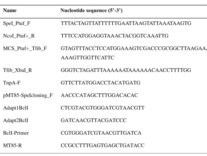
Table 1: Oligonucleotides used in this study
Name Nucleotide sequence (5’-3’)
SpeI_Ptuf_F TTTACTAGTTATTTTTTGAATTAAGTATTAAATAAGTG NcoI_Ptuf+_R TTTCCATGGAGGTAAACTACGGTCAAATTG MCS_Ptuf+_Tfib_F GTAGTTTACCTCCATGGAAAGTCGACCCGCGGCTTAAGAAATT AAAGTTGGTTCATTC Tfib_XbaI_R GGGTCTAGATTTAAAAAATAAAAAACAACCTTTTGG TnpA-F GTTCTTATGGACCTACATGATG pMT85-SpeIcloning_F AACCCATAGCTTTGGACACAC Adapt1BclI CTCGTACGTGGGATCGTAACGTT Adapt2BclI GATCAACGTTACGATCCC BclI-Primer CGTGGGATCGTAACGTTGATCA MT85-R CCGCCTTTGAGTGAGCTGATACC
25
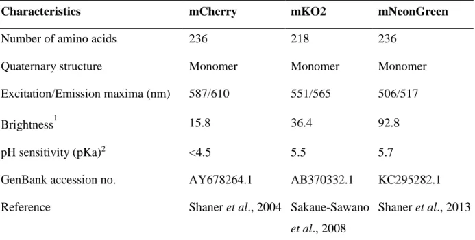
Table 2: Characteristics of selected fluorescent proteins
Characteristics mCherry mKO2 mNeonGreen
Number of amino acids 236 218 236
Quaternary structure Monomer Monomer Monomer
Excitation/Emission maxima (nm) 587/610 551/565 506/517
Brightness1 15.8 36.4 92.8
pH sensitivity (pKa)2 <4.5 5.5 5.7
GenBank accession no. AY678264.1 AB370332.1 KC295282.1
Reference Shaner et al., 2004 Sakaue-Sawano
et al., 2008
Shaner et al., 2013
1
Brightness: Extinction Coefficient (mM-1cm-1) × Fluorescence Quantum Yield 2
26
Table 3: Fold-change increase in green fluorescence intensity associated to mNeonGreen expression in the species Mycoplasma mycoides subsp. mycoides and Mycoplasma bovis
M. mycoides subsp. mycoides M. bovis
Experiment WT1 Neon1 FC2 WT1 Neon1 FC2
1 89 1015 11.4 225 3769 16.8
2 72 1140 15.8 693 7222 10.4
3 65 929 14.3 578 7240 12.5
1
Median fluorescence intensity values
2
Fold-change increase in median fluorescence intensity in the mNeonGreen-expressing strain (Neon) compared to the wild-type strain (WT)