HAL Id: hal-00580703
https://hal.archives-ouvertes.fr/hal-00580703
Submitted on 29 Mar 2011HAL is a multi-disciplinary open access archive for the deposit and dissemination of sci-entific research documents, whether they are pub-lished or not. The documents may come from teaching and research institutions in France or abroad, or from public or private research centers.
L’archive ouverte pluridisciplinaire HAL, est destinée au dépôt et à la diffusion de documents scientifiques de niveau recherche, publiés ou non, émanant des établissements d’enseignement et de recherche français ou étrangers, des laboratoires publics ou privés.
Caroline Deshayes, Horacio Bach, Daniel Euphrasie, Rodgoun Attarian,
Mathieu Coureuil, Wladimir P Sougakoff, Françoise Laval, Yossef Av-Gay,
Mamadou Daffe, Gilles Etienne, et al.
To cite this version:
Caroline Deshayes, Horacio Bach, Daniel Euphrasie, Rodgoun Attarian, Mathieu Coureuil, et al.. MmpS4 promotes glycopeptidolipids biosynthesis and export in Mycobacterium smegmatis. Molecular Microbiology, Wiley, 2010, 78 (4), pp.989. �10.1111/j.1365-2958.2010.07385.x�. �hal-00580703�
For Peer Review
MmpS4 promotes glycopeptidolipids biosynthesis and export in Mycobacterium smegmatis
Journal: Molecular Microbiology Manuscript ID: MMI-2010-09792.R2 Manuscript Type: Research Article Date Submitted by the
Author: 30-Aug-2010
Complete List of Authors: Deshayes, Caroline; University Paris-Descartes, School of Medecine, Inserm U570
Bach, Horacio; University of British Columbia, Medicine Euphrasie, Daniel; INSERM, U570
Attarian, Rodgoun; University of British Columbia, Medicine Coureuil, Mathieu; University Paris-Descartes, School of Medecine, Inserm U570
Sougakoff, Wladimir; University Pierre et Marie Curie, Inserm UMRS-872
Laval, Françoise; CNRS, IPBS
Av-Gay, Yossef; University of British Columbia, Medicine
Daffe, Mamadou; CNRS-University Paul Sabatier Mixed Research Lab (UMR 5089), Department of Molecular Mechanisms of Mycobacterial Infections
Etienne, Gilles; CNRS-University Paul Sabatier Mixed Research Lab (UMR 5089), Department of Molecular Mechanisms of Mycobacterial Infections
Reyrat, Jean-Marc; University Paris-Descartes, School of Medecine, Inserm U570
Key Words: mycobacteria, glycopeptidolipids, cell wall, membrane protein
For Peer Review
1
MmpS4 promotes glycopeptidolipids biosynthesis and export in
2
Mycobacterium smegmatis
3
4
5
6
7
Caroline Deshayes
1, 2, 3,*, Horacio Bach
4, Daniel Euphrasie
2, Rodgoun Attarian
4, Mathieu
8
Coureuil
1, 2, Wladimir Sougakoff
5, Françoise Laval
6, 7, Yossef Av-Gay
4, Mamadou Daffé
6, 7,
9
Gilles Etienne
6, 7and Jean-Marc Reyrat
1, 2, §10
11
12
13
14
15
1INSERM-UMR 570, Unité de Pathogénie des Infections Systémiques, Paris Cedex 15, F-75730,
16
France
17
2Université Paris Descartes, Faculté de Médecine, site Necker, Paris Cedex 15, F-75730, France
18
3Groupe d'Etude des Interactions Hôte-Pathogène, Université d'Angers, 4 rue Larrey, Angers, France
19
4
Department of Medicine, Division of Infectious Diseases, University of British Columbia, 2733
20
Heather St., Vancouver, V5Z 3J5 British Columbia, Canada
21
5
INSERM UMRS-872, équipe 12, Laboratoire de Recherche Moléculaire sur les Antibiotiques
22
(LRMA), faculté de Médecine Pierre et Marie Curie, site Pitié-Salpêtrière, 91 bd de l’Hôpital,
F-23
75634 Paris cedex 13, France
24
6
CNRS, IPBS (Institut de Pharmacologie et de Biologie Structurale), Département Mécanismes
25
Moléculaires des Infections Mycobactériennes. 205 route de Narbonne, F-31077 Toulouse, France
26
7
Université de Toulouse, Université Paul Sabatier (Toulouse III), IPBS. F-31077 Toulouse, France
27
28
29
30
31
32
33
*: corresponding author. Dr Deshayes, C. Groupe d'Etude des Interactions Hôte-Pathogène,
34
Université d'Angers, 4 rue Larrey, Angers, France. deshayes@necker.fr
35
36
§: this paper is dedicated to Dr Jean-Marc Reyrat, who deceased before the submission of the
37
manuscript, for his excellent contribution to the mycobacterial genetics.
38
39
Key words: mycobacteria, glycopeptidolipids, cell wall, membrane protein
40
For Peer Review
Summary. The MmpS family (mycobacterial membrane protein small) includes over one hundred
1
small membrane proteins specific to the genus Mycobacterium that have not yet been studied
2
experimentally. The genes encoding MmpS proteins are often associated with mmpL genes, which are
3
homologous to the RND (resistance nodulation cell division) genes of Gram-negative bacteria that
4
encode proteins functioning as multidrug efflux system. We showed by molecular genetics and
5
biochemical analysis that MmpS4 in M. smegmatis is required for the production and export of large
6
amounts of cell surface glycolipids, but is dispensable for biosynthesis per se. A new specific and
7
sensitive method utilizing single chain antibodies against the surface-exposed glycolipids was
8
developed to confirm that MmpS4 was dispensable for transport to the surface. Orthologous
9
complementation demonstrated that the MmpS4 proteins are exchangeable, thus not specific to a
10
defined lipid species. MmpS4 function requires the formation of a protein complex at the pole of the
11
bacillus, which requires the extracytosolic C-terminal domain of MmpS4. We suggest that MmpS
12
proteins facilitate lipid biosynthesis by acting as a scaffold for coupled biosynthesis and transport
13
machinery.
14
For Peer Review
Introduction
1
The number of complete genome sequences of high GC % Gram-positive bacteria available has
2
increased considerably in the recent years, leading to the accumulation of unprecedented amounts of
3
information. For example, 16 mycobacterial genomes have been sequenced to date and the species
4
sequenced include both rapid and slow growers (Bernal et al., 2001). Mycobacterium is a large genus
5
containing over 150 species, including M. tuberculosis, the etiologic agent of tuberculosis (WHO), M.
6
ulcerans, which causes a devastating necrotic disease of the skin, the so-called Buruli ulcer (WHO),
7
and many opportunistic pathogens (De Groote & Huitt, 2006). Recent sequencing efforts have,
8
through comparative and functional genomics, led to advancements in the characterization of the
9
mycobacterial core genome and of genes involved in virulence. For example, genes encoding a new
10
type of secretion system were characterized, the type VII (Abdallah et al., 2007), in addition to genes
11
required for the biosynthetic of arabino-galactan, a specific attribute restricted to this genus (Crick &
12
Brennan, 2008) and related genera. Sequencing of the mycobacterial genome has also led to the
13
identification of the MmpSL family of membrane proteins, the function of which remains unclear. The
14
name of this protein family is short for mycobacterial membrane protein large (MmpL) and small
15
(MmpS).
16
The genes encoding MmpS are often found closely associated with mmpL genes and nothing is
17
currently known about the function of these proteins. MmpL proteins consist of approximately 1000
18
amino acids organized into twelve predicted transmembrane domains and two large periplasmic
19
domains. MmpL are not restricted to mycobacteria, and are also found in related streptomycetes and
20
Rhodococcus. This family of proteins is distantly related to the resistance nodulation cell division
21
(RND) superfamily (Tekaia et al., 1999), members of which are present in all domains of life and may
22
serve as efflux pumps for toxic environmental molecules (Tseng et al., 1999). Two of the fourteen
23
MmpL members in M. tuberculosis have been shown to be involved in the biosynthesis and/or
24
transport of mycobacterial lipid metabolites. MmpL7 is required together with an ABC transporter and
25
a lipoprotein, for the specific transport of phthiocerol dimycoserate (PDIM) to the cell surface
26
(Camacho et al., 2001, Cox et al., 1999). MmpL8 plays a role in the synthesis of sulfolipid-1 (SL-1),
27
another cell surface polyketide, possibly in the transport of a SL-1 precursor from the cytoplasm to the
28
For Peer Review
periplasm (Converse et al., 2003, Domenech et al., 2004). In M. smegmatis and M. abscessus, the
1
MmpL4a and MmpL4b proteins (previously known as TmtpB and TmtpC, respectively) are
2
implicated in the biosynthesis of cell surface polyketides, the glycopeptidolipids (GPLs) (Recht et al.,
3
2000, Sonden et al., 2005, Medjahed & Reyrat, 2009).
4
As previously mentioned, some mmpL genes are associated with mmpS genes, which encode
5
proteins predicted to have only one N-terminal transmembrane domain with an extracytoplasmic
C-6
terminus (Domenech et al., 2005). Two (M. leprae) to twenty-seven (M. abscessus) such genes may be
7
present in the genome, depending on the species. Most mmpL and mmpS genes are located close to
8
genes involved in the synthesis or modification of polyketides. This close proximity suggests a
9
possible role in the transport and/or biosynthesis of molecules synthesized by the neighbouring
10
synthase gene products (Tekaia et al., 1999). Little is known about MmpS function, but transcriptional
11
regulation of these genes has been observed. For example, the mmpS4 and mmpS5 genes of M.
12
tuberculosis are down regulated during nutrient starvation (Betts et al., 2002). More recently,
13
resistance to azole in a M. tuberculosis mutant strain was correlated with increased transcription of
14
mmpS5-mmpL5 genes (Milano et al., 2009).
15
In M. smegmatis, mmpS4 is organized into a putative operon with the mmpL4a and mmpL4b
16
genes, which have been shown to be involved in GPL biosynthesis (Recht et al., 2000, Sonden et al.,
17
2005). GPLs are the predominant glycolipids found at the surface of many non-tuberculous
18
mycobacteria, including M. avium subsp. avium (Brennan et al., 1981) and M. abscessus
(Lopez-19
Marin et al., 1994), an emerging pathogen predominantly infecting young cystic fibrosis patients.
20
These surface polyketides are required for sliding motility and biofilm formation, and in some cases
21
their level of production is correlated with strain virulence (Byrd & Lyons, 1999, Howard et al.,
22
2006). Upon engineering an in-frame unmarked deletion of the mmpS4 gene in M. smegmatis, reduced
23
levels of GPLs were observed. However, we found that MmpS4 is not required for GPLs localization.
24
This finding was confirmed with a new immunofluorescent method for specific detection of GPLs at
25
the cell surface. Our results suggest that MmpS are not specific for structurally defined lipid
26
molecules. By studying fusions of red fluorescent protein (Rfp) with MmpS4 or two GPL biosynthesis
27
enzymes, we show that MmpS4 is required for the formation of a protein complex at the pole of the
28
For Peer Review
bacillus, presumably via the C-terminal domain. Thus, we suggest that MmpS proteins promote
1
efficient biosynthesis and secretion by acting as a scaffold for the enzymatic and export machinery.
2
This report constitutes the first experimental investigation of a member of the MmpSL family of
3
proteins.
4
For Peer Review
Results
1
The mmpS family in mycobacteria. mmpS genes are specific to mycobacteria and are most often
2
associated with mmpL genes. Indeed, our homology searches in genomes of closely related members
3
of the Actinomycetales, such as Streptomyces and Rhodococcus, show that these bacteria contain
4
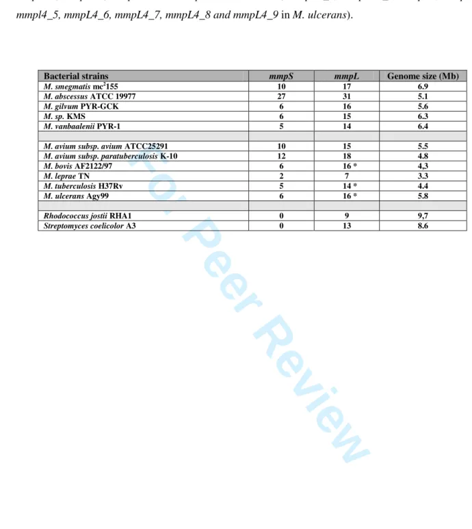
mmpL homologues, but no mmpS genes (Table 1). The MmpS family is currently composed of more
5
than one hundred proteins and new members are added with the release of each new mycobacterial
6
genome sequence. The number of mmpS paralogs per genome varies from two to twenty seven and is
7
correlated with the number of mmpL genes, rather than genome size (Table 1). These members can be
8
organized into five classes, based on the clusters obtained by phylogenetic analysis, corresponding to
9
the M. tuberculosis mmpS paralog numbering (Fig. 1A).
10
MmpS proteins have an overall identity of 22 to 81 % within the same class and 8 to 37 % between
11
members of different classes. Each mmpS gene encodes a predicted protein of approximately 150
12
amino acids in length, anchored in the membrane by a 15-amino-acid transmembrane domain at its
N-13
terminus. It has been suggested that the C-terminus of the protein is extracytosolic (Tekaia et al., 1999,
14
Domenech et al., 2005) and this hypothesis has been supported by recent computer-assisted predicting
15
of membrane protein topology (Fig. 1B). An alkaline phosphatase gene fusion mmpS4-phoA was used
16
to confirm this predicted extracytosolic localization. The activity of bacterial alkaline phosphatase
17
(PhoA) is indeed dependent on it being exported across the plasma membrane. M. smegmatis
18
expressing the mmpS4-phoA fusion showed blue phenotype on plates containing X-P, the chromogenic
19
substrate of PhoA, reflecting an activity of the alkaline phosphatase (Fig. 1C). In contrast, M.
20
smegmatis carrying the empty pJEM11 plasmid was white, clearly confirming the bioinformatical
21
prediction and indicating that the C-terminal domain of MmpS4 is extracytoplasmic.
22
The mmpS3 genes differ from other mmpS genes in encoding predicted proteins of approximately 300
23
amino acids in size. These proteins are thus twice the size of other MmpSs proteins and likely arose
24
from an ancient complete mmpS3 copy through an event of duplication and fusion. mmpS3 is present
25
in both rapid and slow growing mycobacterial species, suggesting that the duplication event occurred
26
in the common ancestor of these two phylogenetic clades. M. smegmatis has ten mmpS genes (Table
27
1), and is specifically enriched in paralogs of the mmpS5 families (Table S1). By contrast, the mmpS4
28
For Peer Review
gene is present as only a single copy in all the sequenced mycobacterial genomes, with the exception
1
of M. abscessus genome, which includes five paralogs.
2
3
MmpS4 is required for sliding motility and biofilm formation. We investigated the function of
4
MmpS4 by generating a mutant strain for this gene in M. smegmatis. The mmpS4, mmpL4a and
5
mmpL4b genes are probably transcribed as an operon, as these genes either overlap or are separated by
6
only few nucleotides. We prevented possible polar effects on downstream genes by constructing an in
7
frame unmarked deletion of the mmpS4 gene by homologous recombination, using a two steps strategy
8
and a counter-selectable marker (Pelicic et al., 1996, Reyrat et al., 1998). The mutant strain was
9
verified by PCR, sequencing and Southern-blotting, all of which confirmed its identity and the
10
occurrence of the anticipated event, a deletion of about 90% of the coding sequence of the mmpS4
11
gene (Fig. S1). The resulting ∆mmpS4 mutant had rough colony morphology readily visible by the
12
naked eye. This mutant was unable to slide (Fig. 2A) or form biofilms (data not shown).
13
Complementation of this mutant strain with the wild-type mmpS4 allele from M. smegmatis restores
14
the wild-type smooth phenotype and the ability to slide (Fig. 2A) and form biofilms. This excluded the
15
possibility that the observed phenotype was due to secondary mutations or a polar effect on
16
downstream genes.
17
In M. smegmatis, changes in sliding motility and biofilm formation are often associated with
18
quantitative or qualitative changes in GPL biosynthesis, due to the lack of biochemical modifications
19
by acetylation, methylation or glycosylation of the GPL backbone for instance (Deshayes et al., 2005,
20
Recht & Kolter, 2001, Sonden et al., 2005). We therefore analyzed GPL production in the mutant
21
strain. As shown in Figure 2B, the ∆mmpS4 strain produced small amounts of GPL-like compounds.
22
Only the major species remained detectable by TLC, whereas the complemented strain produced GPL
23
compounds in amounts similar of those of the wild-type. For the precise quantification of GPL
24
production in the ∆mmpS4 strain, we grew large amounts of cells and purified GPLs, making use of
25
their alkali-stable character (Belisle et al., 1993). In these conditions the mmpS4 mutant produced
26
about 20% as much alkali-stable lipids as the wild-type and complemented strains did, accounting for
27
For Peer Review
the rough morphotype of the mutant strain (Fig. 2C). Complementation of the mutant strain with the
1
mmpS4 wild-type allele of M. smegmatis restored the wild-type level of GPL production (Fig. 2B and
2
2C). The levels of other lipids (triacyl glycerols, glycerol monomycolate, trehalose dimycolate,
3
trehalose monomycolate, phosphatidylinositol mannosides) were similar (Fig. S2). Thus MmpS4 plays
4
a specific role in controlling the level of GPLs biosynthesis in M. smegmatis.
5
6
MmpS4 is required for high level of GPL production. We checked for changes in GPL structure, by
7
analyzing lipid extracts of the ∆mmpS4 strain using MALDI-TOF mass spectrometry, a highly
8
sensitive and accurate method of mycobacterial complex glycolipids detection and characterization
9
(Villeneuve et al., 2003). Total lipid extract prepared from the wild-type strain displayed a major peak
10
at 1257 m/z, corresponding to the main diglycosylated (tri-O-Methyl Rhamnose and di-O-Acetylated
11
6-deoxyTalose) GPL of M. smegmatis mc²155 (Etienne et al., 2005). A similar pattern of GPLs was
12
also detected in the mutant strain, but in relatively smaller amounts and with the peaks 14 Da higher
13
than those of the wild-type (Fig. S3). The major peak was observed at 1271 m/z. This 14 atomic
mass-14
units increase is likely due to an additive O-methylation on the fatty acid moiety, as the only spot
15
detectable by TLC (Fig. 2B) was the most apolar fully O-methylated GPL I (Patterson et al., 2000).
16
Complementation of the ∆mmpS4 strain fully restored the wild-type profile with a major peak at 1257
17
m/z, demonstrating that this 14 atomic mass-units increase was dependent on the absence of the
18
mmpS4 gene.
19
20
MmpS4 is not involved in GPL subcellular localization. We previously showed that sliding motility
21
requires not only the normal level of GPL production but also a localization of these molecules on the
22
cell surface (Sonden et al., 2005). As the ∆mmpS4 strain is not motile, we hypothesized that MmpS4
23
might be involved in GPL export to the cell surface. We tested this hypothesis, by preparing surface
24
and cell-associated (after removal of the surface lipids with glass beads) fractions, extracting lipids
25
from each fraction and analyzing them by TLC (Fig. 3A). As the ∆mmpS4 strain produced less than
26
20% the amount of GPL produced by the wild-type strain, we used larger amounts of cells and
27
For Peer Review
concentrated lipid metabolites through solvent evaporation to visualize them by TLC. A ∆gap mutant
1
strain was used as a control because this mutant produces wild-type levels of GPLs but does not export
2
them to the surface (Sonden et al., 2005). Consistent with quantification results, the ∆mmpS4 strain
3
produced much less GPL than did the wild-type and ∆gap strains, but small amounts of GPL were still
4
present in the surface fraction of the mutant strain. Despite the low level of production of GPL by the
5
mutant, the glycolipid could be visualized in the surface compartment, although barely, for the
6
∆mmpS4 mutant but not for ∆gap strain, demonstrating the presence of GPL at the surface of the
7
∆mmpS4 mutant (Fig. 3A). An overloaded TLC showed selective disappearance in the cellular extract
8
of the lower spots, corresponding to polar GPLs in the ∆mmpS4 mutant. Mass spectrometry analyses
9
unambiguously confirmed the presence of GPLs in the cell surface of the ∆mmpS4 strain while GPLs
10
were not detectable on the cell surface of the ∆gap mutant (Fig. S3).
11
12
Development of a new sensitive immunofluorescent method for lipid localization. To further
13
investigate the in vivo localization of GPLs, we developed a new and specific method. GPL
14
localization in the ∆mmpS4 mutant was characterized by an immunofluorescent method that
15
selectively detected surface-exposed GPLs. Briefly, purified GPLs coupled to ovalbumin were used as
16
antigens to screen a naïve human-derived antibody library using phage display technology. After four
17
biopanning cycles and screening against ovalbumin, four clones with a high affinity for GPLs were
18
selected. The gene encoding the antibody with the highest affinity was fused to the gene encoding the
19
DSred fluorescent protein. The construct was expressed in E. coli, and the fused protein fully retained
20
the fluorescent properties of the DSred as observed by epifluorescence microscopy (Figure 3B). The
21
detection of GPLs was performed in a single step as secondary antibodies were not needed. Bacteria
22
were first non-specifically labelled with FITC. The resultant green staining made it possible to
23
visualize the bacterial bodies and served as a control for evaluating of the number of bacteria present
24
on the slide. Two control strains were used in this experiment: a pks mutant strain producing no GPLs
25
(pks-) (Sonden et al., 2005) used to evaluate labelling specificity, and the ∆gap mutant strain with its
26
complemented companion strain (Sonden et al., 2005). The DSred-antibody labelling perfectly
co-27
For Peer Review
localized with the FITC-labelled bacteria in the case of the wild-type and the complemented ∆gap
1
strain, resulting in a yellow staining after image overlapping (Fig. 3B). The strain producing no GPL
2
and the ∆gap strain were not immunolabelled with the anti-GPL antibody. This antibody was therefore
3
highly specific for GPL and able to bind surface-exposed GPLs only, as the ∆gap strain, which
4
produces wild-type level but does not export them on the surface, was not labelled by the antibody.
5
Interestingly, a weak anti-GPL fluorescence signal was detected in the ∆mmpS4 strain, corresponding
6
to approximately 60% of the relative fluorescence compared to the wild-type strain. A clear signal was
7
observed for the ∆mmpS4 strain but not for the GPL- and ∆gap strains. These results confirm that the
8
∆mmpS4 strain produces smaller amounts of GPL than the wild-type strain but also demonstrate that
9
GPLs are present on the bacterial surface. The ∆mmpS4 complemented strain gave a stronger signal,
10
although not as strong as that of the wild-type strain, in approximately 95% of the bacteria. These
11
experiments demonstrate that GPLs reach the cell surface in the ∆mmpS4 strain but also show that that
12
reintroduction of the mmpS4 gene increases both the total and surface levels of GPLs.
13
14
MmpS4 proteins assemble into a complex at the pole of the bacillus. We investigated the function
15
and location of MmpS4 by constructing gene fusions based on the red fluorescent protein (RFP). The
16
rfp gene was fused to the 5’ or the 3’ end of the mmpS4 gene, yielding RFP-MmpS4 and MmpS4-RFP
17
proteins, respectively. The fusion of RFP to the N-terminal domain did not affect the function of
18
MmpS4. Indeed, the construct encoding RFP-MmpS4 fully complemented the mmpS4 mutant strain,
19
resulting in the production of large amounts of GPLs (data not shown). In contrast, the fusion of RFP
20
to the C-terminal domain of MmpS4 abolished its function, as this construct did not complement the
21
mmpS4 mutant strain. This result suggests that the extracytosolic C-terminal domain of MmpS4 is
22
crucial for its function and therefore does not tolerate fusion with RFP. Nevertheless, it is still possible
23
that the formation of a fused protein may lead to misfolding of MmpS4 and this in turn affects its
24
topology and membrane localization. To dissect the role of the MmpS4 extracytosolic C-terminal
25
domain, truncated proteins were constructed. None of them were able to complement the ∆mmpS4
26
strain (Fig. S4), strengthening our hypothesis that the C-terminal domain of MmpS4 is necessary for
27
For Peer Review
its function. Furthermore, this domain needs to be localized in the extracytosolic compartment. Indeed
1
the intracellular expression of this domain could not restore the mmpS4 function, since the MmpS4
2
protein deleted of its transmembrane region was not able to complement the mutant strain.
3
The strains expressing fusion proteins were observed by fluorescence microscopy and a
4
control strain expressing cytosolic GFP was used (Barker et al., 1998). In strains expressing
MmpS4-5
RFP or the cytosolic GFP, the fluorescence was diffused and spread throughout the bacillus. In
6
contrast, in the strain carrying the RFP-MmpS4 construct, the fluorescence was concentrated into
7
small dot-like area (spot). This punctuate fluorescence was absent from strains expressing the
MmpS4-8
RFP fusion and the cytosolic GFP, but present in approximately 25% of the bacilli expressing the
9
RFP-MmpS4 fusion (Fig. 4A). There was never more than one spot per bacillus and the spots were
10
located either at the site of division or at the pole of the cell (Fig. 4B). The presence of this complex at
11
the division site may enable each of the two daughter cells to inherit half the complex, therefore
12
becoming located at the pole after division, or can be correlated to the presence of the division
13
machinery. Although we can not completely exclude that this apparent MmpS4 localization was an
14
artefact of the RFP fusion, this seems unlikely since a polar or septum localization is rather expected
15
for a complex involved in the synthesis of an envelope constituent. In conclusion, these findings
16
strongly suggest that MmpS4 proteins are not randomly distributed in the membrane but instead
17
assemble into a complex located at the pole of the bacterium. A free C-terminal extracytosolic domain
18
is required for both complex formation and biological function.
19
20
MmpS4 proteins are necessary for the formation of a complex of GPL biosynthesis enzymes at
21
the pole of the bacillus. Based on the data presented herein and previous studies reporting interaction
22
between MmpL and enzymes required for the biosynthesis of small metabolites (Cox et al., 1999,
23
Straight et al., 2007), we suggest that MmpS4 acts as a scaffold for the assembly of the GPL
24
biosynthesis megacomplex (Fig. 5A). Proteins fusions were constructed, fusing Rfp in the C-terminal
25
domain of two GPL synthesis proteins (MbtH and FadD23) predicted to be cytosolic. Their
26
localization was analyzed in the wild-type and ∆mmpS4 mutant strains. Punctuate fluorescence was
27
found at the poles only in the wild-type strain in roughly 25% and 35% of the bacilli for the MbtH-Rfp
28
For Peer Review
and FadD23-RFP constructs, respectively (Fig. 5B and C). These percentages were similar to the
1
percentage obtained for the strain expressing the rfp-mmpS4 fusion. In contrast, the fluorescence was
2
mainly diffuse in the ∆mmpS4 mutant strain. These results show that the absence of MmpS4 proteins
3
affects the localization of other proteins involved in GPL synthesis and reinforce our megacomplex
4
model.
5
6
MmpS4s are functionally exchangeable. MmpS4 orthologs are 50 to 60% identical and many of the
7
conserved amino acids are located in the predicted extracytosolic region (Fig. S5). We assessed the
8
activity of these MmpS4 orthologs, by transforming the ∆mmpS4 strain with a plasmid carrying an
9
ortholog of the mmpS4 gene originating from other species (genes from several different species were
10
used). For M. abscessus, which carries five paralogs of mmpS4, we used the copy (MAB4117c) most
11
similar to mmpS4 of M. smegmatis. This gene mapped to the GPL locus of M. abcessus (Ripoll et al.,
12
2007). We used orthologs from GPL-producing species such as M. abscessus and M. avium subsp.
13
avium (MAV3247), but also from non-GPL-producing organisms such as M. tuberculosis (Rv0451c).
14
In all cases (MAB4117c, MAV3247, and Rv0451c) complementation with the mmpS4 gene restored a
15
smooth wild-type phenotype. The mutant strain complemented with the M. tuberculosis ortholog
16
exhibited wild-type sliding motility, consistent with functional complementation (Fig. 6A). As a
17
control experiment we carried out complementation experiments with the mmpS1 gene of M.
18
tuberculosis (mmpS1Tb). Expression of this mmpS1Tb gene did neither restore the wild-type smooth
19
phenotype, nor the sliding motility, consistent with the notion of orthology for the MmpS4 family
20
(Fig. 6A). To avoid any possibility of non-expression of the mmpS1 gene, its expression in the
21
∆mmpS4/mmpS1Tb strain was confirmed by reverse transcription (Fig. S6). Biochemical analyses of
22
the ∆mmpS4 mutant strain expressing the mmpS4 gene of M. tuberculosis (mmpS4Tb) showed fully
23
functional complementation. The mmpS4 gene of M. tuberculosis therefore conferred to the mutant
24
strain, upon complementation, the ability to produce more GPL, both in the cellular compartment and
25
at the surface (Fig. 6B). Quantitative analyses showed that the mmpS4Tb-complemented strain
26
produced significantly larger amounts of GPLs (2.09 ± 0.72 mg of alkali-stable lipids for 100 mg of
27
For Peer Review
cells, dry weight) than did the ∆mmpS4 mutant strain (0.54 ± 0.33 mg). Furthermore,
1
immunofluorescence experiments with the anti-GPL DSred antibody showed much more GPLs on the
2
surface of the mmpS4Tb complemented strain (Fig. 6C) than on the ∆mmpS4 mutant strain. Indeed,
3
the mmpS4Tb complemented strain produced a stronger signal in approximately 95% of the bacilli.
4
The signal obtained was similar to that for the mmpS4Ms complemented strain (Fig. 3B). As stated
5
above, this complemented strain displayed greater sliding motility compared to the mutant, indicating
6
a gain of function (Fig. 6A). In conclusion, these findings indicate that the mmpS4 gene of a species
7
that does not produce GPL can complement the M. smegmatis ∆mmpS4 mutant strain, both in terms of
8
production and localization of GPLs. Thus, MmpS4 are functionally exchangeable and constitute a
9
true protein family. Our findings also suggest that MmpS proteins are not specific to the lipid
10
compound itself.
11
For Peer Review
Discussion
1
The synthesis and export of complex polyketides present in the outermost structure of the cell
2
envelope such as GPLs require the expression of large biosynthetic pathways, involving proteins
3
encoded by grouped genes or genes dispersed throughout the genome, and the assembly of dedicated
4
machineries (Gokhale et al., 2007). The mmpS family was first identified a decade ago with the
5
sequencing of the first mycobacterial genome (Cole et al., 1998). These genes encode proteins
6
involved in various specific pathways and are often associated with mmpL genes. M. tuberculosis has
7
five mmpS genes and fourteen predicted mmpL genes. Consequently, not all biosynthetic pathways
8
include proteins encoded by mmpS gene. This seems to be the case for PDIM and SL-1 loci for
9
example (Camacho et al., 2001, Converse et al., 2003). The location of the mmpS4 gene within the
10
GPL biosynthesis locus suggests a role in GPL biosynthesis and/or transport to the surface, but our
11
data do not support this hypothesis. Indeed, the ∆mmpS4 mutant strain produced smaller amounts of
12
GPLs than did the wild-type, but these compounds were still targeted to the bacterial surface as shown
13
by biochemical fractionation and the new immunofluorescence sensitive technique for the specific
14
tracking of GPLs at the bacterial surface. The low level of production of GPL in the ∆mmpS4 strain
15
accounts for its phenotype, lack of sliding motility and inability to form biofilms, as all these features
16
are dependant on GPL production (Recht et al., 2000, Kocincova et al., 2008). However, although
17
produced in smaller amounts, the GPLs present in the mutant were chemically identical to those in the
18
wild-type, excluding a possible role of MmpS4 in the synthesis, per se. The MmpS4 protein of M.
19
smegmatis therefore seems to determine the amount of GPL produced. An alternative explanation
20
would be that there was another protein, maybe another MmpS protein that provides a similar function
21
in M. smegmatis, albeit not as efficient as MmpS4. The fact that functional complementation was
22
observed when an orthologous gene (mmpS4Tb) from the same family was introduced into the
23
∆mmpS4 strain, but not with a paralogous gene (mmpS1Tb), makes this last hypothesis unlikely, since
24
only one mmpS4 gene is present in the M. smegmatis genome. The functional complementation with
25
an ortholog from M. tuberculosis was particularly surprising, as this species does not produce GPL. In
26
M. tuberculosis, mmpS4 is organized into an apparent operon with a single mmpL4 gene and this
27
For Peer Review
operon is located close to genes encoding a probable cyclopropane fatty acyl phospholipid synthase
1
and a putative enoyl-CoA hydratase. The lipid metabolite specified by this pathway has not yet been
2
characterized, but a M. tuberculosis mmpL4 mutant has been shown to be strongly attenuated in vivo
3
(Domenech et al., 2005). One possible explanation for this orthologous complementation is that
4
MmpS proteins are not specific for their lipid substrate but are, instead, specific for their cognate
5
MmpL proteins, via molecular interactions involving amino acids conserved throughout the family
6
(Fig. S5).
7
The MmpL members are distantly related to the RND superfamily, but differ from members of
8
this superfamily in several ways. For example, RND proteins have broad substrate specificity, whereas
9
MmpL proteins seem to be much more specific, interacting with only one lipid metabolite. MmpL7
10
and MmpL8 affect specifically PDIM and SL-1 biosynthesis in M. tuberculosis, respectively (Cox et
11
al., 1999, Converse et al., 2003), and MmpL4 affects GPL biosynthesis in M. smegmatis (Recht et al.,
12
2000). Furthermore, whereas RND family members from Gram-negative bacteria act purely as efflux
13
pumps, the RND protein ActII-orf3 from the Gram-positive Streptomyces coelicolor, is involved in the
14
biosynthesis of a blue pigment, γ-actinorhodin, a heterocyclic antibiotic synthesized by a polyketide
15
synthase (Bystrykh et al., 1996). In M. tuberculosis, MmpL8 plays a role in the synthesis of
sulfolipid-16
1 (SL-1), possibly by transporting a precursor of this molecule to the surface (Converse et al., 2003,
17
Domenech et al., 2004). In the same species, MmpL7 has been shown to be required for the transport
18
of PDIM to the cell surface (Cox et al., 1999, Camacho et al., 2001). This transport process requires
19
another protein, DrrC, a component of the putative ABC transporter, DrrABC (Camacho et al., 2001),
20
and secretion into the medium requires LppX, a lipoprotein (Sulzenbacher et al., 2006). Studies based
21
on the two hybrid approach have shown that MmpL7 interacts with PpsE (Jain & Cox, 2005), which
22
interacts with TesA (Rao & Ranganathan, 2004), both of them being enzymes required for PDIM
23
synthesis. It has thus been suggested that MmpL7 interacts with the PDIM synthetic machinery to
24
form a complex that coordinately synthesizes and transports PDIM across the cell membrane (Jain &
25
Cox, 2005). In Bacillus subtilis, a polyketide synthase synthesizing an antibiotic, bacillaene, has been
26
shown to assemble into a single megacomplex at the membrane (Straight et al., 2007). The authors
27
suggested that this organization is established via interaction with an unknown membrane protein.
28
For Peer Review
Based on such models, we suggest that MmpS4 proteins act as a scaffold with MmpL4
1
proteins for the assembly of the GPL biosynthesis megacomplex (Fig. 5A). Several enzymes needed
2
for GPL synthesis/export are predicted to be membrane-bound or -inserted: Atf, a sugar
3
acetyltransferase, Gap, needed for GPL surface localization, MmpL4a and MmpL4b, whose precise
4
role is currently unknown. It is likely that these membrane proteins associate into a complex to
5
maximize GPL biosynthesis and export. In this model, MmpS4 interacts with MmpL4a, MmpL4b and
6
GPL biosynthetic enzymes such as the polyketide synthase (Pks, involved in the lipid moiety
7
synthesis), non-ribosomal protein synthetases (Nrp, involved in the tetrapeptide moiety synthesis),
8
acetyltransferase (Atf), methyltransferases (Mtf) or glycosyltransferases (Gtf), thereby increasing the
9
synthesis and export of GPLs. Our data are consistent with the C-terminal extracytosolic domain of
10
MmpS4 being essential for the function of this protein. The extracytosolic domain of MmpS4 could
11
specifically interact with the extracytosolic domains of MmpL4 proteins, explaining the lack of
12
specificity of MmpS4 towards the lipid moiety. MmpS4 is not randomly distributed throughout the
13
membrane. Instead, it is found principally at the pole of the bacterium. This suggests that MmpS4
14
assembles into a complex that interacts with the biosynthetic machinery. In the absence of MmpS, the
15
biosynthetic complex does not adopt the correct configuration and the enzymes are distributed
16
throughout the cytoplasm (Fig. 5A), resulting in lower levels of GPL production than the wild-type
17
strain. The absence of the scaffold would lead to inappropriate contacts with enzymes of the pathway,
18
leading to the artefactual detection of GPL1271 as the main product. To strengthen our megacomplex
19
model, we constructed fluorescent fusion proteins with two GPL biosynthesis enzymes, MbtH and
20
FadD23. The role of the mbtH genes, whose homologs are found associated with nrp genes, is unclear
21
to date but it has been shown that the mbtH gene is required for the clorobiocin biosyntheis in
22
Streptomyces coelicolor (Wolpert et al., 2007). The FadD23 gene is a close homolog to the M.
23
tuberculosis fadD28 gene involved in acyl transfer of mycocerosic acid (Cox et al., 1999) and is
24
probably involved in the lipid attachment to the GPL tetrapeptide moiety. Our results showed that
25
MmpS4 is required for them to localize as a punctuate spot at the poles of the bacilli. Colocalization
26
studies should be carried out to prove interactions between GPL biosynthesis/export enzymes. The
27
megacomplex model is also supported by the finding that some enzymes predicted to be cytosolic and
28
For Peer Review
involved in PDIM (PapA5, Mas, FadD28) or mycolactone (MlsA1, MlsB) biosynthesis are found in
1
the membrane fraction of M. ulcerans (Tafelmeyer et al., 2008). It can be speculated that the
2
localization of these proteins is due to their anchoring to a biosynthetic membrane megacomplex.
3
The presence of several paralogs, as in M. abscessus, is consistent with the diversification of
4
lipid metabolism. The minimalist species M. leprae has only two mmpS members (mmpS3 and
5
mmpS4), suggesting that other pathways are not required for a strict intracellular lifestyle. The new
6
sensitive immunofluorescent method developed for this study is clearly transposable to other lipid
7
molecules. The method is therefore of considerable interest, because many lipid molecules are poorly
8
immunogenic, making antibody-based detection difficult. This new method allows the sensitive and
9
specific tracking of a single lipid only and is clearly of potential use for biochemical purposes and for
10
investigating lipid trafficking during host cell infection. It is also potentially useful for diagnostic
11
purposes, as some lipids are species- or even strain-specific.
12
In conclusion, according to our current working model of MmpS function, these small
13
transmembrane proteins are not absolutely required for metabolite synthesis and export, but may
14
promote interactions between the various partners involved in the pathway, possibly via their
15
extracytosolic C-terminal domains, therefore stabilizing the complex and optimizing synthesis and
16
export. MmpS proteins display no similarity to membrane fusion proteins of Gram-negative bacteria.
17
However, the close association of mmpS and mmpL genes suggests that MmpS proteins may be the
18
functional homologs of MFPs. Further biophysical studies are required to analyze the assembly and
19
interactions required to stabilize this complex machinery consisting of a dozen enzymes.
20
For Peer Review
Experimental procedures
1
2
Bioinformatical analysis. The NCBI website (http://www.ncbi.nlm.nih.gov/BLAST/) was searched
3
with the BLASTP program, using M. tuberculosis MmpS proteins as queries. The sequences of MmpS
4
proteins from the M. tuberculosis H37Rv, M. bovis AF2122/97, M. ulcerans Agy99 and M. leprae TN
5
genomes were inspected at the Pasteur GenoList website (http://genolist.pasteur.fr/). The sequences of
6
the MmpS proteins of M. abscessus, M. avium subsp. avium 104, M. avium subsp. paratuberculosis
K-7
10, M. gilvum PYR-GCK, M. smegmatis mc2155, M. sp. KMS and M. vanbaalenii PYR-1 were
8
downloaded from the NCBI website. Protein sequences were aligned using the MultAlin program
9
(Corpet, 1988), and a phylogenetic tree was inferred. Secondary structure prediction to identify
10
membrane domains was carried out with Sosui software (Hirokawa et al., 1998) and HMMTOP
11
website (http://www.enzim.hu/hmmtop/).
12
13
Bacterial strains, medium and growth. E. coli DH5α was used for plasmids propagation and was
14
grown in LB medium like M. smegmatis strain mc2155 (Snapper et al., 1990). When required,
15
antibiotics were added to the medium at the following concentrations: hygromycin 50 µg/ml (200
16
µg/ml for E. coli) and kanamycin 25 µg/ml. For the alkaline phosphatase assay, LB plates
17
supplemented with 25 µg/ml kanamycin and 50 µg/ml 5-bromo-4-chloro-3-indolyl phosphate (X-P), a
18
chromogenic alkaline phosphatase substrate, were used (Lim et al., 1995).
19
20
Disruption of the mmpS4 gene of M. smegmatis. We used an in-frame deletion to disrupt the mmpS4
21
gene by homologous recombination with the pPR27 vector to prevent polar effects (Pelicic et al.,
22
1996). The primers used to amplify the upstream and downstream regions for each gene are listed in
23
Table S2. The chromosomal DNA of clones obtained by these procedures was compared with the
24
wild-type DNA by PCR and Southern blotting. The mutant was named ∆mmpS4.
25
26
Construction of the mmpS4 expression plasmids. The wild-type mmpS4 gene coding sequence
27
(accession number AY439015) was amplified by PCR with Pfu Turbo DNA polymerase (Stratagene)
28
For Peer Review
using M. smegmatis mc2155 genomic DNA as a template and the primers mmpS4_trans.5 and
1
mmpS4_trans.3 (Table S2). PCR products were purified with a Qiagen PCR purification kit, digested
2
with XbaI and inserted into the single XbaI site of the dephosphorylated integrative expression vector
3
pNIP40b to generate pNIPmmpS4 (de Mendonca-Lima et al., 2001). Enzymatic digestions were used
4
to select clones in which the mmpS4 gene was inserted in the opposite orientation of the hygromycin
5
resistant gene. One clone was selected, sequenced and named ∆mmpS4/mmps4Ms. A similar strategy
6
was used to insert the mmpS4 genes of M. avium (MAV3247), M. abscessus (MAB4117c), M.
7
tuberculosis (Rv0451c) and the mmpS1 gene (Rv0403c) of M. tuberculosis in pNIP40b yielding
8
pNIPmmpS4Av, pNIPmmpS4Ab, pNIPmmpS4Tb, and pNIPmmpS1Tb respectively. These plasmids
9
were used to electroporate M. smegmatis ∆mmpS4 and transformants were selected on hygromycin
10
and named ∆mmpS4/mmpS4Av, ∆mmpS4/mmpS4Ab, ∆mmpS4/mmpS4Tb and ∆mmpS4/mmpS1Tb
11
respectively. For the construction of plasmid expressing the mmpS4-phoA fusion gene, the pJEM11, a
12
phoA reporter shuttle plasmid that replicates in E. coli and in M. smegmatis (Lim et al., 1995), was
13
generously provided by B. Gicquel. The M. smegmatis mmpS4 gene was PCR amplified (primers
14
mmpS4phoACter.5 and mmpS4phoACter.3, Table S2) and inserted into the BamHI site of pJEM11
15
plasmid to give pJEM11mmpS4. The pJEM11 and pJEM11mmpS4 plasmids were then used to
16
electoporate M. smegmatis strain mc2155.
17
18
Sliding motility assay. A 3 µl aliquot of liquid culture was dispensed onto plates containing 7H9
19
medium plus 0.3% agar with no added carbon source. These plates were then incubated at 37°C for
20
one week.
21
22
Extraction and purification of mycobacterial lipids. Lipids were extracted from cell pellets with a
23
mixture of chloroform and methanol as previously described (Villeneuve et al., 2003). The extracts
24
were dried under vacuum and partitioned between water and chloroform (1:1, vol/vol). The organic
25
phases was extensively washed with distilled water and evaporated to dryness. The lipid extracts were
26
dissolved in chloroform and analyzed by thin-layer chromatography (TLC) on plates coated with
27
For Peer Review
Durasil 25- silica gel (0.25 mm thickness; Macherey-Nagel). The GPLs (250 µg each deposit) were
1
resolved by TLC run in chloroform-methanol (9:1, vol/vol) and visualised by spraying the plates with
2
0.2% anthrone in concentrated sulfuric acid, and heating at 110°C. Identification of GPLs was
3
achieved by matrix-assisted laser-desorption/ionization-time-of-flight (MALDI-TOF) mass
4
spectrometry analysis (Villeneuve et al., 2003).
5
6
Quantification of GPL production. Three 200 mL independent cultures of each strain were grown in
7
LB medium, centrifuged and the pellets were weighted. Lipids were extracted and GPLs were purified
8
as described above. GPL purification was completed by deacylating the lipids with 0.1 M KOH as
9
described by Brennan and Goren (Brennan & Goren, 1979). We expressed the amount of GPLs
10
produced as a function of bacterial pellet weight.
11
12
Surface-exposed material preparation. The surface-exposed material was recovered from
13
mycobacteria cells treated with 10 g of glass beads as previously described (Sonden et al., 2005). The
14
surface-exposed and the cell-associated lipids were extracted with chloroform and methanol from the
15
surface-exposed material and from the residual cells, respectively, and their GPLs components
16
identified by TLC as described above. The amount of lipid spotted on the TLC plates corresponded to
17
the amount of lipid produced by 5 mg of cells, dry weight.
18
19
Immobilization of GPL and selection of single-chain antibodies. We coupled 1 mg of purified GPL
20
to 6 mg of ovalbumin using 1-ethyl-3-[3-dimethylaminopropyl] carbodiimide hydrochloride (Pierce)
21
according to the manufacturer’s instructions. A Tomlinson I single-chain Fv antibodies library was
22
kindly supplied by Geneservice, Cambridge, UK. Fv antibodies were screened according to the
23
instructions supplied with the library. GPL-coupled ovalbumin was used as the antigen for screening.
24
After screening, the gene encoding the antibody identified (sc051) was inserted into the pMal-2X
25
vector (NEB) (Bach et al., 2001) containing the ds-red2 gene (Clontech) downstream from the
26
multiple cloning site. This construct was used to produce a fusion protein consisting of the antibody
27
fused to the fluorescent DS-Red2 protein.
28
For Peer Review
1
Immunofluorescence microscopy. Bacteria were labelled with 10 µg/ml FITC (Sigma) by incubating
2
with gentle shaking for 1h at 37°C. The bacteria were washed three times with PBS and three times
3
with double distilled water and immobilized on cover slip by flaming. The cover slip was covered with
4
100 g of sc051 in PBS and incubated for 30 min. Unbound antibody was then washed away, and the
5
cover slip was washed three times, for three minutes each with double distilled water, and mounted on
6
a glass slide containing FluorSave (Calbiochem). Samples were analyzed by epifluorescence
7
microscopy, as previously described (Sendide et al., 2004). The averages of intensities were measured
8
for each panel and the ∆mmpS4 strain relative fluorescence was calculated compared to the wild-type
9
strain fluorescence.
10
11
Construction of the red fluorescent fusion expression plasmids. For construction of the chimeric
12
rfp-mmpS4 gene, the wild-type mmpS4 gene coding sequence was amplified by PCR with Pfu Turbo
13
DNA polymerase (Stratagene) using M. smegmatis mc2155 genomic DNA as the template and the
14
primers mmpS4rfpNter.5 and mmpS4_trans.3 (Table S2). The rfp gene was amplified using the
15
plasmid pMal-2X as a template and the primers rfpNter.5 and rfpNter.3. The PCR products were
16
purified with a Qiagen PCR purification kit, digested with NdeI and XbaI and inserted into the
17
dephosphorylated integrative expression vector pNip40b at the single XbaI site to generate
pNipRfp-18
mmpS4. Enzymatic digestion was used to select clones on which the rfp-mmpS4 fusion gene was
19
inserted in the opposite orientation to the hygromycin resistant gene. One clone was selected and
20
sequenced. A similar strategy was used to clone the mmpS4-rfp (mmpS4_trans.5 and mmpS4rfpCter.3
21
primers), mbtH-rfp (mbtH_trans.5 and mbtH_rfpCter.3) and fadD23-rfp (fadD23_trans.5 and
22
fadD23_rfpCter.3) chimeric genes. In this case, rfpCter.5 and rfpCter.3 primers were used for the
23
amplification of the rfp gene. The resulting plasmids were named pNipmmpS4-rfp, pNipmbtH-rfp and
24
pNipfadD23rfp. Plasmids were used to electroporate the wild-type and mutant M. smegmatis ∆mmpS4
25
strains and transformants were selected on hygromycin.
26
For Peer Review
Construction of the truncated mmpS4 expression plasmids. For construction of the truncated
1
mmpS4 genes, the M. smegmatis mc2155 mmpS4 gene coding sequence was amplified by PCR as
2
described above using the primers mmpS4_trans.5 and mmpS4-48.3 or mmpS4-82.3 or mmpS4-116.3
3
(Table S2) to generate respectively the plasmids pNipmmpS4-48, pNipmmpS4-82 or pNipmmpS4-116.
4
For the amplification of the extracytosolic domain of mmpS4 gene the primers mmpS4cyto.5 and
5
mmpS4cyto.3 were used.
6
7
RNA isolation and RT-PCR assay. Total RNA was extracted from 10 ml of log phase cultures of M.
8
smegmatis strains grown in LB medium as previously described (Deshayes et al., 2005).
9
Contaminating DNA was removed by digestion with DNase I according to the manufacturer’s
10
instructions (Promega). The DNase I enzyme was removed with two phenol-chloroform–
11
isoamylalcohol extractions, followed by ethanol precipitation. Reverse transcription was carried out
12
with both primers mmpS4-R and mmpS1-R. A PCR run on cDNA was then performed by using the
13
primers mmpS4-F/mmpS4-R and mmpS1-F/mmpS1-R. The PCR products were resolved by horizontal
14
electrophoresis on a 1.5% agarose gel.
15
For Peer Review
Acknowledgements.
1
We thank C. Jeanneau and M. Bertili for bacterial medium preparation. CD is funded by a doctoral
2
grant from “FRM” and a post-doctoral grant form Conseil Général Maine-et-Loire and Angers Loire
3
Métropole. We gratefully acknowledge “CEFIPRA” (grant 3403-B) and INSERM for funding this
4
project. We thank V. Pelicic for critical reading of the manuscript. JMR was Directeur de Recherches
5
at INSERM. We also thank Jeffrey Helm for technical assistance.
6
For Peer Review
Legends to Figures.
1
2
Table 1. Number of mmpS and mmpL orthologs per bacterial species considered. The asterisk
3
indicates the presence of a gene containing frameshift (the mmpL13a and mmpL13b genes in M.
4
tuberculosis, mmpL1a, mmpL1b, mmpL9a and mmpL9b in M. bovis, mmpL1_1, mmpL13_1, mmpL2,
5
mmpL4, mmpl4_5, mmpL4_6, mmpL4_7, mmpL4_8 and mmpL4_9 in M. ulcerans).
6
7
Figure 1. A- Phylogenetic tree of the mmpS paralogs of M. smegmatis (MSMEG), M. avium subsp.
8
avium (MAV), M. bovis (MB), M. ulcerans (MUL), M. leprae (ML) and M. tuberculosis H37Rv (Rv).
9
B- Topology of MmpS4 from M. smegmatis generated though the prediction obtained by the
10
HMMTOP software. The hydrophobic residues are indicated in black, the polar residues are indicated
11
in light blue, the positively charged residues are indicated in dark blue and the negative residues are
12
indicated in red. C- Phenotype on X-P containing LB plates of wild-type M. smegmatis strains
13
carrying pJEM11 or pJEM11mmpS4 reflecting the alkaline phosphatase activity.
14
15
Figure 2. A- Sliding motility of the wild-type and ∆mmpS4 M. smegmatis strains on low agar
16
containing plate. B- TLC analyses of GPLs (250 µg each deposit) produced by the wild-type, ∆mmpS4
17
and ∆mmpS4/mmpS4Ms strains. C- Quantification of alkali-stable lipids in wild-type, mutant and
18
complemented strains. The percentages of alkali-stable lipids differ significantly between the mutant
19
and the wild-type and complemented strains (p <0.01).
20
21
Figure 3: A- TLC analyses of the lipid extracts of the wild-type, ∆mmpS4, and ∆mmpS4/mmpS4Ms
22
strains C, cell-associated (non-surface-exposed) GPLs; S, surface-exposed GPLs. Each deposit
23
corresponds to the estimated amount of lipid produced by 5 mg of cells, dry weight. Apolar and polar
24
GPLs are indicated with open and closed boxes, respectively. B- Immunofluorescence microscopy
25
analyses. Bacteria were labelled with FITC (green) and incubated with purified anti-GPL antibodies
26
coupled to DSred. The merge image is indicated on the panel on the right.
27
For Peer Review
1
Figure 4. A- Percentage of punctuate fluorescence in M. smegmatis producing cytosolic GFP (pG13),
2
the MmpS4-RFP or the RFP-MmpS4 fusion protein. B- Immunofluorescence of M. smegmatis
3
producing the MmpS4-RFP and RFP-MmpS4 fusion proteins. The bar indicates 2.5 µm. The merge
4
image is indicated on the panel on the right.
5
6
Figure 5. A- Hypothetical model of the GPL biosynthesis enzymatic megacomplex stabilized and
7
anchored to the bacterial membrane by MmpS and MmpL proteins. B- Immunofluorescence of M.
8
smegmatis producing the MbtH-Rfp and FadD23-Rfp fusion proteins. The merge image is indicated
9
on the right panel. The bar indicates 2.5 µm. C- Percentage of punctuate fluorescence in the wild-type
10
or ∆mmpS4 strains producing the MbtH-RFP or FadD23-Rfp fusion proteins.
11
12
Figure 6. Orthologous complementation. A- Sliding motility of ∆mmpS4/mmpS4Tb and
13
∆mmpS4/mmpS1Tb strains on low agar containing plate. B- TLC analysis of the lipid extracts from the
14
∆mmpS4/mmpS4Tb strain. C, cell-associated (non-surface-exposed) GPLs; S, surface-exposed GPLs.
15
Apolar and polar GPLs are indicated with open and closed boxes, respectively. C- Immunofluorescent
16
microscopy analyses of the ∆mmpS4/mmpS4Tb strain. Bacteria were labelled with FITC (green) and
17
incubated with purified anti-GPL antibodies coupled to DSred. The merge image is indicated on the
18
panel on the right.
19
20
21
For Peer Review
Table 1. Number of mmpS and mmpL orthologs per considered bacterial species. The asterisk
1
indicates the presence of gene containing frameshift (The mmpL13a and mmpL13b in M. tuberculosis,
2
mmpL1a, mmpL1b, mmpL9a and mmpL9b in M. bovis, mmpL1_1, mmpL13_1, mmpL2, mmpL4,
3
mmpl4_5, mmpL4_6, mmpL4_7, mmpL4_8 and mmpL4_9 in M. ulcerans).4
5
6
7
Bacterial strains mmpS mmpL Genome size (Mb)
M. smegmatis mc2155 10 17 6.9
M. abscessus ATCC 19977 27 31 5.1
M. gilvum PYR-GCK 6 16 5.6
M. sp. KMS 6 15 6.3
M. vanbaalenii PYR-1 5 14 6.4
M. avium subsp. avium ATCC25291 10 15 5.5
M. avium subsp. paratuberculosis K-10 12 18 4.8
M. bovis AF2122/97 6 16 * 4,3
M. leprae TN 2 7 3.3
M. tuberculosis H37Rv 5 14 * 4.4
M. ulcerans Agy99 6 16 * 5.8
Rhodococcus jostii RHA1 0 9 9,7
Streptomyces coelicolor A3 0 13 8.6