HAL Id: hal-02414387
https://hal.archives-ouvertes.fr/hal-02414387
Submitted on 7 Jun 2021
HAL is a multi-disciplinary open access
archive for the deposit and dissemination of
sci-entific research documents, whether they are
pub-lished or not. The documents may come from
teaching and research institutions in France or
abroad, or from public or private research centers.
L’archive ouverte pluridisciplinaire HAL, est
destinée au dépôt et à la diffusion de documents
scientifiques de niveau recherche, publiés ou non,
émanant des établissements d’enseignement et de
recherche français ou étrangers, des laboratoires
publics ou privés.
connectivity in the adult nucleus accumbens core
Marion Deroche, Olivier Lassalle, Laia Castell, Emmanuel Valjent, Olivier
Manzoni
To cite this version:
Marion Deroche, Olivier Lassalle, Laia Castell, Emmanuel Valjent, Olivier Manzoni. Cell-type and
endocannabinoid specific synapse connectivity in the adult nucleus accumbens core. Journal of
Neu-roscience, Society for NeuNeu-roscience, 2019, 40 (5), pp.1028-1041. �10.1523/JNEUROSCI.1100-19.2019�.
�hal-02414387�
Systems/Circuits
Cell-Type- and Endocannabinoid-Specific Synapse
Connectivity in the Adult Nucleus Accumbens Core
Marion A. Deroche,
1,2,3Olivier Lassalle,
1,2,3Laia Castell,
4Emmanuel Valjent,
4and
X
Olivier J. Manzoni
1,2,31Institut de Neurobiologie de la Méditerranée INMED, Institut National de la Santé et de la Recherche Médicale INSERM U1249, 13273 Marseille, France, 2Aix-Marseille University, 13273 Marseille, France,3Cannalab, Cannabinoids Neuroscience Research International Associated Laboratory INSERM-Indiana University, Bloomington Indiana, and4Institut de Génomique Fonctionelle, Centre National de la Recherche Scientifique CNRS, INSERM, University of Montpellier, 34090 Montpellier, France
The nucleus accumbens (NAc) is a mesocorticolimbic structure that integrates cognitive, emotional and motor functions. Although its
role in psychiatric disorders is widely acknowledged, the understanding of its circuitry is not complete. Here, we combined optogenetic
and whole-cell recordings to draw a functional portrait of excitatory disambiguated synapses onto D1 and D2 medium spiny neurons
(MSNs) in the adult male mouse NAc core. Comparing synaptic properties of ventral hippocampus (vHipp), basolateral amygdala (BLA)
and prefrontal cortex (PFC) inputs revealed a hierarchy of synaptic inputs that depends on the identity of the postsynaptic target MSN.
Thus, the BLA is the dominant excitatory pathway onto D1 MSNs (BLA
⬎ PFC ⫽ vHipp) while PFC inputs dominate D2 MSNs (PFC ⬎
vHipp
⬎ BLA). We also tested the hypothesis that endocannabinoids endow excitatory circuits with pathway- and cell-specific plasticity.
Thus, whereas CB1 receptors (CB1R) uniformly depress excitatory pathways regardless of MSNs identity, TRPV1 receptors (TRPV1R)
bidirectionally control inputs onto the NAc core in a pathway-specific manner. Finally, we show that the interplay of TRPV1R/CB1R
shapes plasticity at BLA-NAc synapses. Together these data shed new light on synapse and circuit specificity in the adult NAc core and
illustrate how endocannabinoids contribute to pathway-specific synaptic plasticity.
Key words: endogenous cannabinoids; accumbens; TRPV1R; CB1R
Introduction
The nucleus accumbens (NAc) is a mesocorticolimbic structure
(
Humphries and Prescott, 2010
) that integrates cognitive,
emo-tional and motor functions (
Floresco, 2015
). Although the NAc’s
role in neurological and psychiatric disorders including anxiety,
depression, addiction and intellectual disability (
Goto and Grace,
2008
;
Kasanetz et al., 2010
;
Sesack and Grace, 2010
;
Lafourcade et
al., 2011
;
Jung et al., 2012
;
Neuhofer et al., 2015
,
2018
;
Bosch-Bouju et al., 2016
;
Manduca et al., 2017
) is widely acknowledged
(
Salgado and Kaplitt, 2015
), a detailed understanding of its
phys-iological mechanisms is lacking.
The NAc consists of at least two subregions, a medial “shell”
region and a more lateral “core” component associated with the
extended amygdala and the basal ganglia, respectively (
Zahm and
Brog, 1992
). The core and shell have different morphology
and serve different cognitive functions (
Floresco, 2015
;
Salgado
and Kaplitt, 2015
; reward- and motivation-related learning,
re-spectively). The principal cell type is GABAergic projection
medium-spiny neurons (MSNs) which express either D1 or D2
receptors and play specific roles in NAc-mediated behaviors and
disorders (
Lobo and Nestler, 2011
;
Francis et al., 2015
).
In young mice, MSNs’ intrinsic and synaptic properties
di-verge: D2 MSNs are more excitable than D1. Whether these
spe-cific differences in MSNs persist in adulthood remains unknown.
Received May 14, 2019; revised Dec. 4, 2019; accepted Dec. 5, 2019.
Author contributions: M.A.D., O.L., E.V., and O.J.M. designed research; M.A.D., O.L., and L.C. performed research; M.A.D., O.L., L.C., and E.V. analyzed data; M.A.D. and O.J.M. wrote the first draft of the paper; M.A.D. and O.J.M. edited the paper; M.A.D., E.V., and O.J.M. wrote the paper.
This work was supported by the Institut National de la Sante´ et de la Recherche Me´dicale (INSERM); Fondation pour la Recherche Me´dicale (Equipe FRM 2015 to O.J.M.) and the National Institutes of Health (Grant R01DA043982). We thank Pascale Chavis and members of the Chavis-Manzoni laboratory for helpful discussions, Andrew Scheyer for correcting the manuscript, Steeve Maldera for experimental help, Daniela Neuhofer for initiating the project, and the National Institute of Mental Health’s Chemical Synthesis and Drug Supply Program (Rockville, MD, USA).
The authors declare no competing financial interests.
Correspondence should be addressed to Olivier J. Manzoni at olivier.manzoni@inserm.fr. https://doi.org/10.1523/JNEUROSCI.1100-19.2019
Copyright © 2020 the authors
Significance Statement
We examined the impact of connections from the ventral hippocampus (vHipp,) basolateral amygdala (BLA) and prefrontal cortex
(PFC) onto identified medium spiny neurons (MSNs) in the adult accumbens core. We found BLA inputs were strongest at D1
MSNs while PFC inputs dominate D2 MSNs. Pathway- and cell-specific circuit control was also facilitated by endocannabinoids
that endow bidirectional synaptic plasticity at identified BLA-NAc synapses. These data provide mechanistic insights on synapse
and circuit specificity in the adult NAc core.
NAc MSNs receive and integrate glutamatergic inputs, most
no-tably from the prelimbic region of the prefrontal cortex (PFC),
the ventral hippocampus (vHipp) and basolateral amygdala
(BLA) (
Groenewegen et al., 1999
;
Britt et al., 2012
) but also from
the thalamus, dorsal hippocampus, VTA and insular cortex
(
Stratford and Wirtshafter, 2013
;
Qi et al., 2016
;
Zhu et al., 2016
;
Rogers-Carter et al., 2019
;
Trouche et al., 2019
). These regions
process dissociable types of information and the specific
activa-tion of these pathways can elicit distinct behavioral funcactiva-tions via
interactions with the NAc (
Goto and Grace, 2008
;
Sesack and
Grace, 2010
). The PFC and the NAc interact in behaviors that
require executive attention or working memory (
Christakou et
al., 2001
,
2004
;
Cools et al., 2007
), that place high demands on
attention (
Christakou et al., 2004
), or necessitate the linking of
behaviors across contexts (
Floresco et al., 1999
). The vHipp-NAc
pathway is essential for spatial navigation in relation to goal direct
behavior (
Floresco et al., 1997
;
Ito et al., 2008
;
Mannella et al.,
2013
) and in encoding the temporal dynamics of decision making
(
Cardinal and Howes, 2005
;
Eichenbaum, 2014
;
Abela et al.,
2015
). By contrast, the BLA-NAc pathway plays a large role in
conditioned emotional responses (
Everitt et al., 2003
;
LeDoux,
2003
;
Tye et al., 2011
;
Beyeler et al., 2016
,
2018
) and in forming
associations between stimuli that predict appetitive or aversive
consequences (
Everitt et al., 1991
;
McLaughlin and Floresco,
2007
;
Shiflett and Balleine, 2010
;
Fernando et al., 2013
).
The mesocorticolimbic endocannabinoid (eCB) system
modulates a vast array of synaptic functions (
Robbe et al., 2002
;
Lafourcade et al., 2007
;
Araque et al., 2017
). eCB-mediated
long-term depression was originally discovered in the NAc/ventral
striatum (
Robbe et al., 2002
) and dorsal striatum (
Gerdeman et
al., 2002
). eCB dysfunction is implicated as a major causal factor
in a plethora of synaptopathies linked to the NAc (
Kasanetz et al.,
2010
;
Lafourcade et al., 2011
;
Jung et al., 2012
;
Neuhofer et al.,
2015
,
2018
;
Bosch-Bouju et al., 2016
;
Araque et al., 2017
;
Man-duca et al., 2017
). In the dorsal striatum, chemically induced
eCB-synaptic plasticity shows pathway but not MSN subtype
speci-ficity (
Wu et al., 2015
). Whether eCBs contribute to
pathway-specificity in the NAc and its mechanistic underpinnings is
unknown. Here, we explored the innervation and synaptic
proper-ties of PFC, BLA and vHipp to the NAc core. In addition, we assayed
each pathway for eCB receptors and dissected eCB-plasticity at BLA
inputs. We report that adult D1- are inherently more excitable than
D2 MSNs and that the hierarchy of excitatory inputs depends on the
identity of the postsynaptic target MSN and on circuit specific
prop-erties. Finally, we provide evidence that the eCB system endows
ex-citatory circuits of the NAc with pathway- and cell-specific plasticity.
Together these data reveal a high degree of synapse and circuit
spec-ificity in the adult NAc core.
Materials and Methods
Animals. Animals were treated in compliance with the European
Com-munities Council Directive (86/609/EEC) and the U.S. National Insti-tutes of Health’s Guide for the Care and Use of Laboratory Animals. The French Ethical committee authorized this project (APAFIS#3279-2015121715284829 v5). Male Drd1a-tdTomato mice were from The Jackson Laboratory and female C57BL/6J background mice were pur-chased from the Janvier Laboratory. Mice were acclimated to the animal facility for 1 week and then housed in male and female pairs to enable breeding of hemizygous offspring. Mice were ear punched for identifica-tion and genotyping. Mice were housed at constant room temperature (20⫾ 1°C) and humidity (60%) and exposed to a light cycle of 12 h light/dark (08:00 A.M. to 08:00 P.M.), with ad libitum access to food and water. All experiments were performed on male offspring C57BL/6J mice between P100 and P130.
Injection of the virus. Microinjection needles (32 Ga) were connected to
a 10l Hamilton syringe and filled with purified, concentrated adeno-associated virus (1.98⫻ 1013 infectious units per milliliter) encoding hChR2-EYFP under control of the CaMKII␣ promoter (University of Pennsylvania, Philadelphia). Mice were anesthetized with 150 mg/kg ket-amine and 50 mg/kg xylazine and placed in a stereotaxic frame. Micro-injection needles were bilaterally placed into the vHipp (coordinates: AP⫽ ⫺3.2 mm; ML ⫽ 2.85 mm; DV ⫽ 3.84 mm), basolateral amygdala (coordinates: AP⫽ ⫺1 mm; ML ⫽ 3.1 mm; DV ⫽ 3.9 mm) or prefrontal cortex (coordinates: AP⫽ 2 mm; ML ⫽ 0.3 mm; DV ⫽ 2 mm) and 250 nL of virus was injected with a speed of 100 nL/min. The needles were left in place for an additional 5 min to allow for diffusion of virus particles away from injection site.
Slice preparation. Five to 6 weeks after surgery, adult male mice (P100 –
P130) were deeply anesthetized with isoflurane and killed as previously described (Robbe et al., 2002;Lafourcade et al., 2011;Jung et al., 2012; Neuhofer et al., 2018). The brain was sliced (300m) on the coronal plane with a vibratome (Integraslice, Campden Instruments) in a sucrose-based solution at 4°C containing the following (in mM): 87 NaCl, 75 sucrose, 25 glucose, 2.5 KCl, 4 MgCl2, 0.5 CaCl2, 23 NaHCO3and 1.25 NaH2PO4. Immediately after cutting, slices containing the NAc were stored for 1 h at 32°C in a low-calcium ACSF containing the following (in mM): 130 NaCl, 11 glucose, 2.5 KCl, 2.4 MgCl2, 1.2 CaCl2, 23 NaHCO3,
and 1.2 NaH2PO4, equilibrated with 95% O2/5% CO2and then at room
temperature until the time of recording. EYFP expression was examined in slices containing the virus injection sites to assess placement accuracy.
Electrophysiology. Whole-cell patch-clamp recordings of visualized
NAc medium spiny neurons (MSNs) were made in coronal slices con-taining the NAc as previously described (Robbe et al., 2002;Lafourcade et al., 2011;Jung et al., 2012;Neuhofer et al., 2018). During the recording, slices were placed in the recording chamber and superfused at 2 ml/min with normal ACSF. All experiments were done at 25°C. The superfusion medium contained picrotoxin (100 M) to block GABA type A (GABA-A) receptors, unless specified otherwise. All drugs were added at the final concentration to the superfusion medium.
For whole-cell patch-clamp experiments, neurons were visualized us-ing an upright microscope with infrared illumination. The intracellular solution was based on K⫹gluconate containing the following (in mM): 145 K⫹gluconate, 3 NaCl, 1 MgCl2, 1 EGTA, 0.3 CaCl2, 2 Na⫹ATP, and 0.3 Na⫹GTP, 0.2 cAMP, buffered with 10 HEPES. The pH was adjusted to 7.2 and osmolarity to 290 –300 mOsm. Electrode resistance was 4 – 6 M⍀.
A⫺2 mV hyperpolarizing pulse was applied before each evoked EPSC to evaluate the access resistance, and those experiments in which this parameter changed⬎25% were rejected. Access resistance compensation was not used, and acceptable access resistance was⬍30 M⍀. The poten-tial reference of the amplifier was adjusted to zero before breaking into the cell. Cells were held at⫺70 mV.
Current–voltage (I–V ) curves were made by a series of hyperpolariz-ing to depolarizhyperpolariz-ing current steps immediately after breakhyperpolariz-ing into the cell. Membrane resistance was estimated from the I–V curve around resting membrane potential (Kasanetz et al., 2010;Thomazeau et al., 2014; Mar-tin et al., 2017).
The paired-pulse ratio (PPR) was calculated from the peak current of two closely spaced EPSCs (50 ms), such that the PPR⫽ Peak 2/Peak 1. Quoted PPR values are the average of 30 trials. For the measurements of quantal EPSCs (qEPSCs), transmitter release was desynchronized by substi-tuting calcium with strontium (4 mM) in the superfused ACSF. Asynchro-nous EPSCs were examined during a 200 ms window beginning 5 ms after optical stimulation. Recordings were analyzed if the frequency of events in this 200 ms window were significantly greater than during the 200 ms win-dow preceding the stimulation as previously described (Britt et al., 2012).
Optogenetics. A 473 nm laser (Dragon Laser) coupled to a 50m core
glass silica optical fiber (ThorLabs) was positioned directly above the slice orientated at 30°⬃350m from the recording electrode. At the site of recording discounting scattering a region of⬃0.05 mm2was illuminated
that after power attenuation due to adsorption and scattering in the tissue was calculated as⬃100 mW/mm2(Yizhar et al., 2011). Optically
evoked EPSCs were obtained every 10 s with pulses of 473 nm wavelength light (0 –10 mW, 2 ms).
Figure 1. Adult NAc core D1 MSNs are more excitable than D2 MSNs. A, Typical membrane responses from NAc core D1 or D2 MSNs in reaction to a series of increasing somatic current injections. Sample spike trains in response to depolarizing current from D1 or D2 MSN. B, Summary of all the I–V curves recorded in D1 (black, n⫽30)andD2(white,n⫽26)MSNs(F(interaction 26,1404)⫽8.03, p⬍ 0.0001, F(cell type 1,54)⫽ 2.893, p ⬍ 0.0001, two-way repeated-measures ANOVA). C, The resting membrane potential of D2 MSNS was significantly (Figure legend continues.)
RNAscope ISH assay. Staining for Drd1, Drd2 and Trpv1 mRNAs was
performed using single-molecule fluorescent in situ hybridization (smFISH). Brains from 2 C57BL/6J 8-week-old male mice were rapidly extracted and snap-frozen on dry ice and stored at⫺80°C until use. Ventral striatum coronal sections (14m) were collected directly onto Superfrost Plus slides (Fisher Scientific). RNAscope Fluorescent Multi-plex labeling kit (ACDBio catalog #320850) was used to perform the smFISH assay according to manufacturer’s recommendations. Probes used for staining are Trpv1-C1 (ACDBio catalog #313331), mm-Drd1-C2 (ACDBio catalog #461901-C2) and mm-Drd2-C3 (ACDBio catalog #406501-C3). After incubation with fluorescent-labeled probes, slides were counterstained with DAPI and mounted with ProLong Dia-mond Antifade mounting medium (Thermo Fisher Scientific catalog #P36961). Fluorescent images of labeled cells in the NAc Core (bregma 1.54 mm to 1.42 mm) were captured using sequential laser scanning confocal microscopy (Leica SP8).
Drugs. Drugs were added at the final concentration to the recording
ACSF media. Picrotoxin, strontium, capsaicin, and tetrodotoxin were from Sigma-Aldrich; capsazepine and CP55,940 from Tocris Bioscience; CNQX and SR 141716 were from the National Institute of Mental Health’s Chemical Synthesis and Drug Supply Program.
Data acquisition and analysis. Whole-cell patch-clamp recordings were
performed with an Axopatch-200B amplifier as previously described (Robbe et al., 2002;Kasanetz and Manzoni, 2009;Lafourcade et al., 2011; Jung et al., 2012;Thomazeau et al., 2014,2017;Martin et al., 2017). Data were low pass filtered at 2 kHz, digitized (10 kHz, DigiData 1440A, Mo-lecular Devices), collected using Clampex 10.2 and analyzed using Clampfit 10.2 (all from Molecular Devices). The magnitude of plasticity was calculated and compared 25–30 min after induction.
Spontaneous and quantal AMPAR-mediated EPSCs (sEPSCs/ qEPSCs) were recorded using Axoscope 10 (Molecular Devices). sEPSCs/ qEPSCs were filtered at 2 kHz and digitized at 20 kHz. sEPSCs/qEPSCs amplitude and frequency were analyzed with Axograph X using a double exponential template: f(t)⫽ exp(⫺t/rise) ⫹ exp(⫺t/decay), rise ⫽ 0.5 ms. The threshold of amplitude detection was set at 5 pA.
Statistics. Statistical analysis of data was performed with Prism
(GraphPad Software 6.0) using tests indicated in the main text after outlier subtraction (ROUT test). N values represent cells or individual animals. All values are given as mean⫾ SEM, and statistical significance was set at *p⬍ 0.05.
Results
We recorded a total of 412 MSNs from the NAc core of 291 Tg
(Drd1a-tdTomato) Calak hemizygous adult male mice (P100 –
P130). MSNs were either tdTomato-labeled “D1-positive” MSNs
or tdTomato-unlabeled/presumably D1-negative MSNs. Because
previous studies have consistently shown that unlabeled
D1-negative MSNs are all D2-positive MSNs, in the remainder of the
study, we refer to tdTomato-unlabeled MSNs as D2 MSNs
(
Bertran-Gonzalez et al., 2008
;
Ade et al., 2011
;
Enoksson et al.,
2012
;
Thibault et al., 2013
;
Cao et al., 2018
).
Intrinsic properties of D1 and D2 MSNs in adult
Drd1a-tdTomato mice
In juvenile and adolescent mice, D2 MSNs are more excitable
than D1 MSNs in the NAc (
Grueter et al., 2010
;
Ma et al., 2014
;
Cao et al., 2018
). To our knowledge, the intrinsic properties of D1
and D2 MSNs in adult NAc core have not been described
previously.
The intrinsic properties of current-clamped and visually
iden-tified neighboring D1 and D2 MSNs were compared in NAc core
slices from adult Drd1a-tdTomato mice (
Fig. 1
). The membrane
reaction profiles of D1 and D2 MSNs in response to a series of
somatic current steps differ greatly (
Fig. 1
B). Differences between
MSN subtypes extended to their resting membrane potential,
which was significantly more depolarized in D1 than D2 MSNs
(
Fig. 1
C) and the rheobase was significantly lower in D1 than D2
MSNs (
Fig. 1
D). The hyperexcitability of D1 MSNs was
accom-panied by an increased number of action potentials in response to
somatic currents steps in D1 compared with D2 MSNs (
Fig. 1
E).
Action potential duration was also shorter in D1 MSNs and
ac-tion potential afterhyperpolarizaac-tion (fAHP) was larger in D2
MSNs (
Fig. 1
F–L). Thus, in the NAc core of adult mice, D1 MSNs
are more excitable than D2 MSNs.
Cell-type-specific hierarchy of excitatory inputs in the adult
NAc core
In postsynaptic NAc shell MSNs, optogenetic methods and
tar-geted channelrhodopsin-2 (ChR2) expression to projection
neu-rons from the ventral hippocampus (vHipp), basolateral
amygdala (BLA) and prefrontal cortex (PFC) revealed that vHipp
inputs elicit the largest excitatory currents (vHipp
⬎ BLA ⬎ PFC;
Britt et al., 2012
). At the strict anatomical level, the PFC
prefer-entially projects to the NAc core, in contrast with the vHipp
which preferentially projects to the shell. The BLA projects
equally to both NAc core and shell (
Li et al., 2018
).
However, the functional hierarchy of specific excitatory
syn-aptic inputs to identified MSN subtypes in the NAc core is largely
unknown. Akin to Britt and collaborators (
Britt et al., 2012
), we
targeted ChR2 expression to projection neurons in the vHipp,
BLA and PFC to compare the functional strength and synaptic
properties of these inputs onto identified D1 and D2 MSNs in the
NAc core of adult mice.
Five to 6 weeks after viral infection with
pAAV9-CaMKIIa-hChR2(H134R)-EYFP in the vHipp, BLA or PFC, strong
expres-sion of ChR2-EYFP was observed in the NAc core (
Fig. 2
A). To
best mimic “real life” action potentials and avoid direct
illumina-tion of axon terminals, light pulses (473 nm) were delivered with
a custom-made optical fiber placed
⬃350
m from the recorded
MSNs (
Fig. 2
B). Independently of input, light-evoked EPSCs
(optical EPSCs, oEPSCs) were abolished in the presence of the
ionotropic glutamate receptor antagonist CNQX (20
M) or the
sodium channel blocker tetrodotoxin (TTX, 1
M) (
Fig. 2
C).
These control experiments show that oEPSCs are genuinely
glutamate-mediated EPSCs induced after light-evoked activation
of axonal Na
⫹channels.
In the NAc shell, “regardless of which pathway was optically
stimulated, oEPSCs were observed in
⬎95% of recorded
neu-rons” (quoted from
Britt et al., 2012
). In our experiments, the
same held true (data not shown), regardless of MSN subtypes,
suggesting that each MSN receives innervation from PFC, vHipp
and BLA (
Finch, 1996
;
Groenewegen et al., 1999
;
French and
Totterdell, 2002
,
2003
;
McGinty and Grace, 2009
;
Britt et al.,
2012
).
4
(Figure legend continued.) hyperpolarized compared with that of D1 MSNs (p⫽ 0.0051, Mann–Whitney U test). D, The rheobase, the minimal current required to trigger an action potential, was much lower in D1 MSNs ( p⫽ 0.0025, Mann–Whitney U test). E, The number of evoked action potentials in response to increasing depolarizing current steps was larger in D1 MSNs compared with D2 MSNs (F(interaction 12,648)⫽5.927,p ⬍ 0.0001,F(cell type 1,54)⫽ 27.38, p⬍ 0.0001, two-way repeated measure ANOVA). F, Example of individual AP evoked by
depo-larizing current injection, indicating delay to the first spike, AP threshold, AP amplitude, AP duration, fAHP amplitude and fAHP duration metrics. G, AP duration is shorter in D1 MSNs compared with D2 MSNs (D1 MSNs n⫽ 30; D2 MSNs n ⫽ 26, p ⫽ 0.0354, Mann–Whitney U test). The following action potential properties did not vary between the MSN subtype: H) delay to the first spike, I) AP threshold, J) AP amplitude and K) fAHP amplitude. L, fAHP duration is increased in D2 MSNs compared with D1 MSNs ( p⫽ 0.0009 Mann–Whitney U test). Individual point in scatter dot plots represents one individual neuron. All data are shown as mean⫾SEM. *p⬍ 0.05.
Using increasing optical stimulations and whole-cell
patch-clamp of visually identified MSNs, we built and compared
input-output curves for PFC, BLA and vHipp inputs at both D1 and D2
MSNs (
Fig. 3
; see also
Fig. 6
). We compared evoked oEPSCs in
response to increasing stimulation intensity in different pathways
across D1 and D2 MSNs (
Fig. 3
). We found that, when excitatory
currents were elicited by optical stimulation of PFC fibers, the
largest oEPSCs were observed at D2 MSNs (
Fig. 3
A–D). Similar
Figure 2. Pathway-specific evoked EPSCs in the Nac core after optical stimulation of ventral hippocampal, basal amygdala or prefrontal cortex inputs. A, Representative coronal brain slices showing expression of ChR2-eYFP (green) after injections of (0.25l) AAV9.CaMKIIa.hChR2(H134R)-eYFP.WPRE.hGH (Addgene26969P; 1.98 ⫻ 10 GC/ml)) in the vHipp, BLA, or PFC (left). Image of ChR2-EYFP expressing axons from principal (i.e., CaMKII expressing) cortical neurons (right). B, Illustration of the experimental setup. Synaptic terminals expressing ChR2-EYFP were stimulated with a 473 nm laser coupled to an optical fiber placed 350mfromtherecordingarea.RecordingsofopticallyevokedEPSCswererecordedinthewhole-cellpatch-clampconfigurationinMSNsfrom the NAc core. C, Inward currents evoke by light stimulation in NAc core MSNs. CNQX (20M) and TTX (1M) completely prevented evoked currents recorded in the presence of PTX after optical stimulation of PFC inputs, showing that the oEPSCs depended on presynaptic and postsynaptic glutamate ionotropic AMPAR receptor-mediated currents. Individual oEPSCs amplitude experiments before (pre) and after CNQX (n⫽10)orTTX(n⫽9).Bluedotsindicateopticalstimulations.D,E,Locationofwhole-cellpatch-clampedMSNssortedbysubtypeinnucleusNAccoreofDrd1-tdTomato transgenic mice. D1, Recordings from fluorescently labeled D1-positive MSNs (D); D2, recordings from nonfluorescently labeled MSNs (E).
experiments performed at BLA and vHipp
inputs showed that BLA-D1
⬎ BLA-D2
(
Fig. 3
B–E) and vHipp-D2
⬎ vHipp-D1
(
Fig. 3
C–F ). These data show that in the
adult NAc core, the hierarchy of synaptic
inputs is specific to the cellular identity of
the target MSNs.
Input- and cell-type-specific drive of
action potential firing in the adult
NAc core
What is the relationship between maximal
excitatory synaptic strength and action
potential firing of identified MSNs? We
first sought to determine whether the
pathway/cell specificity extended to the
ability of different pathways to drive
post-synaptic action potentials in identified
MSNs. Current-clamp recordings
re-vealed that optical recruitment of PFC
in-puts elicited postsynaptic action potentials
with greater probability in D2 than D1
MSNs (
Fig. 4
A). In marked contrast, BLA
inputs onto D1 MSNs were more likely to
trigger action potentials than those onto
D2 MSNs (
Fig. 4
B). Finally, there was no
difference at vHipp inputs (
Fig. 4
C).
Per-formed in the absence of inhibition, i.e., in
the presence of picrotoxin, these results
faithfully mirror the aforementioned
cell-type-specificity of the hierarchy of
synap-tic strength.
Input- and cell-type-specific synaptic
properties in the adult NAc core
To investigate cell-type-specific
connec-tivity at glutamatergic inputs into the
NAc, we compared postsynaptic and
pre-synaptic parameters at disambiguated
synapses in the NAc core. First, we
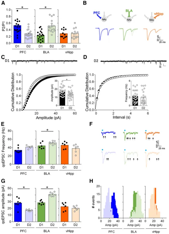
mea-sured the paired-pulse ratio (PPR), a
classical index of neurotransmitter release
probability (
Silver et al., 1998
) (
Fig.
5
A, B). In D1 MSNs, BLA inputs had a low
PPR/high release probability and the
highest PPR/lowest release probability
was found at PFC inputs (D1: BLA
⬎ v
Hipp
⬎ PFC). In contrast, in D2 MSNs,
PFC inputs exhibited the highest release
probability, respectively, whereas the
low-est probability of release was found at BLA
inputs (D2: PFC
⫽ vHipp ⬎ BLA).
In immature mice, miniature EPSCs
properties were similar in D1 and D2
MSNs in early life (P16 –P24,
Cao et al.,
2018
), whereas
⬃P42 sEPSCs are more
Figure 3. Hierarchy of optically driven synaptic inputs depends on the cellular identity of the target MSNs. A, Left, At PFC fibers, the largest excitatory oEPSCs in response to increasing light stimulations were recorded in D2 MSNs (light blue, n⫽17)compared with D1 MSNs (dark blue, n⫽11)(F(interaction 15,416)⫽18.59,p⬍0.0001,F(cell type 1,416)⫽934.4,p⬍0.0001,two-wayrepeated
measure ANOVA). Right, Example oEPSC traces for PFC inputs onto D1 and D2 MSNs. B, Left, At BLA fibers, the largest excitatory oEPSCs in response to increasing light stimulations were recorded in D1 MSNs (dark green, n⫽20)comparedwithD2MSNs(light green, n⫽15)(F(interaction 15,528)⫽5.628,p⬍0.0001,F(cell type 1,528)⫽331.5),p⬍0.0001,two-wayrepeatedmeasureANOVA).
Right, Example oEPSC traces for BLA inputs onto D1 and D2 MSNs. C, Left, At vHipp fibers, the largest excitatory oEPSCs in response to increasing light stimulations were recorded in D2 MSNs (light orange, n⫽ 22) compared with D1 MSNs (dark orange, n ⫽ 24) (F(interaction 15,704)⫽ 4.310, p ⬍ 0.0001, F(cell type 1,704)⫽ 491.3, p ⬍ 0.0001, two-way repeated measure ANOVA). Right,
Wxample oEPSC traces for vHipp inputs onto D1 and D2 MSNs. D, Summary bar histogram showing maximum oEPSCs at PFC inputs onto D1 and D2 MSNs. (D1 MSNs n⫽ 11, D2 MSNs n ⫽ 11, p ⬍ 0.0001, Mann–Whitney U test). E, Summary bar histogram of maximum oEPSCs at BLA inputs in neighboring D1 and D2 MSNs (D1 MSNs n⫽15,D2MSNsn⫽15,p⬍0.0001,Mann–Whitney
U test). F, Summary bar histogram of maximum oEPSCs at vHipp inputs in neighboring D1 and D2 MSNs neighboring (D1 MSNs n⫽
22, D2 MSNs n⫽ 22, p ⬍ 0.0001, Mann–Whitney U test). Individual point in scatter dot plots represents one individual neuron. 4
The line between dots shows neighboring D1 and D2 neurons that were recorded in the same slice with the same optical fiber position. All data are shown as mean⫾SEMorgeometric mean⫾ CI. *p ⬍ 0.05.
frequent in D2 MSNs (
Ma et al., 2012
) in
disagreement with an earlier report of
higher miniature EPSCs in D1 MSNs
(P28 –P56,
Grueter et al., 2010
). We
re-corded sEPSCs in D1 and D2 NAc core
MSNs of adult mice (
Fig. 5
C,D). The
am-plitude of the sEPSCs measured in D1
MSNs was slightly larger than that
mea-sured in D2 MSNs (in agreement with
Grueter et al., 2010
). Both MSN subtypes
exhibited similar distribution and average
interevent intervals.
Next, we measured light-evoked
input-specific quantal EPSCs (qoEPSCs) by
replacing our extracellular medium’s
cal-cium with strontium to desynchronize
transmitter release (
Britt et al., 2012
;
McGarry and Carter, 2017
). qoEPSC
am-plitude provides a direct estimate of
post-synaptic efficacy measure and qoEPSC
frequency was taken as an indirect
indica-tion of the number of connecindica-tions (
Goda
and Stevens, 1994
).
At PFC and vHipp inputs, qoEPSC
fre-quencies were similar across cell types
(
Fig. 5
E, F ). However, qoEPSC
frequen-cies were larger at D2 than
BLA-D1 synapses (
Fig. 5
G,H ). In contrast,
qoEPSC amplitude were larger at PFC-D1
and BLA-D2 synapses compared with the
other afferents (
Fig. 5
G,H ).
The large qoEPSC frequency and
am-plitude observed at BLA-D2 synapses may
at first appear to be at odds with the high
PPR/release probability at these synapses
as estimated by our input/output
experi-ments (
Fig. 3
). Although asynchronous
events are considered
“calcium-inde-pendent” (i.e., no calcium is present),
strontium desynchronization is most
likely the result of strontium’s low-affinity
binding to the calcium sensor underlying
excitation/secretion coupling. Thus,
dif-ferences in calcium-dependent processes
in the terminals (e.g., posttranslational
modification) may account for the
differ-ence between these two techniques.
Another cautious interpretation of the
re-sults is that high quantal size and low Pr
combine to yield a “weak” synapse.
Pathway-specific expression of CB1R and TRPV1R receptors
The eCB system modulates synaptic circuits in the CNS and
no-tably the dorsal and ventral striatum (
Araque et al., 2017
).
Inhib-itory CB1 receptors (CB1Rs) have long been described at
glutamatergic NAc core synapses using standard electrical
stim-ulation (
Robbe et al., 2001
). Stimulation of CB1Rs with a
sub-maximal dose of a synthetic cannabimimetic (CP55940 1
M,
Fig.
6
A, B) inhibited oEPSC in D1 and D2 MSNs regardless of the
input pathway, in support of the idea that functional CB1R are
present at all of these inputs. The degree of inhibition induced by
the single dose used here, however, was pathway dependent.
Thus, CP55940-induced inhibition was larger at BLA than PFC
or vHipp-D1 synapses (
Fig. 6
C) and smaller at vHipp-D2
syn-apses (compared with PFC and BLA,
Fig. 6
D).
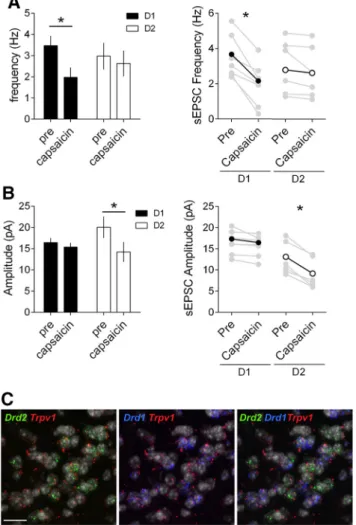
The nonselective cation channel, transient receptor potential
cation channel subfamily V member 1 (TRPV1R), is a
multifac-eted mediator of eCB signaling in the CNS (
Gibson et al., 2008
;
Manduca et al., 2017
;
Bara et al., 2018
), notably in the striatum:
pharmacological activation of TRPV1R suppresses and facilitates
transmitter release in the dorsal striatum (
Musella et al., 2009
)
and inhibits excitatory inputs onto D2 NAc core MSNs (
Grueter
et al., 2010
). Multiple inputs onto D1 and D2 MSNs differentially
expressing various levels of TRPV1R could explain these
appar-ently contradicting results. Thus, we compared the effects of the
specific TRPV1R agonist capsaicin in the disambiguated NAc
core synapse preparation (
Fig. 7
). Although capsaicin inhibited
Figure 4. Pathway-specific drive of action potential in identified NAc core MSNs. A, Left, Comparison of the probability of AP firing versus pulse number shows that PFC inputs preferentially drive the firing of D2 MSNs compared with D1 MSNs (D1 MSNs n⫽ 6, D2 MSNs n⫽6,pulse1:p⫽0.0005,pulse2–5:p⬍0.0001,Fisher’sexacttest).Right,ExampletracesofAPsevokedinD1and D2 MSNs in response to trains of optical stimulation of PFC inputs (2 ms light pulses, 5 pulses at 10 Hz). B, Left, Comparison of the probability of AP firing versus pulse number shows that BLA inputs preferentially drive the firing of D1 MSNs compared with D2 MSNs (D1 MSNs n⫽ 7, D2 MSNs n ⫽ 6, pulse 1–3: p ⬍ 0.0001, pulse 4: p ⫽ 0.0027, pulse 5: p ⫽ 0.0445, Fisher’s exact test). Right, Example traces of APs evoked in D1 and D2 MSNs in response to trains of optical stimulation of BLA inputs. C, Left, Comparison of the probability of AP firing versus pulse number shows no difference at vHipp inputs of the firing of D1 MSNs and D2 MSNs (D1 MSNs n⫽ 6, D2 MSNs n ⫽ 6, pulse 1: p ⫽ 0.0357, pulse 2: p ⫽ 0.1997, pulse 3–5: p ⫽ 1.0000, Fisher’s exact test). Right, Example traces of APs evoked in D1 and D2 MSNs in response to trains of optical stimulation of vHipp inputs. Blue dots indicate time of stimulation. All experiments were performed in the presence of picrotoxin to prevent from feedforward inhibition. All data are shown as mean⫾ SEM or geometric mean ⫾ CI. *p ⬍ 0.05.
Figure 5. Input- and cell-type-specific postsynaptic and presynaptic properties in the NAc core. A, Average paired-pulse ratio (P2/P1) measured in D1 and D2 MSNs (voltage-clamp,⫺70 mV) in response to paired optical stimulations (50 ms interval) of PFC, BLA and vHipp inputs. PFC inputs had a lower PPR/high release probability in D2 MSNs (light blue) compared with D1 MSNs (dark blue) (D1 MSNs n⫽ 12, D2 MSNs n ⫽ 19, p ⫽ 0.0006, Mann–Whitney U test). BLA inputs had a lower PPR/high release probability in D1 MSNs (dark green) compared with D2 MSNs (light green) (D1 MSNs n⫽ 16, D2 MSNs n ⫽ 18, p ⬍ 0.0001, Mann–Whitney U test). vHipp inputs exhibited similar release probability in both D1 (dark orange) and D2 MSNs (light orange) (vHipp: D1 MSNs n ⫽ 15, D2 MSNs n⫽11,p⬎0.9999,Mann–WhitneyUtest).B,ExampletracesofevokedpairedoEPSCresponsestoopticalstimulationsofPFC,BLAandvHippinputsrecordedinD1MSNsandD2MSNs.
C, Cell-type-specific differences in amplitude of sEPSCs in adult core MSNs: Representative traces of whole-cell voltage-clamp recording AMPAR event sEPSC from in D1 and D2 MSNs. D, Left,
Cumulative probability plot of measured sEPSC event amplitudes. Summary bar chart shows mean AMPAR event sEPSC amplitude (D1 MSNs: n⫽32;D2MSNs:n⫽26,p⫽0.0146,Mann–Whitney
U test). Right, Cumulative probability plot of interval between measured sEPSC events. Scatter dot plots represents one cell. Summary bar chart shows mean AMPAR event sEPSC interval. E,
Light-evoked input-specific quantal EPSCs (qoEPSC) in the adult NAc. At BLA inputs, qoEPSCs frequencies were larger in D2 MSNs compared with D1 MSNs (D1 MSNs n⫽ 6, D2 MSNs n ⫽ 7, p ⫽ 0.0734, Mann–Whitney U test). At PFC and vHipp inputs, qoEPSCs frequencies were similar in D1 MSNs and D2 MSNs (PFC: D1 MSNs n⫽ 7, D2 MSNs n ⫽ 6, p ⫽ 0.0361; vHipp: D1 MSNs n ⫽ 6, D2 MSNs n⫽6,p⫽0.1429,Mann–WhitneyUtest).F,RepresentativeqoEPSCsrecordedinD1MSNsandD2MSNsinresponsetopathwayspecificopto-stimulation(arrowsindicatedetectedqoEPSCs).
G, At PFC inputs, qoEPSCs amplitudes were larger in D1 MSNs compared with D2 MSNs (D1 MSNs n⫽7,D2MSNsn⫽6,p⫽0.0350,Mann–WhitneyUtest).AtBLAinputs,qoEPSCsamplitudeswere
larger in D2 MSNs compared with D1 MSNs (D1 MSNs n⫽6,D2MSNsn⫽7,p⫽0.0012,Mann–WhitneyUtest).AtvHippinputs,qoEPSCamplitudesweresimilarinD1MSNsandD2MSNs(D1MSNs
n⫽ 6, D2 MSNs n ⫽ 6, p ⫽ 0.1429, Mann–Whitney U test). H, Histograms for representative D1 cells and D2 cells showing the distribution of qoEPSC amplitude across all trials. Blue dots indicate
PFC-evoked oEPSCs in both D1 and D2 MSNs (
Fig. 7
A, B, left),
the TRPV1R agonist had multifaceted effects on the other
path-ways. Capsaicin had opposite effects on BLA-evoked oEPSCs
de-pending on the MSN subtype: the efficacy of BLA D1 MSN
synapses was enhanced in the presence of the TRPV1 agonist but
reduced at BLA-D2 MSNs synapses (
Fig. 7
A, B, middle). Finally,
at vHipp inputs, TRPV1R activation specifically inhibited vHipp-D1
synapses and had no effect at vHipp synapses onto D2 MSNs (
Fig.
7
A,B, right). These results are summarized in
Figure 7
D.
At excitatory inputs in the NAc core, CB1R are expressed
presynaptically (
Robbe et al., 2001
;
Micale et al., 2009
) and
me-diate presynaptic retrograde LTD (
Robbe et al., 2002
) while
TRPV1R are localized postsynaptically (
Micale et al., 2009
) and
trigger postsynaptic LTD specifically in D2 MSNs (
Grueter et al.,
2010
;
Neuhofer et al., 2018
). We combined electrophysiological
and RNAscope approaches to identify TRPV1R’s loci of
expres-sion in our model. Capsaicin had opposite effects on
spontane-ous’ EPSCs recorded in D1 and D2 MSNs. In D1 MSNs the
TRPV1R agonist reduced the frequency of spontaneous EPSCs
(
Fig. 8
A), but not their amplitude, in support of a presynaptic
localization (
Fig. 8
B). In D2 MSNs, however, capsaicin
re-duced the amplitude of spontaneous EPSCs (
Fig. 8
B), but not
Figure 6. CB1R inhibition is common to all inputs onto D1 and D2 NAc core MSNs. A, Bath application of the synthetic CB1R agonist CP55940 (1M), inhibited oEPSC in D1 (black) and D2 (white) MSNs. CP55940 was applied for 30 min (black bar) after at least 10 min of stable baseline recording. B, Individual and averaged oEPSCs amplitude experiments before (baseline) and 25–30 min after CP55940. (D1 MSNs, black circles: PFC n⫽ 3, p ⫽ 0.0014, BLA n ⫽ 4, p ⫽ 0.0110, vHipp n ⫽ 3, p ⫽ 0.0411; D2 MSNs, white circles: PFC n ⫽ 4, p ⫽ 0.0114, BLA n ⫽ 4, p ⫽ 0.0188, vHipp n ⫽ 4, p⫽ 0.0496 paired t test). C, Summary bar histogram comparing CP55940-induced inhibition of oEPSCs at identified PFC, BLA and vHipp inputs onto D1 (PFC n ⫽ 3, BLA n ⫽ 4, vHipp n ⫽ 3,
F(input 2,7)⫽ 14.32, p ⫽ 0.0034, one-way ANOVA) and D2 MSNs (PFC n ⫽ 4, BLA n ⫽ 4, vHipp n ⫽ 4, F(input 2,9)⫽ 4.207, p ⫽ 0.0513, one-way ANOVA). D, Schematic view of the relative weight
their frequency (
Fig. 8
A), in support of a postsynaptic
local-ization. These functional observations were corroborated by
the distribution of Trpv1, Drd1 and Drd2 mRNAs. Triple
staining indicated that in the NAc core, Trpv1 mRNA was
preferentially enriched in D2 MSNs compared with
D1-expressing MSNs (
Fig. 8
C).
Together, these results reveal that whereas CB1R are
uni-formly expressed across input pathways onto D1/D2 MSNs,
TRPV1R modulate excitatory inputs to the NAc core in a
pathway- and cell-type-specific manner.
Cell-type-specific endocannabinoid-mediated LTD at
amygdala synapses
In the last series of experiments, we focused on the BLA-NAc core
synapses. These synapses are instrumental to reward-seeking
be-havior (
Ambroggi et al., 2008
) and have a role in positive
emo-tional valence (
Beyeler et al., 2018
). Our present observation that
both TRPV1R and CB1R modulate synaptic transmission in a
cell-type-specific manner raised the possibility that the eCB
in-terplay controls the polarity of activity-dependent plasticity at
BLA-NAc synapses.
Figure 7. Divergent input- and cell-specific control of excitatory NAc synapses by TRPV1R. A, Effects of the TRPV1R agonist capsaicin (10M) on oEPSCs at PFC, BLA, and vHipp synapses onto D1 (black circles) and D2 (white circles) MSNs. Capsaicin was applied for 30 min (black bar) after at least 10 min baseline recording. Capsaicin inhibited PFC-evoked oEPSCs similarly in both D1 (black) and D2 (white) MSNs. Capsaicin enhanced BLA-D1 MSN synapses but inhibited BLA-D2 MSN synapses. Finally, capsaicin inhibited vHipp-D1 MSN synapses and has no effect at vHipp-D2 MSN synapses. B, Individual and averaged oEPSCs amplitude experiments 10 min before (baseline) and 25–30 min after capsaicin; D1 MSNs, black circles: PFC n⫽3,p⫽0.0308,BLAn⫽4,p⫽0.0188, vHipp n⫽ 5, p ⫽ 0.0010; D2 MSNs, white circles: PFC n ⫽ 3, p ⫽ 0.0135, BLA n ⫽ 3, p ⫽ 0.0087, vHipp n ⫽ 3, p ⫽ 0.0109 paired t test). C, Summary bar histogram of the maximal effects of capsaicin on oEPSCs at identified inputs onto D1 (PFC n⫽ 3, BLA n ⫽ 4, vHipp n ⫽ 5, F(input 2,9)⫽ 157.4, p ⬍ 0.0001, one-way ANOVA) and D2 MSNs (PFC n ⫽ 3, BLA n ⫽ 3, vHipp n ⫽ 3, F(input 2, 6)⫽ 8.586, p ⫽ 0.0174, one-way ANOVA). D, Schematic view of the relative weight and effects of TRPV1R on oEPSCs at PFC, BLA and vHipp inputs onto D1 and D2 MSNs. n represents the
In the striatum, the principal form of eCB-mediated synaptic
plasticity is LTD (
Gerdeman et al., 2002
;
Robbe et al., 2002
). We
first compared the expression of eCB-LTD at BLA-D1 and -D2
synapses. Using the canonical protocol that elicits eCB-LTD in
NAc MSNs (
Robbe et al., 2002
), we observed a robust LTD in D2
but not D1 MSNs after optical stimulation (
Fig. 9
). This is, to our
knowledge, the first evidence that eCB-LTD can be
optogeneti-cally induced at a single set of identified synapses. Although this
experiment can be interpreted as a lack of eCB-LTD at BLA
in-puts onto D1 MSNs, the current pharmacological
characteriza-tion and previous work in the NAc core (
Neuhofer et al., 2018
)
led us to test the possibility of another, more complex scenario.
Based on the enhancing effects of the TRPV1R agonist at BLA-D1
synapses, we hypothesized that tetanus-induced eCB release
(presumably anandamide) and the ensuing potentiation masked/
prevented the induction of LTD. In support of this idea we
ob-served that in the presence of a TRPV1R antagonist (capsazepine,
CPZ 10
M) the otherwise inefficacious protocol triggered a large
LTD (
Fig. 9
C,D). Interestingly, bath application of the CB1R
antagonist SR 141716 (5
M) prevented the induction of LTD in
the presence of CPZ (
Fig. 9
C,D).
The mirror situation was observed at BLA-D2 synapses.
There, LTD was converted to LTP in the presence of the TRPV1R
antagonist (
Fig. 9
E, F ), a result compatible with the current
find-ing that TRPV1R are inhibitory on BLA-D2 synapses (
Fig. 9
). We
finally controlled for the role of CB1R in BLA-D2 LTD.
Bath-application of the SR 141716 (5
M) blocked post-tetanic
depres-sion and prevented LTD (
Fig. 9
E, F ), showing that CB1R
mediates BLA-D2 LTD.
Discussion
Our principal findings are that in the adult mouse NAc core, the
hierarchy of excitatory inputs depends on the identity of the
post-synaptic target MSN and on circuit specific properties and that
the eCB system endows excitatory circuits of the NAc with
pathway-specific plasticity.
Differential intrinsic properties of adult NAc core MSNs
The data show that D1 and D2 MSNs in adult NAc core have
distinctive intrinsic properties. Compared with D2, D1 MSNs
had a low rheobase, a more depolarized membrane potential and
exhibited a higher propensity to trigger action potentials in
re-sponse to depolarizing current injections. Thus, adult D1 are
more excitable than D2 MSNs.
Subtype specific morphological properties (e.g., soma size or
dendritic arborization,
Gertler et al., 2008
) and differences in
membrane biophysical properties such as membrane resistance,
capacitance, or the expression of voltage-dependent calcium
channels, may explain these dissimilarities (
Nisenbaum et al.,
1994
;
Herna´ndez-Lo´pez et al., 1997
,
2000
).
Divergent intrinsic properties have already been described in
juvenile and adolescent mice, however, at these stages D2- are
more excitable than D1-MSNs (
Kreitzer and Malenka, 2007
;
Gertler et al., 2008
;
Ma et al., 2012
;
Planert et al., 2013
;
Cao et al.,
2018
). Although many factors (e.g., recording site, sample size,
the presence or absence of TTX, the transgenic mouse line used,
the size of the soma or the extent of dendritic arborization) may
explain this discrepancy, it is highly probable that age is the
de-termining factor. In keeping with this idea, in rat NAc core,
pre-pubertal rats and adult rats display different properties (
Belleau
and Warren, 2000
;
Zhang and Warren, 2008
;
Kasanetz and
Man-zoni, 2009
). Additionally, in both rats and mice, NAc dopamine
receptors undergo significant developmental changes during
ad-olescence (
Andersen et al., 1997
;
Andersen and Teicher, 2000
).
The subtype specific evolution of excitability in
juvenile/ado-lescent and adults MSNs may be due to differences in membrane
resistance and conductance. In support of this possibility, early in
development MSNs have very high membrane resistance and do
not express inward rectifying potassium channels (
Tepper et al.,
1998
;
Belleau and Warren, 2000
). The time-scale of this
physio-logical maturation parallels the morphophysio-logical development of
MSNs and dendritic arborization, spine formation and
synapto-genesis which continue until the end of the first postnatal month
(
Tepper et al., 1998
;
Butler et al., 1999
).
Figure 8. MSN-subtype-selective expression and function of TRPV1R in the NAc core. A, Left, Capsaicin reduces the frequency of sEPSC and in D1 but not D2 MSNs. Summary bar histogram comparing the mean sEPSC frequencies in D1 (black, n⫽7)andD2(white,n⫽6)MSNsbefore and after capsaicin (10M). Right, Individual and averaged sEPSCs frequency experiments before (pre) and 25–30 min after capsaicin (D1 MSNs black circles, p⫽ 0.0013 paired t test; D2 MSNs white circles p⫽ 0.1152 paired t test). B, Left, Capsaicin reduces the amplitude of sEPSC in but not D1 MSNs. Summary bar histogram compares the mean sEPSC amplitudes in D1 (black,
n⫽ 7) and D2 (white, n ⫽ 6) MSNs before and after capsaicin. Right, individual and averaged
sEPSCs amplitude (D) experiments before (pre) and 25–30 min after capsaicin (D1 MSNs black circles, p⫽ 0.0559 paired t test; D2 MSNs white circles p ⫽ 0.0008 paired t test). All data are shown as mean⫾ SEM. *p ⬍ 0.05 paired t test. C, Single-molecular fluorescent in situ hybrid-ization for Drd1 (blue), Drd2 (green), and Trpv1 (red) mRNAs in the NAc core. Slides were counterstained with DAPI (white). Scale bar, 12m. Note the segregation of cells expressing
Hierarchy of excitatory afferents depends on MSN subtype in
adult NAc core
Pathway-specific opto-stimulation of excitatory inputs in the
NAc core revealed that there is a hierarchy of synaptic inputs that
depends on the cellular identity of the target MSNs. Most
nota-bly, the BLA and the PFC are the main source of synaptic excitation
on D1 and D2 neurons, respectively. Similarly, BLA and PFC
affer-ents trigger action potentials with a high probability in D1 and D2
MSNs, respectively.
Differences in the number of fibers per afferent or the amount
of glutamate release per fibers may underlie this functional
hier-archy. Based on the recent study of Li and colleagues (
Li et al.,
2018
) that showed similar PFC, BLA, and
vHipp innervations of D1 and D2 MSNs,
we favor the second proposition.
Input- and synapse-specific function
of endocannabinoids
The present data indicate that functional
CB1R are present at all synapses tested,
albeit with differences in the amount of
CB1R-mediated inhibition. The situation
was far more complex with TRPV1R.
In-deed, a TRPV1R agonist inhibited
PFC-evoked oEPSCs in both D1 and D2 MSNs
but led to opposite and MSN
subtype-specific effects at the BLA pathway:
BLA-D1 were enhanced while BLA-D2
synapses were inhibited by capsaicin.
TRPV1R specifically inhibited vHipp-D1
synapses and had no effect on vHipp-D2
synapses. Pharmacological activation of
TRPV1R suppresses or facilitates
neu-rotransmitter release into the dorsal
stria-tum according to their presynaptic or
postsynaptic location (
Musella et al.,
2009
) and our results with sEPSCs also
suggest a differential expression of
TRPV1R at BLA-D1 and BLA-D2
syn-apses. Indeed, capsaicin modified the
fre-quency but not the amplitude of sEPSCs
in D1 MSNs and vice-versa in D2 MSNs.
These data are compatible with
presynap-tic TRPV1R at BLA-D1 and postsynappresynap-tic
TRPV1R at BLA-D2 synapses.
Grueter et
al. (2010)
also reported a postsynaptic
lo-calization of TRPV1R at D2 MSNs.
At the BLA-NAc pathway, TRPV1R
and CB1R modulate synaptic
transmis-sion in a cell specific manner: LTD could
be induced in D2 but not in D1 MSNs in
agreement with a previous report where
the identity of MSNs was not
ascer-tained (
Grueter et al., 2010
). At first
glance, these results may be taken as an
indication that BLA-D2 MSNs are
inca-pable of expressing eCB-LTD. However,
the current pharmacological
character-ization and previous work (
Neuhofer et
al., 2018
) led us to hypothesize that the
tetanus-induced
eCB
(presumably
anandamide) activates TRPV1R and
that the ensuing potentiation masked/
prevented LTD. In support of this idea, we observed that, in
the presence of a TRPV1R antagonist, the previously
ineffi-cient protocol triggered a large LTD. The mirror situation was
observed at BLA-D2 synapses where LTD was converted to
LTP in the presence of the TRPV1R antagonist.
In summary, our study reveals the cell-type- specific synaptic
organization of hippocampal, amygdala and prefrontal inputs to
the PFC. It is tempting to speculate that pathway and cell specific
synaptic strengths correlate with distinct functions and
associ-ated behaviors. Future work will determine whether the PFC-D2
“dominant pathway” is preferentially engaged in executive
func-tions and the role of the BLA-D1 pathway in emotional
behav-Figure 9. TRPV1R delineates cell-type-specific LTD at amygdala synapses in the NAc core. A, Optical stimulation of BLA inputs (10 Hz for 10 min) induced a robust LTD in D2 (white circles, n⫽ 4) but not in D1 MSNs (black circles, n ⫽ 5). B, Individual and averaged oEPSC amplitude before (baseline) and 25–30 min after LTD induction (LTD) in D1 and D2 MSNs (D1 MSNs, p⫽ 0.3311, D2 MSNs, p⫽ 0.0072, paired t test). C, Bath perfusion of the TRPV1R capsazepine (CPZ 10M) allowed the induction of LTD at BLA-D1 synapses (purple, n⫽6;comparedwithcontrolsameasA).WhenCPZwasappliedinthepresenceoftheCB1Rantagonist SR141716 (SR 5M) LTD induction was prevented (orange, n⫽4).D,IndividualandaveragedoEPSCsamplitudebefore(baseline) and 25–30 min after LTD induction (LTD) in D1 MSNs in control, CPZ and CPZ⫹SR(D1CPZ,p⫽0.0056pairedttest;D1CPZ⫹SR,
p⫽ 0.9737). E, Antagonism of TRPV1R converted LTD to LTP at BLA-D2 synapses (purple, n ⫽ 5). Bath application of the CB1R
antagonist SR blocked LTD at BLA-D2 synapses (blue, n⫽ 6). F, Individual and averaged oEPSCs amplitude before (baseline) and 25–30 min after LTD induction (LTD) in D2 MSNs with or without CPZ or SR (D2 CPZ, p⫽ 0.0068; SR, p ⫽ 0.9546 paired t test). n represents the number of mice. All data are shown as mean⫾ SEM. *p ⬍ 0.05 paired t test.
iors. In this context, the observation that two subpopulations of
BLA glutamatergic neurons project to NAc core D1 or D2 MSNs
to generate emotional responses of opposite valence is an
indica-tion of similar parallel dual control circuits at PFC- and/or
vHipp-NAc pathways (
Shen et al., 2019
).
Moreover, these data highlight the versatility of the
endocan-nabinoid system in shaping activity-dependent synaptic plasticity
at BLA-NAc circuits and cortical-limbic circuits in general.
In conclusion, our experiments reveal a high degree of synapse
and circuit specificity in the adult NAc core and illustrate how
endo-cannabinoids contribute to pathway-specific synaptic plasticity.
References
Abela AR, Duan Y, Chudasama Y (2015) Hippocampal interplay with the nucleus accumbens is critical for decisions about time. Eur J Neurosci 42:2224 –2233.
Ade KK, Wan Y, Chen M, Gloss B, Calakos N (2011) An improved bac transgenic fluorescent reporter line for sensitive and specific identifica-tion of striatonigral medium spiny neurons. Front Syst Neurosci 5:32. Ambroggi F, Ishikawa A, Fields HL, Nicola SM (2008) Basolateral amygdala
neurons facilitate reward-seeking behavior by exciting nucleus accum-bens neurons. Neuron 59:648 – 661.
Andersen SL, Teicher MH (2000) Sex differences in dopamine receptors and their relevance to ADHD. Neurosci Biobehav Rev 24:137–141. Andersen SL, Rutstein M, Benzo JM, Hostetter JC, Teicher MH (1997) Sex
differences in dopamine receptor overproduction and elimination. Neu-roreport 8:1495–1498.
Araque A, Castillo PE, Manzoni OJ, Tonini R (2017) Synaptic functions of endocannabinoid signaling in health and disease. Neuropharmacology 124:13–24.
Bara A, Manduca A, Bernabeu A, Borsoi M, Serviado M, Lassalle O, Murphy M, Wager-Miller J, Mackie K, Pelissier-Alicot AL, Trezza V, Manzoni OJ (2018) Sex-dependent effects of in utero cannabinoid exposure on cor-tical function. Elife 7:e36234.
Belleau ML, Warren RA (2000) Postnatal development of electrophysiolog-ical properties of nucleus accumbens neurons. J Neurophysiol 84:2204 – 2216.
Bertran-Gonzalez J, Bosch C, Maroteaux M, Matamales M, Herve´ D, Valjent E, Girault JA (2008) Opposing patterns of signaling activation in dopa-mine D1 and D2 receptor-expressing striatal neurons in response to co-caine and haloperidol. J Neurosci 28:5671–5685.
Beyeler A, Namburi P, Glober GF, Simonnet C, Calhoon GG, Conyers GF, Luck R, Wildes CP, Tye KM (2016) Divergent routing of positive and negative information from the amygdala during memory retrieval. Neu-ron 90:348 –361.
Beyeler A, Chang CJ, Silvestre M, Le´veˆque C, Namburi P, Wildes CP, Tye KM (2018) Organization of valence-encoding and projection-defined neu-rons in the basolateral amygdala. Cell Rep 22:905–918.
Bosch-Bouju C, Larrieu T, Linders L, Manzoni OJ, Laye´ S (2016) Endocannabinoid-mediated plasticity in nucleus accumbens controls vulnerability to anxiety after social defeat stress. Cell Rep 16:1237–1242. Britt JP, Benaliouad F, McDevitt RA, Stuber GD, Wise RA, Bonci A (2012)
Synaptic and behavioral profile of multiple glutamatergic inputs to the nucleus accumbens. Neuron 76:790 – 803.
Butler AK, Uryu K, Rougon G, Chesselet MF (1999) N-methyl-D-aspartate receptor blockade affects polysialylated neural cell adhesion molecule ex-pression and synaptic density during striatal development. Neuroscience 89:1169 –1181.
Cao J, Dorris DM, Meitzen J (2018) Electrophysiological properties of me-dium spiny neurons in the nucleus accumbens core of prepubertal male and female Drd1a-tdTomato line 6 BAC transgenic mice. J Neurophysiol 120:1712–1727.
Cardinal RN, Howes NJ (2005) Effects of lesions of the nucleus accumbens core on choice between small certain rewards and large uncertain rewards in rats. BMC Neuroscience 6:37.
Christakou A, Robbins TW, Everitt BJ (2001) Functional disconnection of a prefrontal cortical– dorsal striatal system disrupts choice reaction time performance: implications for attentional function. Behav Neurosci 115: 812– 825.
Christakou A, Robbins TW, Everitt BJ (2004) Prefrontal cortical–ventral striatal interactions involved in affective modulation of attentional
per-formance: implications for corticostriatal circuit function. J Neurosci 24:773–780.
Cools R, Sheridan M, Jacobs E, D’Esposito M (2007) Impulsive personality predicts dopamine-dependent changes in frontostriatal activity during component processes of working memory. J Neurosci 27:5506 –5514. Eichenbaum H (2014) Time cells in the hippocampus: a new dimension for
mapping memories. Nat Rev Neurosci 15:732–744.
Enoksson T, Bertran-Gonzalez J, Christie MJ (2012) Nucleus accumbens D2- and D1-receptor expressing medium spiny neurons are selectively activated by morphine withdrawal and acute morphine, respectively. Neuropharmacology 62:2463–2471.
Everitt BJ, Morris KA, O’Brien A, Robbins TW (1991) The basolateral amygdala-ventral striatal system and conditioned place preference: fur-ther evidence of limbic-striatal interactions underlying reward-related processes. Neuroscience 42:1–18.
Everitt BJ, Cardinal RN, Parkinson JA, Robbins TW (2003) Appetitive be-havior. Ann N Y Acad Sci 985:233–250.
Fernando AB, Murray JE, Milton AL (2013) The amygdala: securing plea-sure and avoiding pain. Front Behav Neurosci 7:190.
Finch DM (1996) Neurophysiology of converging synaptic inputs from the rat prefrontal cortex, amygdala, midline thalamus, and hippocampal for-mation onto single neurons of the caudate/putamen and nucleus accum-bens. Hippocampus 6:495–512.
Floresco SB (2015) The nucleus accumbens: an interface between cognition, emotion, and action. Annu Rev Psychol 66:25–52.
Floresco SB, Seamans JK, Phillips AG (1997) Selective roles for hippocam-pal, Prefrontal Cortical, and Ventral Striatal Circuits in Radial-Arm Maze Tasks With or Without a Delay. J Neurosci 17:1880 –1890.
Floresco SB, Braaksma DN, Phillips AG (1999) Thalamic– cortical–striatal circuitry subserves working memory during delayed responding on a ra-dial arm maze. J Neurosci 19:11061–11071.
Francis TC, Chandra R, Friend DM, Finkel E, Dayrit G, Miranda J, Brooks JM, In˜iguez SD, O’Donnell P, Kravitz A, Lobo MK (2015) Nucleus accum-bens medium spiny neuron subtypes mediate depression-related out-comes to social defeat stress. Biol Psychiatry 77:212–222.
French SJ, Totterdell S (2002) Hippocampal and prefrontal cortical inputs monosynaptically converge with individual projection neurons of the nucleus accumbens. J Comp Neurol 446:151–165.
French SJ, Totterdell S (2003) Individual nucleus accumbens-projection neurons receive both basolateral amygdala and ventral subicular afferents in rats. Neuroscience 119:19 –31.
Gerdeman GL, Ronesi J, Lovinger DM (2002) Postsynaptic endocannabi-noid release is critical to long-term depression in the striatum. Nat Neu-rosci 5:446 – 451.
Gertler TS, Chan CS, Surmeier DJ (2008) Dichotomous anatomical properties of adult striatal medium spiny neurons. J Neurosci 28:10814–10824. Gibson HE, Edwards JG, Page RS, Van Hook MJ, Kauer JA (2008) TRPV1
channels mediate long-term depression at synapses on hippocampal in-terneurons. Neuron 57:746 –759.
Goda Y, Stevens CF (1994) Two components of transmitter release at a central synapse. Proc Natl Acad Sci U S A 91:12942–12946.
Goto Y, Grace AA (2008) Limbic and cortical information processing in the nucleus accumbens. Trends Neurosci 31:552–558.
Groenewegen HJ, Wright CI, Beijer AV, Voorn P (1999) Convergence and segregation of ventral striatal inputs and outputs. Ann N Y Acad Sci 877:49–63. Grueter BA, Brasnjo G, Malenka RC (2010) Postsynaptic TRPV1 triggers cell type–specific long-term depression in the nucleus accumbens. Nat Neurosci 13:1519 –1525.
Herna´ndez-Lo´pez S, Bargas J, Surmeier DJ, Reyes A, Galarraga E (1997) D1 receptor activation enhances evoked discharge in neostriatal medium spiny neurons by modulating an L-type Ca2⫹ conductance. J Neurosci 17:3334 –3342.
Herna´ndez-Lo´pez S, Tkatch T, Perez-Garci E, Galarraga E, Bargas J, Hamm H, Surmeier DJ (2000) D2 dopamine receptors in striatal medium spiny neurons reduce L-type Ca2⫹ currents and excitability via a novel PLC1– IP3– calcineurin-signaling cascade. J Neurosci 20:8987– 8995.
Humphries MD, Prescott TJ (2010) The ventral basal ganglia, a selection mechanism at the crossroads of space, strategy, and reward. Prog Neuro-biol 90:385– 417.
Ito R, Robbins TW, Pennartz CM, Everitt BJ (2008) Functional interaction between the hippocampus and nucleus accumbens shell is necessary for