HAL Id: hal-02506214
https://hal.archives-ouvertes.fr/hal-02506214
Submitted on 12 Mar 2020
HAL is a multi-disciplinary open access
archive for the deposit and dissemination of
sci-entific research documents, whether they are
pub-lished or not. The documents may come from
teaching and research institutions in France or
abroad, or from public or private research centers.
L’archive ouverte pluridisciplinaire HAL, est
destinée au dépôt et à la diffusion de documents
scientifiques de niveau recherche, publiés ou non,
émanant des établissements d’enseignement et de
recherche français ou étrangers, des laboratoires
publics ou privés.
Hookworm exposure decreases human papillomavirus
uptake and cervical cancer cell migration through
systemic regulation of epithelial-mesenchymal transition
marker expression
Brittany-Amber Jacobs, Alisha Chetty, William Gordon Charles Horsnell,
Georgia Schäfer, Sharon Prince, Katherine Ann Smith
To cite this version:
Brittany-Amber Jacobs, Alisha Chetty, William Gordon Charles Horsnell, Georgia Schäfer, Sharon
Prince, et al.. Hookworm exposure decreases human papillomavirus uptake and cervical cancer cell
mi-gration through systemic regulation of epithelial-mesenchymal transition marker expression. Scientific
Reports, Nature Publishing Group, 2018, 8 (1), �10.1038/s41598-018-30058-9�. �hal-02506214�
Hookworm exposure decreases
human papillomavirus uptake
and cervical cancer cell migration
through systemic regulation of
epithelial-mesenchymal transition
marker expression
Brittany-Amber Jacobs
1, Alisha Chetty
1, William Gordon Charles Horsnell
1,2,3,
Georgia Schäfer
1, Sharon Prince
4& Katherine Ann Smith
1,5Persistent infection with human papillomavirus (HPV) is responsible for nearly all new cervical cancer cases worldwide. In low- and middle-income countries (LMIC), infection with helminths has been linked to increased HPV prevalence. As the incidence of cervical cancer rises in helminth endemic regions, it is critical to understand the interaction between exposure to helminths and the progression of cervical cancer. Here we make use of several cervical cancer cell lines to demonstrate that exposure to antigens from the hookworm N. brasiliensis significantly reduces cervical cancer cell migration and global expression of vimentin and N-cadherin. Importantly, N. brasiliensis antigen significantly reduced expression of cell-surface vimentin, while decreasing HPV type 16 (HPV16) pseudovirion internalization. In vivo infection with N. brasiliensis significantly reduced vimentin expression within the female genital tract, confirming the relevance of these in vitro findings. Together, these findings demonstrate that infection with the hookworm-like parasite N. brasiliensis can systemically alter genital tract mesenchymal markers in a way that may impair cervical cancer cell progression. These findings reveal a possible late-stage treatment for reducing cervical cancer progression using helminth antigens.
Cervical cancer is the fourth most common cancer worldwide, causing an estimated 266,000 deaths in 2012. The major global burden of cervical cancer occurs in low-and middle-income countries (LMICs), which experienced almost 87% of disease-related deaths in this period. It is estimated that by 2035, the incidence of cervical cancer will increase by 58% (from 444,546 to 702,152 cases) in developing regions compared to 6% (from 83,078 to 88,041 cases) in high-income countries (HICs)1. Of significant interest is that nearly 100% of new cervical cancers are attributable to persistent infection with HPV2.
HPV prevalence and cervical cancer incidence varies significantly within the LMIC region. Recent epidemi-ological evidence suggests that shifts in the HPV immune response resulting from concurrent soil-transmitted helminth (STH) infection significantly increased HPV prevalence3. Helminth infections infect over a billion indi-viduals in LMIC4 and result in the loss of 20 million disability-adjusted life years (DALY - the amount of years lost due to illnesses, disability or premature death)5. Amongst the leading STH infections are the hookworms,
1Institute of Infectious Disease and Molecular Medicine, University of Cape Town, Cape Town, 7925, South Africa. 2Institute of Microbiology and Infection, University of Birmingham, B15 2TT, Birmingham, UK. 3Le Studium Institute
for Advanced Studies Laboratory of Molecular and Experimental Immunology and Neurogenetics, CNRS-University of Orleans, Rue Dupanloup, 45000, Orléans, France. 4Department of Human Biology, University of Cape Town,
Cape Town, 7925, South Africa. 5School of Medicine, Cardiff University, Cardiff, CF14 4XN, UK. Correspondence and
requests for materials should be addressed to K.A.S. (email: smithk28@cardiff.ac.uk) Received: 24 May 2018
Accepted: 12 July 2018 Published: xx xx xxxx
www.nature.com/scientificreports/
which cause more morbidity than most other human parasitic infections and infect more than 700 million people worldwide6.
Helminth infections are well documented as providing some benefit to the host, by limiting autoimmune and allergic symptoms in infected individuals7–10. However, helminth infections are also reported to dampen the efficacy of vaccination11 and impair immune responses to co-infection12,13. Helminth infections also influence the risk of cancer but this is understood in only a limited number of contexts. For example, Schistosoma haematobium is classified as a group 1 biological carcinogen and is a conclusive cause of bladder cancer14. Here, antigen from Schistosoma haematobium has been shown to induce urothelial dysplasia and inflammation, whereas the eggs cause squamous cell carcinoma of the urinary bladder15.
In addition to altering HPV prevalence3, infection with STH alters the replication of the oncogenic Kaposi’s sarcoma herpesvirus by driving reactivation from latency16. However, how STH infections may systemically influence cervical cancer cell progression is unknown. In this study, we used the rodent model of human hook-worm Nippostrongylus brasiliensis to identify how STH may influence cancer cell biology. We show here that N. brasiliensis exposure significantly reduced cervical cancer cell migration and the uptake of HPV. These data demonstrate that helminth infection can systemically modify cancer cell progression by significantly altering epithelial-mesenchymal transition (EMT) marker expression.
Results
N. brasiliensis L3 antigen decreases HeLa migration in vitro.
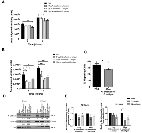
The impact of exposure to STH oncer-vical cancer cell proliferation and metastasis is unknown. Using an established in vitro two-dimensional scratch motility assay, we demonstrate that exposure of the HPV type 18 (HPV18) positive human cervical cell line HeLa to increasing concentrations of antigen isolated from the third-stage larvae (L3) of the hookworm N. brasiliensis L3 antigen for 8 hours, significantly decreased cervical cancer migration (Fig. 1A and B). Antigens from the solely gastrointestinal nematode H. polygyrus contain immune-modulatory components17 and were expected to impact HeLa migration. However, somatic antigen derived from the adult stage of H. polygyrus or the excretory/ secretory products of this nematode had no effect on the migration of HeLa cells (Fig. 1C). Transwell migration assays also show that, compared to untreated cells, 10 μg N. brasiliensis L3 antigen significantly inhibited HeLa cell migration by 1.36 fold (Fig. 1D). To determine if the impact of N. brasiliensis L3 antigen on cervical cancer cell migration was associated with changes in markers of EMT, we assessed the expression of E-cadherin, N-cadherin and vimentin by western blotting. Although we could not detect E-cadherin in HeLa cells, we found that expo-sure of HeLa cells to 10 μg N. brasiliensis L3 antigen resulted in a trend toward reduced expression of N-cadherin and vimentin by 12 and 24 hours, compared to untreated cells. Treatment of HeLa cells to a high dose of 50 μg N. brasiliensis L3 antigen significantly reduced expression of N-cadherin and vimentin by 24 hours, compared to untreated cells (Fig. 1E and F). There was no change in the proliferative ability of HeLa cells exposed to increasing concentrations N. brasiliensis L3 antigen (Fig. 1G).
N. brasiliensis L3 antigen decreases Ca Ski and C33-A migration in vitro.
The impact of N.bra-siliensis antigen on cervical cancer cell migration was validated in the HPV16 positive human cervical cancer cell line Ca Ski. Indeed, 10 μg of N. brasiliensis L3 antigen significantly decreased Ca Ski cell migration by 4 and 8 hours in vitro (Fig. 2A). To determine whether the HPV status of cervical cancer cells influences the effect of N. brasiliensis L3 antigen on cervical cancer cell migration we repeated the above experiments in the HPV negative human cervical cancer cell line C33-A. Our results show that 0.1 µg, 1 µg and 10 μg N. brasiliensis L3 antigen significantly reduced the migration of C33-A cells in scratch motility assays (Fig. 2B). The same results were obtained for 10 μg N. brasiliensis L3 antigen in transwell migration assays (Fig. 2C). Furthermore, both 10 μg and 50 μg N. brasiliensis L3 antigen reduced the expression of N-cadherin and vimentin in C33-A cells, compared to untreated cells (Fig. 2D and E).
N. brasiliensis infected mice have reduced genital tract EMT marker expression.
While our datademonstrate that the expression of N-cadherin and vimentin is significantly reduced in HeLa and C33-A cervical cancer cell lines following exposure to N. brasiliensis L3 antigen in vitro, it is not known how N. brasiliensis infec-tion impacts on the expression of these markers in non-cancerous tissue. We thus investigated this and demon-strate that infection significantly reduced the expression of vimentin in the female genital tract, while resulting in a trend toward decreased expression of N-cadherin, when compared to that of naïve mice (Fig. 3A–C). Indeed, the densitometric analyses of the western blots show that the average expression of vimentin and N-cadherin in infected mice are 0.62 and 0.55 compared to 1.29 and 0.73 in naïve mice respectively.
N. brasiliensis L3 antigen decreases HeLa cell-surface vimentin expression.
Gastrointestinalhel-minths have been implicated in increasing the susceptibility of cervical tissue to the cancer-causing virus HPV3. Interestingly, cell-surface vimentin expression has been shown to act as a restriction factor on HeLa cells for HPV pseudovirions18. Although our western blot data demonstrate that N. brasiliensis L3 antigen significantly reduces the expression of vimentin in cervical cancer cells (Figs 1G and 2D), it is not known how antigen exposure alters cell-surface vimentin expression or the uptake of HPV pseudovirions. Using flow cytometry, we demonstrate that 12 hours post-treatment of HeLa cells with 50 µg of N. brasiliensis L3 antigen, the expression of cell-surface vimentin was significantly decreased when compared to untreated cells (Fig. 4A and B). When we screened the N. brasiliensis L3 antigen by limulus amebocyte lysate (LAL) assay we found it to contain low levels of endotoxin (2.80EU/ml) and we also demonstrate that endotoxin (from 1 µg of lipopolysaccharide) had no significant impact on the expression of cell-surface vimentin (Fig. 4B).
N. brasiliensis L3 antigen decreases HPV16 pseudovirion internalisation in HeLa cells.
Due toFigure 1. N. brasiliensis L3 antigen decreases HeLa migration and EMT marker expression. (A) Scratch
motility assay showing HeLa cells exposed to 10 µg N. brasiliensis L3 antigen imaged at 4 and 8 hours post-wound formation and (B). Total area migrated calculated relative to post-wound area at time 0 hours for HeLa cells exposed to 0.1, 1 and 10 µg N. brasiliensis L3 antigen. (C) Scratch motility assay showing HeLa cells exposed to 10 µg H. polygyrus somatic antigen or 10 µg H. polygyrus excretory/secretory products (HES) imaged at 4 and 8 hours post-wound formation. (D) Transwell migration of HeLa cells determined at 24 hours following exposure to 10 µg N. brasiliensis L3 antigen or an untreated control. (E) Western blot analysis of N-cadherin, vimentin and p38 (loading control) was performed 12 or 24 hours following exposure of HeLa cells to 10 μg or 50 µg N. brasiliensis L3 antigen or an untreated control and (F). Densitometry readings were obtained using ImageJ in order to determine the changes in protein expression of N-cadherin and vimentin normalised to p38 and expressed as a fold-change relative to control wells. (G) Live cell counts over a 3-day period following exposure of HeLa cells to 0.1 µg, 1 µg and 10 µg N. brasiliensis L3 antigen or an untreated control. The figures shown are the result of (G). Two independent experiments, (A–D and F). Three independent experiments or (E). Representative of two independent experiments. The data was analysed using GraphPad Prism 6.01 and a parametric unpaired t-test was performed where ns = not significant, *p < 0.05 **p < 0.01 and ***p < 0.001. Error bars represent Standard Error of the Mean.
www.nature.com/scientificreports/
brasiliensis L3 antigen (Fig. 5), we assessed whether this may correlate with an expected increase in pseudovirion internalization. However, using flow cytometry, we demonstrate that 10 μg and 50 µg of N. brasiliensis L3 antigen significantly decreased the uptake of fluorescently labelled HPV16 pseudovirions by HeLa cells when compared to untreated cells (Fig. 5A and B).
Discussion
Despite specific helminths being irrefutable causes of cancer, certain other helminths have been shown to modu-late the host immune response by promoting immune-regulatory mechanisms19. While the exact mechanism that these helminths employ to achieve this is still emerging, the potential for their use in impeding cancer progres-sion is worth thorough investigation. Epidemiological studies have revealed a positive correlation between STH infection and susceptibility to the oncogenic virus HPV3. However, it is currently unknown how exposure to STH helminths influences cervical cancer cell growth and metastasis. For these reasons, this study aimed to identify a possible mechanism by which helminths alter cervical cancer behaviour.
Figure 2. N. brasiliensis L3 antigen decreases HPV negative and positive cervical cancer cell migration. (A)
Scratch motility assay showing Ca Ski cells exposed to 0.1 µg, 1 µg or 10 µg N. brasiliensis L3 antigen and imaged at 4 and 8 hours post-wound formation and the total area migrated calculated relative to wound area at time 0 hours. (B) Scratch motility assay showing C33-A cells exposed to 0.1 µg, 1 µg and 10 µg N. brasiliensis L3 antigen imaged at 4 and 8 hours post-wound formation. (C) Transwell migration of C33-A cells determined at 24 hours following exposure to 10 µg N. brasiliensis L3 antigen or an untreated control. (D) Western blot analysis of N-cadherin, vimentin and p38 (loading control) was performed 12 or 24 hours following exposure of C33-A cells to 10 μg or 50 µg N. brasiliensis L3 antigen or an untreated control and (E). Densitometry readings were obtained using ImageJ in order to determine the changes in protein expression of N-cadherin and vimentin normalised to p38 and expressed as a fold-change relative to control wells. The figures shown are the result of (A–C). Three independent experiments, (E). Two independent experiments, or (D). Representative of two independent experiments. The data was analysed using GraphPad Prism 6.01 and a parametric unpaired t-test (A–C) or two-way ANOVA (D) was performed where ns = not significant, *p < 0.05 **p < 0.01 and ***p < 0.001. Error bars represent Standard Error of the Mean.
Figure 3. Exposure to N. brasiliensis decreases the expression of murine genital tract EMT markers. (A)
Western blot analysis of N-cadherin, vimentin and p38 (loading control) within the female genital tract was performed 9 days following N. brasiliensis infection or in naïve BALB/c mice (B). Densitometry readings were obtained using ImageJ in order to determine the changes in protein expression of N-cadherin and vimentin as a ratio to p38 expression. The data shown is result of (B and C). Two independent experiments or (A). Representative of two independent experiments with n ≥ 4 mice per group. The data was analysed using GraphPad Prism 6.01 and a parametric unpaired t-test was performed where ns = not significant, *p < 0.05 **p < 0.01 and ***p < 0.001. Error bars represent Standard Error of the Mean.
Figure 4. N. brasiliensis L3 antigen decreases HeLa cell-surface vimentin expression. (A) Representative
flow cytometry plots and (B). Graphical representation of cell-surface vimentin expression by HeLa cells 12 hours following exposure to 10 µg or 50 µg N. brasiliensis L3 antigen, 1 µg LPS or an untreated control. The figure shown is a result of two independent experiments. The data was analysed using FlowJo Version 10 and GraphPad Prism 6.01 and a parametric unpaired t-test was performed where ns = not significant, *p < 0.05 **p < 0.01 and ***p < 0.001. Error bars represent Standard Error of the Mean.
www.nature.com/scientificreports/
While N. brasiliensis L3 antigen had no significant effect on cervical cancer cell proliferation, high con-centrations of the antigen significantly decreased the expression of EMT markers, N-cadherin and vimentin. Interestingly, the excretory/secretory products or somatic antigen from a solely gastrointestinal nematode H. polygyrus had no significant effect on the migration of HeLa cells, despite having a known immune-modulatory potential17. This suggests a selective inhibitory effect of hookworm antigen from N. brasiliensis on cervical cancer cell migration, which is likely to result from differences in the parasite lifecycle and the composition of their anti-genic components. N-cadherin, a transmembrane cell adhesion molecule (NCBI Gene ID: 1000), and vimentin, an intermediate filament protein (NCBI Gene ID: 7431), are both expressed in mesenchymal cells and therefore, are used as markers of cells which are undergoing or have undergone EMT. Both N-cadherin and vimentin have been associated with the transition of epithelial cells to a more invasive and metastatic mesenchymal cell type20, which is associated with a poor outcome in cancer progression21,22. As a consequence of this decrease in EMT marker expression, a significant decrease in the migration of N. brasiliensis L3 antigen treated HeLa cells was observed in a two-dimensional scratch motility assay and a transwell migration assay. However, while the clinical relevance of this 1.36 fold decrease in migration requires further investigation, this result is comparable to what has previously been defined as a significant decrease in HeLa migration following drug treatment23. This observed decrease in EMT marker expression and migration was confirmed in another HPV positive cell line Ca Ski as well as a HPV negative cell line C33-A. Furthermore, the importance of this result was confirmed in vivo where genital tract tissue from N. brasiliensis infected mice displayed drastically reduced N-cadherin and vimentin expression compared to that of naïve mice.
As far as we are aware, this is the first study to reveal a role for hookworm exposure in reducing cervical cancer cell migration, through modulation of cell surface EMT markers. A report in 2005 described increased vacuolation and loss of cell contact by HeLa cells following exposure to the excretory/secretory products of the sheep nematode parasites Haemonchus contortus and Ostertagia circumcincta24. Interestingly, both products were reported to increase the permeability of Caco-2 cell monolayers, through re-distribution of the tight junction proteins zona occludens-1 and occludin25. We hypothesise that components of helminth antigens can alter the permeability of Caco-2 cells and the migration and invasion of cervical cancer cells through modulation of cell surface markers common to formation of tight junctions and the EMT process. More investigation is required to determine the precise mechanisms underlying how N. brasiliensis exposure significantly reduces EMT marker expression.
While an increase in cellular vimentin has been associated with a poor outcome in cancer progression20, cell-surface vimentin has been implicated as a restriction factor for HPV pseudovirion entry18. A recent epide-miological study by Gravitt et al., also revealed a positive correlation between STH infection and susceptibility to HPV3. For this reason, it was expected that the decrease in cell-surface vimentin expression observed when HeLa cells were exposed to N. brasiliensis L3 antigen would increase the susceptibility of HeLa cells to HPV16 pseudovirion internalisation. This was however, not the case and instead a significant decrease in pseudovirion internalisation was observed. Differing HPV subtypes have multiple receptors and restriction factors and there-fore changes in the expression of only one of these proteins may not have an overall effect on virus internaliza-tion26. The sample size of the Gravitt et al., pilot study limited their ability to stratify HPV subtypes into their oncogenic potential (e.g. HPV16 and HPV18 strains), whereas stratification of parasitic species revealed high levels of Ascaris lumbricoides, Trichuris trichiura, Giardia lamblia and then Ancylostoma duodenale infection3. The protective effect of a hookworm (similar to Ancylostoma duodenale and Necator americanus) we note on HPV16 uptake may therefore have been missed. Interestingly, both this study and the Reese et al., paper16 impli-cate the t-helper type 2 cytokines (e.g. IL-4) associated with helminth infection in altering viral control. Further
Figure 5. N. brasiliensis L3 antigen decreases HPV16 pseudovirion internalisation in HeLa cells. (A)
Representative flow cytometry plots and (B). Graphical representation of Alexa-488 labelled HPV16
pseudovirion uptake by HeLa cells 12 hours following exposure to 10 µg or 50 µg N. brasiliensis L3 antigen or an untreated control. This figure is a result of three independent experiments. The data was analysed using FlowJo Version 10 and GraphPad Prism 6.01 and a parametric unpaired t-test was where ns = not significant, *p < 0.05 **p < 0.01 and ***p < 0.001. Error bars represent Standard Error of the Mean.
investigation is required in order to determine how N. brasiliensis modifies the expression of HPV receptors, including cell-surface vimentin and whether this is mediated systemically through type-2 responses.
The data presented here provides evidence that hookworm exposure could have a protective impact on cer-vical cancer cell progression, through inhibiting migration, EMT marker expression and HPV internalisation. Further in-depth analysis may reveal novel candidates for targeting and treating late-stage cancer.
Methods
All methods were performed in accordance with The University of Cape Town’s guidelines and regulations.
N. brasiliensis L3 antigen preparation.
N. brasiliensis was maintained and used as described27. Briefly, N. brasiliensis L3 larvae were hatched and collected from the faecal material of Wistar rats, 5–7 days post-infection. Larvae were washed in fungizone before homogenization in Phosphate Buffered Saline (PBS) containing 1% protease inhibitor cocktail (Sigma). Following centrifugation, the supernatant was collected before quantification of protein by nanodrop.Cell culture.
The human cervical epithelial cell lines, HeLa, Ca Ski and C33-A were purchased from the American Type Culture Collection (ATCC) and maintained in the appropriate specified media supplemented with 10% heat-inactivated fetal bovine serum (FBS), 100 µg penicillin and 100 µg streptomycin. Cells were main-tained at 37 °C in an atmosphere of 5% CO2 and 95% humidity.Growth curve assay.
The short-term growth of cells was monitored over a 3-day period as previously described28. 5 × 10⁴ cells were seeded per well, in triplicate, in a 24-well plate and N. brasiliensis L3 antigen (0.1 µg,1 µg or 10 µg) or 1X PBS was added to each of the required wells. Live cells were counted 24, 48 and 72 hours post-treatment.
Scratch motility assay.
Cervical cancer cell migration was determined using a two-dimensional scratch motility assay29. 5 × 10⁴–2 × 10⁵ cells were seeded in triplicate, in a 24-well plate and allowed to reach confluency.Following serum starvation in medium containing 0% FBS for 24 hours, a sterile 2 µl pipette tip was used to make a vertical scratch in the cell monolayer of each well. Cells were treated with N. brasiliensis L3 antigen (0.1 µg, 1 µg or 10 µg), 10 µg H. polygyrus adult somatic antigen, 10 µg H. polygyrus excretory/secretory products (HES) or 1X PBS and subsequently viewed at 0, 4 and 8 hours post-wound formation. At each time point the total area of the scratch was imaged using ZoomBrowser Ex and pictures were taken using a non-phase contrast lens at 10X magnification (Canon PowerShot S50).
Transwell migration assay.
Cervical cancer cell migration was further characterized through an estab-lished transwell migration assay29, utilising 24-well hanging-inserts fitted with an 8 µm pore size membrane (Millicell Cell Culture Inserts Category No. MCEP24H48). 1 × 10⁵ serum starved cells were seeded in triplicatein media containing 1% FBS, onto the apical surface of each hanging-insert and placed into wells containing 10% FBS. Following the addition of N. brasiliensis L3 antigen (10 µg) or 1X PBS, the plates were incubated for 24 hours and the lower surface of the insert fixed with methanol and stained with crystal violet. Excess crystal violet was washed off the insert using distilled water before release of the crystal violet stain into 50% acetic acid. The absorbance of this solution was then quantified at 595 nm using a Rayto RT-2100C Microplate Reader.
Western blotting.
Western blotting was performed as previously described30. Primary antibodies used were as follows: mouse monoclonal anti-N-cadherin (Cell Signalling, #14215), rabbit polyclonal anti-vimentin (Cell Signalling, #3932) and rabbit polyclonal anti-p38 (Sigma-Aldrich, M0800). Signal was detected using peroxidase-conjugated goat anti-mouse or anti-rabbit antibodies (1:5000) and visualised by enhanced chemilu-minescence (ECL) (Thermo Scientific, #34080 or Advansta, R-03031-D25). Densitometry readings were obtained using ImageJ in order to determine changes in protein expression.Western blotting on murine genital tract tissue.
The female genital tract (excluding the ovaries) was removed from N. brasiliensis infected or naïve BALB/c mice. Prior to infection, female BALB/c mice were treated with 2 mg Medroxyprogesterone acetate (Depo Provera®
) subcutaneously to synchronize estrous cycles. A week later, mice were infected subcutaneously with 500 L3 N. brasiliensis larvae and killed at day 9 post-infection. Tissue samples were sonicated in PBS containing 1% protease inhibitors before centrifugation. The protein con-centration of the supernatant was determined and 20 µg of protein per sample was used to run a SDS-PAGE gel for western blot analysis.Flow cytometry on HeLa cell-surface vimentin expression.
Flow cytometry was performed on 1 × 10⁵ cells treated with N. brasiliensis L3 antigen (10 µg or 50 µg), 1 µg lipopolysaccharide or 1X PBS inquadru-plicate at 12 hours post-treatment18. Lidocaine-EDTA was used to lift the cells before staining with rabbit poly-clonal vimentin H-84 antibody (Santa Cruz Biotechnology) and R-phycoerythrin-conjugated donkey anti-rabbit IgG (Jackson ImmunoResearch Laboratories, Inc), followed by acquisition on a BD LSR II Flow Cytometer and FlowJo, LCC software analysis.
Flow cytometry on HPV16 pseudovirion internalisation.
In order to determine the effect of N. bra-siliensis L3 antigen on HPV internalisation, HeLa cells were exposed to fluorochrome labelled HPV16 pseudo-virions and analysed by flow cytometry. HPV16 pseudovirion preparation, labeling, and quality controls were performed as previously described18. 1 × 10⁵ cells were seeded per well, in quadruplicate, in a 24-well plate andN. brasiliensis L3 antigen (50 µg or 10 µg) or 1X PBS added to each of the required wells for 12 hours. After this incubation period, 400 ng of Alexa-488 labelled HPV16 pseudovirions were added to each well (4 pg per cell) and
www.nature.com/scientificreports/
incubated for 1 hour at 4 °C and then 30 minutes at 37 °C. Cells were then lifted using Trypsin-EDTA acquired on the BD LSR II Flow Cytometer and analysed using FlowJo, LCC software.
Ethics.
Section 20 dispensation to carry out animal research at the University of Cape Town was granted by the South African Department of Agriculture Fisheries and Food and by the UCT Health Sciences Animal Ethics Committee (Project number 014/027). All experimental protocols were approved by the University of Cape Town Animal Ethics Committee and were performed by researchers accredited by the South African Veterinary Council.Statistics.
Data were assessed for normality and equal variance and were log transformed if required using the GraphPad Prism software (La Jolla, CA). For comparison between two groups, an unpaired T-test was used; where more than three groups were being tested, a parametric one-way analysis of variance with Tukey’s multiple comparison was used. Two-way ANOVA analysis was performed for groups split on two independent variables. NS on graphs denotes no statistical differences, *P < 0.05, **P < 0.01, and ***P < 0.001.Data availability.
All data generated or analysed during this study are included in the published article.References
1. International Agency for Research on Cancer. GLOBOCAN 2012: Estimated Cancer Incidence, Mortality and Prevalenve Worldwide
in 2012, http://globocan.iarc.fr/Pages/fact_sheets_cancer.aspx (2017).
2. American Cancer Society. The Cancer Atlas, http://canceratlas.cancer.org/risk-factors/infection/ (2017).
3. Gravitt, P. E. et al. Soil-Transmitted Helminth Infections Are Associated With an Increase in Human Papillomavirus Prevalence and a T-Helper Type 2 Cytokine Signature in Cervical Fluids. J Infect Dis 213, 723–730, https://doi.org/10.1093/infdis/jiv498 (2016). 4. WHO/TDR. Research priorites for helminth infections, http://www.who.int/tdr/publications/helminth_infections/en/ (2017). 5. Mathers, C. D., Ezzati, M. & Lopez, A. D. Measuring the burden of neglected tropical diseases: the global burden of disease
framework. PLoS Negl Trop Dis 1, e114, https://doi.org/10.1371/journal.pntd.0000114 (2007).
6. Hotez, P. J. et al. Hookworm infection. N Engl J Med 351, 799–807, https://doi.org/10.1056/NEJMra032492 (2004).
7. Cooper, P. J., Sandoval, C., Chico, M. E. & Griffin, G. E. Geohelminth infections protect against severe inflammatory diarrhoea in children. Trans R Soc Trop Med Hyg 97, 519–521 (2003).
8. Feary, J., Britton, J. & Leonardi-Bee, J. Atopy and current intestinal parasite infection: a systematic review and meta-analysis. Allergy 66, 569–578, https://doi.org/10.1111/j.1398-9995.2010.02512.x (2011).
9. Aravindhan, V. et al. Decreased prevalence of lymphatic filariasis among subjects with type-1 diabetes. Am J Trop Med Hyg 83, 1336–1339, https://doi.org/10.4269/ajtmh.2010.10-0410 (2010).
10. Correale, J. & Farez, M. Association between parasite infection and immune responses in multiple sclerosis. Ann Neurol 61, 97–108,
https://doi.org/10.1002/ana.21067 (2007).
11. Cooper, P. J., Espinel, I., Paredes, W., Guderian, R. H. & Nutman, T. B. Impaired tetanus-specific cellular and humoral responses following tetanus vaccination in human onchocerciasis: a possible role for interleukin-10. J Infect Dis 178, 1133–1138 (1998). 12. Chen, C. C., Louie, S., McCormick, B., Walker, W. A. & Shi, H. N. Concurrent infection with an intestinal helminth parasite impairs
host resistance to enteric Citrobacter rodentium and enhances Citrobacter-induced colitis in mice. Infect Immun 73, 5468–5481,
https://doi.org/10.1128/IAI.73.9.5468-5481.2005 (2005).
13. Su, L. et al. Coinfection with an intestinal helminth impairs host innate immunity against Salmonella enterica serovar Typhimurium and exacerbates intestinal inflammation in mice. Infect Immun 82, 3855–3866, https://doi.org/10.1128/IAI.02023-14 (2014). 14. Bouvard, V. et al. A review of human carcinogens–Part B: biological agents. Lancet Oncol 10, 321–322 (2009).
15. Botelho, M. C., Machado, J. C., Brindley, P. J. & Correia da Costa, J. M. Targeting molecular signaling pathways of Schistosoma haemotobium infection in bladder cancer. Virulence 2, 267–279 (2011).
16. Reese, T. A. et al. Helminth infection reactivates latent gamma-herpesvirus via cytokine competition at a viral promoter. Science 345, 573–577, https://doi.org/10.1126/science.1254517 (2014).
17. Maizels, R. M. et al. Immune modulation and modulators in Heligmosomoides polygyrus infection. Exp Parasitol 132, 76–89,
https://doi.org/10.1016/j.exppara.2011.08.011 (2012).
18. Schafer, G. et al. Vimentin Modulates Infectious Internalization of Human Papillomavirus 16 Pseudovirions. J Virol 91, https://doi. org/10.1128/JVI.00307-17 (2017).
19. Godkin, A. & Smith, K. A. Chronic infections with viruses or parasites: breaking bad to make good. Immunology 150, 389–396,
https://doi.org/10.1111/imm.12703 (2017).
20. Huber, M. A., Kraut, N. & Beug, H. Molecular requirements for epithelial-mesenchymal transition during tumor progression. Curr
Opin Cell Biol 17, 548–558, https://doi.org/10.1016/j.ceb.2005.08.001 (2005).
21. Nakajima, S. et al. N-cadherin expression and epithelial-mesenchymal transition in pancreatic carcinoma. Clin Cancer Res 10, 4125–4133, https://doi.org/10.1158/1078-0432.CCR-0578-03 (2004).
22. Bhat, A. A., Ahmad, R., Uppada, S. B., Singh, A. B. & Dhawan, P. Claudin-1 promotes TNF-alpha-induced epithelial-mesenchymal transition and migration in colorectal adenocarcinoma cells. Exp Cell Res 349, 119–127, https://doi.org/10.1016/j.yexcr.2016.10.005
(2016).
23. Li, W. et al. Non-thermal plasma inhibits human cervical cancer HeLa cells invasiveness by suppressing the MAPK pathway and decreasing matrix metalloproteinase-9 expression. Sci Rep 6, 19720, https://doi.org/10.1038/srep19720 (2016).
24. Przemeck, S., Huber, A., Brown, S., Pedley, K. C. & Simpson, H. V. Excretory/secretory products of sheep abomasal nematode parasites cause vacuolation and increased neutral red uptake by HeLa cells. Parasitol Res 95, 213–217, https://doi.org/10.1007/ s00436-004-1280-8 (2005).
25. Rehman, Z. U. et al. Excretory/secretory products of adult Haemonchus contortus and Teladorsagia circumcincta which increase the permeability of Caco-2 cell monolayers are neutralised by antibodies from immune hosts. Vet Parasitol 221, 104–110, https:// doi.org/10.1016/j.vetpar.2016.03.017 (2016).
26. Letian, T. & Tianyu, Z. Cellular receptor binding and entry of human papillomavirus. Virol J 7, 2, https://doi.org/10.1186/1743-422X-7-2 (2010).
27. Camberis, M., Le Gros, G. & Urban, J., Jr. Animal model of Nippostrongylus brasiliensis and Heligmosomoides polygyrus. Curr
Protoc Immunol Chapter 19, Unit19 12, https://doi.org/10.1002/0471142735.im1912s55 (2003).
28. Willmer, T., Hare, S., Peres, J. & Prince, S. The T-box transcription factor TBX3 drives proliferation by direct repression of the p21(WAF1) cyclin-dependent kinase inhibitor. Cell Div 11, 6, https://doi.org/10.1186/s13008-016-0019-0 (2016).
29. Su, W. H., Chuang, P. C., Huang, E. Y. & Yang, K. D. Radiation-induced increase in cell migration and metastatic potential of cervical cancer cells operates via the K-Ras pathway. Am J Pathol 180, 862–871, https://doi.org/10.1016/j.ajpath.2011.10.018 (2012). 30. Abrahams, A., Mowla, S., Parker, M. I., Goding, C. R. & Prince, S. UV-mediated regulation of the anti-senescence factor Tbx2. J Biol
Acknowledgements
We thank Prof. Virna Leaner for providing the C33-A cell line and Dr. Tarryn Wilmer for her technical assistance. We thank the European Commission for provision of a Marie Sklodowska-Curie global fellowship (K.S.), the National Research Foundation of South Africa (K.S., B.J., G.S., S.P.), the Cancer Association of South Africa (G.S., S.P.), the poliomyelitis research foundation (G.S., W.H. and A.C.), the Medical Research Council of South Africa (S.P.), the University of Cape Town (S.P.) and the Deutsche Forschungsgemeinschaft (W.H. and A.C.) for funding.
Author Contributions
B.J., K.S. and S.P. conceived the experiments, B.J. and A.C. conducted the experiments. B.J. and K.S. analysed the results. All authors reviewed the manuscript.
Additional Information
Competing Interests: The authors declare no competing interests.
Publisher's note: Springer Nature remains neutral with regard to jurisdictional claims in published maps and
institutional affiliations.
Open Access This article is licensed under a Creative Commons Attribution 4.0 International
License, which permits use, sharing, adaptation, distribution and reproduction in any medium or format, as long as you give appropriate credit to the original author(s) and the source, provide a link to the Cre-ative Commons license, and indicate if changes were made. The images or other third party material in this article are included in the article’s Creative Commons license, unless indicated otherwise in a credit line to the material. If material is not included in the article’s Creative Commons license and your intended use is not per-mitted by statutory regulation or exceeds the perper-mitted use, you will need to obtain permission directly from the copyright holder. To view a copy of this license, visit http://creativecommons.org/licenses/by/4.0/.