RESEARCH PAPER
Purification, cloning and functional characterization of
a fructan 6-exohydrolase from wheat (Triticum aestivum L.)
Liesbet Van Riet1, Vinay Nagaraj2, Wim Van den Ende1, Stefan Clerens3, Andres Wiemken4andAndre´ Van Laere1,* 1
Laboratory of Molecular Plant Physiology, Institute of Botany and Microbiology, K.U. Leuven, Kasteelpark Arenberg 31, B-3001 Leuven, Belgium
2
The Biodesign Institute at Arizona State University, 9709, ECG 334, Tempe, Arizona 85287-9709, USA 3
Laboratory of Neuro-endocrinology and Immunological Biotechnology, Zoological Institute, K.U. Leuven, Naamsestraat 59, B-3000 Leuven, Belgium
4
Zurich Basel Plant Science Center, Botanische Institut der Universita¨t Basel, Hebelstrasse 1, CH 4056 Basel, Switzerland
Received 12 May 2005; Accepted 25 October 2005
Abstract
Fructans, b2-1 and/or b2-6 linked polymers of fructose, are produced by fructosyltransferases (FTs) from sucrose. They are important storage carbohydrates in many plants. Fructan reserves, widely distributed in plants, are believed to be mobilized via fructan exohy-drolases (FEHs). The purification, cloning, and func-tional characterization of a 6-FEH from wheat (Triticum aestivum L.) are reported here. It is the first FEH shown to hydrolyse exclusively b2-6 bonds found in a fructan-producing plant. The enzyme was purified to homogen-eity using ammonium sulphate precipitation, ConA affinity-, ion exchange-, and size exclusion chromatog-raphy and yielded a single band of 70 kDa following SDS-PAGE. Sequence information obtained by mass spectrometry of in-gel trypsin digests demonstrated the presence of a single protein. Moreover, these unique peptide sequences, together with some ESTs coding for them, could be used in a RT-PCR based strategy to clone a 1.7 kb cDNA. Functionality tests of the cDNA performed after heterologous expression in the yeast Pichia pastoris showed—as did the native enzyme from wheat—a very high activity of the pro-duced protein against bacterial levan, 6-kestose, and phlein whilst sucrose and inulin were not used as substrates. Therefore the enzyme is a genuine 6-FEH. In contrast to most FEHs from fructan-accumulating
plants, this FEH is not inhibited by sucrose. The relative abundance of 6-FEH transcripts in various tissues of wheat was investigated using quantitative RT-PCR.
Key words: 6-FEH, fructan exohydrolase, fructans, grasses, invertases, levan, Pichia expression, Triticum aestivum, wheat.
Introduction
Fructans, which occur in several bacterial groups and in
about 45 000 angiosperm species (Hendry, 1993) function,
just like starch, as reserve carbohydrates in plants. For a
general review on fructans and their metabolism in fructan
plants we refer to Vijn and Smeekens (1999) and Ritsema
and Smeekens (2003). Dicotyledonous plants accumulate
fructans that consist mainly of b2-1 bound fructose units,
whereas monocotyledonous species accumulate fructans
with mainly b2-6 linkages. Temperate grasses like wheat
(Triticum aestivum L.) or barley (Hordeum vulgare L.),
mainly store mixed-type fructans (graminans) with b2-6
fructose linkages, some branch points with b2-l linked
fructoses, and a terminal glucose molecule (Bancal et al.,
1992; Bonnett et al., 1994, 1997). Lolium perenne L.
fructans contain predominantly b2-6 linkages derived from
neokestose, while Dactylis glomerata L. (Bonnett et al.,
1994, 1997; Yamamoto and Mino, 1985) and Phleum
* To whom correspondence should be addressed. E-mail: [email protected]Abbreviations: FEH, fructan exohydrolase; MS, mass spectrometry; Q-TOF, Quadrupole-Time-of-flight; PAUP, Phylogenetic Analysis Using Parsimony; EST, Expressed sequence tag.
doi:10.1093/jxb/erj031 Advance Access publication 5 December, 2005
ª The Author [2005]. Published by Oxford University Press [on behalf of the Society for Experimental Biology]. All rights reserved.
pratense L. (Cairns et al., 1999) accumulate linear b2-6
linked fructans. Different functions are attributed to
fructans in grasses: they are mobilized from stems for
grain-filling at the end of the growing season, or for other
sink activities (Prud’homme et al., 1992; Bonnett and
Incoll, 1993; Schnyder, 1993; Willenbrink et al., 1998;
Morvand-Bertrand et al., 2001). Complete breakdown of
fructan chains requires the combined action of b2-1 and
b2-6 hydrolysing activities.
In wheat, Bancal et al. (1992) suggested that the
graminan-type fructans are ‘trimmed’ by 1-FEH activity.
Since 1-FEH activity is also present during fructan
bio-synthesizing stages it could play a role in reducing the b2-1
linkages in favour of short branches or b2-6 linkages.
How-ever, the identified 1-FEH isoforms in wheat do not show
high activity against bifurcose or other small branched
fructans (Van den Ende et al., 2003a).
Although attempts have been made to purify FEH from
grasses (Yamamoto and Mino, 1985; Henson, 1989;
Simpson et al., 1991; Bonnett and Simpson, 1995), only
few FEHs have been purified to homogeneity (Henson and
Livingston, 1996; Marx et al., 1997; Van den Ende et al.,
2003a). Only recently has the cDNA of 1-FEH isoforms
from grass species been cloned (Van den Ende et al.,
2003a; Nagaraj et al., 2005). To our knowledge no 6-FEH
cDNA from a fructan plant has ever been cloned.
However, recently, a 6-FEH from non-fructan plants was
characterized (Van den Ende et al., 2003b; De Coninck
et al., 2005). This 6-FEH was proposed to fulfil a role in
pathogen defence.
It is striking that FEH cDNAs show more similarity with
cell wall invertases than with vacuolar invertases. They also
contain the conserved WECPD motif, which is typical for
cell wall-like invertases (Tymowska-Lalanne and Kreis,
1998). Even though FEH activity has been reported in
vacuolar compartments (Wagner and Wiemken, 1986;
Wiemken et al., 1986) it was proposed that fructan
exo-hydrolysing enzymes, contrary to fructan synthesizing
en-zymes, are derived from cell wall-like invertase ancestors
(Van den Ende et al., 2002). However, most of the FEHs
characterized up to now have a low isoelectric point, while
typical cell wall invertases can have a high pI for improved
binding to the cell wall.
As a contribution to understand the complex regulation
of fructan synthesis and breakdown, 6-FEH activity has
been investigated in wheat. One 6-FEH isoform was
purified, its cDNA cloned, and its activity confirmed by
heterologous expression in Pichia pastoris. As can be
expected in a hexaploid species, several other isoforms are
present and the characterization of one of them is
in-sufficient to understand fructan breakdown in all
physio-logical stages and all tissues. However, since the isoform
was isolated from spikes with developing seeds, it might be
useful, for example, in an RNAi-based approach to limit
fructan breakdown in ripening grains in order to increase
the content of the pre-biotic fructans in the major staple
food of large parts of the world.
Material and methods
Plant material
Wheat (Triticum aestivum L. cv. Pajero) was sown and grown in local fields with sandy loam soil in a temperate climate during the growing seasons 2002, 2003, and 2004. Spikes and other tissues at different morphological stages were harvested, stored atÿ80 8C and used for purification, activity, and expression measurements of the 6-FEH.
Enzyme extraction and purification
Three kilograms of spike tissue (roughly at soft dough stage) were homogenized in a Waring blender with 3.0 l 50 mM sodium acetate buffer (NaAc) pH 5 containing 0.02% sodium azide, 1 mM PMSF, 0.1% Polyclar AT, 1 mM 2-mercaptoethanol, 10 mM NaHSO3, and
20% (NH4)2SO4. The homogenate was filtered through cheesecloth
and centrifuged for 20 min at 12 000 g. Proteins in the supernatant were pelleted in 80% (NH4)2SO4. The precipitate was redissolved in 400 ml
50 mM NaAc, pH 5 containing 1 mM CaCl2, 1 mM MgCl2and 1 mM
MnCl2. This fraction was applied to a ConA Sepharose column
(253100 mm). The bound proteins were eluted with 500 mM a-methyl-D-mannoside. This eluted fraction, containing only glycosy-lated proteins, was passed over a monoQ column in 50 mM TRIS-HCl pH 7.5 and applied on a monoS column (both columns: Pharmacia Biotech HR5/5, Uppsala, Sweden) in 20 mM HEPES buffer pH 8.2 and eluted from the latter with a linear gradient from 0–0.3 M NaCl. The fractions containing b2-6 hydrolysing activity were pooled and applied to a Superdex-75 column (Pharmacia Biotech, Upsala, Sweden) in 50 mM NaAc pH5.
After SDS-PAGE in 12.5% (w/v), the polyacrylamide gel was stained with Coomassie Brilliant Blue-R250. Whenever possible, enzymes were kept on ice and 0.02% sodium azide (w/v) was added to all buffers to prevent microbial growth.
Enzyme activity measurements and substrates
Proteins were diluted in 50 mM NaAc pH 5 and incubated with substrates for different time intervals at 30 8C. Enzyme amounts and/ or incubation times were adjusted to result in the linear production of fructose during the incubation period. The reaction was terminated by heating at 90 8C for 5 min. The substrates used were 10 mM in-ulin (Sigma), 10 mM sucrose or 10 mM low molecular weight levan (Mr 8000, DP 72) in 50 mM NaAc buffer pH 5. For testing the
substrate specificities of both the native and the heterologous 6-FEH, a concentration of 1 mM was used for all fructan substrates (Table 1). Reaction products were analysed by anion exchange chromatogra-phy with pulsed amperometric detection (AEC-PAD) as described by Van den Ende and Van Laere (1996). Proteins were determined by the method of Sedmak and Grossberg (1977).
1-Kestose and 1,1 nystose were prepared from neo-sugarP (Beghin-meiji Industries, Paris, France) by preparative reversed-phase HPLC (nucleosil 7 C 18, 250312.7 mm) with water as a solvent and a flow rate of 2 ml minÿ1. Manually collected fractions were pooled and lyophilized. Low Mr levan and 6-kestose were
generous gifts from Dr M Iizuka (Iizuka et al., 1993). Neokestose and phlein were kindly provided by Dr NJ.Chatterton. Levanbiose (F2) was purified as described by Timmermans et al. (2001). The mean DP, phlein and inulin was estimated from the fructose/glucose ratio after mild acid hydrolysis in 60 mM HCl at 70 8C for 75 min. Subsequently, molar concentrations were estimated.
Q-TOF analysis on tryptic fragments
The SDS-PAGE protein band was subjected to MS identification. The Coomassie Brilliant Blue stained protein band was excised, trypsi-nized, extracted, desalted, and analysed on Q-TOF as previously described (Van den Ende et al., 2003a). Sequence information was derived from the MS/MS spectra with the aid of MaxEnt3 (decon-voluting and deisotoping of data) and PepSeq software from the MicromassBioLynx software package.
RNA isolation, RT-PCR and cloning
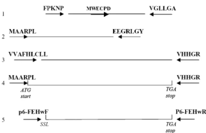
Total RNA was prepared from different wheat tissues, stored at ÿ80 8C, by using the RNeasy Plant Mini kit (Qiagen, Valencia, CA, USA). Degenerate primers were designed based on the Q-TOF MS/MS internal sequences: VGLLGA, FPKNP, and the conserved amino acid sequence WECPD from cell wall-type invertases. All primers used in the cloning scheme (Fig. 1) are presented in Table 2. RNA, prepared from wheat inflorescences at the same stage as used for the purification of the protein, was transcribed with one step RT-PCR (Access RT-PCR system, Promega Madison, WI, USA) using the primers FPKNP and VGLLGA. Only a faint band with the expected Mr appeared and semi-nested PCR was performed with
HALFFOR (Step 1 in Fig. 1). The clear resulting band was cut out, purified with the Qiaquick gel extraction kit (Qiagen, Valencia, CA, USA) and ligated in the TOPO-TA vector and transformed into E. coli (TOPO-TA cloning kit, Invitrogen, Groningen, The Netherlands). Plasmids from a number of clones were extracted using the Qiaprep spin miniprep kit (Qiagen, Valencia, CA, USA). After sequencing, a new specific primer was designed based on the obtained sequence EEGRLGY and combined with a primer MAARPL at the 59 end in step 2 (Fig. 1). This primer was distilled from ESTs from Triticum (accession number BJ211844 and CD932035) which coded for Q-TOF MS/MS identified internal peptides. The following PCR pro-cedure was used: first single-strand cDNA was synthesized, using the AMV reverse transcriptase (10 u llÿ1) (Promega, Madison, WI, USA). The cDNA was used as a template for the PCR reaction with the forward and reverse primer in the PCR reaction. The ‘Expand long template PCR system’ kit (Boehringer Mannheim, Mannheim, Germany) with supplements of MgCl2and parameters were followed
as prescribed by the suppliers. The clear PCR band was cut out of the gel and purified using the ‘geneclean SPIN kit’ (Q-Biogene, USA) and ligated into pGEMT-vector (Promega, Madison, USA) and transformed into E. coli. Plasmids from a number of clones were extracted, using the E.Z.N.A plasmid miniprep kit (Peqlab, Germany) and fully sequenced.
This obtained sequence was satisfactory since it overlapped perfectly with the previously obtained partial sequence and the identified internal peptides were present. To clone the 39 end in step 3 a primer at the stop codon was designed based on homologous ESTs from Triticum BJ243363 and CN010968. This primer was combined with the primer VVAFHCLL in a one step RT-PCR. By overlapping all the sequences of the previous PCR products, the full sequence could be predicted. As a control, a one step RT-PCR was performed with the primers at the start codon MAARPL and at the stopcodon VVHGR (step 4). After ligation into the TOPO-XL (Topo–XL cloning kit, Invitrogen) vector and transformation into E. coli, the plasmids were fully sequenced. During the whole cloning strategy PCR-products were analysed by electrophoresis in a 1% TAE gel and stained with ethidium bromide. The sequence was deposited in the EMBL sequence library (accession no. AM075205)
The cDNA was analysed using the software tools available on the internet (www.expasy.com). Alignment of the sequences was performed using Clustal X (version 1.80) and subsequent cladistic analysis was carried out without an outgroup in PAUP Version 4.0b10. A heuristic search strategy was used to find minimum-length trees: searches were conducted with 10 000 random-addition replicates (tree bisection–reconnection (TBR), MulTrees on). The level of support for individual clades was determined by a bootstrap analysis (NPB) (Felsenstein, 1985). The bootstrap as implemented by PAUP was carried out with 1000 pseudoreplicates and 10 ran-dom addition sequence repetitions.
Expression in Pichia
To construct the expression plasmid p6FEHW containing the mature protein, a primer with a restriction site for EcoRI was designed for the N-terminus as predicted by comparison with other homologous FEHs and the SignalP predict protein program (Bendtsen et al., 2004). A primer coding for the stop codon was designed for the C-terminus and contained the XbaI restriction site (Table 2). The cDNA was obtained after a two-step high fidelity RT-PCR (Prostar ultra high fidelity RT-PCR system, Stratagene, Ca, USA) (step 5 in Fig. 1). Two independent RT-PCR reactions were performed using the follow-ing procedure: the RNA was first heated for 5 min at 65 8C. The
Fig. 1. Cloning scheme (step 1–4) of the wheat 6-FEH cDNA. Step 5: cloning of the cDNA that codes for the mature protein which was expressed in Pichia pastoris. Primers are not drawn to scale; the tip of the arrowhead indicates the position of the end of the primers.
Table 1. Comparison of the substrate specificities (1 mM) of the native wheat 6-FEH and the heterologously expressed wheat 6-FEH cDNA in Pichia pastoris
Results are shown as values relative to the activity with bacterial levan (B) as substrate. The value for 100% corresponds with 13 650 nmol mgÿ1 protein minÿ1for the native protein and 26 600 nmol mgÿ1protein minÿ1 for the Pichia-derived protein. Inulin from Cichorium intybus L. (C.i.) was used. Experiments were carried out three times and the SE was always <5% of the mean.
Substrate Native activity (%) Heterologous activity (%) Levan (B) b2-6 DP672 100 100 6-kestose b2-6 DP 3 101 85 n-kestose b2-6 DP 3 39 45 Phlein b2-6 DP 4–12 95 120 Levanbiose b2-6 DP 2 63 57 1-kestose b2-1 DP 3 1 1 Nystose b2-1 DP 4 1 1 Inulin (C.i.) b2-1 DP >10 1 1 Sucrose DP 2 1 1 Levan (B) + 5–200 mM suc 100 100
mastermix was added and the RT reaction proceeded for 30 min at 48 8C. By placing the mix on ice, the RT reaction was stopped and 2 ll of cDNA was used for the subsequent PCR step. For the first five cycles, an annealing temperature of 58 8C and an extension time of 90 s were used. For the next 30 cycles an annealing temperature of 68 8C was programmed. The bands of about 1700 bp were cut out the gel and purified with Qiaquick gel extraction kit. These purified PCR-products and the PicZaA vector (Invitrogen, Groningen, The Netherlands) were digested with EcoRI and XBaI. Again, these mixtures were purified with the Qiaquick PCR purification kit (Qiagen, Valencia, Ca, USA). After dephosphorylation of the digested PicZaA the cDNA was ligated into this vector, resulting in the expression plasmid p6FEHW with the 6-FEH coding sequence in frame behind the a-signal. This plasmid was transformed into E. coli competent cells. As a control an empty PicZaA vector was also transformed into E. coli. Cells were plated on a 23Yeast Tryptone medium, supplemented with 30lg mlÿ1 zeocine as a selection marker. Positive colonies were used for vector amplification. Inserts were sequenced after cloning and contained the desired sequence. Pichia pastoris strain X33 was subsequently transformed using electroporation at 1500 V (Genepulser, BIORAD, Ca, USA) with both 20 lg of PmeI linearized p6FEHW and empty PicZaA.
Expression of the recombinant proteins in P. pastoris was performed as described in Hochstrasser et al. (1998) and Altenbach et al. (2004) using 50 mM NaAc, pH 5 buffer for the desalting of the column (Fast Desalting Column, Pharmacia, Upsala, Sweden). The expressed proteins were analysed by SDS-PAGE and activity was measured as described above.
Analysis of gene expression (quantitative RT-PCR)
Primers were designed to amplify the specific 6-FEH, 1-FEHw2, and ubiquitin transcripts (Table 2). DNase-treated RNA from several wheat tissues was reverse transcribed, using the AMV reverse transcriptase (10 U llÿ1) (Promega, Madison, WI, USA). The cDNA was used as a template for the real-time PCR reaction with a Gene Amp 5700 Sequence Detection System (Applied Biosystems, CA, USA). The following thermal profile, was programmed: 1 cycle 50 8C for 2 min, 1 cycle 95 8C for 10 min, and 40 cycles 15 s at 95 8C and 75 s at 60 8C. The gene-specific forward and reverse pri-mers and a 1:3 dilution of the cDNA were added to the SYBR Green PCR master mix (Applied Biosystems, CA, USA). Ubiquitin transcript levels in the different samples were used to normalize the amounts of 6-FEH and 1-FEHw2.
Results
Total FEH activity
Total 1-FEH and total 6-FEH activity was measured in
different tissues of wheat (Fig. 2). The 1 and 6-FEH
activities are roughly comparable. Most 1-FEH and 6-FEH
activities were measured in the stem of adult wheat plants.
FEH activity is highest in the penultimate internode. Roots
of adult plants contained some 6-FEH and 1-FEH activity.
In the inflorescences FEH activity was lower, but still
largely sufficient to explain the rate of in vivo degradation
from 470 to 70 lmol fructose equivalents g
ÿ1FW in 28 d
(M Lambaerts, unpublished result).
Purification of 6-FEH
After several attempts to purify a 6-FEH from stems, it was
not possible to separate it from 1-FEH and invertase
contaminants. Therefore, wheat inflorescences (spikes) at
the ‘soft dough’ stage were used as a source of enzymes for
the purification of 6-FEH. The latter were easily obtainable
and contained sufficient 6-FEH activity that could be
separated completely from contaminating 1-FEH and most
sucrose hydrolysing activity using the purification
proced-ure as described. Several peaks with 6-FEH activity were
eluted with a linear NaCl gradient during ion exchange
chromatography on monoQ. However, low 6-FEH
ac-tivity combined with a high invertase acac-tivity hindered
further complete purification. By contrast with most of
the 1-FEH activity, which was retained by the column,
the majority of the 6-FEH did not bind to the monoQ
column at this pH. Further purification on monoS and
Superdex columns resulted in a pure fraction containing
only 6-FEH activity. This b2-6 fructan hydrolysing
frac-tion was precipitated with acetone and loaded on a
SDS-PAGE. Wheat 6-FEH appears to be a glycoprotein of
about 70 kDa as estimated by SDS-PAGE (Fig. 3). Three
Table 2. Primers used for cDNA cloning, Pichia expression and RT-PCR of the 6-FEH from wheatRestriction sites are indicated in bold. In the degenerate primers the following symbols are used: N=(A, C, G or T), Y=(C, T).
Primer Sequence FPKNP 59-TTCCCNAAGAACCCNGCC-39 VGLLGA 59-CGCTCCNAGYAAGCCNACAAC-39 WECPD 59 GAATGTGGGARTGYCCNGA-39 MAARPL 59-CCATGGCCGCCAGACTCCCTCTC-39 EEGRLGYA 59-CGCGTAGCCAAGTCGCCCCTCCTC-39 VVAFHLCLL 59-GYCGTCGCGTTCCACCTCTGCCTYCTC-39 VVHGR 59-ATCACCTGCCATGGACAAC-39 p6-FEHwF 59-AATCCCGAATTCAGCTCGCTAGTCCGCCATG-39 p6-FEHwR 59-GAACGGTCTAGATCACCTGCCATGCACAACG-39 6-FEHWF 59-GTGGCAGAGCCTGGTACA-39 6-FEHWR 59-TTGCATAGTAGTCCTGGGTCA-39 1-FEHW2F 59-CCGCGTTAGTACGGGATA-39 1-FEHW2R 59-GCCTGATGTTGATCTATGTCG-39 UBIWF 59-CCTTCACTTGGTTCTCCGTCT-39 UBIWR 59-AACGACCAGGACGACAGACACA-39
specific peptides were identified after Q-TOF analysis
in a trypsinized SDS-PAGE band: EVQVQNVAFPKNP,
YDDVADTFVPEVDVER, and VVGLLGAQVNAGGVNK.
Hydrolytic properties
No products other than fructose could be detected after
incubations of the pure enzyme with low molecular weight
bacterial levan (results not shown). Of all fructans tested,
the purified enzyme most efficiently hydrolysed b2-6
linkages (Table 1). Bacterial levan, DP 4-12 phlein,
6-kestose and levanbiose are the best substrates, while b2-l
type fructans (inulin, 1-kestose, and 1,1-nystose) and
sucrose are poor substrates or are not utilized at all.
Significant activity was also detected with neokestose.
In-cubations of the enzyme with levan in combination with
different sucrose concentrations did not result in a decrease
of 6-FEH activity (Table 1).
Cloning strategy
The low activity of this 6-FEH suggested a low
concentra-tion of the corresponding mRNA. The use of a small
dilution (1:3) of total RNA (concentrated 700 lg ml
ÿ1)
from the wheat spikes in larger reaction volumes (50 ll)
improved the PCR reaction. Spikes from the same stage
were used as for the purification of the enzyme. Partial
sequences were obtained using the degenerate primers
based on the internal peptides and homologous sequences
from ESTs. These sequences showed overlapping stretches.
By combining all overlapping parts the full-length
se-quence could be predicted. A PCR with primers at the
start and stop codon produced the cDNA of the propeptide
and confirmed the complete sequence. Attempts to clone
the 39 end containing the poly-A tail failed.
The 6-FEH cDNA contained one open reading frame of
590 amino acids, starting at the ATG start codon and
end-ing at nucleotide 1773 before the TGA stop codon (Fig. 4).
Five potential glycosylation sites (NXS/T) could be
de-tected. The conserved b-fructosidase motif NDPNG could
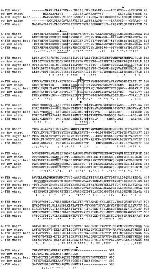
Fig. 3. SDS-PAGE of the native (N) and the Pichia-expressed (H) wheat 6-FEH. Lane 1: molecular mass marker proteins with their masses indicated at the left. The 6-FEH has a predicted mass of 70 kDa (arrow). Fig. 2. Total 6-FEH activity (black bars) and total 1-FEH activity (white bars) after incubation with 10 mM bacterial levan and 10 mM inulin, respectively. Data are means of three replicates. The protein content gÿ1FW was 0.877, 0.987, and 0.629 lg for the inflorescence in heading stage, booting stage, and milky stage, respectively; 0.351 lg for the seedling roots; 0.896 lg for the seedling leaves; 0.0336 lg for the adult roots; 0.0186 lg for the peduncle part of the stem, and 0.0150 lg for the penultimate part of the stem.Fig. 4. Alignment of the deduced amino acid sequence of 6-FEH, 1-FEH, and invertase from wheat and cell wall invertase 4 from maize and the sugar beet 6-FEH. The tryptic fragments found after Q-TOF MS/MS analysis are presented in bold. The potential glycosylation sites are underlined. The start and end of the expressed protein is indicated with an arrow. The b-fructosidase motifs and the cysteine catalytic sites are boxed. The three carboxylic acids that are thought to be crucial for enzyme activity are indicated with a black square. Asterisks indicate identical residues, colons indicate conserved substitutions, and periods indicate semi-conserved substitutions.
be distinguished and also the cysteine catalytic motif
(MWECPD) is present with a proline as 5th residue. The
two carboxylic acids that are thought to be important
for enzyme activity are present in these regions, together
with the third important carboxyl acid in the FRDP region
(Fig. 4) (Verhaest et al., 2004). Its cDNA-derived pI and
molecular mass is 6.5 and 65 kDa, respectively. Since the
cDNA-derived pI does not correspond with the
chromato-graphic behaviour of the enzyme, it is assumed that this is
a very highly homologous isoform with a different pI. A
theoretical tryptic digest of the protein sequence contained
the three peptides that were obtained by Q-TOF mass
spectrometry after purification and tryptic digest of the
native protein.
Homology with other glycosyl hydrolases
Similar to other plant FEHs, the 6-FEH described here is
related to cell wall invertases (CWINV Triticum aestivum
52%, CWINV Hordeum vulgare 51%, CWINV T.
mono-coccum 67% similarity). Similarity to vacuolar invertases
and fructan synthesizing enzymes from grass species is less
(42–46% similarity). Surprisingly, the similarity with
1-FEH from wheat is lower (52%) than with cell wall
in-vertases from non-fructan plants (Incw4 Zea mays 68%,
apoplastic invertase Oryza sativa 69% similarity).
Relatedness with other 6-FEHs from non-fructan plants
are: sugar beet 6-FEH 46% and Arabidopsis thaliana
CWINV3 50%. Similarities with microbial fructan
hydro-lases are much lower (below 25%).
The deduced amino acid sequence from the wheat
propeptide 6-FEH is aligned in Fig. 4 with related
trans-lated cDNAs from an apoplastic invertase and 1-FEH from
wheat and CWINV from maize. As a comparison, the
simi-larities with a translated 6-FEH cDNA from a non-fructan
plant are also presented. All four sequences contain the
cell wall like b-fructosidase motif and the catalytic site
(Tymowska-Lalanne and Kreis, 1998).
A phylogenetic bootstrap tree of some plant cell wall
type fructosyl transferases and hydrolases is presented in
Fig. 5. Tree bootstrap percentages (>50%) are indicated
Fig. 5. Phylogenetic bootstrap tree of the cell wall-like invertase translated cDNAs. The bootstrap percentages (>50%) are indicated next to the branches (1000 replicates). Accession numbers are between brackets. cDNAs from which the functionality is proven by purification of the enzyme or heterologous expression are indicated in italics. The wheat 6-FEH is indicated in bold.
next to the branches and highly support the obtained tree.
The 6-FEH under investigation cluster together in the same
clade with CW invertases from rice and Incw4 from maize.
The cell wall invertases from wheat and other invertases
from maize and rice, are more distantly related.
Heterologous expression in Pichia
The heterologously expressed 6-FEH was very active and
stable. A 1000-fold dilution of the Pichia pastoris
super-natant was necessary to measure activities in a linear range.
SDS-PAGE resulted in a similar band as the native protein
(Fig. 3). The heterologously expressed protein showed
essentially the same characteristics as the native protein
(Table 1). It hydrolysed b2-6 linkages exclusively.
Negli-gible activity was measured against inulin-type fructans
and sucrose. There was no sucrose inhibition. Incubation
of the Pichia supernatant, expressing the empty PicZaA
vector, resulted in no detectable products with all substrates
tested (results not shown).
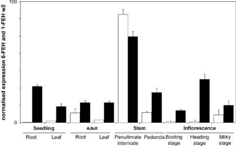
Expression pattern
The expression of the mRNA that codes for the 6-FEH was
measured, using real-time PCR. As a comparison the
mRNA from 1-FEHw2 was also analysed. The 6-FEH is
expressed in several tissues and developmental stages
such as leaves and roots from young as well as older
stems and roots (Fig. 6). As with 1-FEH, expression is
maximal in stem tissue, especially the penultimate
inter-node. 6-FEH transcripts are also found in the
inflores-cences of adult plants. Minor expression of 1-FEH was
found in inflorescences after anthesis (‘milky stage’) and
adult roots. Other tissues gave almost no signal.
Discussion
Enzymatic properties
In this study a 6-FEH from wheat was identified. The
presence of 6-FEHs in grasses has been reported (see
Introduction), but this is the first 6-FEH from a fructan
plant that has been characterized at the molecular level.
The 6-FEH protein was purified to homogeneity as
confirmed by SDS-PAGE and Q-TOF mass spectrometry.
It is a glycoprotein and hydrolyses exclusively b2-6
fructosyl linkages. Like all plant FEHs characterized so
far, wheat 6-FEH is an exohydrolase using a multi-chain
mechanism of hydrolysis since only fructose and no glucose
or short levans appear as long as the substrate does not
become limiting. By contrast, the previously described
6-FEHs from Lolium perenne (Marx et al., 1997) and Avena
sativa (Henson and Livingston, 1996), also demonstrated
some 1-FEH activity. It cannot be excluded that these
reported b2-1 fructosidase activities are not an intrinsic
property of the enzymes, but results from contamination by
a 1-FEH. Indeed, most previously reported 6- as well as
1-FEH enzymes have comparable molecular weights
be-tween 68 and 70 kDa and will probably not be separated
by SDS-PAGE (Bonnett and Simpson, 1993; Marx et al.,
1997; Claessens et al., 1990; Van den Ende et al., 2003a).
The present enzyme is not active against sucrose,
demonstrating that it is a genuine 6-FEH (EC 3.2.1.154)
and not a b-fructosidase. Contrary to most FEHs described
so far (Yamamoto and Mino, 1985; Simpson et al., 1991;
Prud’homme et al., 1992; Bonnett and Simpson, 1993,
1995; Marx et al., 1997; Van den Ende et al., 2000, 2003a),
but comparable to a 6-FEH from sugar beet (Van den
Ende et al., 2003b) and a 1-FEH from chicory (Claessens
et al., 1990) this 6-FEH is not inhibited by sucrose.
Sequence homology
According to some conserved motifs and similarities in the
sequences of FEHs and cell wall-like invertases it was
proposed that FEHs are derived from apoplastic invertases
rather than vacuolar invertases. Although the vacuolar
invertases have been proposed to be the putative precursors
of fructan synthesizing enzymes (Van den Ende et al., 2002),
phylogenetic analysis reveals that the 6-FEH clusters with the
cell wall invertases from maize (Incw4) and rice (O. sativa
INV1), two non-fructan plants. The cysteine catalytic motif
with a proline as the fifth residue, as described in most cell
wall-like enzymes (Tymowska-Lalanne and Kreis, 1998),
occurs in the three sequences. However, the tryptophan in
this same motif is replaced by a valine. The clade containing
the FEHs is more distantly related to the genuine cell wall
invertases from the same species. In view of the recent
report of 6-FEHs in non-fructan plants, it would be very
inter-esting to check functionality of these highly related cell
wall invertases from the non-fructan plants Oryza sativa
and Zea mays by means other than homology, for example,
heterologous expression in Pichia. Based on amino acid
sequences it is still impossible to predict whether a putative
fructosidase is a specific fructan exohydrolase, a specific
invertase, or a general fructosidase.
Expression pattern
During grain-filling, fructans in heterotrophic parts of the
stem are depolymerized by the rise of fructan
exohydrolys-ing activitiy (Gebbexohydrolys-ing, 2003; Schnyder, 1993; Willenbrink
et al., 1998). Also other source–sink modifications, such as
cutting or grazing, induce FEH activity (Prud’homme et al.,
1992). Following these reports, FEH is anticipated to be
expressed in the stem of adult plants. The expression of
1-FEH w2 corresponds well with the above-mentioned
reports. On the other hand, the 6-FEH is expressed in stems
and also in photosynthetically active tissues like leaves
of one-week-old seedlings and adult plants. Very young
tissues that are not expected to accumulate significant
amounts of fructans, like the roots of one-week-old
seed-lings or very young inflorescences before anthesis,
never-theless contain 6-FEH mRNA. These expression patterns,
suggest that this 6-FEH might fulfil another function in
addition to the breakdown of b2-6 linkages during the
grain-filling period at the end of the growing season.
The measured FEH activities do not correspond with the
transcript levels. Although, some caution is needed in
comparing these results. The activities presented comprise
total FEH activity, which is a mixture of the different
1-FEH isoforms that are present in wheat (Van den Ende
et al., 2003a). The total 6-FEH activity includes different
6-FEH isoforms which are reported in the different
elution peaks during the purification procedure. The several
6-FEH isoforms can be present in different tissues and may
fulfil different functions or different cellular localizations.
Since neither the cellular nor the histological localization of
the 6-FEHs in cereals has been demonstrated convincingly
up to now, it is difficult to relate fructan dynamics with
gene expression and enzymatic activity at the organ level.
General roles and localization of 6-FEHs in fructan and non-fructan plantsThe rather ubiquitous expression of 6-FEH and the fact
that it is not inhibited by sucrose does not support a specific
role for this enzyme in reserve mobilization. Although it
is generally accepted that fructans and fructan
metabol-izing enzymes occur in the vacuole (Wagner et al., 1983;
Wiemken et al., 1986), fructans and fructan-degrading
activity outside the vacuole have been reported (Livingston
and Henson, 1998; Wang and Nobel, 1998). The high
homology of this 6-FEH with rice invertase (Hirose et al.,
2002) and maize CwInv4 (78%), an unbound apoplastic
invertase with a low pI (Kim et al., 2000) and the fact that it
is not inhibited by sucrose, which is present in high
concentrations in the vacuole, might point to an apoplastic
localization. In this view this enzyme can be compared with
the 6-FEHs described in sugar beet and Arabidopsis (Van
den Ende et al., 2003b; De Coninck et al., 2005). Functions
ascribed to FEHs in grass-type species till now are: (i)
hydrolysing the fructan pools whenever energy is needed
(Prud’homme et al., 1992; Morvand-Betrand et al., 2001;
Simpson et al., 1991; Willenbrink et al., 1998), (ii)
hydro-lysing fructans to stimulate frost tolerance by membrane
stabilization (Demel et al., 1998; Hincha et al., 2002;
Vereyken et al., 2003), and (iii) prevention of b2-1 linked
chain formation by selectively trimming of some substrates
(graminans) (Bancal et al., 1992).
Since the discovery of 6-FEHs in non-fructan plants,
new functions for these FEHs (which are also not inhibited
by sucrose) were suggested (Van den Ende et al., 2003b;
De Coninck et al., 2005). Assuming these 6-FEHs are
localized in the apoplast, (i) they might fulfil a
defence-related function. The 6-FEH activity could prevent the
formation of exogenous levans, formed by bacterial plant
pathogens. In this way they might prevent bacterial diseases
or diminish the toxicity of the levans (Cairns, 2003) formed
by the intruding bacteria. (ii) Since fructans in oat (Avena
sativa) are reported to be situated in the apoplast after
treatment at
ÿ3 8C (Livingston and Henson, 1998), the
latter FEHs could depolymerize these fructans in order to
stabilize the membrane during frost and other stresses (see
above) or remove them after stress release.
Conclusion
In this study a 6-FEH from wheat was purified and cloned.
Since this is the first 6-FEH cDNA from a fructan plant, the
precise function remains elusive. Therefore, it would be
interesting to identify and clone more 6-FEHs from wheat
and other fructan plants and determine their localization
in cells and tissues. Some 6-FEHs are inhibited by sucrose
and others are not. It is not unlikely that there are two types
of 6-FEHs in fructan plants, with different function and
localization. Studies involving immunocytochemical
local-ization might elucidate the apoplastic and/or vacuolar
nature of these enzymes. If the 6-FEH proves to be
im-portant for the breakdown of fructans in the developing
grains, the tissue from which it was isolated, it might be
very useful in transgenic approaches to increase the content
of health-promoting fructans in wheat kernels, flour and
bread, one of the worlds important staple foods.
Acknowledgements
We would like to thank Peter Schols for the help offered during the phylogenetic analysis and Rudy Vergauwen for technical assistance. Thanks are due to James Tosh for critical reading and valuable suggestions on the first draft. This work was supported by grants of the Research Council of the K.U. Leuven (OT/01/26) and the Fund for Scientific Research-Flanders (Belgium) (FWO-Vlaanderen) (G0177.02).
References
Altenbach D, Nu¨ esch E, Meyer AD, Boller T, Wiemken A. 2004. The large subunit determines catalytic specificity of barley sucrose:fructan 6-fructosyltransferase and fescue sucrose:sucrose 1-fructosyltransferase. FEBS Letters 567, 214–218.
Bancal P, Carpita NC, Gaudille`re JP.1992. Differences in fructan accumulated in induced and field-grown wheat plants: an elonga-tion-trimming pathway for their synthesis. New Phytologist 120, 313–321.
Bendtsen JD, Nielsen H, von Heijne G, Brunak S.2004. Improved prediction of signal peptides: SignalP 3.0. Journal of Molecular Biology 340, 783–795.
Bonnett GD, Incoll LD.1993. Effects on the stem of winter barley of manipulating the source and sink during grain-filling. 1. Changes in accumulation and loss of mass from internodes. Journal of Experimental Botany 44, 75–82.
Bonnett GD, Simpson RJ. 1993. Fructan-hydrolyzing activities from Lolium rigidum Gaudin. New Phytologist 123, 443–451. Bonnett GD, Simpson RJ.1995. Fructan exohydrolase activities
from Lolium rigidum that hydrolyse b-2,1- and b-2,6-glycosidic linkages at different rates. New Phytologist 131, 199–209. Bonnett GD, Sims IM, Simpson RJ, Cairns AJ.1997. Structural
diversity of fructan in relation to the taxonomy of the Poaceae. New Phytologist 136, 11–17.
Bonnett GD, Sims IM, St John JA, Simpson RJ.1994. Purification and characterization of fructans with b-2,1 glycosidic linkages and b-2,6 glycosidic linkages suitable for enzyme studies. New Phytologist 127, 261–269.
Cairns AJ.2003. Fructan biosynthesis in transgenic plants. Journal of Experimental Botany 54, 549–567.
Cairns AJ, Nash R, Machado De Carvalho M, Sims IM.1999. Characterization of the enzymatic polymerization of 2,6-linked fructan by leaf extracts from timothy grass (Phleum pratense). New Phytologist 142, 79–91.
Claessens G, Van Laere A, De Proft M. 1990. Purification and properties of an inulinase from chicory roots (Cichorium intybus L.). Journal of Plant Physiology 136, 35–39.
De Coninck B, Le Roy K, Francis I, Clerens S, Vergauwen R, Halliday AM, Smith S, Van Laere A, Van den Ende W.2005. Arabidopsis AtcwINV3 and 6 are not invertases but are fructan
exohydrolases (FEHs) with different substrate specificities. Plant, Cell and Environment 28, 432–443.
Demel RA, Dorrepaal E, Ebskamp MJM, Smeekens JCM, de Kruijff B. 1998. Fructans interact strongly with model membranes. Biochimica et Biophysica Acta–Biomembranes 1375,36–42.
Felsenstein J.1985. Confidence limits on phylogenies: an approach using the bootstrap. Evolution 39, 783–791.
Gebbing T.2003. The enclosed and exposed part of the peduncle of wheat (Triticum aestivum): spatial separation of fructan storage. New Phytologist 159, 245–252.
Hendry G. 1993. Evolutionary origins and natural functions of fructans. A climatological, biogeographic and mechanistic ap-praisal. New Phytologist 123, 3–14.
Henson CA.1989. Purification and properties of barley stem fructan exohydrolase. Journal of Plant Physiology 134, 186–191. Henson CA, Livingston DP.1996. Purification and characterization
of an oat fructan exohydrolase that preferentially hydrolyzes b-2,6 fructans. Plant Physiology 110, 639–644.
Hincha DK, Zuther E, Hellwege EM, Heyer AG.2002. Specific effects of fructo-and gluco-oligosaccharides in the preservation of liposomes during drying. Glycobiology 12, 103–110.
Hirose T, Takano M, Terao T. 2002. Cell wall invertases in developing rice caryopsis: molecular cloning of OsCIN1 and analysis of its expression in relation to its role in grain filling. Plant Cell Physiology 43, 452–459.
Hochstrasser U, Lu¨scher M, De Virgilio C, Boller T, Wiemken A. 1998. Expression of a functional sucrose-fructan 6-fructosyltrans-ferase in the methylotrophic yeast Pichia pastoris. FEBS Letters 440,356–360.
Iizuka M, Yamaguchi H, Ono S, Minamiura N.1993. Production and isolation of levan by use of levansucrase immobilized on the ceramic support SM-10. Bioscience, Biotechnology and Bio-chemistry 57, 322–324.
Kim JY, Mahe´ A, Guy S, Brangeon J, Roche O, Chourey PS, Prioul JL.2000. Characterization of two members of the maize gene family, Incw3 and Incw4, encoding cell-wall invertases. Gene 245,89–102.
Livingston DP, Henson CA. 1998. Apoplastic sugars, fructans, fructanexohydrolase, and invertase in winter oat: responses to second-phase cold hardening. Plant Physiology 116, 403–408. Marx SP, No¨sberger J, Frehner M. 1997. Hydrolysis of fructan
in grasses: A b-(2,6)-linkage specific fructan-b-fructosidase from stubble of Lolium perenne. New Phytologist 135, 279–290. Morvand-Betrand A, Boucaud J, Le Saos J, Prud’homme MP.
2001. Roles of the fructans from the elongating leaf bases in the regrowth following defoliation of Lolium perenne L. Planta 213, 109–120.
Nagaraj VJ, Galati V, Lu¨scher M, Boller T, Wiemken A.2005. Cloning and functional characterization of a cDNA encoding barley soluble acid invertase (Hv INV1). Plant Science 168, 249–258.
Prud’homme MP, Gonzalez B, Billard JP, Boucaud J. 1992. Carbohydrate content, fructan and sucrose enzyme activities in roots, stubble and leaves of ryegrass (Lolium perenne L.) as affected by source/sink modification after cutting. Journal of Plant Physiology 40, 282–291.
Ritsema T, Smeekens S.2003. Fructans: beneficial for plants and humans. Current Opinion in Plant Biology 6, 223–230.
Schnyder H. 1993. The role of carbohydrate strorage and re-distribution in the source-sink relations of wheat and barley during grain-filling: a review. New Phytologist 123, 233–245.
Sedmak JJ, Grossberg SE.1977. A rapid, sensitive and versatile assay for protein using Coomassie Brilliant Blue G250. Analytical Biochemistry 79, 544–552.
Simpson RJ, Walker RP, Pollock CJ.1991. Fructan exohydrolase activity in leaves of Lolium temulentum. New Phytologist 199, 499–507.
Timmermans JW, Slaghek T, Iizuka M, De Roover J, Van Laere A, Van den Ende W.2001. Isolation and structural analysis of new fructans produced by chicory. Journal of Carbohydrate Chemistry 20, 375–395.
Tymowska-Lalanne Z, Kreis M.1998. Expression of the Arabi-dopsis thaliana invertase gene family. Planta 207, 259–265. Van den Ende W, Clerens S, Vergauwen R, Van Riet L, Van
Laere A, Yoshida M, Kawakami A. 2003a. Fructan 1-exohydrolases: b (2,1) trimmers during graminan biosynthesis in stems of wheat? Purification, characterization, mass mapping and cloning of two fructan 1-exohydrolase isoforms. Plant Phy-siology 131, 621–631.
Van den Ende W, De Coninck B, Clerens S, Vergauwen R, Van Laere A.2003b. Unexpected presence of fructan 6-exohydrolases (6-FEHs) in non-fructan plants: characterization, cloning, mass mapping and functional analysis of a novel ‘cell-wall invertase-like’ specific 6-FEH from sugar beet (Beta vulgaris L.). The Plant Journal 36, 697–710.
Van den Ende W, Michiels A, De Roover J, Verhaert P, Van Laere A.2000. Cloning and functional analysis of chicory root fructan 1-exohydrolase I (1-FEH): a vacuolar enzyme derived from a cell-wall invertase ancestor? Mass fingerprint of the 1-FEH I enzyme. The Plant Journal 24, 447–456.
Van den Ende W, Michiels A, De Roover J, Van Laere A.2002. Fructan biosynthetic and breakdown enzymes in dicots evolved from different invertases: expression of fructan genes through-out chicory development. The Scientifical World Journal 2, 1273–1287.
Van den Ende W, Van Laere A.1996. Fructan synthesizing and degrading activities in chicory roots (Cichorium intybus L.) during
growth, storage and forcing. Journal of Plant Physiology 149, 43–50.
Vereyken IJ, Chupin V, Islamov A, Kuklin A, Hincha DK, de Kruijff B. 2003. The effect of fructan on the phospholipid organization in the dry state. Biophysical Journal 85, 3058–3065. Verhaest M, Van den Ende W, Yoshida M, Le Roy K, Peeraer Y, Sansen S, De Ranter CA, Van Laere A, Rabijns A. 2004. Crystallization and preliminary X-ray diffraction study of fructan 1-exohydrolase IIa from Cichorium intybus. Acta Crystallo-graphica D 60, 553–554.
Vijn I, Smeekens SCM. 1999. Fructan: more than a reserve carbohydrate? Plant Physiology 120, 351–359.
Wagner W, Keller F, Wiemken A. 1983. Fructan metabolism in cereals: induction in leaves and compartmentation in proto-plasts and vacuoles. Zeitschrift fu¨r Pflanzenphysiologie Bd 112, 359–372.
Wagner W, Wiemken A. 1986. Properties and subcellular localization of fructan hydrolase in the leaves of barley (Hordeum vulgare L. cv. Gerbel). Journal of Plant Physiology 123, 429–439.
Wang N, Nobel PS. 1998. Phloem transport of fructans in the Crassulacean acid metabolism species Agave deserti. Plant Physiology 116, 709–714.
Wiemken A, Frehner M, Keller F, Wagner W. 1986. Fructan metabolism, enzymology and compartmentation. Current Topics in Plant Biochemistry and Physiology 5, 17–37.
Willenbrink J, Bonnett GD, Willenbrink S, Wardlaw IF.1998. Changes of enzyme activities associated with the mobilization of carbohydrate reserves (fructans) from the stem of wheat during the kernel filling. New Phytologist 139, 471–478.
Yamamoto S, Mino Y.1985. Partial purification and properties of phleinase induced in stem base of orchardgrass after defoliation. Plant Physiology 78, 591–595.