HAL Id: hal-00005904
https://hal.archives-ouvertes.fr/hal-00005904
Submitted on 11 Jul 2005HAL is a multi-disciplinary open access archive for the deposit and dissemination of sci-entific research documents, whether they are pub-lished or not. The documents may come from teaching and research institutions in France or abroad, or from public or private research centers.
L’archive ouverte pluridisciplinaire HAL, est destinée au dépôt et à la diffusion de documents scientifiques de niveau recherche, publiés ou non, émanant des établissements d’enseignement et de recherche français ou étrangers, des laboratoires publics ou privés.
Ral-GDS/Xral branch of the Ras signalling pathway.
Stéphanie Lebreton, Laurent Boissel, Jacques Moreau
To cite this version:
Stéphanie Lebreton, Laurent Boissel, Jacques Moreau. Control of embryonic Xenopus morphogenesis by a Ral-GDS/Xral branch of the Ras signalling pathway.. Journal of Cell Science, Company of Biologists, 2003, 116, pp.4651-4662. �10.1242/jcs.00763�. �hal-00005904�
Control of embryonic Xenopus morphogenesis by a Ral-GDS/XralB branch of the Ras signalling pathway.
Stéphanie Lebreton, Laurent Boissel and Jacques Moreau*
Institut Jacques Monod, CNRS, Universités Paris VI et Paris VII, Mécanismes Moléculaires du Développement. 2 Place Jussieu, 75251 Paris cedex 05, France.
To whom correspondence should be addressed
Submitted to: Journal of Cell Science
Running title: Study of the RalB pathway in Xenopus early development
Phone: (033) 01 44 27 82 91 Fax. : (033) 01 44 27 52 65 e-mail : [email protected]
Summary
Ras proteins mediate biological responses through various effectors and play a key role in relaying the Fibroblast Growth Factor (FGF) mesoderm induction signal during embryogenesis of the frog, Xenopus laevis. One Ras effector pathway involves the activation of the small G protein Ral. In the present study, we have investigated the role of key components in the RalB branch of FGF and Ras signalling during early Xenopus development. Treatment of animal caps with bFGF, which converts prospective ectoderm to mesoderm, activates XralB. The Ras 12V37G mutant, which can bind to Ral-GDS but not Raf, also activates XralB as well as causing developmental defects and cortical F-actin disassembly. A similar phenotype is induced by Ral-GDS itself. FGF induced expression of several signature mesodermal genes, by contrast, is independent of XralB signalling. This and other data suggest that the RalB branch of Ras and FGF signalling regulates the actin cytoskeleton and morphogenesis in a transcriptionally-independent manner. We also find XralB to be specifically activated in the marginal zone of Xenopus embryos, and find that disruption of the RalB pathway in this region causes gastrulation defects. We conclude that RalB signalling is autonomously required by mesodermal cells to effect essential morphogenetic changes during Xenopus gastrulation.
Introduction
Mesoderm induction depends on multiple diffusible extracellular factors that induce specific programs of gene expression and morphogenesis. Prior to overt gastrulation, a number of mesoderm-specific transcription factors are transcribed in induced mesoderm (for reviews (Stennard et al., 1997) and (Chan and Etkin, 2001)). Shortly thereafter, mesodermal cells participate in stereotyped gastrulation movements (for review (Gerhart and Keller, 1986)) that are achieved via coordinated changes in the cytoskeleton of individual cells (Selchow and Winklbauer, 1997; Wacker et al., 1998). Despite intensive study, the intracellular signalling pathways connecting mesoderm induction to the direct modification of the cytoskeleton by effector molecules remain unclear.
FGFs can induce the expression of many mesodermal markers (Kimelman and Kirschner, 1987), (Green et al., 1992), (Whitman and Melton, 1992) (LaBonne and Whitman, 1994), (Amaya et al., 1991), and expression of a dominant negative FGF receptor dramatically perturbs gastrulation (Amaya et al., 1991). Mesodermal gene induction by FGFs has been shown to be achieved via the Ras protein, acting through both the MAPK and PI3K pathways (Whitman and Melton, 1992), (LaBonne and Whitman, 1994), (Umbhauer et al., 1995), (Carballada et al., 2001). Ras is also known to modulate the formation of the actin cytoskeleton in a variety of cell types, but the specific Ras pathway involved is unknown (Bar-Sagi and Feramisco, 1986). A role for FGF signalling in Drosophila embryonic cell motility has been found, seen in the requirement for the FGF receptor heartless molecules for mesodermal cell migration (Gisselbrecht et al., 1996), (Beiman et al., 1996). More recently, FGF4 and FGF8 have been shown to control directional movement of streak cells in the chick (Yang et al., 2002). These data demonstrate that FGFs are able to control both gene expression and cellular behavior (for reviews see (Manske and Bade, 1994), (Montell, 1999), (Boilly et al., 2000)) and they raise the possibility that during mesoderm development, Ras
not only mediates the induction of gene expression by FGF signalling, but also controles the cellular behaviour.
As indicated above, the Ras protein initiates several cellular processes including proliferation, differentiation and modulation of the cytoskeleton (Reuter and Der, 2000). Although the Raf serine/threonine kinase is the best-studied Ras effector protein, at least two other effectors, PI3K (Rodriguez-Viciana et al., 1997) and Ral-GDS (Kikuchi et al., 1994), are known to mediate Ras signalling. Moreover, a Ras mutant with an inactive effector domain can be complement by another distinct Ras effector-domain mutant (White et al., 1995), demonstrating that different Ras effectors can cooperate to transform cells (Urano et al., 1996). Recently PI3K was shown to synergize with the extracellular signal regulated kinase (ERK) pathway in the induction of Xenopus mesoderm (Carballada et al., 2001). More recently, oncogenic activity was demonstrated for the Ras 12V37G mutant, which binds and activates Ral-GDS, but not Raf (Hamad et al., 2002). Much remains to be learned about the roles of Ras and its various effectors during early development . Of particular interest is whether or not Ras directly influences cellular morphology via modulation of the cytoskeleton.
In previous studies, we have characterised XralB, a small G protein of the Ras family, isolated by differential screening of a subtractive cDNA library (Moreau et al., 1999). This protein cycles between an inactive GDP-bound and an active GTP-bound conformation. Ral proteins are activated by the Ras effector, Ral-GDS (Kikuchi et al., 1994)(Wolthuis et al., 1998b) and have been directly implicated in various cellular mechanisms, such as endocytosis (Nakashima et al., 1999) and regulation of the actin cytoskeleton (Ohta et al., 1999). By microinjecting different mutant forms of XralB into Xenopus embryos, we have shown that expression of an inactivate form of XralB, XralB S28N, blocks gastrulation movements, whereas a constitutively active form, XralB G23V, causes disruption of the actin cytoskeleton (Moreau et al., 1999).
In this paper, we delineate a cascade linking FGF signalling to embryonic morphogenesis. We show that exposure of animal caps to bFGF causes activation of XralB, and that overexpression of an activated form of XralB causes cortical F-actin disassembly. We also show that early developmental arrest are caused by ectopic expression of a mutant form of Ras able to bind Ral-GDS, but not Raf, or by ectopic expression of Ral-GDS itself. By contrast, we find that FGF-induced expression of a number of signature mesodermal genes requires signal transduction by Raf, but not XralB. Finally, we find that XralB is preferentially activated in the marginal zone of Xenopus embryos, and that disruption of the XralB pathway is most effective when targeted to this region.
Materials and Methods
Protein extraction, electrophoretic analysis, immunoblotting and pull-down
To monitor the expression of injected mRNA, proteins were extracted from embryos and analysed as previously described (Gusse et al., 1989). Protein extracts were separated by SDS–PAGE on a 15% acrylamide gel and transferred to a Hybond P membrane (Amersham). The membrane was probed overnight at 4°C with anti–rat RalB goat antibodies (Sc1531, Santa Cruz) diluted 1/125 in 1X PBS – 1% BSA (wt/vol). The secondary antibody, peroxidase-linked rabbit anti-goat Ig was diluted 1/1250 (Chemicon). Signals were detected by chemiluminescence (ECL+, Amersham).
To analyse Ral-GTP levels, protein extracts from embryos were incubated with RalBP1 agarose (Upstate Biotechnology, ref. 14-415) for 30 min. at 4° C. Immunoprecipitates were separated on a 15% acrylamide gel and transferred to a P Hybond membrane (Amersham). Precipitated endogenous Ral was probed with an anti-RalA antibody (Upstate Biotechnology, ref. 20-189) diluted 1/750 in 1X PBS – 0.05% Tween -20 and 5% milk powder (wt/vol). Signals were detected by chemiluminescence (ECL, Amersham) and quantified by ImageQuant (Molecular Dynamics).
Embryos and microinjections
Xenopus frogs were imported from South Africa (South Africa Farms, Fish Hoek) or from the
CNRS frog colony (Rennes). Animals were housed and fed as described (Gurdon et al 1984). Embryos were fertilized in vitro and chemically dejellied with 2% (wt/vol) cysteine-HCl, pH 7.8/7.9, then maintained in 1 X MBS (Modified Barth’s solution pH (7.6) (Gurdon and Wickens, 1983) until microinjection. Microinjection was performed in a solution of 1 X MBS and 3% Ficoll (wt/vol), followed by overnight incubation at 16°C in 0.1 X MBS and 3% Ficoll, then at 15-22°C until they reached appropriate stages. Embryonic stages were
determined according to Nieuwkoop and Faber (Nieuwkoop and Faber, 1956). In vitro transcribed RNA (4.6 nl) was microinjected into 2-cell stage embryos in the animal hemisphere or into 4-cell stage embryos in the marginal zone, animal pole or vegetal pole.
cDNA cloning and in vitro transcription of RNA for injection
pSP64-Ras 12V35S and 12V37G were obtained by cloning the SalI-BamHI insert from pDCR-12V35S and 12V37G (White et al., 1996) into the SalI-BamHI sites of the pSP64 vector (Promega). pSP64-Ral-GDS was obtained by subcloning of the BamHI insert from pCEP4-Ral-GDS (White et al., 1996). pRN3-RalBD-GST was obtained by subcloning of the HindIII-SalI insert from pGEXT4T3-GST-RalBD (Wolthuis et al., 1998b). The 3’untranslated regions of the pRN3 RalB clones S28N, G23V were deleted by removal of BamHI/NotI, followed by end-filling and religation. mRNAs were transcribed from pSP64T (Promega) or pRN3 (Lemaire et al., 1995) vectors using the SP6 or T3 mMessage mMachine (Ambion).
Confocal microscopy
Animal caps were observed using a Leica TCS-4D confocal imaging system (Leica Instruments, Heidelberg, Germany) fitted with a 63X objective (NA 1.4). For FITC and TRITC, an argon–krypton ion laser was used set to 488 and 568 nm, respectively. For each optical section, double fluorescence images were acquired sequentially to avoid potential signal contamination by linkage-specific fluorescence emission “cross-talk.” Signal noise was reduced by line averaging of four frames. A focal series was collected for each specimen. The focus step between sections was 1µm.
Embryos were prepared for confocal microscopy analysis as described previously (Moreau et al., 1999).
Animal cap assay
Animal caps were manually dissected from MBT-stage embryos. These explants were incubated with or without FGF (100 ng/ml) in 1X MMR, 1% BSA for 4 hours at 16°C.
RNA isolation and reverse transcriptase -PCR assay
Total RNA extraction was carried out as previously described (Moreau et al 1999), except that extracts were treated with 1 U of DNase I RQ1 (Promega) for 30 minutes at 37° C after the LiCl precipitation step. First strand cDNA was prepared from total RNA using the reverse transcriptase M-MLV (Gibco-BRL, ref. 28025-013) and Oligo dT primer (Pharmacia). PCR and analysis of cDNA was performed in 50 µl of solution containing 5µl of cDNA, 1X Taq buffer, 3 mM MgCl2, 0.1mM (each) dNTPs, 200 ng of each specific primer, and 1 U of Taq polymerase (Invitrogene), using the following PCR conditions: 93° C for 3 min, followed by a variable number of cycles at 93° C for 45 s, 52° C for 50 s. and 72° C for 1 min. Ornithine decarboxylase (ODC), Xbra, Xwnt11, Xnot, Xty2 PCR amplification was carried out for a total of 27 cycles, and Xwnt8, Xsnail and Xcad3 amplification was for a total of 35 cycles. T h e p r i m e r s e q u e n c e s w e r e a s f o l l o w s : O D C f o r w a r d 5 ’ – GTCAATGATGGAGTGTATGGATC-3’, reverse 5’- TCCATTCCGCTCTCCTGAGCAC-3’; Xbra forward 5’ – GTGTAGTCTGTAGCAGCA-3’, reverse 5’-GGATCGTTATCACCTCTG- 3’; Xwnt 11 forward 5’ – GTGAGAGAGGTCTGAGCTGG-3’, reverse 5’- ACATGACATAGCAGCACC- 3’; Xnot forward 5’ – CAGAGCAGCTGGAGAAGCTG-3’, reverse 5’- CAGTGTGATCTGAGCTGTTC- 3’; Xty2 forward 5’ – ATCTGCTCCACGACGGAC-3’, reverse 5’- GAAGAACTGCTACTTGTCC-3’; Xcad3 forward 5’ –CAGCGCAGAACTACGTCTCC-3’, reverse
5’-CCCAGTCCCAGATGGATGTG- 3’; Xwnt8 forward 5’ –AGATGACGGCATTCCAGA-3’, r e v e r s e 5 ’ - T C T C C C G A T A T C T C A G G A - 3 ’ .
Results
XralB is activated by FGF
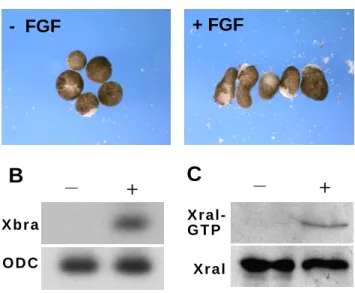
Activation of Ral proteins by growth factors, including those signalling through receptor tyrosine kinases (EGF, FGF, etc.), has been documented in a variety of contexts (Wolthuis et al., 1998a), (Suzuki et al., 2000) (Nakashima et al., 1999). Considering the role of FGFs in mesoderm induction, and our previous observations that XralB function is required for gastrulation, we wished to determine whether the FGF pathway regulates XralB. Animal cap explants were dissected at the blastula stage and cultured for 4 hours in the presence or absence of bFGF (Fig. 1A). After incubation, mRNA was extracted to verify the activation of the pan-mesodermal gene marker Xbra by bFGF (Fig. 1B). To determine the relative abundance of endogenous GTP-bound XralB, protein was also extracted and analysed by affinity purification (pull-down) using GST fused to the Ral binding domain of RalBP1 (GST-RalBP1) (Fig. 1C). Animal caps incubated in the presence of bFGF became elongated (Fig. 1A), and displayed an increase in Xbra transcript levels (Fig. 1B) and GTP-bound XralB (Fig. 1C). Thus, the Ral pathway can be activated by FGF signalling during early
Xenopus development.
Ras activates the XralB protein via Ral-GDS binding
Ras stimulates multiple, distinct signalling cascades. Indeed, White et al (White et al., 1995) have shown that a mutant form of Ras, Ras 12V37G, does not transform cells, but can be complemented for transformation by another Ras mutant, Ras 12V35S. Ras 12V35S binds specifically to Raf kinase but not to Ral-GDS, whereas Ras 12V37G only binds to Ral-GDS (White et al., 1996). We utilized these Ras mutants to assess the relative roles of Ras signalling via Raf and Ral-GDS in the activation of XralB in Xenopus embryos.
Whole embryos were injected with Ras 12V35S (0.5 ng/blastomere), or Ras 12V37G (0.5 ng/blastomere) RNA at the two-cell stage, grown until the 128/256-cell stage and protein was extracted and analysed by Ral-GTP pull-down. We found the level of
activated XralB to be specifically increased in embryos injected with Ras 12V37G (Fig. 2). Xral-GTP levels in embryos injected with Ras 12V35S were similar to the basal levels observed in uninjected embryos. To confirm the specificity of XralB activation by Ras 12V37G, we asked whether a dominant-negative form of RalB, XralB S28N, can interfere with XralB activation when co-injected with Ras 12V37G. In such embryos, levels of XralB-GTP are similar to those of control embryos (Fig. 2). These results indicate that Ras can activate XralB independently of Raf kinase.
Ras can activate two independent targets
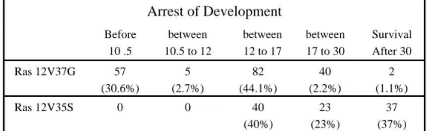
We also examined the effect of the Ras 12V35S and 12V37G mutants on development and morphogenesis. All embryos injected with Ras 12V35S (Fig. 3A and Table 1) appeared normal through the end of gastrulation and 37% survived to stage 32, though many displayed developmental defects such as a shorter axis or open neural folds. Embryos injected with Ras 12V37G displayed earlier defects. Development was normal through the midblastula transition (MBT), but then blastomeres near the injection area became necrotic, characterised by large bleached cells (Fig. 3A), and 30.6% of embryos (n = 186) underwent developmental arrest prior to gastrulation (stage 10.5) (Fig. 3A). More than 96.6% of Ras 12V37G-injected embryos failed to survive beyond neurulation (Table 1) and only 1.1% reached the tadpole stage. To investigate if Ras 12V37G had the same target as XralB, we examined whether the expression of this Ras mutant disrupted the integrity of the actin cytoskeleton, as does the constitutively active form of XralB, XralB G23V (Moreau et al., 1999), and we compared the effects of Ras 12V37G on the actin cytoskeleton with those of Ras 12V35S. Apical blastomeres from the animal hemisphere, before and after stage 8, were observed by confocal microscopy following F-actin staining with Rhodamin-Phalloidin. Actin cytoskeleton analysis in embryos injected with Ras 12V37G was carried out on embryos that did not display cell necrosis.
As expected, embryos injected with Ras 12V37G had severely disrupted cortical and nuclear actin cytoskeletons (Fig. 3B), whereas blastomeres of embryos injected with Ras 12V35S were similar to uninjected controls. To better characterise the timing of F-actin disruption, we performed a kinetic study (Fig. 4). We found that F-actin is intact until the 2000-cell stage (Fig. 4D) and becomes damaged near MBT (Fig. 4 F). This timing of Ras 12V37G-induced disruption differs from that of constitutively active XralB G23V, which induces disruptions as early as the 256-cell stage (corresponding to the technical limit of analysis). Nevertheless, the ability of either Ras 12V37G or XralB G23V to disrupt the actin cytoskeleton, together with the ability of Ras 12V37G to induce activation of XralB, suggests that Ras 12V37G induced disruption of the cytoskeleton is mediated by XralB. To confirm this apparent specificity, we co-injected the dominant-negative XralB S28N mutant with Ras 12V37S at a ratio of six mRNA molecules to one. Co-injection of XralB S28N rescued early morphological defects, as embryos had no necrotic cells (Fig. 3A) and survived longer. The development of these embryos was blocked during gastrulation, but this was expected, as gastrulation arrest results from injection of XralB S28N alone. We also observed partial rescue of the cortical actin array when XralB S28N was co-injected with Ras 12V37G (Fig. 3B). We conclude that Ras signalling via XralB and independent of Raf can cause alterations in the actin cytoskeleton as well as cell necrosis and early embryonic death. As Raf is known to be an essential mediator in the activation of several Xenopus mesodermal genes by Ras (McNicol et al., 1993), our results indicate that Ras/Raf/MAPK and Ras/XralB signalling can act on independent targets during early Xenopus.
Ral-GDS is a putative embryonic exchange factor of Xral
Having established that Ras can affect early Xenopus morphogenesis via XralB activation, we wished to determine which molecule mediates this event. A good candidate for this function is Ral-GDS, a Ral-GEF-related molecule, which specifically binds to Ras GTP and activates Ral in a Ras-dependent manner (reviewed in (Wolthuis and Bos, 1999)). To
evaluate the role of Ral-GDS during Xenopus embryonic development, we injected murine Ral-GDS mRNA (1.5 ng) into each blastomere of two-cell stage embryos. From the 64-cell stage, depigmentation of the animal hemisphere was observed in these embryos and during blastula stages ectodermal lesions arise, leading to arrested development at the end of blastula stage (Fig. 5A). A lower amount of Ral-GDS mRNA (750 pg) induces hyperpigmented cells. The same spectrum of phenotypes, including opposing effects on cell pigmentation, are seen in response to high and low doses of the constitutively active XralB G23V (Moreau et al., 1999). As described for XralB G23V, in embryos injected with low doses of Ral-GDS, patches of hyperpigmentation are maintained throughout development until the tadpole stage. At hatching, some hyperpigmented cells are often lost with the vitelline membrane. Thus, the phenotypic consequences of either Ral-GDS or XralB G23V misexpression are indistinguishable.
We went on to address whether, like Ras 12V37G or XralB G23V, Ral-GDS misexpression causes disruptions to the actin cytoskeleton. Animal blastomeres of embryos injected with 1.5 ng of Ral-GDS indeed lose their cortical F-actin cytoskeleton (Fig. 5B). In contrast to Ras 12V37G, Ral-GDS-induced damage is already apparent at cleavage stages, as is also seen in XralB G23V-injected embryos (Moreau et al., 1999). To confirm that this Ral-GDS-induced phenotype was dependent on the XralB protein, we asked whether it could be rescued by co-injection of XralB S28N RNA. Embryos co-injected with XralB S28N and Ral-GDS, at a ratio of seven mRNA molecules to one, displayed a fainter depigmented phenotype (Fig. 5A). As expected, the high quantity of XralB S28N also blocked embryonic development beyond the gastrula stage (Fig. 5A), however no early ectodermal lesions appeared in rescued embryos and survival up to the time of the XralB S28N-induced block was significantly enhanced. Co-injection of XralB S28N with Ral-GDS also protected embryos against F-actin cortical disruption (Fig. 5B). In addition, coinjection of XralB S28N rescues the hyperpigmentation phenotype arising from lower doses of injected Ral-GDS (data not shown). Finally, we examined the activation of the XralB by Ral-GDS, using the
pull-down assay to precipitate GTP-bound Xral from embryonic lysates. Embryos injected with 750 pg and 1.5 ng of Ral-GDS RNA, respectively, displayed 1.47-fold and 2.8-fold increases in Ral-GTP levels relative to uninjected embryos (Fig. 5C). These data demonstrate that disruption of the actin cytoskeleton and cellular depigmentation are induced by Ral-GDS, that these events require functional XralB signalling, and that the XralB protein is activated by Ral-GDS. The Ras pathway is therefore able to achieve morphogenetic changes during early development through the activation of XralB via Ral-GDS.
XralB is activated in the mesodermal marginal zone during gastrulation
In Xenopus, all mesoderm and some endoderm arises from equatorially situated cells of cleavage-stage embryos, a region known as the marginal zone. Furthermore, large molecules residing in- or delivered to the marginal zone cytoplasm of early-stage blastomeres are inherited by this cell lineage. Consistent with this, mesoderm-specific gene products and activities are often concentrated in the marginal zone. Because the mesoderm inducer bFGF activates XralB, and because the Ras/Raf effector, MAPK is specifically activated in the marginal zone, we wished to assess the relative activity of XralB in this region. We divided stage 10.5 embryos into three parts, corresponding to the animal hemisphere, marginal zone and vegetal hemisphere (Fig. 6A). Protein was extracted from pools of 30 such explants, and Xral-GTP levels were assessed in pull-down assays. Levels of endogenous Xral-GTP were clearly highest in medial zone explants (Fig. 6B). To confirm this, we increased the level of XralB in targetted zones by direct microinjection of XralB mRNA (Fig. 6C). RNA encoding wild-type XralB (500pg/embryo) was injected into the animal hemisphere, marginal zone or vegetal hemisphere of four cell-stage embryos. Protein from whole embryos (n=10) at stage 10.5 was extracted and analysed in pull-down assays. The pull-down results clearly show that the highest levels of XralB-GTP are obtained from embryos injected in the marginal zone. This indicates that, like MAPK activation, XralB activation is most concentrated in the mesodermal region of Xenopus embryos.
Loss of XralB function in presumptive mesoderm causes arrest of gastrulation
We previously reported that microinjection of XralB S28N mRNA in Xenopus embryos causes abnormal embryonic development (Moreau et al., 1999). Developmental alterations caused by XralB S28N were seen to vary with the XralB S28N RNA concentration injected, and a diversity of phenotypes was even observed to arise from a fixed quantity of microinjected RNA. In more severely affected embryos, the blastopore failed to close during gastrulation. The least affected embryos either had incompletely closed neural tubes or were bent dorsally at the tailbud stage. In light of our finding that endogenous XralB activation is concentrated in the embryonic marginal zone, we decided to assess the relative effects of disrupting XralB signalling with ectopic XralB S28N that was targetted to different embryonic regions. Four-cell embryos were injected with 750 pg of XralB S28N in each blastomere at the apical animal hemisphere, the marginal zone or the bottom of the vegetal hemisphere. We confirmed the location of XralB S28N using ß-galactosidase mRNA as a co-injected tracer. When RNA encoding XralB S28N was microco-injected into animal (Fig. 7A) or vegetal hemispheres (Fig. 7C), the majority of embryos survived gastrulation and beyond. Respectively, only 38.1% (n = 79) and 25% (n = 45) of such embryos failed to develop normally through the neurula stage. However, when XralB S28N was microinjected in the marginal zone, most embryos (81%; n =153) exhibited incomplete blastopore closure (Fig. 7B) and developmental arrest between stages 10.5 and 11.5. Histological sections of marginal-zone-injected embryos reveal that the coinjected ß-galactosidase is expressed in mesoderm and that arrest occurs when these labelled cells are undergoing invagination (data not shown). These observations support the idea that the most severe gastrulation defects arise when XralB S28N is targetted to prospective mesoderm and further suggest that XralB function is specifically required by mesodermal cells to achieve the morphological changes associated with invagination. To verify that the effect of XralB S28N during gastrulation was not due to a specific toxicity of XralB protein in the marginal zone, wild type XralB (4 x 750
pg) was injected. No developmental disturbances were observed in these control embryos (Fig. 7E) and over 90% (n=35) developed normally to the tadpole stage.
As an independent test for an essential role of XralB activity in the marginal zone, we examined the effect of another protein capable of disrupting Ral signalling. The Ral binding domain (RalBD) region of the effector protein RLIP, corresponding to the amino acids 397 to 518 of RLIP76 (Wolthuis et al., 1998a), has been shown to be sufficient for direct binding to GTP-bound Ral in vitro. This RLIP peptide fused to either the glutathion-S-transferase (GST) protein or the myc peptide was used in injection experiments. As a control, we coinjected embryos at the four-cell stage with either 4 x 500 pg of GST-RalBD or 4 x 300 pg of myc-RalBD mRNAs, together with ß-galactosidase, in the apical animal hemisphere, in the vegetal hemisphere and in the marginal zone. Whole embryos injected with ß-galactosidase, GST, or myc-tag mRNA alone did not show any morphological changes (data not shown). However, as observed with XralB S28N, RalBD injected in the marginal zone caused an arrest of gastrulation (Fig. 7D) between stages 10.5 and 11.5. In summary, these data demonstrate a region-specific effect of titrating XralB targets (with constitutively inactivated XralB S28N) or XralB effector sites (with RalBD). When expressed in the marginal zone, these proteins disrupt development at the time of mesodermal cell migration, whereas expression in other regions has much milder consequences.
XralB signalling is not required for the induction of signature mesoderm genes
In Xenopus, FGF is able to induce the conversion of prospective animal cap ectoderm into mesoderm (Slack et al., 1987), (Kimelman and Kirschner, 1987) and FGF signalling through the Ras pathway is required for formation of most mesoderm (Amaya et al., 1991), (LaBonne and Whitman, 1994), (Kroll and Amaya, 1996). Induction of the expression of a number of mesodermal genes by FGFs or Ras has been shown to depend on the MAPK and PI3K pathways (Whitman and Melton, 1992), (LaBonne and Whitman, 1994), (Umbhauer et al., 1995) (Carballada et al., 2001). We wished to assess whether or not XralB signalling is
similarly required for the activation of these signature mesodermal genes. Four cell-stage embryos were injected in the animal hemisphere with mRNA encoding either XralB S28N or Raf KD, dominant-negative constructs to disrupt Ras/XralB signalling or Ras/Raf signalling respectively (Fabian et al., 1993). When embryos reached stage 8, animal caps were excised and explants were cultured for 4 hours in the presence or absence of bFGF. Expression levels of several mesodermal genes including the pan-mesodermal marker Xbra (Smith et al., 1991), Xcad3 (Northrop and Kimelman, 1994), Xsnail (Sargent and Bennett, 1990), Xty2 (Nutt et al., 2001) Xnot (Dassow et al., 1993), Xwnt 8 (Christian et al., 1991) and 11 (Saka et al., 2000) were assessed by RT-PCR. Whereas the dominant-negative form of Raf inhibited expression of all markers tested, excepted expression of Xcad3 (Fig. 8), their expression was not significantly diminished by overexpression of XralB S28N (Fig. 8). Thus, Raf signalling, but not XralB signalling, is essential for the induction of several key mesodermal genes by bFGF. Considering our other data implicating XralB signalling in the control of morphogenesis, these results suggest that mesoderm patterning and mesoderm morphogenesis are controlled to a degree by independent pathways.
Discussion
Ras is the upstream element of the RalB pathway
The Ral-GDS factor interacts with Ras (Hofer et al., 1994) and has been shown to be the guanine nucleotide dissociation stimulator protein for Ral in COS cells (Urano et al., 1996), (Kishida et al., 1997) and fibroblasts (Matsubara et al., 1999). Another small G protein, Rap1, contains the same effector domain as Ras (Spaargaren and Bischoff, 1994). The study of the activation of Ral in human platelets suggested that Ral-GDS could be the effector protein of Rap1 rather than Ras (Wolthuis et al., 1998a). The potential interaction of Ral-GDS with either activated Ras or Rap1 suggests that Ral can be activated by at least two pathways. However, although Rap1/Ral-GDS interaction has been seen in the two-hybrid system and in vitro, Rap1 fails to co-immunoprecipitate with Ral-GDS in co-transfected cells, so compartmentalization of the proteins was suggested (Nancy et al., 1999). Here we have demonstrated that Ras activates Ral through the Ral-GDS effector in Xenopus embryos. It has been reported that Ras recruits Ral-GDS to the plasma membrane, which can then induce the activation of Ral in COS cells (Matsubara et al., 1999). Constitutive binding of Ral-GDS to membranes has also been reported (Vojtek and Der, 1998). We have confirmed these latter data with the observation that mutation of the RalB membrane targeting sequence inhibits the depigmentation activity of RalB G23V (data not show). It is therefore conceivable that phenotypes arising from overexpression of wild type Ral-GDS via titration of factors necessary for the membrane localization of endogenous Ral-GDS.
The XralB effector acts downstream of bFGF, Ras and Ral-GDS
We demonstrate that bFGF, Ras and Ral-GDS can all activate XralB. Furthermore, misexpression of Ral-GDS or the constitutively-active RalB G23V causes the same phenotype. This phenotypic identity suggests that in early Xenopus embryos, XralB is the key
target of Ral-GDS. The phenotypes arising from Ras 12V37G misexpression, by contrast, differ from one another as well as from the Ral-GDS/RalB G23V phenotype. The complete mesoderm induction by bFGF is likely achieved through its activation of multiple parallel pathways. In addition to activating XralB, bFGF activates Raf/MAPK and PI3K via Ras. The Ras 12V37G misexpression phenotype is qualitatively similar to the Ral-GDS/RalB G23V phenotype, but its onset is later. This could reflect a regulatory mechanism that normally delays Ral-GDS activity, such as late regulation of maternal Ral-GDS mRNA translation or a derepression of its activity by a post-translational modification of the Ral-GDS protein. Indeed, a negative regulation of Ral-GDS has been demonstrated to occur through the phosphorylation of its catalytic domain (Rusanescu et al. 2001), and positive regulation of Ral-GDS occurs via formation of a complex with the N-terminus of PI3-K-dependent kinase 1 (Tian et al., 2002).
Ras 12V37G and Ras 12V35S have distinct effects on development (Fig. 3 and Table 1). Ras 12V37G induces large necrotic blastomeres in the injected area and embryos undergo developmental arrest by the end of the blastula stage, whereas embryos injected with Ras 12V35S have normal blastomeres and survive until the neurula stage. Hence, two distinct signalling pathways are controlled by Ras. One, corresponding to Ras 12V35S, activates mesoderm induction by the classic MAP kinase pathway and causes post-gastrulation phenotypes. The other, corresponding to Ras 12V37G, destabilizes the actin cytoskeleton from the MBT stage onward, followed by cell necrosis and early arrest. Consistent with this, a Raf/MAPK-independent target of Ras has been described to induce membrane ruffling (Joneson et al., 1996) for a review see (Ridley, 1994).
The RalB pathway is required for early embryogenesis.
It is now clear that Ras controls several cellular functions by acting on various downstream pathways. However, only the Raf/MAP kinase cascade and more recently
Ras/PI3K signalling (Carballada et al., 2001) have been investigated in early development. Previously, we demonstrated the involvement of the Ral protein during embryogenesis. Our current data strongly suggests a requirement for the RalB cascade during gastrulation. We show that expression in the marginal zone of either of two distinct inhibitors of RalB signalling causes arrest during gastrulation. The cellular mechanisms through which the RalB pathway participates in early development are not yet understood but our results clearly suggest that the RalB pathway affects cell behaviour by controlling actin cytoskeleton integrity. The presence of a distinct blastopore groove in embryos injected with XralB S28N indicates that gastrulation is succesfully initiated in the absence of RalB signalling. Gastrulation defects arise later, and only when RalB signalling is disrupted in presumptive mesoderm. Cells expressing injected RalB S28N are found in the region of involuting mesoderm of such embryos. These blocked embryos also frequently acquire ectodermal folds in their animal hemispheres. This could be a secondary consequence of blocked invagination. If invagination were reduced, an excess of surface tissue that would normally invaginate or replace invaginated tissue might be expected to arise, and constraint of this excess tissue within a limited surface would inevitably lead to folding or lesion formation. We therefore conclude that RalB signalling is essential for the involution of mesoderm at the blastopore lip, in an autonomous fashion. Considering that RalB signalling can induce actin cytoskeleton disassembly, we further propose that RalB is required to effect cytoskeletal changes that drive or enable the cellular shape changes required for involution.
RalB and its partners, Ral-GDS and the putative effector RLIP, are not present in the
Saccharomyces cerivisae genome or in other unicellular eukaryotes (Bauer et al., 1999). This
pathway, therefore, may have a general role in the regulation of multicellular behaviour, as we have seen for Xenopus gastrulation. Indeed, in Drosophila development, RalB has also been implicated in the control of cell shape via regulation of the actin cytoskeleton (Sawamoto et al., 1999a) (Sawamoto et al., 1999b) and is also required for the initiation of border cell migration during Drosophila oogenesis (Lee et al., 1996).
In conclusion, we propose that the RalB pathway is activated in the marginal zone during gastrulation and participates in the control of the dynamic equilibrium between F- and G-actin and we further propose that this function is essential for mesoderm involution during gastrulation. It appears, therefore, that at least three Ras-dependent pathways are required during development including the cascade of interactions between Ras/Ral-GDS/RalB, the Ras/Raf cascade and the Ras/PI3K cascade (Fig. 9). Whereas the Ras/Raf and Ras/PI3K pathways regulate mesodermal gene expression, the Ras/Ral pathway are likely to act directly on F-actin, independent of gene transcription. This last point is strongly supported by the facts that the constitutively active RalB G23V perturbs the organisation of F-actin at cleavage stages, well before the onset of zygotic gene transcription, and that the dominant negative form of RalB S28N fails to inhibit expression of most mesodermal genes tested.
The idea that distinct pathways control cell behaviour and gene expression is supported by a variety of data. In mouse fibroblasts migration and cellular morphogenesis in response to FGF signalling occurs independently of Ras/Raf/MAP kinase (Liu et al., 1999). Also, cell migration during tracheal morphogenesis (Skaer, 1997) and (Ribeiro et al., 2002) or directional migration of mesodermal cells (Gisselbrecht et al., 1996) can occur in the absence of gene activation. We propose that the actin cytoskeleton dynamics controlling these morphogenetic changes depends in part on RalB signalling in response to FGFs, without gene activation. Understanding the molecular events connecting RalB activation to modification of the actin cytoskeleton is a key area for future research. A key player will likely be RLIP, a putative effector of Ral, which is a modular protein containing a Rac/CDC42-GAP domain. Activated Ral protein might recruit RLIP to the membrane where it interacts with the Rac/CDC42 protein. Rac/CDC42 is known to control rearragments of F-actin to induce filopodia and lamellipodia (Nodes and Hall, 1995; Tapon and Hall, 1997). Thus activated Ral may cause localized alterations to the actin cytoskeleton via membrane localization, and activation of an RLIP/Rac/CDC42 cascade culminating in the conversion of F-actin to G-actin. In this way, Ral signalling downstream of FGF, Ras and Ral-GDS may regulate
morphogenesis at the cellular, and ultimately multicellular, level, in a transcriptionally independent manner.
Acknowledgements
We thank Michael White, Jacques Camonis and Johannes Boss for the gift of plasmids used in this study. We thank Gerard Geraud for the confocal analyses and, Anne-Lise Haenni and Benjamin Feldman for critical reading of this manuscript. This work was supported by the CNRS, “l’Association pour la Recherche sur le Cancer” and the “Fondation pour la Recherche Médicale.”
References
Amaya, E., Musci, T. J. and Kirschner, M. W. (1991). Expression of dominant
negative mutant of the FGF receptor disrupts mesoderm formation in Xenopus embryos. Cell
66, 257-270.
Bar-Sagi, D. and Feramisco, J. R. (1986). Induction of membrane ruffling and
fluid-phase pinocytosis in quiescent fibroblasts by ras protein. Science 233, 1061-1068.
Bauer, B., Mirey, G., Vetter, I. R., Garcia-Ranea, J. A., Valencia, A.,
Wittinghofer, A., Camonis, J. H. and Cool, R. H. (1999). Effector recognition by the small
GTP-binding proteins Ras and Ral. The Journal of Biological Chemistry 274, 17763-17770.
Beiman, M., Shilo, B. Z. and Volk, T. (1996). Heartless, a Drosophila FGF receptor
homolog, is essential for cell migration and establishment of several mesodermal lineages.
Genes and Development 10, 2993-3002.
Boilly, B., Vercoutter-Edouarta, A. S., Hondermarcka, H., Nurcombeb, V. and LeBourhisa, X. (2000). FGF signals for cell proliferation and migration through different
pathways. Cytokine Growth Factor Review 11, 295-302.
Carballada, R., Yasuo, H. and Lemaire, P. (2001). Phosphatidylinositol-3 kinase
acts in parallel to the ERK MAP kinase in the FGF pathway during Xenopus mesoderm induction. Development 28, 35-44.
Chan, A. P. and Etkin, L. D. (2001). Patterning and lineage specification in the
amphibian embryo. Current topics in developmental biology 51, 1-67.
Christian, J. L., Mahon, J. A. M., Mahon, A. P. M. and Moon, R. T. (1991).
Xwnt-8, a Xenopus Wnt-1/int-1 related gene responsive to mesoderm-inducing growth factors, may play a role in ventral mesodermal patterning during embryogenesis. Development 111, 1045-1055.
Dassow, G. V., Schmidt, J. E. and Kimelman, D. (1993). Induction of the Xenopus
organizer : Expression and regulation of Xnot, a novel FGF and activin-regulated homeo box gene. Genes and Development 7, 355-366.
Fabian, J. R., Morrison, D. K. and Daar, I. O. (1993). Requirement for Raf and
MAP Kinase fuction during the meiotic maturation of Xenopus oocytes. The Journal of Cell
Biology 122, 645- 652.
Gerhart, J. and Keller, R. (1986). Region-specific cell activities in amphibian
gastrulation. Palo Alto.
Gisselbrecht, S., Skeath, J. B., Doe, C. Q. and Michelson, A. M. (1996). heartless
encodes a fibroblast growth factor receptor (DFR1/DFGF-R2) involved in the directional migration of early mesodemal cells in the Drosophila embryo. Genes and Development 10, 3003-3017.
Green, J. B. A., New, H. V. and Smith, J. C. (1992). Responses of embryonic
xenopus cells to activin and FGF are separated by multiple dose thresholds and correspond to distinct axes of the mesoderm. Cell 71, 731-739.
Gurdon, J. B. and Wickens, M. P. (1983). The use of Xenopus oocytes for the
expression of cloned genes. In Methods in Enzymology, vol. 101 (ed. A. Press), pp. 370-382.
Gusse, M., Ghysdael, J., Evan, G., Soussi, T. and Mechali, M. (1989).
Translocation of a store maternal cytoplasmic c-myc protein into nuclei during early development. Molecular and Cellular Biology 9, 5395-5403.
Hamad, N. M., Elconin, J. H., Karnoub, A. E., bai, W., Rich, J. N., Abraham, R. T., Der, C. J. and Counter, C. M. (2002). Distinct requirements for Ras oncogenesis in
human versus mouse cells. Genes and Development 16, 2045-2057.
Hofer, F., Fields, S., Schneider, C. and Martin, S. G. (1994). Activated Ras
interacts with the Ral guanine nucleotide dissociation stimulator. Proc. Natl. Acad. Sci. USA
91, 11089-11093.
Joneson, T., White, M. A., Wigler, M. H. and Bar-Sagi, D. (1996). Stimulation of
membrane ruffling and MAP Kinase activation by distinct effectors of Ras. Science 271, 810-812.
Kikuchi, A., Demo, S. D., Ye, Z.-H., Chen, Y.-W. and Williams, L. T. (1994).
ralGDS family members interact with the effector loop of p21. Molecular and Cellular
Biology 14, 7483-7491.
Kimelman, D. and Kirschner, M. (1987). Synergic induction of mesoderm by FGF
and TGF-ß and the identification of an mRNA coding for FGF in the early Xenopus embryo.
Cell 51, 869-877.
Kishida, S., Koyama, S., Matsubara, K., Kishida, M., Matsuura, Y. and Kikuchi, A. (1997). Colocalization of Ras and Ral on the membrane is required for Ras-dependent Ral
activation through Ral GDP dissociation stimulator. Oncogene 15, 2899-2907.
Kroll, K. L. and Amaya, E. (1996). Transgenic Xenopus embryos from sperm
nuclear transplantations reveal FGF signaling requirements during gastrulation. Development
122, 3173-3183.
LaBonne, C. and Whitman, M. (1994). Mesoderm induction by activin requires
FGF-mediated intracellular signals. Development 120, 463-472.
Lee, T., Feig, L. and Montell, D. J. (1996). Two distinct roles for Ras in a
developmentally regulated cell migration. Development 122, 409-418.
Lemaire, P., Garrett, N. and Gurdon, J. B. (1995). Expression cloning of Siamois, a
Xenopus homeobox gene expressed in dorsal-vegetal cells of blastulae and able to induce a complete secondary axis. Cell 81, 85-94.
Liu, J., Huang, C. and Zhan, X. (1999). Src is required for cell migration and shape
changes induced by fibroblast growth factor 1. Oncogene 18, 6700-6706.
Manske, M. and Bade, E. G. (1994). Groth factor-induced cell migration: biology
and methods of analysis. Inter. Rev. Cytol. 155, 49-96.
Matsubara, K., Kishida, S., Matsuura, Y., Kitaytama, H., Noda, M. and Kikuchi, A. (1999). Plama membrane recruitment of RalGDS is critical for Ras-dependent Ral
McNicol, A. M., Muslin, A. J. and Williams, L. T. (1993). Raf-1 kinase is essential
for early Xenopus development and mediates the induction of mesoderm by FGF. Cell 73, 571-583.
Montell, D. J. (1999). The genetics of cell migration in Drosophila melanogaster and
Caenorhabditis elegans development. Development 126, 3035-3046.
Moreau, J., Lebreton, S., Iouzalen , N. and Mechali, M. (1999). Characterization of
Xenopus RalB and its involvement in F-actin control during early development. Developmental biology 209, 268-281.
Nakashima, S., Morinaka, K., Koyama, S., Ikeda, M., Kishida, M., Okawa, K., Iwamatsu, A., Kishida, S. and Kikuchi, A. (1999). Small G protein Ral and its downstream
molecules regulate endocytosis of EGF and insulin receptors. EMBO J. 18, 3629-3642.
Nancy, V., Wolthuis, R. M., Tand, M. F. d., Janoueix-Lerosey, I., Bos, J. L. and Gunzburg, J. d. (1999). Identification and characterization of potential effector molecules of
the Ras-related GTPase rap2. Journal of Biological Chemistry 274, 8737-8745.
Nieuwkoop, P. D. and Faber, J. (1956). Normal table of Xenopus laevis.
North-Holland, Amsterdam.
Nodes, C. D. and Hall, A. (1995). Rho, rac and cdc42 GTPases regulate the assembly
of multimolecular focal complexes associated with actin stress fibers, lamllipodia, and filopodia. Cell 81, 53-62.
Northrop, J. L. and Kimelman, D. (1994). Dorsal-ventral differences in Xcad-3
expression in response to FGF-mediated induction in Xenopus. Developmental biology 161, 490-503.
Nutt, S. L., Dingwell, D. S., Holt, C. E. and Amaya, E. (2001). Xenopus Sprouty2
inhibits FGF-mediated gastrulation movements but does not affect mesoderm induction and patterning. Genes and Development 15, 1152-1166.
Ohta, Y., Suzuki, N., Nakamura, S., Hartwig, J. H. and Stossel, T. P. (1999). The
small GTPase RalA targets filamin to induce filopodia. Proc. Natl. Acad. Sci. USA 96, 2122-2128.
Reuter, G. W. and Der, C. J. (2000). The Ras branch of small GTPases:Ras family
members don't fall far from the tree. Current Opinion in Cell Biology 12, 157-165.
Ribeiro, C., Ebner, A. and Affolter, M. (2002). In vivo imaging reveals different
cellular functions for FGF and Dpp signaling in tracheal branching morphogenesis.
Developmental Cell 2, 677-683.
Ridley, A. J. (1994). Membrane ruffling and signal transduction. BioEssays 16,
321-327.
Rodriguez-Viciana, P., Warme, P. H., Khwaja, A., Marte, B. M., Pappin, D., Das, P., Waterfield, M. D., Ridley, A. and Downward, J. (1997). Role of phosphoinositide 3-OH
Kinase in cell transformation and control of the actin cytoskeleton by Ras. Cell 89, 457-467.
Rusanescu G., Gotoh T., Tian X. and Feig L.A. (2001) Regulation of Ras signaling
Saka, Y., Tada, M. and Smith, J. C. (2000). A screen for targets of te Xenopus
T-box gene Xbra. Mechanism of Development 93, 27-39.
Sargent, M. G. and Bennett, M. F. (1990). Identification in Xenopus of a structral
homologue of the Drosophila gene snail. Development 109, 967-973.
Sawamoto, K., Winge, P., Koyama, S., Hirota, Y., Yamada, C., Miyao, S.,
Yoshikawa, S., Jin, M. H., Kikuchi, A. and Okano, H. (1999a). The Drosophila RalGTPase
regulates developmental cell shape changes through the jun NH(2)-terminal kinase pathway.
J. Cell. Biol., 361-372.
Sawamoto, K., Yamada, C., Shosei, K., Hirota, Y., Akiko, T., Kikuchi, A. and Okano, H. (1999b). Ectopic expression of constitutively activayed GTPase inhibits cell shape
changes during Drosophila eye development. Oncogene 18, 1967-1974.
Selchow, A. and Winklbauer, R. (1997). Structure and cytoskeletal organization of
migratory mesoderm cells from the Xenopus gastrula. Cell motility and the cytoskeleton 36, 12-29.
Skaer, H. (1997). Morphogenesis: FGF branches out. Current Biology 7, R238-241. Slack, J. M. W., Darlington, B. G., K., H. J. and S.F., G. (1987). Mesoderm
induction in early Xenopus embryos by heparin-binding growth factors. Nature 326, 197-200.
Smith, J. C., Price, B. M., Green, J. B., Weigel, D. and Herrmann, B. G. (1991).
Expression of a Xenopus homolog of Brachyury (T) is an immediate-early response to mesoderm induction. Cell 67, 79-87.
Spaargaren, M. and Bischoff, J. R. (1994). Identification of the guanine nucleotide
dissociation stimulator for Ral as a putative effector molecule of R-ras, H-ras, K-ras, and Rap.
Proc. Natl. ca. Sci. USA 91, 12609-12613.
Stennard, F., Ryan, K. and Gurdon, J. B. (1997). Markers of vertebrate mesoderm
induction. Current Biology in Genetics and Development 7, 620-627.
Suzuki, J., Yamazaki, Y., Guang, L., Karziro, Y. and Koide, H. (2000).
Involvement of Ras and Ral in chemotactic miration of skeletal myoblasts. Mol. Cell. Biol.
20, 4658-4665.
Tapon, N. and Hall, A. (1997). Rho, Rac and Cdc42 GTPases regulate the
organization of the actin cytoskeleton. Current Opinion in Cell Biology 9, 86-92.
Tian, X., Rusanescu, G., Hou, W., Schaffhausen, B. and Feig, L. A. (2002). PDK1
mediates growth factor-induced Ral-GEF activation by a kinase-independent mechanism. The
EMBO journal 21, 1327-1338.
Umbhauer, M., Marshall, C. J., Mason, C. S., Old, R. W. and Smith, J. C. (1995).
Mesoderm induction in Xenopus caused by activation of MAP kinase. Nature 376, 58-62.
Urano, T., Emkey, R. and Feig, L. A. (1996). Ral-GTPases mediate a distinct
downstream signaling pathaway from Ras that facilitates cellular transformation. The EMBO
journal 15, 810-816.
Vojtek, A. B. and Der, C. J. (1998). Increasing complexity of the Ras signaling
Wacker, S., Brodbeck, A., Lemaire, P., Niehrs, C. and Winklbauer, R. (1998).
Patterns and control of cell motility in the Xenopus gastrula. Development 125, 1931-1942.
White, M. A., Nicolette, C., Minden, A., Polverino, A., Aelst, L. V., Karin, M. and Wigler, M. H. (1995). Multiple Ras functions can contribute to mammalian cell
transformation. Cell 80, 533-541.
White, M. A., Vale, T., Camonis, J. H., Schaefer, E. and Wigler, M. H. (1996). A
role for the Ral guanine nucleotide dissociation stimulator in mediating Ras-induced transformation. J. Biol. Chem. 271, 16439-16442.
Whitman, M. and Melton, D. A. (1992). Involvement of p21ras in Xenopus
mesoderm induction. Nature 357, 252-254.
Wolthuis, R., M. F. and Bos, J., L. (1999). Ras caught in another affair: the exchange
factors for Ral. Current Opinion in Genetics and Development 9, 112-117.
Wolthuis, R. M., Franke, B., Triest, M. V., Bauer, B., Cool, R. H., Camonis, J. H., Akkerman, J. W. and Bos, J. L. (1998a). Activation of the small GTPase Ral in platelets.
Mol. Cell. Biol. 18, 2486-2491.
Wolthuis, R. M. F., Zwartkruis, F., Moen, T. C. and Bos, J. L. (1998b).
Ras-dependent activation of the small GTPase Ral. Curr. Biol. 8, 471-474.
Yang, X., Dormann, D., Münsterberg, A. E. and Weijer, C. J. (2002). Cell
movement patterns during gastrulation in the chick are controlled by positive and negative chemotaxis mediated by FGF4 and FGF8. Developmental Cell 3, 425-437.
Figure legends
Figure 1
FGF activates the XralB protein. Animal caps explanted from post-MBT embryos were
cultured for 4 h in the presence (+) or absence (-) of 100 ng/ml bFGF (A) and analysed by RT-PCR for Xbra expression (B) or by immunoblotting to detect the GTP form of Xral (C). Active GTP was affinity purified from lysates of 15 animal caps (C) using the Ral-binding domain of RalBP1, and detected with anti-Ral antibodies. The result is representative from two separate experiments.
Figure 2
Activation of endogenous XralB by Ras 12V37G. Protein from embryos injected with 2 x
500 pg of Ras 12V35S, Ras 12V37G or Ras 12V37G in combination with 3 ng of XralB S28N, were extracted, the Ral-GTP was immunoprecipitated with RalBD-conjugated glutathion-sepharose, and total Ral protein in whole-embryo lysates were detected subsequent to SDS-PAGE by immunoblotting with specific antibodies, as described in the Materials and Methods. The signal from each Ral-GTP band from experiments was quantified by densitometry and analysed by Image-Quant. The values express the ratio of the immunoprecipitated Ral-GTP signal / total Ral protein signal.
Figure 3
Morphogenetic pertubations induced by the Ral-GDS binding, Raf non-binding, Ras 12V37G. Shown are phenotypic effects at the blastula (stage 8) and neural plate stages (stage
14) resulting from Ras mutant mRNA injections. A - mRNAs encoding Ras 12V35S and 12V37G (500 pg/blastomere) were injected into the animal pole of each blastomere in two-cell embryos. The white arrow indicates patches of abnormal two-cells in embryos injected with
Ras 12V37G. Embryos co-injected with Ras 12V37G and XralB S28N were developmentally arrested during gastrulation but did not display necrotic cells. B - Analysis of the cortical actin cytoskeleton in embryos injected with either 12V35S or 12V37G, and rescue effect of XralB S28N on Ras 12V37G. Embryos were injected with Ras 12V35S or Ras 12V37G mRNA (500 pg/blastomere), or co-injected with Ras 12V37G (500 pg/blastomere) and XralB S28N (3 ng/blastomere), respectively. The white arrow shows the reconstituted cortical actin cytoskeleton in embryos co-injected with Ras 12V37G and RalB S28N mRNAs. The actin cytoskeleton of animal caps was analysed at the MBT stage. Scale bars represent 50 µm; confocal optical sections are 1 µm.
Figure 4
Onset of Ras 12V37G induced actin disruption after the mid-blastula transition.
Embryos were injected in the animal hemisphere with Ras 12V37G mRNA (500 pg/blastomere) (B, D and F) and compared to uninjected embryos (A, C and E). Cortical actin was analysed at the 500-cell stage (A-B), the 2000-cell stage (C-D), and the MBT (E-F).
Figure 5
Morphogenetic pertubations and XralB activation induced by Ral-GDS.
A – Phenotypic effects of Ral-GDS mRNA and rescue by the XralB S28N mutant. Embryos either injected with either Ral-GDS mRNA (1.5 ng/blastomere) or co-injected with XralB S28N mRNA (4 ng/blastomere) in the animal pole of each blastomere of two-cell stage embryos. In embryos coinjected with XralB S28N, the white arrow shows ectodermal roll corresponding to the incomplete closure of the blastopore at the neurula stage. B –Analysis of cortical actin cytoskeleton of embryos injected with Ral-GDS and rescue effect of XralB S28N. Embryos were injected with Ral-GDS mRNA (1.5 ng/blastomere) alone or in combination with XralB S28N (4 ng/blastomere each) mRNAs. The animal cap actin
cytoskeleton was analysed after the MBT stage. The white arrow shows the reconstituted cortical actin cytoskeleton in embryos co-injected with Ral-GDS and RalB S28N mRNAs. Scale bars represent 50 µm and confocal optical sections are 1 µm. C – Xral activation was analysed by pull-down as described in the Material and Methods and Figure 2. Precipitated Ral-GTP and total Ral protein from whole-embryo lysate were detected after immunoblotting with specific antibodies.
Figure 6
Activation of XralB in the marginal zone of Xenopus embryos. A and C - Experimental
scheme showing animal cap (AN), marginal zone (MZ), endoderm (EN) and vegetative pole (VG) domains that were dissected (A) or injected (C). Protein from explants (B) or whole, injected embryos (D) were extracted and analysed for RalB-GTP content by pull-down, as described in the Material and Methods and in Figure 1.
Figure 7
Targeted disruption of RalB signalling in prospective mesoderm causes gastrulation defects. A - Effect of XralB S28N on early development. Embryos were coinjected in each
blastomere of 4-cell stage embryos with XralB S28N (500 pg/blastomere), and ß-Galactosidase (500 pg/blastomere) RNAs, in the animal apical hemisphere (A), in the marginal zone (B) or in the bottom of the vegetal hemisphere (C). D – Effect of the Ral binding domain of RLIP on early development. Embryos at the 4-cell stage were injected in the marginal zone with 500 pg/blastomere of mRNA encoding the Ral binding domain of RLIP (RalBD). These embryos remained blocked during gastrulation, even when control embryos had reached stage 22. E – Embryos injected in the marginal zone with mRNA encoding wild-type XralB (4 x 750 pg). The site of RNA expression was monitored by detection of co-injected ß-Galactosidase expression. B and D show embryos corresponding to sibling controls at stage 17. Embryos had X-gal-stained cells in the marginal zone.
Figure 8
bFGF-induced expression of key mesoderm genes is independent of RalB signalling.
RT-PCR analysis of mRNA extracted from animal caps cultured until end of gastrulation. Embryos at the four-cell stage were injected into the animal hemisphere of each blastomere with either 4 x 750 pg of XralB S28N or Raf KD RNAs. Animal caps were dissected at the midblastula stage and cultured, with bFGF (+) (100ng/ml) or without (-) bFGF, until the siblings embryos reached the gastrulation (stage 12).
Figure 9
Ras signalling is mediated by three independent effector proteins during Xenopus mesoderm induction and gastrulation. Ras activation leads to signalling through the
Raf/MAP kinase and PI3K pathways to activate gene expression and through the Ral-GDS/Ral/RLIP pathway to regulate assembly and disassembly of the actin cytoskeleton in marginal-zone-derived cells during gastrulation.
Table I - Comparison of effects on development of both constitutively activated form of Ras specific to Raf (Ras 12V35S) and specific to RalGDS (Ras 12V37G).
Arrest of Development Before 10 .5 between 10.5 to 12 between 12 to 17 between 17 to 30 Survival After 30 Ras 12V37G 57 (30.6%) 5 (2.7%) 82 (44.1%) 40 (2.2%) 2 (1.1%) Ras 12V35S 0 0 40 (40%) 23 (23%) 37 (37%) Note. Each blastomere of two-cell embryos was injected with 500 pg of mRNA.
Fig 1 + FGF - FGF
+
C
X r a l X r a l -G T PB
X b r a O D C+
uninjected 35S 37G 37G/Ral S28N Ras 12V XRal-GTP Total XRal uninjected 35S 37G 37G/Ral S28N Ras 12V 1 2 Ral-GTP/Ral total B Fig. 2
Raf PI3K RalGDS MAPK Ral "RLIP" Mesodermal genes expression XBra Actin cytoskeleton
Fig.9
Control Ras 12V37G A C D F G H B E Fig. 4
RalGDS Control
RalGDS/ S28N
Cleavage stage Neurula stage
RalGDS RalGDS/ S28N XRal-GTP Total XRal Uninjected RalGDS 750 1500 C A B 1.0 0.5 1.5 Ral -GTP / Ral total Uninjected RalGDS 750 1500 Fig 5
Xral-GTP Total Xral
B
AN MZ EN AN EN MZ ENA
Xral-GTP Total XralD
AN MZ VGC
AN VG MZ Fig. 6Fig. 7
Fig. 8
Embryo
_ +
Xbra
_ + _ +
Control Raf KD XralB S28N
Embryo Embryo Xwnt11 Xcad3 Xsnail Xty2 Xnot Xwnt 8 ODC