HAL Id: inserm-00130207
https://www.hal.inserm.fr/inserm-00130207
Submitted on 6 Mar 2007
HAL is a multi-disciplinary open access
archive for the deposit and dissemination of sci-entific research documents, whether they are pub-lished or not. The documents may come from teaching and research institutions in France or abroad, or from public or private research centers.
L’archive ouverte pluridisciplinaire HAL, est destinée au dépôt et à la diffusion de documents scientifiques de niveau recherche, publiés ou non, émanant des établissements d’enseignement et de recherche français ou étrangers, des laboratoires publics ou privés.
dexamethasone or anti-IL-6 antibody-induced apoptosis.
Karène Mahtouk, Michel Jourdan, John de Vos, Catherine Hertogh,
Geneviève Fiol, Eric Jourdan, Jean-François Rossi, Bernard Klein
To cite this version:
Karène Mahtouk, Michel Jourdan, John de Vos, Catherine Hertogh, Geneviève Fiol, et al.. An inhibitor of the EGF receptor family blocks myeloma cell growth factor activity of HB-EGF and potentiates dexamethasone or anti-IL-6 antibody-induced apoptosis.. Blood, American Society of Hematology, 2004, 103 (5), pp.1829-37. �10.1182/blood-2003-05-1510�. �inserm-00130207�
An inhibitor of the EGF receptor family blocks myeloma cell
growth factor activity of HB-EGF and potentiates
dexamethasone or anti-IL-6 antibody-induced apoptosis
Short title: Inhibitor of EGF receptor family in myeloma.
Karène Mahtouk, Michel Jourdan, John De Vos, Catherine Hertogh, Geneviève Fiol, Eric Jourdan, Jean-François Rossi, Bernard Klein
INSERM U475 and Unit for Cellular Therapy, CHU Montpellier, 99 Rue Puech Villa, 34197 Montpellier, France
This work was supported by grants from the Ligue Nationale Contre le Cancer (équipe labellisée), Paris, France.
Corresponding Author: Dr. Bernard Klein,
INSERM U475, 99 Rue Puech Villa, 34197 Montpellier, France. Tel : 33-4 67 33 78 88
Fax : 33-4 67 33 79 05
Klein@montp.inserm.fr
Total word count : 5234 Abstract word count : 213 Scientific heading : Neoplasia.
Abstract
We previously found that some myeloma cell lines express the heparin-binding epidermal growth factor-like growth factor (HB-EGF) gene. As the proteoglycan syndecan-1 is an HB-EGF coreceptor as well as a hallmark of plasma cell differentiation and a marker of myeloma cells, we studied the role of HB-EGF on myeloma cell growth.
The HB-EGF gene was expressed by bone marrow mononuclear cells of 8/8 patients with myeloma, particularly by monocytes and stromal cells, but not by purified primary myeloma cells. 6/9 myeloma cell lines and 9/9 purified primary myeloma cells expressed ErbB1 or ErbB4 genes coding for HB-EGF receptor. In the presence of a low IL-6 concentration, HB-EGF stimulated the proliferation of the six ErbB1+ or ErbB4+ cell lines, through the PI-3K/AKT pathway. A pan-ErbB inhibitor blocked the myeloma cell growth factor activity and the signaling induced by HB-EGF. This inhibitor induced apoptosis of patients' myeloma cells cultured with their tumor environment. It also increased patients’ myeloma cell apoptosis induced by an anti-IL-6 antibody or dexamethasone. The ErbB inhibitor had no effect on the interaction between MM cells and stromal cells. It was not toxic for non-myeloma cells present in patients’ bone marrow cultures or for the growth of hematopoietic progenitors. Altogether, these data identify ErbB receptors as putative therapeutic targets in multiple myeloma.
Klein@montp.inserm.fr
Introduction
Multiple myeloma (MM) is a B cell neoplasia characterized by the accumulation of clonal malignant plasma cells in the bone marrow. In the majority of patients, malignant plasma cells require mediators delivered by the tumor environment to survive and proliferate. Interleukin-6 (IL-6) is one of these mediators produced in the bone marrow environment1,2. However, IL-6 alone can support the growth of myeloma cells in vitro only in patients with extramedullary proliferation3. In patients with intramedullary MM, additional factors produced by the tumor environment are necessary to promote the survival of primary myeloma cells, together with IL-63. Searching for such intercellular communication signals with DNA arrays, we found that some myeloma cell lines expressed heparin-binding epidermal growth factor-like growth factor (HB-EGF) gene4 and that autocrine HB-EGF was involved in the IL-6-induced survival of two myeloma cell lines5.
HB-EGF is one of the 11 members of the epidermal growth factor (EGF)-related peptide growth factor family. This family binds to and induces the homo- or heterodimerization of three receptors: the EGF receptor ErbB1, ErbB3, and ErbB4. ErbB2 is a fourth member unable to bind EGF ligands. ErbB2 is a preferred heterodimerization partner for all other ErbB members and increases ErbB receptor signaling6. The EGF family includes EGF, transforming growth factor-α, amphiregulin, and epigen, which bind to ErbB1, and HB-EGF, betacellulin, and epiregulin, which bind to both ErbB1 and ErbB47. It also includes the four neuregulins. Neuregulin 1 and 2 bind to both ErbB3 and ErbB4 and neuregulin 3 and 4 only to ErbB48. ErbB receptors can cross-communicate with other receptors, including growth hormone receptor9, G-protein-coupled
receptors10, gp130 IL-6 transducer11,12 or IGF-1 receptor13,14. Recently, interferon-α
(IFN-α) was shown to trigger ErbB3 phosphorylation in the KAS-6/1 myeloma cell line15.
An abnormal expression of ErbB receptors and/or EGF family ligands has been reported in numerous cancers16-18. In particular, ErbB1 is expressed or overexpressed in a wide variety of solid human tumors, including non-small cell lung (NSCL), prostate, breast, colorectal, and ovarian cancers19. The ErbB4
gene is also amplified in cancer and is associated with a poor prognosis20. The
ErbB2 gene is frequently amplified in epithelial cancers, mainly melanoma,
breast, and ovarian cancers, leading to abnormal EGF signaling21. Expression of ErbB3 is seen in many of the same tumor types that overexpress ErbB222. The involvement of the EGF/ErbB family in cancer has encouraged the development of two families of ErbB inhibitors in clinical studies: monoclonal antibodies and ErbB tyrosine kinase inhibitors. ErbB kinase inhibitors are small molecules that compete with adenosine triphosphate binding to the ErbB kinase domains. They block ErbB kinase activity specifically, but not the activity of other receptors23. Antibodies to ErbB2 are currently used in the treatment of breast and ovarian cancers. Antibodies to ErbB1 and inhibitors of ErbB kinase activity are under active investigation in phase I–III trials for a variety of tumors24.
In this study, we show that HB-EGF was a growth factor for six out of nine myeloma cell lines that expressed ErbB1 and/or ErbB4 HB-EGF receptors, but not for three nonresponsive cell lines. HB-EGF activity required a low concentration of IL-6 and was blocked by an anti-IL-6 antibody. HB-EGF triggered the PI-3K/AKT pathway, but not the JAK/STAT and MAPK pathways. The biological activity of HB-EGF was blocked by PD169540, a pan-ErbB kinase
inhibitor analog to the CI-1033 clinical-grade inhibitor25. PD169540 ErbB inhibitor induced primary myeloma cells apoptosis in short-term culture and strongly potentiated dexamethasone (DEX) or anti-IL-6 monoclonal antibody (MoAb)-induced apoptosis. Altogether, these data provide a framework for targeting the ErbB pathway in novel, biologically based therapeutics in multiple myeloma.
Materials and Methods
Myeloma cell lines and reagents
The human myeloma cell lines (HMCLs) were obtained in our laboratory and their characteristics have been reported previously4,26,27. Eight are IL-6-dependent myeloma cell lines (XG-1, XG-2, XG-5, XG-6, XG-7, XG-11, XG-16, XG-19). Their growth is dependent on addition of exogenous IL-6. Upon removal of IL-6, myeloma cells progressively apoptose3,28. One is autonomously growing (RPMI 8226). The HMCLs were routinely maintained in RPMI 1640, 10% fetal calf serum (FCS) and 2 ng/ml of IL-6. The experiments were performed in RPMI 1640 with 5% FCS.
Bone marrow or peripheral blood samples were obtained from patients with intramedullary myeloma or with plasma cell leukemia (PCL), after they had provided informed consent. Recombinant HB-EGF and IL-6 were purchased from R&D Systems (Minneapolis, MN), the PD153035 and AG1478 ErbB1 inhibitors from Alexis Biochemicals (San Diego, CA), the LY294002 PI-3 kinase inhibitor from Sigma (St Louis, MO), the PD169540 pan-ErbB kinase inhibitor was a generous gift from Pfizer Global Research and Development (Ann Arbor, MI) and the B-E8 anti-IL-6 MoAb from Dr. Wijdenes (Diaclone, Besancon, France).
Cell proliferation assay
Cells were cultured for 5–7 days (depending on the cell lines) in 96-well flat-bottomed microtiter plates at 104 cells/well in 200 µl of RPMI 1640 culture medium and 5% FCS. Various concentrations of cytokines or growth factors or inhibitors of cytokine/growth factor were added at the beginning of the culture in six culture wells per group. At the end of the culture, cells were pulsed with tritiated thymidine (Amersham Pharmacia Biotech, Orsay, France) for 12 hours, then harvested and counted as reported previously29.
Detection of apoptotic cells
Myeloma cells were cultured for 3 days in 24-well flat-bottomed microtiter plates at 105 cells/well in 1 ml of RPMI 1640 medium with 5% FCS and various
cytokines/growth factors or inhibitors. At the end of the culture, cells were washed once in PBS and suspended in Annexin-V FITC solution (Boehringer, Mannheim, Germany). Fluorescence was analyzed with a FACScan flow cytometer.
Reverse transcriptase-polymerase chain reaction
We generated cDNA with 2 µg of total RNA using the Superscript II reverse transcriptase (Life Technologies) and oligo d(T)12-18 (Amersham Pharmacia Biotech,
Orsay, France) as primer. Each 25-µl PCR reaction contained 1 µl of the first strand cDNA, 1 µM of each primer (sense and antisense) designed for annealing to different exons, 0.2 mM of each dNTP, 1.5 mM MgCl2, 1X polymerase buffer and two units of
Taq polymerase (Life Technologies). The following primers were used: HB-EGF, 5 ′-TGG TGC TGA AGC TCT TTC ′-TGG (sense) and 5′-GTG GGA ATT AGT CAT GCC CAA (antisense); β actin, 5′-TCC CTG GAG AAG AGC TAC GA (sense) and 5′-AGC ACT GTG TTG GCG TAC AG (antisense); ErbB1, 5′-CAG CGC TAC CTT GTC ATT CAG (sense) and 5′-TCA TAC TAT CCT CCG TGG TCA (antisense); ErbB2, 5′-CGC TTT GTG GTC ATC CAG AAT G (sense) and 5′-TCG TGT TCA CAC TGG CAC GTC (antisense); ErbB3, 5′-TGG CCC GAG ACC CAC CAC GGT ATC TG (sense) and 5′-AGT TAC GTT CTC TGG GCA TTA GCC TT (antisense); ErbB4, 5′-AGT TTT CAA GGA TGG CTC GAG ACC CTC (sense) and 5′-AGC TTA CAC CAC AGT ATT CCG GTG TCT (antisense).
The sizes of the PCR products were HB-EGF (605 bp), β actin (194 bp), ErbB1 (726 bp), ErbB2 (821 bp), ErbB3 (1129 bp) and ErbB4 (1013 bp). The amplification profile was 1 min at 95°C, 1 min at 62°C (HB-EGF) or 60°C (β actin, ErbB1, ErbB2, ErbB3
and ErbB4), 1 min at 72°C, followed by a final extension of 10 min at 72°C. The cycle number was 25 for β actin and 30 for HB-EGF, ErbB1, ErbB2, ErbB3 and ErbB4. Reaction products were electrophoresed on a 1.3% agarose gel.
Purification of primary myeloma cells and CD14 monocytes
Malignant plasma cells were purified (>95% myeloma cells) from nine patients with MM (five with intramedullary myeloma and four with plasma cell leukemia), with the MI15 anti-CD138 MoAb and anti-mouse IgG1 MACS microbeads (Miltenyi Biotech, Paris, France) as described30. Bone marrow CD14 monocytes were purified (>98% CD14 cells) from the bone marrow of three patients with MM with anti-CD14 MACS microbeads (Miltenyi Biotech).
Bone marrow stromal cells (BMSCs) culture
BMSCs were obtained from five patients with MM. Bone marrow mononuclear cells were obtained after Ficoll-Hypaque density centrifugation and cultured in DMEM medium supplemented with 20% FCS at a concentration of 106 cells/ml. After 3 days,
nonadherent cells were removed and fresh culture medium with 20% FCS was added. The culture medium was substituted with fresh medium and 20% FCS once a week. When a confluent layer of adherent cells was obtained, the cells were trypsinized and cultured in DMEM supplemented with 10% FCS.
Adhesion assay of myeloma cells to BMSCs and IL-6 production
BMSCs obtained from one patient were immortalized with a hTERT-GFP retrovirus produced in our laboratory. After selection with G418, 100% of the stromal cells expressed GFP as assayed with FACS. Stromal cells were trypsinized and plated in 24-well culture plates at a density of 2.5 x 10⁴ cells/well and incubated overnight at 37°C with or without the PD169540 pan ErbB inhibitor. 1.5 x 10⁵ XG-11 myeloma
cells per well were added and cultured for 24 hours. Non-adherent cells were
counted and adherent cells were trypsinized and counted. In the two cell populations, GFP+ stromal cells were quantified using FACS analysis. The IL-6 production by BMSCs after myeloma cell adhesion was assayed as described31. Briefly, both
confluent BMSCs and myeloma cells were preincubated with or without PD169540. Myeloma cells and BMSCs were cocultured for 24H at 37°C, with or without PD169540, in four different culture wells per group. The culture supernatant was recovered at the end of the culture and assayed for IL-6 with an ELISA (Beckman-Coulter, Marseilles, France).
Immunoprecipitation
Myeloma cells (20 × 106) were lysed in Tris, pH 7.4, 50 mM, NaCl 150 mM, EDTA 5 mM, Na PPi 10 mM, Na3VO4 1 mM, NaF 160 mM, PMSF 2.5 mM, leupeptin 10 µM, pepstatin 2 µM, aprotinin 10 mg/l, 1% NP 40, deoxycholate 5 g/L. The lysates were immunoprecipitated with an anti-ErbB4 (clone H4.72.8, Neomarkers, Union City, CA) or anti-ErbB1 (clone LA-22, Upstate biotechnology, Lake Placid, NY) antibody. The precipitated material was separated on a 7.5% SDS-PAGE and analyzed by Western blotting with an anti-ErbB4 (clone C-18, Santa Cruz Biotechnology, Santa Cruz, CA) or anti-ErbB1 antibody (clone LA-1, Upstate Biotechnology).
Western Blot
Myeloma cells were starved overnight in RPMI 1640 culture medium in the presence of 1% BSA, without cytokines. Cells were then stimulated with HB-EGF (5 µg/ml), IGF-1 (1 µg/ml) or IL-6 (20 ng/ml) for 15 min at 37°C. Cells were lysed in 10 mM Tris-HCl (pH 7.05), 50 mM NaCl, 50 mM NaF, 30 mM sodium pyrophosphate (NaPPi), 1% triton X-100, 5 µM ZnCl2, 100 µM Na3VO4, 1 mM DTT, 20 mM β-glycerophosphate,
20 mM p-nitrophenolphosphate (PNPP), 20 µg/ml aprotinin, 2.5 µg/ml leupeptin, 0.5 mM PMSF, 0.5 mM benzamidine, 5 µg/ml pepstatin, and 50 nM okadaic acid.
Lysates were resolved in 7.5% sodium dodecyl sulfate-polyacrylamide by gel electrophoresis (SDS-PAGE) and transferred to a nitrocellulose membrane (Schleicher and Schuell, Kassel, Germany). Membranes were blocked for 2 hours at room temperature in 140 mM NaCl, 3 mM KCl, 25 mM Tris-HCl (pH 7.4), 0.1% tween 20 (TBS-T), 5% non-fat milk, and then immunoblotted with the primary antibodies: anti-phospho-STAT3, anti-phospho-AKT and anti-phospho-MAPK rabbit polyclonal antibodies from New England Biolabs (Beverly, MA), at a 1:1000 dilution. As a control for protein loading, we used anti-STAT3 mouse monoclonal antibody from Transduction Laboratories (Lexington, KY), anti-AKT rabbit polyclonal antibody from New England Biolabs, and anti-MAPK rabbit polyclonal antibody from Santa Cruz Biotechnology. The primary antibodies were visualized with goat anti-rabbit (SIGMA, St Louis, MO) or goat anti-mouse (Biorad, Hercules, CA) peroxidase-conjugated antibodies by an enhanced chemiluminescence detection system.
Mononuclear cells culture
Mononuclear cells from eight patients with MM were cultured for 5 days at 5 × 10⁵ cells/ml in RPMI 1640 medium, 5% FCS, 1 ng/ml IL-6, without (control) or with the PD169540 pan-ErbB inhibitor (1 µM), the B-E8 anti-IL-6 MoAb (10 µg/ml) or 10–6 M Dexamethasone (DEX) used alone or in combination. In each culture group, the viability and cell counts were assayed and myeloma cells were stained with an anti-CD138-PE MoAb (Beckman-Coulter, Marseilles, France).
Assay of hematopoietic colony-forming cells
Bone marrow mononuclear cells from five MM patients were grown in a semi-solid culture medium with hematopoietic cytokines (GF H4434, StemCell Technologies, Vancouver, BC, Canada).Cells were cultured without (control) or with the PD169540 pan-ErbB kinase inhibitor (1 µM), the B-E8 anti-IL-6 MoAb (10 µg/ml) or 10–6 M DEX
used alone or in combination. The number of granulocyte-macrophage colonies was countedon day 14 of incubation.
Statistical analysis
Results were compared with a Student t test or a Wilcoxon test for pairs.
Results
Gene expression of EGF receptor family members and HB-EGF in myeloma cell lines.
Gene expression of the four members of the EGF receptor family was studied in nine myeloma cell lines. Eight are IL-6-dependent myeloma cell lines (XG-1, XG-2, XG-5, XG-6, XG-7, XG-11, XG-16, XG-19) and one is an autonomously growing cell line (RPMI 8226). The A431 epidermoid carcinoma cell line was used as a control. In agreement with previous studies32, a high ErbB1, ErbB2 and ErbB3 expression,
unlike ErbB4, was found in A431 cells (Figure 1A). ErbB2 was expressed in all cell lines and ErbB3 in six out of nine cell lines. The gene expression was always weaker than that found in the A431 cells. Three myeloma cell lines - XG-5, XG-7, and XG-11 - expressed ErbB1. Four out of nine myeloma cell lines expressed ErbB4, with a high expression in XG-1 and XG-6 cells and a weak one in XG-7 and XG-19 cells. ErbB4 protein was detected by immunoprecipitation in XG-1 and XG-6 cells and was probably too low to be detected in XG-7 and XG-19 cells (Figure 1B). ErbB1 was detected by immunoprecipitation in A431, but protein was below the detection limit in XG-5, XG-7 and XG-11 cells (Figure 1B). In agreement with our previous report4, we found HB-EGF expression in two out of nine myeloma cell lines (Figure 1A).
Sensitivity of myeloma cell lines to HB-EGF.
To investigate the effect of HB-EGF on myeloma cell growth, we first used the XG-1 and XG-6 cell lines, which cannot survive and proliferate without adding IL-6 when cultured at a low cell density26. HB-EGF induced the proliferation of XG-1 cells and had no effect on XG-6 cells (Figure 2). One difference between XG-1 and XG-6 cells is a low production of autocrine IL-6 by XG-1 cells33. This production of autocrine IL-6 by XG-1 cells is too weak to support the growth of XG-1 cells alone but contributed to
the HB-EGF-induced proliferation since it was abrogated by anti-IL-6 MoAb (Figure 2). In the presence of a low IL-6 concentration (5 pg/ml), HB-EGF dramatically increased the IL-6-induced proliferation of XG-1 and XG-6 myeloma cells 14- and 12-fold, respectively. Anti-IL-6 MoAb abrogated this effect (Figure 2). With a high IL-6 concentration, HB-EGF increased the proliferation of XG-1 cells threefold and did not increase that of XG-6 cells, which was likely already at its maximum level. A significant effect was found with 100 ng/ml of HB-EGF and a maximum effect with 100–1000 ng/ml, depending on the myeloma cell lines (results not shown). Using annexin-V, we observed that HB-EGF protected myeloma cells from apoptosis (Figure 3). Again, the myeloma cell survival activity of HB-EGF required a low concentration of IL-6 and was inhibited by adding an anti-IL-6 antibody. Results obtained with XG-1 and XG-6 cells were extended to the six IL-6-dependent myeloma cell lines that expressed the ErbB1 or ErbB4 gene. Their proliferation was highly stimulated by adding HB-EGF (Figure 4A). No effect was found in the other three cell lines, which failed to express ErbB1 or ErbB4 (Figure 4B). These data indicate that HB-EGF enhances survival and proliferation of IL-6-dependent MM cell lines.
We next looked for signaling molecules activated by HB-EGF. We used the XG-7 myeloma cell line that is the most sensitive to HB-EGF (Figure 4A). HB-EGF induced AKT phosphorylation but not STAT3 phosphorylation (Figure 5A, lane 3). HB-EGF, used alone or with a small amount of IL-6, did not induce MAPK phosphorylation in myeloma cells after a short exposure (15 min, Figure 5A, lane 3) or longer exposures (60 min, 2 hours, 24 hours; results not shown). As a control, we used IL-6, which activated the JAK/STAT, MAPK, and PI3-K/AKT pathways (Figure 5A, lane 2), and IGF-1, which activated the MAPK and PI3-K/AKT pathways (Figure 5A, lane 4), in
agreement with previous reports29,34,35. Indeed, the PI-3K/AKT pathway was critical for HB-EGF-induced myeloma cell proliferation since the PI-3K inhibitor LY294002 inhibited this proliferation (Figure 5B).
Inhibition of HB-EGF-induced myeloma cell proliferation by ErbB inhibitors.
We then looked for the ability of ErbB inhibitors to block the HB-EGF-induced growth of myeloma cells. We used the PD169540 pan-ErbB kinase inhibitor, an analog of the CI-1033 clinical-grade inhibitor25, which at a 1-µM concentration is known to inhibit specifically ErbB1, ErbB2, and ErbB4 kinase activities, without affecting a large panel of other kinases (Dr. Leopold, Pfizer Laboratories, personal communication). Accordingly, PD169540 blocked ErbB1 phosphorylation induced by HB-EGF in A431 cells (Figure 6A). PD169540 reversed the HB-EGF-induced survival of myeloma cells (Figure 6B) and inhibited the HB-EGF-induced proliferation of the six myeloma cell lines that were sensitive to HB-EGF (Figure 6C). This inhibitor had no toxic effect on the growth induced by high IL-6 concentration of XG-6 cells, that failed to express the
HB-EGF gene (Figure 6D). In the two cell lines expressing the HB-EGF gene, XG-1
and XG-7, PD169540 inhibitor partially blocked the proliferation induced by high IL-6 concentration, in agreement with our previous results obtained with anti-HB-EGF antibodies or CRM197 mutated diphtheria toxin (Figure 6D)4,5. These data
demonstrate again that autocrine HB-EGF produced by some myeloma cell lines contributes to their growth induced by recombinant IL-6. As expected, the ErbB inhibitor blocked HB-EGF-induced AKT phosphorylation in XG-7 cells (Figure 6E). Its specificity is shown by its lack of effect on IGF-1-induced Akt phosphorylation (Figure 6E). Interestingly, it also inhibited IL-6-induced-AKT phosphorylation. This observation is in agreement with the inhibition of IL-6-dependent proliferation of this
HB-EGF-producing cell line by ErbB inhibitor (Figure 6D) and indicates a cooperation between gp130 and ErbB transduction pathways.
We then tested other two inhibitors known to block ErbB1 tyrosine kinase activation, AG1478 and PD153035, on the three ErbB1-positive myeloma cell lines, XG-5, XG-7 and XG-11. These two inhibitors also blocked the HB-EGF-induced proliferation of the three ErbB1 myeloma cell lines in a manner similar to PD169540 (Figure 7). As all myeloma cell lines express the ErbB2 gene and as ErbB2 can participate in the HB-EGF-induced signaling36, we also tested the Herceptin anti-ErB2 antibody37. We
found that this antibody did not affect HB-EGF-induced myeloma cell proliferation (results not shown).
The pan ErbB kinase inhibitor potentiates dexamethasone-induced or anti-IL-6 MoAb-induced apoptosis of patient myeloma cells.
We first looked for the expression of ErbB receptors in primary purified myeloma cells from five patients with intramedullary myeloma and four patients with plasma cell leukemia. As indicated in Figure 8A, seven out of nine purified primary myeloma cells expressed ErbB4. Eight out of nine expressed ErbB1 and ErbB2 and six expressed
ErbB3. Thus, nine out of nine primary myeloma cells expressed either ErbB1 and/or ErbB4 and therefore might be sensitive to HB-EGF. Purified myeloma cells from eight
out of nine patients did not express the HB-EGF gene but the bone marrow mononuclear cells of eight out of eight MM patients expressed the HB-EGF gene (Figure 8B). Bone marrow CD14 monocytes were purified from three patients and bone marrow stromal cell lines were derived from five patients. It is noteworthy that
HB-EGF expression was found in three out of three purified bone marrow CD14 cells
as well as in five out of five stromal cell lines (Figure 8C).
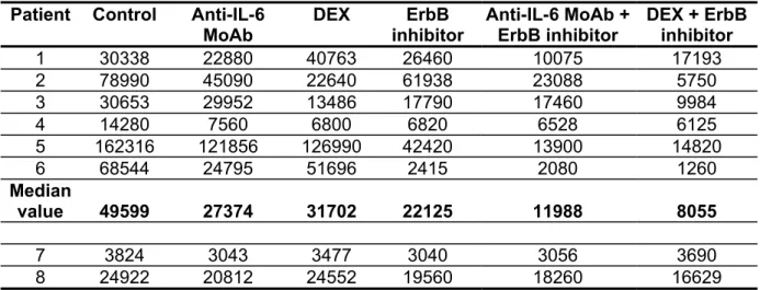
We then investigated the effect of the pan-ErbB inhibitor on the survival of primary myeloma cells. Detailed results obtained with eight patients are shown in Table 1. In six out of eight patients, the B-E8 anti-IL-6 MoAb or the pan ErbB kinase inhibitor significantly reduced the number of viable myeloma cells (respectively by 45% and 55%, P≤.05; table 1 and figure 9A). The effect of DEX was less marked and not significant (P=.07). Of major interest, the pan-ErbB inhibitor dramatically increased both anti-IL-6 MoAb-induced or DEX-induced apoptosis. We observed a 76% reduction of the number of viable myeloma cells with anti-IL-6 MoAb and the ErbB inhibitor (P=.0028) and a 84% reduction with DEX and the ErbB inhibitor (P=.0028) (Figure 9A). In two patients, the three compounds had no effect on myeloma cell survival, either used alone or in combination (Table 1). Interestingly, the pan ErbB inhibitor had no effect on the survival of other bone marrow non myeloma cells (Figure 9B). We then looked for the effect of the pan-ErbB kinase inhibitor on the growth of hematopoietic precursors present in the bone marrow of five MM patients using a methylcellulose semi-solid assay (Figure 10A). None of the three, pan-ErbB kinase inhibitor, DEX or anti-IL-6 MoAb, significantly affected it, either used alone or in combination (P>.5).
Effect of HB-EGF inhibitor on the adhesion of myeloma cells to BMSCs and IL-6 production.
As HB-EGF is expressed by MM patients’ stromal cells and can be involved in juxtacrine intercellular communication38, we investigated whether the PD169540 could affect the adhesion of myeloma cell to BMSCs. About 20% myeloma cells adhered to BMSCs and pretreatment of both myeloma cells and BMSCs with the pan-ErbB inhibitor did not affect myeloma cell adhesion to stromal cells (Figure 10B). Given that binding of myeloma cells to BMSCs had been proven important to
promote IL-6 production by stromal cells and myeloma cell survival31,39, we next investigated whether the PD169540 could affect the secretion of IL-6 by BMSCs cocultured with myeloma cells. IL-6 secretion was 889±17 pg/ml without ErbB inhibitor and 810±43 pg/ml with ErbB inhibitor. No IL-6 was detected in the supernatant of myeloma cells (results not shown). Therefore the pan-ErbB inhibitor did not significantly affect the IL-6 production by stromal cells cultured with myeloma cells.
Discussion
As we initially found that the HB-EGF gene was expressed in some myeloma cell lines and could stimulate their growth4,5, our aim was to investigate whether this
observation was a common feature in multiple myeloma and whether ErbB inhibitors could be potentially useful in treating this disease.
Firstly, we show here that HB-EGF expression is not a salient feature of primary myeloma cells. There is no HB-EGF gene expression in the purified myeloma cells of eight out of nine MM patients. HB-EGF expression was found in the myeloma cells of only one patient with plasma cell leukemia. An autocrine production of HB-EGF by myeloma cells could eventually make it possible to obtain a myeloma cell line in vitro, explaining why two myeloma cell lines out of nine express the HB-EGF gene. Interestingly, the bone marrow tumor environment of all eight MM patients expressed the HB-EGF gene. Monocytes were purified from three patients with MM and were found to express the HB-EGF gene. This was expected because stimulated monocytes are known to produce HB-EGF40 and monocytes are highly activated in MM patients' bone marrow, in particular producing high levels of IL-641. HB-EGF expression was also found in the five stromal cell lines derived from the bone marrow of patients with MM. We could not directly assay for a production of HB-EGF protein because a sensitive HB-EGF ELISA is not commercially available. Thus, these current data emphasize that the bone marrow environment of patients with MM produces HB-EGF, in particular monocytes and stromal cells and that primary myeloma cells rarely express the HB-EGF gene.
Secondly, we found that myeloma cells from six out of nine myeloma cell lines and myeloma cells from nine out of nine patients expressed the two HB-EGF receptor genes, ErbB1 and/or ErbB4. Using RT-PCR, we did not detect ErbB expression in
polyclonal plasmablasts generated from normal peripheral blood B cells using the methodology we recently reported42. In agreement with this study, we found that an ErbB inhibitor did not affect the generation of plasmablasts (results not shown). These results are intriguing and suggest that ErbB is either expressed on bone marrow plasmocytes, unlike plasmablasts, or involved in plasma cell oncogenesis. We are now trying to obtain purified bone marrow plasma cells to investigate this point, but these cells are rare cells (0.25% of bone marrow cells) and for ethical reasons, it is difficult to obtain enough normal bone marrow to purify them. The level of ErbB gene expression on myeloma cell lines was low compared to A431 cells. In particular, ErbB1 protein was not detectable by immunoprecipitation in two myeloma cell lines, unlike in A431 cells. ErbB4 was detected in two out of the four cell lines expressing the ErbB4 gene. Despite this low ErbB expression, recombinant HB-EGF stimulated the growth of the six ErbB1+ and/or ErbB4+ cell lines, unlike the three ErbB1-/ErbB4- cell lines. We found that recombinant HB-EGF activity required a low concentration of IL-6 and was blocked by an antibody to IL-6. Recombinant HB-EGF alone stimulated the proliferation of XG-1 cells but this was due to their previously reported ability to produce a small amount of autocrine IL-633, since HB-EGF-induced growth was blocked by an anti-IL-6 MoAb. These data are in agreement with recent data showing the ability of distinct cell surface receptors to cross-communicate. This cooperation between HB-EGF/ErbB and IL-6/gp130 pathways to trigger myeloma cell growth might be explained by the interaction of ErbB transducing elements with gp130, as it was reported previously in epithelial cancer cells. In a prostate carcinoma cell line, Qiu et al. firstly demonstrated receptor crosstalk between the gp130 and ErbB3 via a gp130/ErbB2/ErbB3 complex11. Badache et al. showed that IL-6-induced MAPK/PI3K activity depends on the cooperation with an EGFR
autocrine loop in T47D breast cancer cells. In response to IL-6 treatment, the protein phosphatase Shp-2 and the docking molecule Gab1, which are constitutively associated with ErbB1, are recruited to gp130/JAK complexes and subsequently phosphorylated12. In addition, EGF was shown to enhance OSM-induced inhibition of cellular proliferation in the MCF-7 breast carcinoma cell line, where gp130 is constitutively associated with ErbB2 and ErbB343. Recently, several examples of receptor crosstalk have been reported in multiple myeloma. IFN-α stimulation can induce tyrosine phosphorylation of gp13044 or ErbB315 in a myeloma cell line. Podar et al. showed that gp130 and IGF-1 receptor are colocalized in caveolae, which are cholesterol-rich plasma membrane microdomains45. These data suggest that these caveolin-pits might play an important role in receptor interactions in multiple myeloma, as gp130 IL-6 transducer, IGF1-R, and eventually ErbB receptors and coreceptors might be colocalized in these membrane structures. Thus, the present study shows that HB-EGF is a third major myeloma cell growth factor, as are IL-6 and IGF-146. Further studies are necessary to clarify how a low concentration of IL-6 and HB-EGF work together to promote myeloma cell growth.
The two known myeloma cell growth factors, IL-6 and IGF-1, activate three major transduction pathways in myeloma cells: the JAK/STAT pathway induced by IL-6, the MAPK, and the PI3-K/AKT pathway induced by both IL-6 and IGF-129,34,35,46,47. An
activation of these three pathways in our myeloma cells by IL-6 or IGF-1 was confirmed here. In agreement with the known activation of PI-3K/AKT by the ErbB receptor family in various cells16, HB-EGF induced AKT phosphorylation in myeloma cells. It did not activate STAT3 or MAPK phosphorylation. MAPK phosphorylation was looked for from 15 min to 24 hours after HB-EGF triggering, excluding a delayed activation. This lack of MAPK activation by HB-EGF was unexpected. Indeed, the
ras/MAPK pathway is frequently activated by EGF ligands in various cells16. We will explore whether other signaling pathways known to be activated by HB-EGF in various cells (PCLγ, PKC, Sarc kinase)48 could be involved in HB-EGF-induced
myeloma cell growth.
A major point was to determine whether inhibitors of ErbB kinases could promote apoptosis of primary myeloma cells and block the HB-EGF myeloma cell growth activity. As myeloma cells expressed ErbB4 and/or ErbB1, we used the PD169540 inhibitor, which inhibits kinase activity of ErbB1, ErbB2 and ErbB4, without affecting a large panel of other kinases. This inhibitor is close to the CI-1033 investigated in patients with neck, breast, and NSCL cancers23. This pan-ErbB kinase inhibitor blocked the HB-EGF-induced proliferation of the six sensitive cell lines at a 1-µM concentration, which ensures specificity on ErbB members (Dr. Leopold, Pfizer Laboratories, personal communication). Two other ErbB1 inhibitors (AG1478 and PD153035) also blocked the HB-EGF-induced stimulation of the three ErbB1 myeloma cell lines. Despite ErbB2 expression in myeloma cells, we found no inhibition of HB-EGF-induced myeloma cell proliferation by the Herceptin anti-ErbB2 MoAb. As ErbB2 participated in HB-EGF response in various cells36, this suggests that ErbB2 expression in myeloma cells is too weak to be functional. The PD169540 pan ErbB inhibitor did not affect the IL-6-induced proliferation of the XG-6 myeloma cells that did not express the HB-EGF gene. It had no toxicity on the 14-day growth of bone marrow hematopoietic progenitors. As HB-EGF is a membrane protein that can be involved in juxtacrine stimulation and is produced by bone marrow stromal cells, we looked for a putative role of PD169540 on the interaction of myeloma cells and BMSCs in accordance with the model reported by Uchimaya et al31. We found that
the ErbB inhibitor had no effect on myeloma cell adhesion to stromal cells or on IL-6 production by stromal cells cocultured with myeloma cells.
It is of major interest that the pan-ErbB kinase inhibitor induced a 55% reduction in the number of viable primary myeloma cells cultured for several days together with their bone marrow environment, which expresses the HB-EGF gene. In association with DEX, it induced an 84% reduction in myeloma cell number, whereas DEX alone induced only a 36% reduction. A similar additive effect was observed with an anti-IL-6 MoAb. Interestingly, this toxicity was specific to myeloma cells and no reduction in the number of non-myeloma cells was observed. Several ErbB inhibitors are in various stages of clinical development and some are producing significant clinical responses. The compound that is at the most advanced stage of development is IRESSA (ZD1839), which targets EGFR. This compound has shown an acceptable tolerability and promising clinical efficacy in phase I trials and is currently in phase III trials in advanced NSCL and breast cancers49. CI-1033 is another clinical-grade irreversible tyrosine kinase inhibitor that is unique because it targets the three ErbB members with kinase activity25. It has potent and long-lasting in vivo activity in a range of tumor models. For example, it has demonstrated antitumor activity against A431 and H125 tumors in vivo50. Data from three phase I trials in patients with head
and neck, breast and NSCL cancers showed that CI-1033 is generally well tolerated51. A phase II study in patients with advanced ovarian cancer and a randomized phase II trial with patients with NSCL after a first or second line of chemotherapy are being done52.
In conclusion, our data are very encouraging and provide a framework for clinical evaluation of ErbB inhibitors in patients with MM, alone and coupled with dexamethasone or anti-IL-6 MoAb, now available for clinical use53. A first step will be
to investigate in preclinical studies whether ErbB inhibitors can block the growth of myeloma cell lines and primary myeloma cells with SCID mice models of human multiple myeloma27,54. These current data are in agreement with the recent
development of novel biologically based treatment approaches that target mechanisms whereby MM cells grow and survive in their bone marrow environment. The most relevant example is the proteasome inhibitor PS341, which was identified as a promising new therapeutic agent in multiple myeloma55.
Acknowledgments. We thank Dr. Leopold (Pfizer Global Research and Development, Ann Arbor, Michigan 48105, USA) for providing us with the PD169540 pan-ErbB inhibitor.
1. Klein B, Zhang XG, Jourdan M, et al. Paracrine rather than autocrine regulation of myeloma-cell growth and differentiation by interleukin-6. Blood. 1989;73:517-526
2. Kawano M, Hirano T, Matsuda T, et al. Autocrine generation and essential requirement of BSF-2/IL-6 for human multiple myeloma. 1988;332:83-85
3. Gu ZJ, De Vos J, Rebouissou C, et al. Agonist anti-gp130 transducer monoclonal antibodies are human myeloma cell survival and growth factors. Leukemia. 2000;14:188-197
4. De Vos J, Couderc G, Tarte K, et al. Identifying intercellular signaling genes expressed in malignant plasma cells by using complementary DNA arrays. Blood. 2001;98:771-780
5. Wang YD, De Vos J, Jourdan M, et al. Cooperation between heparin-binding EGF-like growth factor and interleukin-6 in promoting the growth of human myeloma cells. Oncogene. 2002;21:2584-2592.
6. Graus-Porta D, Beerli RR, Daly JM, Hynes NE. ErbB-2, the preferred heterodimerization partner of all ErbB receptors, is a mediator of lateral signaling. EMBO J. 1997;16:1647-1655.
7. Harris RC, Chung E, Coffey RJ. EGF receptor ligands. Exp Cell Res. 2003;284:2-13.
8. Falls DL. Neuregulins: functions, forms, and signaling strategies. Exp Cell Res. 2003;284:14-30.
9. Yamauchi T, Ueki K, Tobe K, et al. Tyrosine phosphorylation of the EGF receptor by the kinase Jak2 is induced by growth hormone. Nature. 1997;390:91-96
10. Prenzel N, Zwick E, Daub H, et al. EGF receptor transactivation by G-protein-coupled receptors requires metalloproteinase cleavage of proHB-EGF. Nature. 1999;402:884-888
11. Qiu Y, Ravi L, Kung HJ. Requirement of ErbB2 for signalling by interleukin-6 in prostate carcinoma cells. Nature. 1998;393:83-85
12. Badache A, Hynes NE. Interleukin 6 inhibits proliferation and, in cooperation with an epidermal growth factor receptor autocrine loop, increases migration of T47D breast cancer cells. Cancer Res. 2001;61:383-391.
13. Roudabush F, Pierce K, Maudsley S, Khan K, Luttrell L. Transactivation of the EGF receptor mediates IGF-1-stimulated shc phosphorylation and ERK1/2
activation in COS-7 cells. J Biol Chem. 2000;275:22583-22589
14. Hurbin A, Dubrez L, Coll J, Favrot M. Inhibition of apoptosis by
amphiregulin via an Insulin-like Growth Factor-1 Receptor-dependant Pathway in Non-small Cell Lung Cancer Cell lines. The journal of biological chemistry.
2002;277:49127-49133
15. Walters D, French J, Arendt B, Jelinek D. Atypical expression of ErbB3 in myeloma cells: cross-talk between ErbB3 and the interferon-α signaling complex. Oncogene. 2003;22:3598-3607
16. Olayioye MA, Neve RM, Lane HA, Hynes NE. The ErbB signaling network: receptor heterodimerization in development and cancer. EMBO J. 2000;19:3159-3167.
17. Yarden Y. The EGFR family and its ligands in human cancer. signalling mechanisms and therapeutic opportunities. Eur J Cancer. 2001;37 Suppl 4:S3-8.
18. Holbro T, Civenni G, Hynes NE. The ErbB receptors and their role in cancer progression. Exp Cell Res. 2003;284:99-110.
19. Nicholson R, Gee J, Harper M. EGFR and cancer prognosis. Eur J Cancer. 2001;34:S9-S15
20. Carpenter G. ErbB-4: mechanism of action and biology. Exp Cell Res. 2003;284:66-77
21. Slamon DJ, Godolphin W, Jones LA, et al. Studies of the HER-2/neu proto-oncogene in human breast and ovarian cancer. Science. 1989;244:707-712.
22. Bodey B, Bodey B, Jr., Groger AM, et al. Clinical and prognostic
significance of the expression of the c-erbB-2 and c-erbB-3 oncoproteins in primary and metastatic malignant melanomas and breast carcinomas. Anticancer Res. 1997;17:1319-1330.
23. Fry DW. Mechanism of action of erbB tyrosine kinase inhibitors. Exp Cell Res. 2003;284:131-139.
24. Mendelsohn J, Baselga J. The EGF receptor family as targets for cancer therapy. Oncogene. 2000;19:6550-6565
25. Slichenmyer WJ, Elliott WL, Fry DW. CI-1033, a pan-erbB tyrosine kinase inhibitor. Semin Oncol. 2001;28:80-85.
26. Zhang XG, Gaillard JP, Robillard N, et al. Reproducible obtaining of human myeloma cell lines as a model for tumor stem cell study in human multiple myeloma. Blood. 1994;83:3654-3663
27. Rebouissou C, Wijdenes J, Autissier P, et al. A gp130 interleukin-6 transducer-dependent SCID model of human multiple myeloma. Blood.
1998;91:4727-4737
28. Jourdan M, De Vos J, Mechti N, Klein B. Regulation of Bcl-2-family
proteins in myeloma cells by three myeloma survival factors: interleukin-6, interferon-alpha and insulin-like growth factor 1. Cell Death Differ. 2000;7:1244-1252
29. De Vos J, Jourdan M, Tarte K, Jasmin C, Klein B. JAK2 tyrosine kinase inhibitor tyrphostin AG490 down-regulates the MAPK and STAT pathways and induces apoptosis in myeloma cells. Br J Haematol. 2000;109:823-828
30. Sun RX, Lu ZY, Wijdenes J, et al. Large scale and clinical grade purification of syndecan-1+ malignant plasma cells. J Immunol Methods. 1997;205:73-79
31. Uchiyama H, Barut BA, Mohrbacher AF, Chauhan D, Anderson KC. Adhesion of human myeloma-derived cell lines to bone marrow stromal cells stimulates interleukin-6 secretion. Blood. 1993;82:3712-3720
32. Stoll SW, Kansra S, Peshick S, et al. Differential utilization and localization of ErbB receptor tyrosine kinases in skin compared to normal and malignant keratinocytes. Neoplasia. 2001;3:339-350.
33. Jourdan M, Zhang XG, Portier M, et al. IFN-alpha induces autocrine production of IL-6 in myeloma cell lines. 1991;147:4402-4407
34. Shi Y, Hsu JH, Hu L, Gera J, Lichtenstein A. Signal pathways involved in activation of p70S6K and phosphorylation of 4E-BP1 following exposure of multiple myeloma tumor cells to interleukin-6. J Biol Chem. 2002;277:15712-15720.
35. Hideshima T, Nakamura N, Chauhan D, Anderson KC. Biologic sequelae of interleukin-6 induced PI3-K/Akt signaling in multiple myeloma. Oncogene.
2001;20:5991-6000.
36. Iwamoto R, Yamazaki S, Asakura M, et al. Heparin-binding EGF-like growth factor and ErbB signaling is essential for heart function. Proc Natl Acad Sci U S A. 2003;100:3221-3226
37. Leyland-Jones B. Trastuzumab: hopes and realities. Lancet Oncol. 2002;3:137-144.
38. Iwamoto R, Mekada E. Heparin-binding EGF-like growth factor: a juxtacrine growth factor. Cytokine Growth Factor Rev. 2000;11:335-344
39. Lokhorst HM, Lamme T, de Smet M, et al. Primary tumor cells of myeloma patients induce interleukin-6 secretion in long-term bone marrow cultures. Blood. 1994;84:2269-2277
40. Nakano T, Raines EW, Abraham JA, Klagsbrun M, Ross R.
Lysophosphatidylcholine upregulates the level of heparin-binding epidermal growth factor-like growth factor mRNA in human monocytes. Proc Natl Acad Sci U S A. 1994;91:1069-1073.
41. Portier M, Rajzbaum G, Zhang XG, et al. In vivo interleukin-6 gene expression in the tumoral environment in multiple myeloma. 1991;21:1759-1762
42. Tarte K, De Vos J, Thykjaer T, et al. Generation of polyclonal plasma cells from peripheral blood B cells: a normal counterpart of malignant plasma cells. The two first authors contributed equally to the work. (Submitted). 2001
43. Grant SL, Hammacher A, Douglas AM, et al. An unexpected biochemical and functional interaction between gp130 and the EGF receptor family in breast cancer cells. Oncogene. 2002;21:460-474.
44. French J, Walters D, Jelinek D. Transactivation of gp130 in myeloma cells. J Immunol. 2003;170:3717-3723
45. Podar K, Tai Y, Cole C, et al. Essential role of caveolae in interleukin-6- and insulin-like growth factor I-triggered Akt-1-mediated survival of multiple myeloma cells. J Biol Chem. 2003;278:5794-5801
46. Ferlin M, Noraz N, Hertogh C, et al. Insulin-like growth factor induces the survival and proliferation of myeloma cells through an IL-6-independent transduction pathway. Br J Haematol. 2000;111:626-634
47. Qiang YW, Kopantzev E, Rudikoff S. Insulinlike growth factor-I signaling in multiple myeloma: downstream elements, functional correlates, and pathway cross-talk. Blood. 2002;99:4138-4146.
48. Jorissen RN, Walker F, Pouliot N, et al. Epidermal growth factor receptor: mechanisms of activation and signalling. Exp Cell Res. 2003;284:31-53.
49. Ranson M. ZD1839 (Iressa): for more than just non-small cell lung cancer. Oncologist. 2002;7:16-24
50. Driscoll D, Steinkampf R, Patmore S, Elliott W, Klohs W. Effect of epidermal growth factor receptor tyrosine kinase inhibitor PD183805 on vascular endothelial growth factor secretion from several tumor models. Proc. Am.
Assoc.Cancer Res. 1999;40:121
51. Shin DM, Nemunaitis J, Zinner RG, et al. A phase I clinical and biomarker study of CI-1033, a novel pan-ErbB tyrosine kinase inhibitor in patients with solid tumors. Proc.Am.Soc.Clin.Oncol. 2001;21:82
52. Sridhar S, Seymour L, Shepherd F. Inhibitors of epidermal-growth-factor receptors: a review of clinical research with a focus on non-small-cell lung cancer. Lancet Oncol. 2003;4:397-406
53. Trikha M, Corringham R, Klein B, Rossi J. Targeted Anti-IL-6 Monoclonal Antibody Therapy for Cancer: A Review of the rationale and clinical evidence. Clinical Cancer Research. 2003;in press
54. Yaccoby S, Barlogie B, Epstein J. Primary myeloma cells growing in SCID-hu mice: a model for studying the biology and treatment of myeloma and its manifestations. Blood. 1998;92:2908-2913
55. Richardson PG, Barlogie B, Berenson J, et al. A phase 2 study of bortezomib in relapsed, refractory myeloma. N Engl J Med. 2003;348:2609-2617
Table 1. Effect of the pan-ErbB inhibitor on the survival of primary myeloma cells.
Myeloma cell count/ml.
Patient Control Anti-IL-6 MoAb DEX ErbB inhibitor Anti-IL-6 MoAb + ErbB inhibitor DEX + ErbB inhibitor 1 30338 22880 40763 26460 10075 17193 2 78990 45090 22640 61938 23088 5750 3 30653 29952 13486 17790 17460 9984 4 14280 7560 6800 6820 6528 6125 5 162316 121856 126990 42420 13900 14820 6 68544 24795 51696 2415 2080 1260 Median value 49599 27374 31702 22125 11988 8055 7 3824 3043 3477 3040 3056 3690 8 24922 20812 24552 19560 18260 16629
Non-myeloma cell count/ml. Patient Control Anti-IL-6
MoAb DEX ErbB inhibitor Anti-IL-6 MoAb + ErbB inhibitor DEX + ErbB inhibitor 1 344663 302120 334238 323540 314925 307808 2 221010 254910 257360 248062 216912 244250 3 259347 230048 206514 282210 282540 230016 4 325720 292440 293200 303180 233472 243875 5 177684 218144 213010 167580 186100 185180 6 271456 265205 188304 227585 257920 198740 Median value 265402 260058 235185 265136 245696 236946 7 156176 166957 186523 196960 156944 146310 8 315078 199188 285448 220440 201740 213371
Mononuclear cells from eight patients with MM were cultured for 5 days in RPMI 1640 medium, 5% FCS, 1 ng/ml IL-6, with or without the PD169540 ErbB inhibitor (1 µM), B-E8 anti-IL-6 MoAb (10 µg/ml) and DEX (10-6 M). At day 5 of culture, the viability and cell counts were assessed and the percentage of CD138+ viable myeloma cells was determined by flow cytometry. Results are expressed as number of myeloma and non-myeloma cells per milliliter. The median values were calculated from the 6 responder patients. Two patients did not significantly respond to the three compounds, either used alone or in combination.
Figure legends.
Figure 1. Expression of EGF receptor family members in myeloma cell lines.
A. ErbB1, ErbB2, ErbB3, ErbB4 and HB-EGF gene expression was assayed with RT-PCR in nine myeloma cell lines. The A431 epidermoid carcinoma cell line was used as a control. Amplification of β-actin shows the equivalence of the cDNA loading and amplification. Results are of one experiment representative of three. B. Myeloma cells or A431 cells were lysed and the lysates were immunoprecipitated with the H4.72.8 anti-ErbB4 or the LA-22 anti-ErbB1 antibody. The precipitated material was separated on a 7.5% SDS-PAGE and analyzed by Western blotting with a second anti-ErbB antibody (anti-ErbB4, clone C-18 or anti-ErbB1, clone LA-1). Results are of one experiment representative of three.
Figure 2. Sensitivity of myeloma cell lines to HB-EGF.
XG-1 and XG-6 cells were IL-6-starved for 3 hours and cultured in RPMI1640 culture medium and 5% FCS with either 5 pg/ml IL-6, or 5 pg/ml IL-6 and 1 µg/ml HB-EGF, or 500 pg/ml IL-6, or 500 pg/ml IL-6 and 1 µg/ml HEGF (with or without 10 µg/ml B-E8 anti-IL-6 MoAb). Cells were cultured for 6 days and were pulsed for 12 hours with tritiated thymidine at the end of the culture. Data are means ± SD of the tritiated thymidine incorporation determined on sixplicate culture wells and are those of one experiment representative of four.
Figure 3. HB-EGF reduces the apoptosis induced by IL-6 removal.
XG-1 cells were IL-6 starved for 3 hours and cultured in RPMI 1640 culture medium and 5% FCS with either 5 pg/ml IL-6, or 5 pg/ml IL-6 and 1 µg/ml HB-EGF, or 500 pg/ml IL-6, or 500 pg/ml IL-6 and 1 µg/ml HB-EGF (with or without 10 µg/ml B-E8 anti IL-6 MoAb). Cells were cultured for 3 days and stained with FITC-annexin V to
determine the percentage of apoptotic cells. Results are of one experiment representative of three.
Figure 4. Correlation between ErbB1/ErbB4 expression and HB-EGF activity on
myeloma cells.
ErbB1/ErbB4 positive myeloma cells (A) and ErbB1/ErbB4 negative myeloma cells
(B) were cultured for 5-7 days in RPMI 1640, 5% FCS and 5 pg/ml IL-6, without (open bars) or with (black bars) HB-EGF and then pulsed with tritiated thymidine for 12 hours at the end of the culture. Data are expressed as means ± SD of tritiated thymidine incorporation determined on sixplicate culture well. * Indicates that the mean value is statistically significantly different from that obtained with 5 pg/ml IL-6 alone, using a Student t test (P≤.05).
Figure 5. HB-EGF activity is mediated by activation of the PI-3K/AKT pathway.
A. XG-7 myeloma cells were starved overnight in culture medium without serum or cytokines. Cells were then stimulated with either IL-6 (20 ng/ml), IGF-1 (1 µg/ml), or HB-EGF (5 µg/ml) for 15 min at 37°C. Lysates from unstimulated (Co) or stimulated myeloma cells were immunoblotted with anti-phospho-specific STAT3 antibody (upper panel) and then reprobed with STAT3 antibody (lower panel), anti-phospho-specific AKT antibody (upper panel) and reprobed with anti-AKT antibody (lower panel), anti-phospho-specific MAPK antibody (upper panel) and reprobed with anti-MAPK antibody (lower panel). B. XG-5 and XG-7 myeloma cell lines were IL-6-starved for 3 hours and cultured in RPMI 1640 culture medium and 5% FCS with 5 pg/ml IL-6 and 1 µg/ml HB-EGF without (open bars) or with (black bars) the PI-3K inhibitor LY294002 (25 µM). Cells were cultured for 6 days and pulsed with tritiated thymidine at the end of the culture. Mean tritiated thymidine incorporations were determined in sixplicate culture wells and results are expressed as percentages of
the mean proliferation without ErbB inhibitor. *Indicates that the mean value is statistically significantly different from that obtained with 5 pg/ml IL-6 and HB-EGF without inhibitor, using a Student t test (P≤.05).
Figure 6. The myeloma cell growth factor activity of HB-EGF is inhibited by a
pan-ErbB kinase inhibitor.
A. A431 cells were stimulated with HB-EGF, with or whithout PD169540, for 15 min prior to lysis and immunoprecipitation of ErbB1. Tyrosine phosphorylation of ErbB1 was assayed using an anti-phosphotyrosine antibody (4G10, upstate biotechnology, Lake Placid, NY) and ErbB1 was quantified with an anti-ErB1 antibody (clone LA-22). B. Myeloma cells were IL-6-starved for 3 hours and cultured in RPMI 1640 culture medium and 5% FCS with either 5 pg/ml IL-6 or 5 pg/ml IL-6 and 1 µg/ml HB-EGF, without or with 1 µM of the PD169540 ErbB inhibitor. Cells were cultured for 3 days and stained with FITC-annexin-V to determine the percentage of apoptotic cells. C. Myeloma cell lines were IL-6-starved for 3 hours and cultured in RPMI 1640 culture medium and 5% FCS with 5 pg/ml IL-6 and 1 µg/ml HB-EGF without (open bars) or with (black bars) the PD169540 ErbB inhibitor (1 µM). Cells were cultured for 5-7 days and pulsed with tritiated thymidine at the end of the culture. Mean tritiated thymidine incorporations were determined in sixplicate culture wells and results are expressed as percentages of the mean proliferation without ErbB inhibitor. *Indicates that the mean value is statistically significantly different from that obtained with 5 pg/ml IL-6 and HB-EGF without inhibitor, using a Student t test (P≤.05). D. 1, XG-6 and XG-7 cells were IL-XG-6 starved for 3 hours and cultured in RPMI1XG-640 culture medium and 5% FCS with high concentration of IL-6 (500 pg/ml), without (open bars) or with (black bars) the PD169540 ErbB inhibitor (1 µM). Mean tritiated thymidine incorporations were determined in sixplicate culture wells and results are expressed
as percentages of the mean proliferation without ErbB inhibitor. *Indicates that the mean value is statistically significantly different from that obtained without inhibitor, using a Student t test (P≤.05). E. XG-7 myeloma cells were starved overnight in culture medium without serum or cytokines with or without the PD169540 ErbB inhibitor. Cells were then stimulated with either no cytokine (Co), or IL-6 (20 ng/ml), or HB-EGF (5 µg/ml), or IGF-1 (1 µg/ml) for 15 min at 37°C. Lysates from unstimulated (Co) or stimulated myeloma cells were immunoblotted with anti-phospho-specific AKT antibody (upper panel) and reprobed with anti-AKT antibody (lower panel).
Figure 7. The myeloma cell growth factor activity of HB-EGF is inhibited by two
ErbB1-specific inhibitors.
ErbB1 positive myeloma cell lines (XG-5, XG-7 and XG-11) were IL-6-starved for 3
hours and cultured in RPMI 1640 culture medium and 5% FCS with 5 pg/ml IL-6 and 1 µg/ml HB-EGF without or with PD169540 (1 µM), AG1478 (1 µM) and PD159540 (1 µM). Cells were cultured for 6 days and pulsed with tritiated thymidine at the end of the culture. Mean tritiated thymidine incorporations were determined in sixplicate culture wells and results are expressed as percentages of the mean proliferation without ErbB inhibitor. *Indicates that the mean value is statistically significantly different from that obtained with 5 pg/ml IL-6 and HB-EGF without inhibitor, using a Student t test (P≤.05).
Figure 8. Expression of the ErbB receptors and HB-EGF in purified primary
myeloma cells and in the tumor environment.
A. ErbB1, ErbB2, ErbB3, ErbB4, and HB-EGF expression was assayed by RT-PCR on primary myeloma cells. These cells were purified (≥95%) from nine patients (five with intramedullar myeloma and four with plasma cell leukemia). The A431
epidermoid carcinoma cell line was used as a control. B. HB-EGF gene expression was analyzed by RT-PCR in unselected mononuclear cells from eight patients with MM. (C) HB-EGF gene expression was analysed by RT-PCR in CD14 purified monocytes (>98%) from 3 patients and in stromal cells derived from 5 patients. Amplification of β-actin shows the equivalence of the cDNA loading and amplification. Results are of one experiment representative of three.
Figure 9. The PD169540 ErbB inhibitor potentiates dexamethasone and
anti-IL-6-induced apoptosis of primary myeloma cells.
Mononuclear cells from eight patients with MM were cultured at 5 × 10⁵ cells/ml in RPMI 1640 medium and 5% FCS with either the PD169540 ErbB inhibitor (1 µM) or anti-IL-6 MoAb (B-E8, 10 µg/ml) or dexamethasone (DEX, 10–6 M), alone or in
combination, for 5 days. As endogenous production of IL-6 is highly variable in short-term culture of patients’ bone marrow, 1 ng/ml recombinant IL-6 was added to eliminate this source of variability. In each culture group, viability and cell counts were assayed and myeloma cells were stained with an anti-CD138-PE antibody. Results are median values of the numbers of myeloma (A) or non-myeloma cells (B), compared to that of the control group, which assigned the value of 100%. Only the six responder patients listed in Table 1 are represented there.
* Indicates a statistically significant difference of the median value compared to that of the control group using a Wilcoxon test for pairs (P≤.05).
**Indicates a statistically significant difference of the median value compared to that obtained with the anti-IL-6 MoAb or the DEX groups using a Wilcoxon test for pairs (P≤.05).
Figure 10. The PD169540 ErbB inhibitor has no effect on the growth of the
hematopoietic progenitors or on the adhesion of MM cells to stromal cells.
A. Bone marrow mononuclear cells from five patients with MM were grown in a semi-solid culture medium with hematopoietic cytokines for 14 days. Cells were treated with either the PD169540 pan ErbB inhibitor (1µM) or anti-IL-6 MoAb (B-E8, 10µg/ml) or 10-6 M dexamethasone (DEX), alone or in combination. The number of granulocyte-macrophage colonies was counted on day 14 of incubation. Data are means ± SD of the number of colonies determined on two different culture plates for each condition. B. XG-11 cells were cultured with either BMSC-coated (black bars) or noncoated culture plates (white bars), without or with 1µM PD169540 for 24h. Non-adherent cells were counted and Non-adherent cells were trypsinized and counted. In the two cell populations, GFP+ stromal cells were quantified with FACS analysis. The percentage of bound plasma cells was calculated as follows: percentage bound cells =((input cells)-(non adherent cells)/(input cells))X100. Results are of one experiment representative of three.