HAL Id: hal-02413350
https://hal.archives-ouvertes.fr/hal-02413350
Submitted on 25 Nov 2020HAL is a multi-disciplinary open access archive for the deposit and dissemination of sci-entific research documents, whether they are pub-lished or not. The documents may come from teaching and research institutions in France or abroad, or from public or private research centers.
L’archive ouverte pluridisciplinaire HAL, est destinée au dépôt et à la diffusion de documents scientifiques de niveau recherche, publiés ou non, émanant des établissements d’enseignement et de recherche français ou étrangers, des laboratoires publics ou privés.
External Stimuli for Drug Delivery and Tissue
Engineering Applications
Damien Mertz, Sebastien Harlepp, Jacky Goetz, Dominique Begin, Guy
Schlatter, Sylvie Bégin-colin, Anne Hebraud
To cite this version:
Damien Mertz, Sebastien Harlepp, Jacky Goetz, Dominique Begin, Guy Schlatter, et al.. Nanocom-posite Polymer Scaffolds Responding under External Stimuli for Drug Delivery and Tissue Engi-neering Applications. Advanced Therapeutics, Wiley, 2020, pp.1900143. �10.1002/adtp.201900143�. �hal-02413350�
1 DOI: 10.1002/ (adtp.201900143R1)
Article type: Review
Nanocomposite Polymer Scaffolds Responding Under External Stimuli for Drug Delivery and Tissue Engineering Applications
Damien Mertz*, Sebastien Harlepp, Jacky Goetz, Dominique Bégin, Guy Schlatter, Sylvie Bégin-Colin, Anne Hébraud
Dr. Damien Mertz, Prof. Sylvie Bégin-Colin,
Institut de Physique et Chimie des Matériaux de Strasbourg (IPCMS), UMR-7504 CNRS-Université de Strasbourg, 23 rue du Loess, BP 34 67034, Strasbourg Cedex 2, France. E-mail: [email protected]
Dr. Sébastien Harlepp, Dr. Jacky Goetz,
INSERM UMR_S1109, Tumor Biomechanics, Strasbourg, Université de Strasbourg, Fédération de Médecine Translationnelle de Strasbourg (FMTS), 67000 Strasbourg, France Dr. Dominique Bégin, Prof. Guy Schlatter, Dr. Anne Hébraud
Institut de Chimie et Procédés pour l'Energie, l'Environnement et la Santé (ICPEES), UMR-7515 CNRS-Université de Strasbourg, 25 rue Becquerel, 67087 Strasbourg, Cedex 2, France.
Keywords: responsive nanocomposites, external stimuli, polymer scaffolds, drug release,
tissue engineering
Abstract. The blossoming development of nanomaterials and polymer science has opened the
way towards new biocompatible scaffolds responding remotely to external stimuli (magnetic
field, light, electric field). Such smart scaffolds are envisioned as new implantable tissues
displaying multiple therapeutic and imaging functionalities. They hold great promises to
achieve a controlled delivery of therapeutics for various diseases, or to ensure a
stimuli-induced cellular response for bone, cardiac, or muscle tissue engineering.
Table of content
1. Introduction
2. Criteria and key issues to design smart stimuli responsive scaffolds for biomedicine
2.1 Biomedical issues: biocompatibility and immune response
2.2 Fabrication criteria for biomedical applications
2
3 . From nanoparticles and polymers as building blocks to responsive nanocomposite scaffolds
3.1 Inorganic nanomaterials as ideal building blocks for signal conversion
3.2 Polymer scaffolds as host integrative matrix
3.3 Formulate polymer scaffolds and NPs: process, issues and physico-chemical features
4. Applications of nanocomposite scaffolds for controlled drug delivery
4.1 Drug delivery upon static magnetic field by magnetomechanical effects
4.2 Drug delivery upon an alternating magnetic field by magnetothermal effects
4.3 Photoresponsive scaffolds with drug delivery activated by near infra-red light
4.4 Electroresponsive scaffolds for transdermal drug delivery
5 . Applications in Tissue Engineering
5.1 Bone regeneration
5.2 Soft actuators controlled by external fields and muscle tissue engineering
5.3 Cardiovascular tissue regeneration induced by external fields
5.4 Neuronal regeneration
6. Conclusion and Perspectives
Acronym List :
AMF: alternating magnetic field
CNTs: carbon nanotubes
DOX : doxorubicin
ECM: extracellular matrix
ELS: electrospun
EP: electroporation
GO: graphene oxide
IO NPs: iron oxide NPs
3 MH: magnetic hyperthermia
MRF: magneto-rheological fluid
MRI: magnetic resonance imaging
MS: mesoporous silica
NIR: near infra-red light
NPs: nanoparticles
PAN: poly(acrylonitrile) PCL: poly( -caprolactone) PEG: poly(ethylene glycol)
PEO: poly(ethylene oxide)
PLGA: poly(lactide-co-glycolide)
PLLA: poly(L-lactic acid)
PNIPAM: poly(N-isopropylacrylamide)
PU: polyurethane
PVA: poly(vinylalcohol)
PVP: poly(vinyl pyrrolidone)
SAR: specific absorption rate
SMF: static magnetic field
SPR: surface plasmon resonance
US: ultra-sound
1. Introduction
The design of smart and remotely responsive nanocomposite scaffolds emerged this last
decade as a powerful strategy for applications in nanomedicine, biomaterials and tissue
engineering.[1–4] For instance, in some antitumoral treatments, important advantages are
4
achieving a sustained delivery of therapeutics to a specific diseased tissue, these implants can
ensure a local delivery of antitumor drugs to fight cancer, with an optimized dose and
minimum loss of drugs administered avoiding side effects. In the area of tissue engineering,
the controlled release of therapeutics (growth factors, anti-inflammatory agents or pain
killers), is also highly needed.[4,6,7] However, another emerging strategy is the possibility of
remotely activating by external fields, tissue actuation for bone, cardiac, neuronal or muscle
regeneration which should result in very attractive applications.[1–3,8,9] Other emerging
applications for which nanotechnology hold great promises also concerns the development of
integrated systems or devices releasing antimicrobial agents to fight against biofilm formation
at implant surface[10] or insulin for glucose regulation in the case of diabetes, where an
implanted biomaterial would be more convenient than injection.[11,12] Hence, the development
of smart new implants, remotely controlled by application of external fields could be very
attractive to treat these diseases.
To respond to these biomedical challenges, the combination of a polymer scaffold
with remotely responsive inorganic nanoparticles (NPs) emerges as a powerful approach to
design a new generation of smart implantable scaffolds. Indeed, the incorporation of remotely
responsive NPs brings numerous key advantages. First, the presence of NPs acting as
nanofillers, help to substantially improve the mechanical properties of the polymer scaffolds.
One of the main limitations of polymer scaffolds such as hydrogels for biomedical
applications is probably their weak mechanical properties, in terms of viscoelasticity (low
elastic modulus). Such mechanical features render them quite difficult to handle, or to
stabilize after injection in vivo. Therefore, the incorporation of NPs within the scaffold could
help solve the mechanical stability issue.[2,13,14] Moreover, iron oxide, gold or carbon based
NPs render the composite materials responsive to a wide range of external stimuli such as
magnetic field, infrared light or electric field.[15–19] For instance, the application of magnetic
5
polymer matrix. When this matrix is a lower critical solution temperature (LCST) responsive
polymer, i.e. a polymer that undergoes coil-to-globule transition above a certain temperature,
local heating can then trigger different physical responses such as swelling, shrinking,
distortions, content release etc.[20,21] At least, the localization of NPs at the implant surface,
inducing roughness or nanostructuration could also be highly attractive to ensure strong
suitable adhesion between hydrogels and even between tissues.[22]
The topic of this review article is hence at the cutting edge of various scientific and
biomedical fields: nanotechnology, polymer chemistry, biomaterial science and medicine.
Herein, we address the design of nanocomposite scaffolds for biomedical applications
responding to external fields: magnetic, light and electric (Figure 1). We focus on the design
of nanocomposite scaffolds, from the synthesis of the building blocks to their assembly into a
functional biomedical device. After discussing briefly the various criteria and specifications
needed for these type of implants, the building blocks are described in detail: the responsive
NPs (iron oxide, carbon and gold) are used to achieve remote optimized responses (magneto-,
photo-thermal effects or electric conduction) while the polymer matrix such as hydrogels and
electrospun (ELS) fibers provides the desired scaffold properties i.e., mechanical stability,
thermal responsivity, hydrophilicity/hydrophobicity. Then, we describe methods (and related
issues) to assemble both inorganic and polymer materials to create externally responsive
nanocomposite polymer scaffolds. We provide key examples that illustrate the medical
potential of such scaffolds for remote drug delivery and for tissue engineering (bone, muscle,
cardiac and neuronal regeneration). Noteworthy, scaffolds incorporated with bioactive glasses
and hydroxyapatites for bone regeneration[2,23] or with silver NPs for antimicrobial
applications[10,24] not used as remote stimuli-responsive NPs, were reviewed previously
6
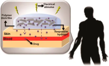
Figure 1. Scheme illustrating the components for the design of smart stimuli-responsive
nanocomposite polymer scaffolds and their challenges for drug delivery and tissue
engineering applications.
2. Criteria and key issues to design smart stimuli responsive scaffolds for biomedicine 2.1. Biomedical issues: biocompatibility and immune response
Human biocompatibility of engineered scaffolds is a key determinant of success for
biomedical applications. Polymers, for example, hold great promise due to their
biocompatibility, as well as their degradation/absorption rates in physiological settings.[25]
When designing nano-engineered scaffolds and aiming for biomedical applications such as
tunable drug delivery, one needs to carefully study how the human body would react to such
scaffolds and anticipate several issues such as biocompatibility, anti-microbial mechanisms,
immune surveillance and also the potential release of toxic, genotoxic and immunogenic
materials.
One way to reach optimal biocompatibility is to design smart and biomimetic
7
replace or supplement.[26] In that sense, the interface between local stem cells and the material
is a key determinant of both the material and stem cells’ fate.[27]
Another key factor to successful biocompatibility (i.e., function and survival of the
biomaterial) is to design materials that are likely to trigger controlled immunological
outcomes that ultimately serve the desired biomedical application.[28] Such tasks however
require a careful understanding of inflammatory and immune responses that would favor the
design of immunomodulatory materials, that can be obtained by tuning extracellular matrix
components. Implantation of biomaterials is rapidly followed by a chain reaction with
activation of coagulation and complement systems, as well as the recruitment of platelets and
immune cells (including polymorphonuclear neutrophils, monocytes and resident
macrophages). Designing materials that shape specific cell and tissue responses locally and
allow, for example, to complete physiological wound healing can control immune reaction. In
addition, allowing recruitment of cells, by surface coating that participate in the homeostasis
of the targeted tissue is likely to facilitate implantation of the biomaterial.[29] Immune
response is further stimulated upon implantation of degradable biomaterials where
degradation products are likely to elicit an additional recruitment of immune components. It is
therefore essential to design smart biomaterials whose degradation products can be controlled.
Along this line, it is essential to control the local and distant dissemination of NPs in the case
of nanocomposite scaffolds (NP dissemination to be expanded). Indeed, when the NPs are
disseminated within the organism after leaking out, one important question is the fate of the
NPs within the organism.
2.2 Fabrication criteria for biomedical applications
Regarding the fabrication criteria, probably one of the first questions that comes to mind is:
do we need a stable or degradable implant over time? This criterion design is crucial in the
fabrication of the implantable device and on its functionalization. In the case of long-term
8
bring a flexible solution to render these implants responsive or to formulate it with
stimuli-responsive nanocomponents. The main medical issue is to ensure a long term (decade)
biocompatible interface.
When the implant is a self-assembled polymer scaffold, it is usually designed to be
degradable or resorbable. However, the majority of soft scaffolds are made of synthetic
polymers which are not degradable and even if the scaffold can be biochemically
disassembled, there is a risk of dissemination or retention of the synthetic polymers in some
organs of the body. This approach requires the need for biodegradable polymer structure
which can be used as a suture or a sacrificial templating scaffolds for tissue reconstruction.
There are some examples of natural or synthesized biodegradable polymers based on
poly(L-lactic acid) (PLLA) and poly(lactide-co-glycolide) (PLGA), whose degradation with time can
be tailored with the copolymer composition and chain length.[30] Scaffolds made of proteins,
polypeptides or polysaccharides such as dextran or chitosan, which can be colonized by cells
and degraded by enzymes, seem to be relevant candidates for such biodegradability and
non-toxicity criteria.[31] Some polymer systems are also designed to have an accelerated erosion by
using T-degradable polymers which can be useful for a desired remotely controlled
degradation.[32] At last, in such architectures the incorporation of heat nanomediators endowed
with hyperthermia properties induced by external fields would be helpful to control these
degradation profiles.
In addition, interaction of cells and tissues with the scaffold to achieve the efficient
scaffold integration in the body, are key issues for fabrication criteria. Bringing optimized
mechanical properties is a key point for biomaterial design which should be designed to
mimic the tissue and cellular micro-environment (extra cellular matrix denoted ECM). The
ideal criteria in terms of mechanical properties for a polymer scaffold are to possess a high
mechanical strength combined with a suitable elasticity (high values of elastic modulus) based
9
limiting protein adherence is also an important aspect to prevent inflammatory responses, to
ensure cell integration. High amounts of water in hydrogels or the chemical functionalization
of the scaffold with low fouling polymers allow limited protein adhesion.[33,34]
2.3 Biomedical implementation of the remotely responsive scaffold
In this section, we discuss the advantages and drawbacks of the potential external fields and
the different responsive nanosystems that may be used in the clinics. Major external fields that
are useful for biomedical applications are near infra-red (NIR) light, X-Ray, magnetic fields,
voltage applied or ultrasonic waves. Even if these stimuli are of different physical natures, the
responses from the scaffold-loaded NPs are essentially a local heating, a higher electrical
conduction or an actuation effect within the host polymer matrix. Other effects (not detailed in
this review), are also reported such as generations of ultra-sound (US) by magnetic NPs[35,36]
and also photoacoustic effects upon photo-irradiation of CNTs.[37] Thus, we will first make a
brief summary on the different physical behaviors due to the different external stimuli: NIR
excitation, alternating magnetic field (AMF), static magnetic field (SMF), X-Ray, US
propagation and electroporation (EP).
In the case of NIR excitation, most nanomaterials absorb and scatter light in the NIR
region especially designed gold NPs through plasmonic resonance and carbon-graphite
structure through vibrational relaxation. Upon NIR irradiation, the excited electrons return
back to the ground state by releasing energy as electromagnetic field or heat, and this heating
will be called photo induced hyperthermia. The NIR irradiation is a noninvasive and
easy-going technology, allowing a controlled and directed excitation (endoscopic excitation of the
region of interest). The typical range of excitation goes from 0.2 to 5 W/cm2 in vivo for 10
minutes on average.[38–41] Nevertheless, this technique suffers from the limited penetration
depth due to scattering and absorption of the tissues. Furthermore, the main drawbacks
associated with the use of NIR-light responsive nanomaterials such as carbon nanotubes or
10
light excitation parameters to avoid tissue over-heating that may lead to inflammation or
necrosis problems.
The application of a static magnetic field (SMF) in combination with radiofrequency
pulses are already commonly used to image soft tissues in the body in magnetic resonance
imaging (MRI). An alternating magnetic field (AMF) applied to injected magnetic NPs is also
used for magnetic hyperthermia treatment applications. This technology is non-invasive and
presents no risks for healthy tissues and total penetration of the full body. The drawbacks are
the complexity of use, the cost and the specific implementation for a surgeon. The typical
ranges of use in frequency of the magnetic field goes from 100<f<437 kHz; 30<time<60min;
B<100mT.[42,43] Current issues with the use of magnetic nanomaterials as stimuli-responsive
systems is that they should be as least cytotoxic as possible. That’s why, even if metallic NPs
or cobalt or zinc-doped ferrites would be highly preferred for their high saturation
magnetization, they would induce important toxicity because of the release of heavy metals.
Iron oxide NPs (IO NPs) appear thus as one of the best magnetic NP candidates for such
biomedical issues, as will be detailed in the next paragraph 3.1.
X-Ray irradiation is linked to high energy electromagnetic beam inducing ionization
radiation. Thus, heavy metal NPs such as gold NPs will enhance the radiation dose through an
increased absorption of X-rays, and this will lead to a subsequent ejection of electrons, known
as the Auger cascade.[44–46] This irradiation is noninvasive, penetrates the full body but the
main drawbacks include risks for healthy tissues, complexity of use, a high cost and a specific
implementation. The typical range of use of this irradiation is 25 Gy of 6 MeV radiation in the
presence of gold NPs.
Ultrasound (US) waves are mainly used to compromise the integrity of drug delivery
liposomes. This process is achieved through two distinct pathways. Either by thermal release
when the US waves are absorbed by the material or by mechanical disrupter. The mechanical
11
This fluid will then oscillate at the US frequency and depending on the amplitude of these
oscillations, it is called stable cavitation which is when the oscillations are below the bubble
resonant radius, or inertial cavitation leading to the collapse of the fluid drop. The collapse
leads either to sono-chemistry generating high temperatures or to shockwaves where the
pressure reaches 10000 atmospheres. This technique is noninvasive, penetrates the full body
and presents only moderate risks for healthy tissues, and as a plus it can be combined with
imaging. This technique is also well suited for lysosome delivery. The typical acoustic
amplitude ranges from less than mW/cm2 (for inertial cavitation) to several W/cm2 (heating).
The frequency ranges from 0.5 to 1.5 MHz and the duty cycle from 1 to 100%. The exerted
pressure varies thus from 100 kPa to MPa.[47,48]
Penultimately, electroporation (EP) is a method suitable to increase the permeability of
the cell membrane and thus it allows molecules to enter the so-called permeable cell. Short
electrical pulses of high field strength are applied to reach EP achievement. The technique is
invasive and mostly used to treat solid tumors. It needs surgery to locate the electrodes close
to the tumor and the EP presents risks for the surrounding healthy tissues. The EP is usually
reached for a potential of 400 V and the time varies from 3 to 12 minutes before NPs
delivery.[49–51]
Finally, each of the external stimuli has its advantages and its drawbacks, depending
on the therapeutic action and of the instrumentation and setup used by the clinicians. These
features of the different stimuli are summarized in the following table (Table 1).
Table 1. Summary of the pro and cons of external stimuli or fields potentially useful in the
clinics
External stimulus / Invasiveness
Advantages Drawbacks Experimental conditions References
NIR light Non invasive Easy to use, applying laser Limited penetration From 0.2 to 5W/cm2 in vivo for 10 minutes on
12 beam (endoscope, direct irradiation) to the region of interest
depth into the tissues. average. AMF Non invasive No risk for healthy tissue Full body penetration Complex to use, costly, specific implementation for a surgeon 100<f<437 kHz, 30<t<60min B<100mT [42,43,52] X-Ray Non invasive Full body penetration
Risk for healthy tissue, complex to use, costly, specific implementation for a surgeon, 25 Gy of 6 MeV radiation in the presence of gold NPs [44–46] US Non invasive Full body penetration Well suited for lysosome drug delivery Combination possible with imaging Moderate risk for healthy tissues Acoustic amplitude From mW/cm2 (inertial cavitation) to several W/cm2 (heating) Frequency from 0.5 to 1.5MHz
Time from minute to hours Pressure 100 kPa to MPa
[48,53] EP Invasive Local solid tumor treatment Surgery needed, risk for healthy tissues.
400V electroporation using pincher electrodes The timing of electroporation was 3 to 12 minutes before nanoparticle treatment [49–51]
3. From nanoparticles and polymers as building blocks to smart responsive
nanocomposite scaffolds
The design of suitable responsive nanocomposite scaffolds for drug delivery and tissue
engineering applications requires first a well-controlled chemical design both of the inorganic
NPs and the polymer scaffolds. Then, achieving a suitable formulation between the NPs and
the polymer scaffold and controlling the response of this smart NP-polymer scaffold upon
external stimuli (magnetic, electric fields or light) are also key issues to control the scaffold
biomedical function (drug delivery or cell actuation/guidance) once implanted within the body.
To perform such tasks, the development of smart nanocomposite scaffold relies both on a
13
chemical design of polymer assemblies such as hydrogels or polymer ELS fibers.
Nevertheless, combined strategies are being developed to give the advantageous features of
both the polymer scaffold and of the NPs, but such strategies are various and complex, and
according to the process used, they have their pros and cons which are highlighted in this
section.
3.1 Inorganic nanomaterials as ideal building blocks for signal conversion
In this section, we describe in detail the design of nanomaterials having a remote response
under external stimuli that can be incorporated in a polymer matrix for biomedical
applications. The main advantage of such nanocomponents, is that they convert one physical
signal into another. Thus, understanding the structure and properties of inorganic NPs is of
paramount importance to control the NPs response upon external stimuli and then to obtain
the desired remote response of the scaffold once the smart device is implanted in a human
body.
3.1.1 Iron oxide NPs and response upon magnetic fields
Superparamagnetic IO NPs were commercially used as T2 contrast agents for
MRI[54,55] and are also developed for a localized therapy achieved by magnetic hyperthermia
(MH).[15] The principle of MH is to submit suspensions of magnetic NPs having suitable
magnetic properties to an AMF with given amplitude and frequency. The resulting elevated
temperature promotes the selective destruction of abnormal cells (when NPs are internalized
into these cells) that are reported to be more thermally sensitive than healthy cells. Moreover,
magnetic hyperthermia enhances the sensitivity of tumor cells towards chemo or
radio-therapy and was also demonstrated to trigger a thermally-induced release of drugs or to act on
cell membranes.[56–58]
These NPs release heat locally under such stimulation through three main mechanisms
depending on their size: Néel relaxation, Brown relaxation and hysteresis loss.[59,60] Néel and
14
hysteresis loss (shift of domain walls)[62] concerns multi-domain particles which display larger
sizes than monodomain superparamagnetic NPs. Néel and Brown relaxation mechanisms
depend mainly on: the magnetocrystalline anisotropy (i.e. a favored orientation of the NP
magnetic moment), the NP volume and the viscosity of the surrounding media. Brownian
relaxation will be typically predominant with bigger size magnetic NPs and lower viscosities
whereas the Néel relaxation will dominate for smaller size NPs and in media having higher
viscosities. The interest of superparamagnetic NPs is that they easily form stable colloidal
suspensions due to their superparamagnetic properties and absorb much more power at
clinically recommended magnetic fields and frequencies. It is indeed important to adjust the
parameters of the magnetic field applied as for a clinically safe application of MH therapy, the
product of the frequency and the amplitude of the magnetic field (H*f) should not be higher
than 5 x 10⁹ A m-1s-1. [63,64]
The positive results obtained by a German company MagForce Nanotechnology
(hospital Charité in Berlin)[65–67] by using MH combined with drug delivery for cancer
treatment demonstrated the potential of magnetic NPs for such therapy. Furthermore,
MagForce obtained 35 M€ from the European Investment bank to develop MH. Nevertheless,
currently the low heating power of usual magnetic NPs makes that large amounts of NPs are
injected locally for MH treatment and these NPs have to be further eliminated by the body.
Therefore most works currently are aiming at increasing the heating power of individual
superparamagnetic NPs.[68–70]
The heating performance is usually assessed by measuring the specific absorption rate
(SAR), named also specific loss power (SLP), which is the power dissipated by magnetic NPs
per unit of mass (Wg-1).[71,72] SAR = C/m * dT/dt with C, the water specific heat per unit of
volume (J K-1L-1), m the iron massic concentration in suspension (Fe g/L) and dT/dt the initial
increase in measured temperature (initial slope of the heating curve). The term Specific Loss
15
the idea of a stored magnetic power released into a thermal power. SAR or SLP (in W/g)
values are highly dependent of the intrinsic features of the NPs (structure and composition)
and also on extrinsic parameters such as the frequency (f) and the amplitude of the magnetic
field (H).[15] For superparamagnetic NPs, SAR/SLP values usually increase with the
frequency or the magnetic field amplitude applied. A standardization protocol has been
proposed very recently for determining accurate, reliable and reproducible SLP values, thus
adequately evaluating the NPs potential.[73]
To envision a clinical translation of MH as therapeutic treatment, magnetic NPs have
to be designed in order to obtain the highest SAR/SLP values which should allow to
administer the lowest NPs dose at clinically safe low frequency and amplitude of the magnetic
field applied.[61,74] The level of heat released by NPs is highly dependent on their structural
and magnetic properties (high Ms and high magnetocrystalline anisotropy)[75–78] and first
works revealed that iron oxide NPs with a spherical shape and a mean diameter in the range
12-20 nm are clinically suitable for MH therapy.[79] Thus, synthesis methods of iron oxide
NPs[80] were deeply investigated to improve the MH performance of NPs. The magnetic
properties of NPs have been optimized by tuning mainly their shape and their composition
(doped iron oxide or core-shell structures). Among synthesis methods, the thermal
decomposition method[81,82] was shown to be particularly suitable for controlling the size,
shape and composition of NPs, as well as the polyol method. Thus for example, nanocube
iron oxide NPs[83–85] with an optimal size of 19 nm were found to have high SAR values
(2453 W.g-1 at 29 kA.m-1 and 520 kHz).[74] Recently, they boosted the heating power of the
cubic NPs by doping with cobalt.[86] Core-shell NPs consisting of a core with a high magnetic
anisotropy and of a shell with a small magnetic anisotropy displayed a very important
anti-tumor effect induced by MH.[70] Otherwise when iron oxide NPs with a mean diameter of ~22
nm coated with dendron molecules display a SAR of 120 and 395 W.g-1Fe with a field
16
same NPs coated with a silica shell presented a SAR of 120 and 209 W.g-1Fe respectively.[88]
Doping of iron oxide with cobalt, zinc or manganese is also a way to improve heating
properties.[68,89] However, the possible toxicity of these doping elements has restricted their in
vivo development so far. Nano-engineering the shape of iron oxide nanomaterials (Figure 2)
is for instance a way to tailor high SAR values and it allows the use of less cytotoxic magnetic
NPs.[87]
The heating efficiency of an ensemble of magnetic NPs depends not only on the
structural and magnetic properties of NPs but also on the magnitude and frequency of the
applied magnetic field, the media viscosity and NPs concentration and aggregation state.[90–92]
Therefore, IO NPs displaying high heating power are currently available for MH. In addition,
current investigations showed that unlike the majority of inorganic NPs, IO NPs can be
metabolized by the human body through biodegradation inside the lysosomal compartment
17
Figure 2. TEM images of a) nanospheres, b) nanoplatelets, c) nanocubes and d) nanooctopods
associated respectively to SAR values of 395, 177, 238 and 462 W.g-1 at f=795 kHz and
H=12 kA m-1. Reproduced with permission.[87] Copyright 2019, IOP Publishing.
3.1.2 Carbon-based materials and responses under NIR light conversion and electric
fields
Carbon nanotubes (CNTs) and graphene have remarkable mechanical, thermal and electron
conductivity properties. Especially regarding this latter property, they are relevant key
components to develop electrically conductive artificial tissues which allow us to envision
many applications for drug release or tissue regeneration.[4,19] However, one first main issue
encountered with these carbon based materials is their long dimensions (several tens of
microns scale length) which may display similar toxicity to asbestos and thus requires
procedures to shorten their dimensions. To solve this problem, CNTs have been treated in a
strong oxidizing media under ultrasound to decrease their length and thus their
length/diameter ratio.[94] Furthermore, in the last decade, graphene has emerged as an exciting
new material. After its first "characterization" following a mechanical exfoliation,[95] the most
common approach to achieve graphite exfoliation is the use of strong oxidizing agents to
obtain graphene oxide (GO), a more hydrophilic carbon material.[96] Graphene can be also
obtained under US in an ammoniac media.[97] More recently another method was developed
consisting of exfoliation assisted by US in an organic liquid[98] or with a surfactant in water
which allowed obtaining graphene with a high purity.[99]
Another main issue with CNTs or graphene surfaces is their hydrophobicity and the
need of efficient strategies to cover their surfaces with a hydrophilic coating. There are
several strategies with surface chemical modifications to introduce hydrophilic functional
(amines, hydroxyls or carboxyls) groups[100,101] and improve dispersions in aqueous media.
18
density. To improve CNTs dispersions handling and increase the potential for versatile
chemical modifications, we have recently proposed the coating of such hydrophobic carbon
based materials with mesoporous silica (MS).[102] In this work, a surfactant mediated process
for MS coating was achieved on individual sliced CNTs and exfoliated graphene layers. It
ensured an efficient separation and dispersion in aqueous media of the CNT and graphene as
well as a high antitumor drug loading. We also showed in a parallel study, that this MS layer
can be selectively etched from the structure after protein coating allowing the development of
a new and alternative method to coat CNTs homogenously with proteins.[103]
3.1.3 Gold NPs for NIR plasmonic and electrical response.
Gold NPs are also promising theranostic NPs because, when correctly designed, they may
provide imaging by NIR fluorescence imaging and therapy through radiosensitization and
photothermal therapy. Indeed, due to their unique optical properties and biocompatibility,
gold NPs, when irradiated with NIR light, absorb the light and convert it locally into
heating.[104] The fact that the NIR light is rather suitable to ensure the lowest absorption and
highest penetration depth in biological tissues and to ensure a high local release of heat makes
them very promising for photothermal cancer therapy.[105] Some pre-clinical studies have also
evidenced the radiosensitizing effect of gold NPs whose presence in tumors has been shown
to enhance the efficacy of external radiotherapy.[45] Photothermal ablation is currently clinically developed in USA by the company “Nanospectra Biosciences”, and the safety of
their gold/silica NPs has been demonstrated. However these interesting properties of gold NPs
depend strongly on their size and shape.[106,107]
The effect of the gold NPs morphology has been widely investigated and the
photothermal properties of gold NPs with different morphology, such as gold nanorods,
nanostars, nanorings, nanocages, and hollow nanoshells, have been investigated.[108–111]
Non-spherical morphologies were found necessary to observe a suitable effect in NIR region.
19
photothermal therapy because, once gold NPs are internalized in cells, they form
self-assembled large clusters directly inside cells, which leads to a surface plasmon resonance shift
from the visible to the NIR region.[112,113] This effect was also observed in gold nanoparticles
coated with periodic mesoporous organosilica (PMO) as several cores are encapsulated inside
the nanoPMOs shell.[114,115] Such gold NPs clusters in the cells[116] were also suggested to
result in laser-induced bubble formation that is more effective for eliminating cells.[112]
Since only NIR light can penetrate inside living tissues, gold nanoshells, gold
nanorods, and gold nanocages which have surface plasmon resonance absorption in the range
650–900 nm are ideal for in vivo imaging or phototherapy. The preparation of gold nanorods
with different aspect ratios (length divided by width), which allows tuning their plasmonic
absorption peak in the NIR region, is simple and well established. In addition the gold
nanorods size is rather small favouring their internalization by cancer cells.[117–119]
3.2 Polymer scaffolds as host integrative matrix
There are different types of polymer scaffolds that can play the role of the matrix holding the
responsive NPs. Such polymeric systems display various physico-chemical features: tunable
mechanical stability, elasticity, response to biological media (low adherence, biodegradation),
response to thermal changes through T-transitions or T-swelling. In this section, two different
types of polymer matrixes, commonly used as scaffolds, are described: hydrogels, and ELS
fibers and their features as scaffolds are presented.
3.2.1 Hydrogels
A hydrogel corresponds to a hydrophilic 3D polymer network that can swell up to
hundreds/thousands of times. There are usually two possible cohesion states in hydrogels held
by chemical or physical crosslinking.[120] Chemical gels are bound by various types of
chemical linkages established through covalent crosslinking or polymerization reactions
20
(H bond-hydrophobic domains, Van der Waals interactions). Chemically-assembled gels are
particularly stable as their covalent bonds are resistant to breaking under biological conditions
(unless desired) whereas physical gels are usually more prone to be used as resorbable
hydrogels. Importantly, one of the most attractive features of polymer hydrogels is that they
can be chemically tailored to adjust their physicochemical properties: swelling, mechanical
elasticity, molecule diffusion, degradability upon cellular or tissues conditions etc. Indeed,
hydrogels can be chemically designed to respond to numerous chemical stimulations: pH, T
and/or ionic strength.[121,122] Such designs was essentially performed so that they respond to
local biological stimuli such as: the acidic pH of cancer tissues, or the cellular endosomes.
Therefore, regarding drug delivery applications, hydrogels scaffolds were used for the release
of various products for therapeutic applications. Their cross-linking levels allowed the ability
of tuning the porosity and the diffusion of the therapeutics species depending on their size
(small molecules, proteins).[123] Another feature of hydrogels for the engineering and
industrial process aspects is that they can be molded with any required shape and
dimensions.[124]
Regarding implantation of the hydrogels, the design of in vivo injectable polymer
scaffolds has become an emerging field in this last decade because of their high potential for a
range of biomedical applications including regenerative medicine/tissue engineering or drug
releasing smart implants.[125–128] Polymer hydrogels are well suited to address the features
required for the design of in situ injectable gels which are: i) a stringent biocompatibility as
the gel must be non-toxic and non-immunogenic, ii) a rapid in vivo gelation (less than 1 min.)
at the injection site at 37 °C, iii) mimicking the complex tissue environment with suitable
mechanical properties close to the ECM iv) stability and stiffness over time until potential
long-term biodegradation within the body.
In the last few decades, there have been some very interesting approaches of in situ
poly(N-21
isopropylacrylamide) (PNIPAM) or Pluronics co-polymers that are solubilized below body T
and gelates above the T threshold. Hence, this strategy may be of interest to solve the problem
of direct implantation of the scaffold. Furthermore, as conventional hydrogels, they are
obtained though chemical or physical methods i.e. respectively via the formation of covalent
or non-covalent bonds. Current chemical methods include either photo-polymerization of
preformed acrylate- or methacrylate-modified polymers (PEG, hyaluronic acid,
alginate)[129,130] or covalent cross-linking of complementary groups in water-soluble polymers
based on various chemistries such as Michael-type additions[131,132] or Schiff’s base
formation.[133,134] Alternatively, physical gels are obtained through the formation of
non-covalent bonds which can be, for instance, of a hydrophobic nature as in the case of
thermosensitive polymers including PNIPAM[135,136] or various block copolymers of
PEG[137,138] which undergo a transition from an hydrophilic state to an hydrophobic state when
the temperature reaches the physiological temperature.
3.2.2 Electrospun fibers
The ECM of biological tissues (bone, muscle, tendon etc…) is a 3D fibrous structure with
protein-based fibers having an average diameter ranging from a few tens to hundreds of
nm.[139] Therefore, the development of scaffolds mimicking the native structure of ECM is of
prime importance in tissue engineering in order to favor cell adhesion and proliferation
leading to an efficient repair of tissues.[140]
Electrospinning has been identified as a process of choice allowing the fabrication of
such scaffolds with various polymers.[141] During electrospinning,[142] a polymer solution in a
semi-dilute entangled regime is pumped towards a metallic needle which is subjected to an
electric field in the order of 1 kV/cm. The charged droplet exiting the needle takes the shape
of the so-called Taylor cone from which a jet is propelled towards a collector generally
connected to the ground. During its flying, the jet is subjected to vigorous
22
decrease of the jet diameter. At the same time, these whipping movements induce the
evaporation of the solvent resulting finally in the pseudo-random deposition of a continuous
nanofiber having an average diameter ranging between few tens of nm to few microns as a
function of the processing and material parameters. The deposited nanofiber takes the
morphology of a non-woven nanofibrous mat also called a scaffold. When decreasing the
number of polymer chain entanglements in the solution, the resulting less viscous jet breaks
into smaller droplets. Under these conditions, solid microparticles are deposited and the
process is named electrospraying.[142]
Although electrospinning is a well-known process allowing the fabrication of
non-woven scaffolds, it has been demonstrated that various hierarchically controlled fibrous
structures can be obtained[143] such as core-shell nanofibers[144], hairy fibers[145], aligned
fibers[146] as well as micro-patterned 3D scaffolds[147] (Figure 3). All these features render the
ELS biocompatible materials as promising candidates for various biomedical applications
such as biomaterials for tissue engineering[141] and drug delivery purposes.[143] For the latter
case, well-designed scaffolds can embed drugs which can be released in a controlled manner.
Indeed, the versatility of the process of electrospinning allows the encapsulation of active
compounds through diverse strategies such as fiber surface modification, direct
electrospinning of the drug within the polymer solution, core-shell electrospinning or
electrospraying of drug loaded particles simultaneously with the electrospinning of the
scaffold. Depending on the used strategy, the morphology of the scaffold and the kind of
23
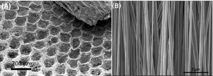
Figure 3. Examples of structured electrospun scaffolds: (A) Organotype culture of cells from
a chicken bone embryo onto a honeycomb PCL/Hydroxyapatite ELS scaffold produced by
electrospinning/electrospraying onto a micropatterned collector. Reproduced with
permission.[149] Copyright 2018, American Chemical Society. (B) Aligned silk fibroin ELS
scaffolds obtained by electrospinning onto a rotating collector as synthetic nerve guide.
Reproduced under the terms of the CCBY license.[150] Copyright 2018, Holder name.
3.3 Formulate polymer scaffolds and NPs: process, issues and physico-chemical features
Another level of functionality can be reached by the combination of responsive NPs with
hydrogels or ELS fibers within a polymer composite scaffold. With the aim of obtaining
implants that will respond to external stimuli such as NIR light, magnetic field or electric
voltage, it is thus of paramount importance to develop powerful processes of scaffold
formulations between the polymer and the NPs.
There are different ways to formulate polymer scaffolds and NPs. Various chemical
driving forces are involved for the NPs formulation and controlling NPs/polymer interactions
is one of the key issues to deal with. It is indeed crucial to chemically tailor the NP surface in
order to provide physical or chemical crosslinking of the polymer matrix and obtain a
homogeneous NP dispersion while avoiding NP aggregation or leaking.[151,152] In this section,
we review the methods allowing the incorporation of different types of inorganic NPs with
24
3.3.1 Nanocomposite hydrogels
There are different possible strategies to formulate NPs in a hydrogel polymer scaffold which
are respectively:
i) Blending the NPs and the polymer;
ii) Synthesizing the NPs in situ within the polymer scaffold
iii) Grafting covalently the NPs to the surface of the polymer scaffold
i) Blending the NPs and the polymer
The simplest method is probably to blend the NPs suspension in an aqueous solution of the
polymer precursor and then trigger the gelation. Gelation can be obtained according to various
polymer chemistries (e.g.) polycondensation, photopolymerization, coordination of alginate
with calcium forming egg boxes, or via H-bond network formation like in the agarose case.
For instance, in a work by Bannerman et al.,[153] the authors synthesized magnetic cryogels by
blending poly(vinyl alcohol) (PVA) and magnetic IO NPs through freeze thawing procedures
to formulate a fully biocompatible magnetic hydrogel. The NPs can also be dispersed with the
monomers and then the polymerization of monomers is induced by different triggers (heat,
light induction etc) forming the composite gel. For instance, Frimpong et al.[154] dispersed
iron oxide NPs in NIPAM monomer solution and triggered gel formation by radical
polymerization. In another example, Servant et al., prepared poly(methacrylic acid) hydrogels
loaded with graphene by the in situ polymerization of methacrylic acid.[155] The graphene
layers were initially dispersed in water by a ball milling process. However, while the blending
technique is particularly simple to proceed, one of the main problems associated with this
technique is the difficulty to disperse the NPs homogenously. Some works reported thus the
possibility to crystallize the NPs in situ within the polymer scaffolds.
25
The second method used is an in situ post-synthesis approach where the nucleation of NPs is
achieved within a polymer matrix. For instance, Ilg et al.,[156] triggered the in situ
precipitation of iron salts into IO NPs within the polymer gel. In another work, Wang et al.,
designed a magnetic composite polymer scaffold by forming a complex between iron (II and
III) cations and amine groups of chitosan by increasing the pH of the scaffold.[157] Similarly,
gold NPs can be embedded in gel precursors or grown inside the hydrogel by precipitation of
gold salts.[158,159] Here too, such in situ synthesis approaches can display some inconvenience.
While the seed nucleation should ensure a homogenously dispersed distribution of NPs, one
main issue with such a method is that the reaction conditions of nucleation (basic or
reductive) to form the NPs may be too harsh both for the polymer architecture and also for the
cells or biological medium that may interact with such structure.
iii) Grafting covalently the NPs to the polymer scaffold
A third possible approach is the covalent crosslinking by the NPs themselves where NPs act a
new points of polymer crosslinking especially in hydrogels.[160] The great advantage of this
method is the high chemical and mechanical stability and also the limited leaking out of the
NPs. The increase of the mechanical properties is highly needed for biomaterial and
regenerative medicine applications. Hence, in a work by Skardal et al., cross-linked thiol
functionalized gold NPs in a blended hyaluronic acid/gelatin hydrogel were shown to have a
great stiffness as compared to non-cross-linked gold NPs.[161] Carbon-based materials are also
particularly suitable nanomaterials to crosslink hydrogels. In a work by Gaharwar et al., the
authors investigated the effect on their mechanical properties of the covalent crosslinking of
CNTs within a poly(glycerol sebacate) hydrogel.[162] With incorporation of 1% CNTs
followed by covalent crosslinking, the authors measured increased values of the ultimate
tensile strength (275 vs 122 kPa ) and of the elastic modulus (1014 vs 198 kPa) as compared
to the poly(glycerol sebacate) hydrogel alone (without CNTs). In another work, covalently
26
recreate more rigid tissues.[163,164] Such covalent cross-linking is known also to have an effect
over cell interactions with the scaffold surface.
3.3.2 Nanocomposite ELS fibers
There are also several possible routes to prepare nanocomposite ELS fibers leading to
different locations of the NPs in the scaffold (Figure 4):
i) Blend electrospinning
ii) In situ particle synthesis
iii) Use of simultaneous electrospray and electrospinning
iv) Post-electrospinning impregnation
In the two first strategies, the NPs are distributed inside the fibers. They are thus protected
from the environment and consequently, leaking of the NP out of the scaffolds is low.
However, their contact with the environment is also limited. Therefore, it would not be the
method of choice for chemically triggered drug release, sensing or catalysis for example. In
the last two strategies, NPs are either trapped in the ELS matrix during the electrospinning
process, physically adsorbed or chemically grafted at the surface of the nanofibers. They will
be accessible to the environment but they are more prone to leaching from the fibers.
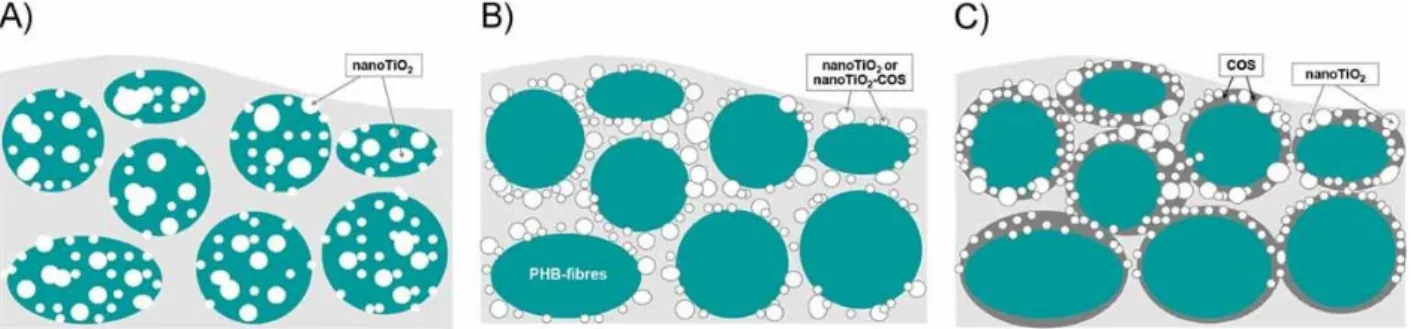
Figure 4. Location of TiO2 NPs in a poly(3-hydroxybutyrate) (PHB) nanofibrous mat as a
function of the following strategies: A) blend electrospinning of the dispersion of nanoTiO2 in
PHB solution, B) simultaneous electrospinning of a PHB solution and electrospray of a
27
impregnation of the PHB nanofibers in a nanoTiO2-COS suspension. Reproduced with
permission.[165] Copyright 2013, Wiley.
i) Blend electrospinning
Blend electrospinning is the easier way to obtain NP/polymer composite scaffold. The
pre-prepared NPs are simply dispersed in the polymer solution prior to electrospinning. They are
thus distributed in the whole nanofiber section. If the nanofiber is not degradable, the NP
should not leach out of the nanofibers, however, as most of the particles are not on the fiber
surface, they are not directly accessible and a drug, released from the NP, still has to diffuse
through the polymer matrix before being delivered to the environment. In this strategy the
main difficulty is to avoid aggregation or sedimentation of the NP in the polymer solutions
prior to electrospinning and ensure an even dispersion in the polymer matrix.
Magnetic IO NPs were electrospun by blending the NP suspension with the polymer
solution prior to electrospinning.[166,167] In order to improve the dispersibility of NPs, Shan et
al.[168] prepared the Fe3O4 NP by co-precipitation in the presence of PLLA. The synthesized
PLLA surface-coated NP later electrospun in PLLA solutions were homogeneously
distributed along the nanofibers. Coaxial electrospinning was also performed with
magneto-rheological fluid (MRF) as the core and a poly(ethylene terephthalate) as the shell.[169] IO NPs
were still mobile after electrospinning in the MRF encapsulated inside the fibers. Their
response to an external magnetic field was thus instantaneous, increasing the mechanical
properties of the nanofibers thanks to dipole-dipole interactions between the NPs.
Other types of NPs were also blended to the polymer solutions before electrospinning. For
instance, mesoporous silica NPs (MSNPs) loaded with a drug were also electrospun by blend
electrospinning with PLLA[170] or PLGA[171] with concentration of 15 wt% and 25 wt%,
respectively, of well dispersed MSNPs in the final fibers. Another example is the preparation
28
Finally, graphene oxide (GO) was also incorporated into PVA[173] or PLGA[174] with the effect
of improving the mechanical properties and protein adsorption ability of the fibrous mat.
ii) In-situ nanoparticle synthesis
Metallic salts can be included in the polymer solutions prior to electrospinning and reduced in
NPs in situ just before or after the electrospinning step. The size of the NPs and their uniform
dispersion of the NPs in the polymer matrix is one of the major challenges of this process.
Gold NPs were reduced in situ by addition of tea polyphenol to a polyacrylonitrile (PAN)
solution in DMF, just prior to electrospinning. The authors showed that the PAN played the
role of a stabilizer through the chelating effect between gold and the cyano groups, allowing
the production of well dispersed gold NPs with smaller NPs sizes, as when the gold NPs are
synthesized by addition of tea polyphenol in water.[175] In a similar manner, Celebioglu et
al.[176] have prepared PVA nanofibers containing silver NPs by in situ reduction of AgNO3
directly by the PVA, used as reducing agent, prior to electrospinning. They obtained
8 ± 0.5 nm NPs but observed some aggregation. Addition of 7.5 to 25 wt% of -cyclodextrin (HP CD) in the formulation, as reducing and stabilizing agent, allowed them to control the size and the dispersion of the silver NPs in the fibers. In the presence of HP CD well dispersed particles as small as 1.8 ± 0.4 nm were obtained. These composite nanofibrous
mats have also shown interesting antibacterial properties.
In another example, in situ synthesis of magnetic IO NPs was conducted by adding a 2:1
molar ratio of ferric and ferrous chloride to an aqueous PEO or poly(vinyl pyrrolidone) (PVP)
solution containing sodium borohydride to reduce the ions to NPs. Electrospinning was
performed directly after the NPs synthesis to obtain composite polymer/magnetite nanofibers.
Smaller magnetite NPs of 10 ± 4 nm were obtained in PVP, probably due to the stabilizing
effect of PVP for the NPs as compared to 27 ± 4 nm in the PEO fibers.[177]
29
Electrospinning and electrospraying can be performed simultaneously[178] from both sides of a
rotating collector, or in alternating layers[179] leading to a composite fibrous scaffold with
particles trapped in it. For example, electrically conductive composite CNT/polyurethane
(PU) nanofibrous scaffolds for cardiac tissue engineering were developed by simultaneous
electrospray of multiwall carbon nanotubes and electrospinning of PU nanofibers.[180] The
CNT adhered on the PU nanofibers forming an interconnected electrically conductive
web-like structure with increased mechanical properties as compared to the pure PU nanofibrous
mats. Finally, alternating electrospinning and electrospraying on a microstructured collector
also allows the control of the localization of the NPs in the scaffolds by the Electrostatic
Template-Assisted Deposition (ETAD) of NPs on ELS fibers. This technique was developed
by Nedjari et al.[181] who prepared microstructured hydroxyapatite-poly( -caprolactone) (PCL) scaffolds for bone regeneration and could easily be adapted to other kind of NPs.
iv) Post-electrospinning impregnation
Another method to prepare nanofibrous scaffolds containing NPs is post-electrospinning
impregnation. An advantage of this method is that the NPs will be deposited on the surface of
the nanofibers and thus, will be in direct contact with the environment. However, it may also
lead to leaching during use.
Superparamagnetic Fe3O4 NPs were assembled at the surface of ELS polyurethane
(PU) fibers by simply dipping into a NP colloidal suspension in the presence of ethylene
glycol. The interactions between the NPs and the PU fibers are very strong, probably due to
hydrogen bonding between the amide groups of the PU backbone and the hydroxyl groups
capped on the surface of the superparamagnetic NPs.[182]
This method can also be used for the preparation of metallic NPs. For example, silver
NPs containing nanofibers were also obtained by dipping of poly(acrylic acid) (PAA)/PVA
crosslinked ELS nanofibers in AgNO3 aqueous solution enabling the complexation of the Ag+
30
for 30 min in a sodium borohydride (NaBH4) solution to reduce the ions in Ag0 NPs.[183] The
same group also prepared Fe0NP/PAA/PVA composite nanofibers using the same method.[184]
In a similar manner, gold NPs were also attached to the surface of ELS fibers using Layer by
Layer (LbL) deposition of 10 bilayers of lysozyme and tannic acid, followed by impregnation
of the scaffolds in chloroauric acid solution and in situ reduction to gold NPs by the tannic
acid present in the LbL[185]
4. Applications of nanocomposite scaffolds for controlled drug delivery
Probably one of the great advantages to use remote action by external stimuli (magnetic,
electric fields or light) over other local mechanisms of release (pH, local etc) is the possibility to perform “on-demand” pulsatile release which ensures the drug to be dosed spatially and
temporally. Usual release methods involve the high diffusion of the drugs as a burst release
and this approach of remote pulsatile release may be more efficient in future applications to
envision long term controlled drug delivery. Here, in this paragraph, we review the possibility
of drug releases based on the ability of the polymer scaffold to respond to SMF, AMF, NIR
light or electric field (voltage). The examples presented in this section are mainly based on
hydrogels and on ELS fibers.
4.1 Drug delivery upon static magnetic field by magnetomechanical effects
i) Magnetic guiding of anticancer drugs under SMF field
The transport of an anticancer molecule loaded within magnetic microgels is a concept that
was developed since the early 1980s. For instance, Chen et al. achieved the guiding of
magnetic PVP hydrogel microparticles (c.a. 1 micron size) called ferrogel with a magnet for
their retention in a rabbit tumor.[186] The ferrogel was loaded with an antitumoral drug,
bleomycin A5 hydrochloride (BLM), and was injected intra-arterially. A 0.5 T magnet was
placed at the tumor location for 24 h and the microparticles accumulated there magnetically.
This approach showed a drastic tumor size reduction over 2 weeks as compared to the tumor
31
ferrogel led to an efficient tumor reduction, it shows that the concept of the magnetic guiding
in vivo is working, which could be interesting to reduce secondary effects by treating the
tumor locally.
ii) Scaffold with remote controlled release of antitumor drugs under SMF applied
Besides the magnetically induced transport of the magnetic carriers, the application of a SMF
(or DC magnetic field) is also a way to act mechanically on a magnetic ferrogel and to trigger
therapeutic agent diffusion. In a work by Zhao et al.,[6] alginate based ferrogels incorporating
Fe3O4 NPs were shown to be efficient for the release of various therapeutics (mitoxantrone,
plasmid DNA, and SDF-1α, a chemokine) in vitro under SMF (Figure 5, left). Under the
magnetic field applied, the magnetic ferrogel is submitted to large deformation (over 70% of
its initial dimensions) which generates a water flow through the gel and thus drug diffusion.
The gel was stimulated 2 minutes every 30 minutes or 2 h (depending on the agent used) and
the drug release profiles showed a well-controlled release achieved specifically when the
magnetic field is applied. Additionally, such a powerful strategy was applied successfully for
the controlled delivery of mouse mesenchymal stem cells in vitro and in vivo. (Figure 5,
right).
Conversely, the application of a SMF can help also to block drug diffusion by
magnetically shrinking the hydrogel. Hence, Liu et al. developed a PVA/IO NPs hydrogel
nanocomposite whereupon when a DC magnetic field was applied, the magnetic NPs come
closer in space, reversibly aggregate and retain the embedded drugs within the gel by blocking
drug diffusion.[187,188] Once the field is removed, the diffusion of the drug restarts out of the
gel. The specificity of this system over other sources of external stimuli is that a SMF is
required to block the drug diffusion whereas usually the drug is released upon application of
32
Figure 5. At the left. Graphs showing the cumulative release profiles of various therapeutics
(A) mitoxantrone, (B) plasmid DNA, and (C) the chemokine SDF-1α under SMF. At the
right A) scheme of the cell release upon DC field from the macroporous gel, B) graphs
showing the % of fibrobast cells release with time and cycled magnetic field application and
C) microscopy images showing the proliferation of the cells released with time. Reproduced
with permission.[6] Copyright 2011, National Academy of Sciences.
4.2 Drug delivery upon an alternating magnetic field by magnetothermal effects
4.2.1 Magnetothermally induced remote release of molecules under AMF field
Various works demonstrated the possibility of a controlled pulsatile drug delivery by AMF
applied for different applications. Hence, Satarkar et al. investigated the application of a
radiofrequency field to a PNIPAM scaffold loaded with magnetic NPs and Vit. B12 as a
33
kA.m-1) reached the LCST of 32 °C of the PNIPAM polymer and ensured the release of the
Vit. B12 drug by shrinking effect. This effect can be well controlled as demonstrated by the
pulsatile release achieved upon cyclic stimulations.
Similarly, Hoare et al. developed biocompatible and biodegradable in situ injectable
superparamagnetic IO NPs based PNIPAM hydrogels, made by the condensation of
aldehyde-grafted dextran with IO NPs functionalized with PNIPAM-hydrazide.[190] The in situ gelation
is easy and rapid, and the cross-linking with the magnetic NPs ensured an important increase of the storage modulus (elastic modulus ≥60 kPa). These composite hydrogels were
demonstrated to be biocompatible in vitro and in vivo and were loaded with bupivacaine (an
anesthetic agent). Controlled pulsatile release of bupivacaine drug could be achieved through
application of several pulses of 5 minutes of AMF (f= 200 kHz, I=30 A, P=1.3 kW) leading to
34
Figure 6. A) Scheme of the biocompatible and biodegradable in situ injectable
superparamagnetic IO NPs based PNIPAM hydrogels, made by the condensation of
aldehyde-grafted dextran with IO NPs functionalized with PNIPAM-hydrazide B) Storage modulus in
the presence or in the absence of IO NPs. C) Controlled pulsatile release of bupivacaine
through application of 5 minutes pules of AMF (f= 200 kHz, I=30 A, P=1.3 kW). Reproduced
with permission.[1] Copyright 2014, Elsevier.
In a last example, Kim et al. prepared cross-linked PNIPAM ELS nanofibers
containing doxorubicin (DOX) and magnetic NPs.[191] The application of an AMF (f = 166
kHz, I = 480 A, P = 362 W) on the electrospun mat containing 31% of magnetic NPs led to an
increase of the fibers temperature from 25 to 45 °C, above the LCST of PNIPAM, resulting in
fiber deswelling and release of DOX in the environment. The temperature reached by the
magnetothermal effect could be controlled by choosing the appropriate concentration of
magnetic NPs in the fibers. Furthermore, swelling and deswelling of the fibers being
reversible, on-off switchable release could be obtained.
4.2 .2 Magnetic hyperthermia therapy combined with magnetothermal drug delivery
Regarding the potential for biomedical applications and anticancer treatments, the possibility
to combine localized hyperthermia and drug delivery was recently achieved from such
polymer/NPs composite implants to destroy cancer cells at elevated T above 42 °C. Hence,
methacrylate-based PEG magnetic nanocomposites were designed as potential implants for
the thermal ablation in cancer treatment along with drug delivery.[192] The authors showed that
build-up parameters of the hydrogels such as the ethylene glycol amount and the crosslinking
level were determinants for their swelling behavior. Upon AMF, they showed that the iron
oxide content and field amplitude influenced the resulting T profiles and thermal transfers.