HAL Id: inserm-00849267
https://www.hal.inserm.fr/inserm-00849267
Submitted on 6 Oct 2013
HAL is a multi-disciplinary open access
archive for the deposit and dissemination of
sci-entific research documents, whether they are
pub-lished or not. The documents may come from
teaching and research institutions in France or
abroad, or from public or private research centers.
L’archive ouverte pluridisciplinaire HAL, est
destinée au dépôt et à la diffusion de documents
scientifiques de niveau recherche, publiés ou non,
émanant des établissements d’enseignement et de
recherche français ou étrangers, des laboratoires
publics ou privés.
Statistical Analysis of White Matter Integrity for the
Clinical Study of Specific Language Impairment in
Children
Emmanuel Vallee, Olivier Commowick, Camille Maumet, Aymeric Stamm,
Elisabeth Le Rumeur, Catherine Allaire, Jean-Christophe Ferré, Clément de
Guibert, Christian Barillot
To cite this version:
Emmanuel Vallee, Olivier Commowick, Camille Maumet, Aymeric Stamm, Elisabeth Le Rumeur,
et al.. Statistical Analysis of White Matter Integrity for the Clinical Study of Specific Language
Impairment in Children. MICCAI 2013 Workshop on Computational Diffusion MRI, Sep 2013, Japan.
pp.187-195, �10.1007/978-3-319-02475-2_17�. �inserm-00849267�
the Clinical Study of Typical Specific Language
Impairment in Children
Emmanuel Vall´ee, Olivier Commowick, Camille Maumet, Aymeric Stamm, Elisabeth Le Rumeur, Catherine Allaire, Jean-Christophe Ferr´e, Cl´ement de Guibert, and Christian Barillot
Abstract Children affected by Specific Language Impairment (SLI) fail to develop a normal language capability. To date, the etiology of SLI remains largely unknown. It induces difficulties with oral language which cannot be directly attributed to in-tellectual deficit or other developmental delay. Whereas previous studies on SLI focused on the psychological and genetic aspects of the pathology, few imaging studies investigated defaults in neuroanatomy or brain function. We propose to in-vestigate the integrity of white matter in SLI thanks to diffusion Magnetic Res-onance Imaging. An exploratory analysis was performed without a priori on the impaired regions. A region of interest statistical analysis was performed based, first, on regions defined from Catani’s atlas and, then, on tractography-based regions. Both the mean fractional anisotropy and mean apparent diffusion coefficient were compared across groups. To the best of our knowledge, this is the first study fo-cusing on white matter integrity in specific language impairment. 22 children with SLI and 19 typically developing children were involved in this study. Overall, the tractography-based approach to group comparison was more sensitive than the clas-sical ROI-based approach. Group differences between controls and SLI patients
in-Emmanuel Vall´ee· Olivier Commowick · Camille Maumet · Aymeric Stamm · Jean-Christophe Ferr´e· Christian Barillot
Inria, INSERM, VisAGeS U746 Unit/Project, F-35042 Rennes, France Elisabeth Le Rumeur· Jean-Christophe Ferr´e
University Hospital, Department of Neuroradiology, F-35033 Rennes, France Catherine Allaire
Centre Toul-arC’hoat, F-29150 Chateaulin, France Cl´ement de Guibert
European University of Brittany-Rennes 2, LAS-EA 2241, F-35043, Rennes, France and Univer-sity Hospital, Regional center for language and learning disorders, F-35033 France
Contact: e-mail: Olivier.Commowick@inria.fr
58 Emmanuel Vall´ee et al.
cluded decreases in FA in both the perisylvian and ventral pathways of language, comforting findings from previous functional studies.
1 Introduction
Some children fail to develop their language capability for no obvious reason. Their pathology typically induces difficulties with oral language, and cannot be attributed to intellectual deficit or other developmental delay. This developmental disorder is known as Specific Language Impairment [2].
The literature [2, 18] does not show evidence of brain lesion or educational is-sues, so the origin of the trouble is not well understood. While this pathology has been studied in its psychological or genetic aspects [3, 4], few studies have been con-ducted on the neuroanatomical and functional aspects. The current neuroanatomical hypothesis is a maturation default and/or an abnormal functional specialization of the networks dedicated to language.
SLI covers a large and heterogeneous clinical spectrum. In the context of this study, we focus on Typical-Specific Language Impairment (T-SLI), a subtype of the pathology, in which the trouble mainly affects the structural aspects of language (including morphosyntax and phonology) [9].
A functional MRI study was recently conducted by de Guibert et al. [9] on a group of subjects similar to ours. It revealed an abnormal lateralization of language function in patients. While the left hemisphere has usually a predominant role in language tasks, their study revealed a reduced activity in the supramarginal region of the left hemisphere (Geshwind area) and an increased activity in the right hemi-sphere close to the counterpart of Broca’s region.
Earlier, Kim et al. published a study [11] on language impairment in children, using diffusion MRI. They measured the Fractional Anisotropy in six regions of the brain, and found a significant reduction in the genu of the corpus callosum. It can have an importance in language impairments as it is the main bundle that connects the two hemispheres and therefore manages connections between the two hemi-spheres, as shown by Preis et al. in [17]. Although their results are interesting, the study was led on very young children (mean 3.8 years old). It is then hard to differ-entiate a persistent language impairment from a transitional language retardation. Also, the specificity of the trouble can be questioned, as other non-language related troubles were not diagnosed. Filippi et al [7] led a diffusion imaging study on devel-opmental delay in children. The study revealed an alteration of white matter bundles in affected children. Mao et al. [14] also conducted a diffusion imaging study where they showed that language impaired subjects had a decreased white matter integrity in the left frontal and medial temporal areas.
To the best of our knowledge, no previous study investigated white matter in-tegrity in T-SLI. Understanding the neurological basis of SLI is of great interest as the specificity of the trouble allows to target a single impaired function (language) while most other developmental disorders are characterized by complex patterns of
clinical signs. In this paper, we aim at studying this integrity through the group com-parison of diffusion weighted images between patients suffering from T-SLI and controls. Two different strategies were assessed for this exploratory comparison: classical region of interest (ROI) analysis and tractography-based analysis where ROIs are drawn from fiber bundles seeded from an atlas. We present in Section 2 the material and the processing pipeline. Then, we present in Section 3 the main findings of our study before discussing and concluding on these results.
2 Material and Methods
2.1 Participants
22 children diagnosed with T-SLI and 19 typically developing children were in-volved in this study. Two controls and one patient were excluded as diffusion MRI data was not acquired for these subjects. This resulted in a group of 21 children with T-SLI aged from 7 to 18 years old (mean age = 11.4± 3.3, 9 males, 3 left-handed) and 17 control children aged from 8 to 18 years old (mean age = 12.5± 3.1, 9 males, 1 left-handed). The T-SLI and control groups were similar for sex and handedness, and no significant between-group difference was found for age (two-sample t-test: p=0.30). None of the subjects exhibited any neurological abnormalities or audi-tory deficit, or was affected by communication, behavioral or attentional disorders. Visual inspection of anatomical 3D T1 and FLAIR images by an experienced neu-roradiologist showed no significant abnormalities. For all children with T-SLI, im-pairment in the morphosyntaxic or phonological component of language as well as preserved skills in non-verbal scores were assessed by neuropsychological tests [9].
2.2 Data acquisition
Acquisitions were performed on a 3 T whole-body scanner (Achieva, Philips Med-ical Systems) using an 8-channel head coil. AnatomMed-ical imaging included a 3D T1-weighted image with a Fast Field Echo sequence (TR = 9.9 ms, TE = 4.6 ms, flip angle = 8◦, acquired matrix size: 256 x 256, voxel size: 1 x 1 x 1 mm3, 160 slices) and a FLAIR sequence (TR = 11 ms, TE = 125 ms, flip angle = 90◦, ac-quired matrix size: 352 x 233, voxel size: 0.34 x 0.34 x 4 mm3, 34 slices). Diffu-sion images were acquired with 15 gradient directions, a b-value of 1000 s.mm−2 (TR = 10 ms, TE =64 ms, flip angle = 90◦, acquired matrix size: 128 x 128, voxel size: 2 x 2 x 2 mm3, 60 slices).
60 Emmanuel Vall´ee et al.
2.3 Processing Pipeline
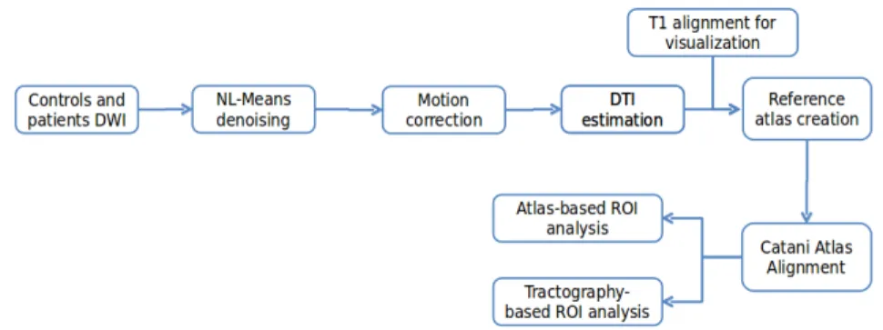
Fig. 1: Overall processing pipeline for DTI-based study of Specific Language Impairment.
We have performed an exploratory study on those two populations to infer differ-ences in white matter organization related to T-SLI. To this end, we first investigated based analysis where images are brought onto a reference frame and voxel-wise comparisons of the diffusion parameters are performed. The indices compared included scalar values derived from the diffusion tensor: Apparent Diffusion Coef-ficient (ADC), or Fractional Anisotropy (FA), and the full-tensor itself in the log-Euclidean framework. However, this approach did not lead to conclusive results. Instead, we follow a ROI approach, based either on an atlas or on tractography re-sults, following the pipeline presented in Fig. 1.
This pipeline consists first of preprocessing steps to improve the quality of diffu-sion tensor images. First, noise removal was performed on the DWI images utilizing the non-local means method (NL-Means) assuming Rician noise in the images [20]. Then, motion between successive diffusion images of each patient was corrected by registering all diffusion volumes on the reference B0 image of each patient, looking for a global rigid transform [16]. To keep the gradients and tensors aligned with the data, the linear parts of the rigid transformations (rotations) were applied to the gradients. Then, brain masking of the DWIs was performed. The threshold value was automatically computed from the diffusion images, after averaging the values of DW images, excluding the B0 image. Here, we used a cutoff at the first quartile of the intensity histogram of this average volume. If we apply this simple thresholding method, the brain mask can have holes where the values were low. A closing opera-tion was therefore applied to the mask image to retrieve the missing values from the original image. Finally, tensors were estimated utilizing Fillard et al. log-Euclidean estimation method [8], ensuring that the estimated tensors are always positive defi-nite.
All of the DTI volumes were then aligned into a single DTI atlas using Guimond et al. unbiased atlas construction method [10]. Each of the required registrations consisted of first finding a global affine transformation, followed by a full tensor-based non linear registration [19], which is a block-matching tensor-based registration with a generalized correlation coefficient as the similarity measure. For visualization pur-poses, all T1 images were also brought to this reference frame utilizing the obtained transformations. We then used Catani’s atlas [6] to define ROIs for each patient or control, by non-linearly registering the FA image provided with Catani’s atlas onto the FA image of our common frame. The analysis was then performed on each ROI choosing from two options:
• Compare mean FA and ADC values inside of the ROIs defined by Catani’s atlas • Perform tractography on each aligned DTI, based on Catani seeding ROIs, and use the envelopes of the obtained tracts as ROIs to compare mean FA and ADC In summary, while the first approach amounts to an automated classical ROI-based analysis, the second approach could lead to more sensitive results as it fo-cuses on the tracts extracted from each individual patient rather than on predefined regions. To build the tracts from the tensor images, we chose a deterministic ap-proach that is widely used in tractography, called Fiber Assignment by Continuous Tracking (FACT), introduced by Mori et al. [15].
For each region, two-sample t-tests were performed to compare the patient and control groups. To avoid false positive detections, the results were corrected for multiple comparisons using FDR correction [1] with a q-value threshold of 0.05. For explanatory purposes, results displaying a trend to significance with an uncorrected p-value smaller than 0.05 were also retained for discussion.
3 Results
3.1 ROI-Based Analysis
Using ROI-based analysis, no regions were declared as significantly different from patients to controls after FDR correction. For exploratory purposes, we present re-gions with a trend to significance with an uncorrected p-value p < 0.05. This is reasonable as we computed 30 tests but the results have to be interpreted with care as they show a tendency.
Out of the 30 regions, three, illustrated in Fig. 2, presented a trend to significance: the Anterior Segment Left, the Anterior Segment Right and the Inferior Longitudi-nal Fasciculus Left. The Anterior Segment is part of the Arcuate Fasciculus which is the main fasciculus involved in language. The inferior longitudinal fasciculus is also related to language [13].
The ADC in the Anterior Segment Right was higher in patients than controls (p=0.008). The FA values were lower in patients than controls in the Anterior Seg-ment Left (p=0.035) and in the Inferior Longitudinal Fasciculus Left (p=0.009).
62 Emmanuel Vall´ee et al.
Fig. 2: Regions as defined in Catani’s Atlas that differ from controls to patients; p <0.05, uncorrected; Blue: Anterior Segment Right, Red: Anterior Segment Left, Green: Inferior Longitudinal Fasciculus Left
3.2 Tractography-Based Analysis
The tractography-based analysis using Catani’s atlas showed significant differences in terms of FA and only some trends for ADC. Out of 30 regions under study, 10 presented a significant between-group difference. In all detected regions, a decrease in FA was observed in patients by comparison to controls as described in Table 1.
Bundle name FA Control FA Patient q-Value Anterior Segment Right 0.375±0.0134 0.362±0.0168 0.0129 Arcuate Left 0.397±0.0127 0.384±0.0167 0.0126 Inferior Longitudinal Fasciculus Left 0.423±0.0154 0.406±0.0181 0.0046 Inferior Longitudinal Fasciculus Right 0.41±0.0202 0.393±0.0206 0.0153 Inferior Occipito Frontal Fasciculus Right 0.406±0.0184 0.387±0.0191 0.0031 Long Segment Left 0.411±0.013 0.394±0.0249 0.0163 Optic Radiations Left 0.427±0.0249 0.406±0.0241 0.0115 Optic Radiations Right 0.43±0.0511 0.395±0.0317 0.0113 Posterior Segment Right 0.407±0.0292 0.387±0.017 0.0135 Anterior Commissure 0.375±0.0214 0.358±0.0187 0.0119
Table 1: Fibers bundles showing significant FA decrease in patients; q < 0.05 FDR corrected
Most of the significantly different regions are related to language. While some fiber bundles such as Optic Radiations and Anterior Commissure are not related to language, the other ones (Anterior Segment Right, Inferior Longitudinal Fasciculus Left and Right, Inferior Occipito-Frontal Fasciculus Right, Long Segment Left and Posterior Segment Right) are directly involved in this function. We illustrate in Fig. 3 the fiber bundles related to language with an altered integrity in children diagnosed with T-SLI.
Fig. 3: Language related tracts showing integrity decrease in patients; q < 0.05, FDR corrected; Anterior Segment Right, Arcuate Left, Inferior Longitudinal Fas-ciculus Left and Right, Inferior Occipito Frontal FasFas-ciculus Right, Long Segment Left and Posterior Segment Right issued from tractography.
4 Discussion and conclusion
We proposed two ROI-based approaches to analyze white matter integrity from Dif-fusion Weighted Images in children diagnosed with T-SLI. While the first one uti-lized ROIs defined directly from an atlas, the second approach used these regions as seeds for tractography which were in turn used for comparison of scalar indices. As expected, this second approach revealed a better sensitivity to detect differences as it focuses on the individual fiber bundles of the subjects.
Overall, this study revealed that several fiber bundles are impacted by T-SLI. The ROI based analysis exhibited interesting trends of differences in regions related to language. An increase of ADC correlated with a decrease of FA in patients was observed. This phenomenon is associated to brain maturation and myelination as described in [12]. The exploratory pipeline we followed revealed alteration of in-tegrity of white matter in the perisylvian and ventral pathways, which are related to language functions [13, 5].
64 Emmanuel Vall´ee et al.
A previous study led by Kim et al. on language impairment [11] revealed a de-crease of FA in the corpus callosum. Filippi et al. noted an inde-crease of ADC in the cortical regions and a decrease of FA in white matter bundles (corpus callosum, and subcortical white matter of the frontal and parieto-occipital lobes) of children with developmental delay [7]. These pathologies can be compared to T-SLI as oral lan-guage is also impacted. Their findings are consistent with our results showing ADC decrease in the occipital, postcentral and temporal lobe of the left hemisphere. They also noted a decrease of FA in patients, comparable to the general tendency that we observed during the comparison of FA along fiber tracts.
Interestingly, the findings of this study also confirm previous functional studies on a similar dataset that had highlighted a different cortex functional organization in patients [9]. The hypo and hyper-activated cortical regions (Geschwind and Broca) revealed in the f-MRI study are connected through the white matter fiber bundles that differ from control to patient subjects (Arcuate and Inferior Longitudinal Fas-ciculus). Considering that alterations in ADC and FA in white matter fiber tracts are sensitive markers of white matter integrity, one can argue that several white matter bundles are impacted in children with T-SLI.
Future works will include refinements of the methodology and particularly using more advanced diffusion models (such as orientation distribution functions) to better capture the white matter organization. Also, the developed pipeline is by essence generic and will be applied to the study of other pathologies such as dementia or Parkinson’s disease.
References
1. Benjamini, Y., Hochberg, Y.: Controlling the false discovery rate: A practical and powerful approach to multiple testing. J. of the Royal Stat Society. Series B (Methodological) 57(1), 289–300 (1995)
2. Bishop, D.V.M.: Uncommon understanding: Development and disorders of language compre-hension in children, vol. viii. Psychology Press/Erlbaum (UK) (1997)
3. Bishop, D.V.M.: Genetic and environmental risks for specific language impairment in chil-dren. Philosophical Transactions of the Royal Society of London. Series B 356(1407), 369– 380 (2001). DOI 10.1098/rstb.2000.0770
4. Bishop, D.V.M.: What causes specific language impairment in children? Current Directions in Psychological Science 15(5), 217–221 (2006). DOI 10.1111/j.1467-8721.2006.00439.x 5. Catani, M., Mesulam, M.: The arcuate fasciculus and the disconnection theme in language
and aphasia: History and current state. Cortex 44(8), 953–961 (2008). DOI 10.1016/j.cortex. 2008.04.002
6. Catani, M., Thiebaut de Schotten, M.: A diffusion tensor imaging tractography atlas for virtual in vivo dissections. Cortex 44(8), 1105–1132 (2008)
7. Filippi, C.G., Lin, D.D.M., Tsiouris, A.J., et al.: Diffusion-tensor MR imaging in children with developmental delay: preliminary findings. Radiology 229(1), 44–50 (2003)
8. Fillard, P., Pennec, X., Arsigny, V., Ayache, N.: Clinical DT-MRI estimation, smoothing, and fiber tracking with log-euclidean metrics. IEEE TMI 26(11), 1472 –1482 (2007). DOI 10. 1109/TMI.2007.899173
9. de Guibert, C., Maumet, C., Jannin, P., Ferr´e, J.C., Tr´eguier, C., Barillot, C., Le Rumeur, E., Allaire, C., Biraben, A.: Abnormal functional lateralization and activity of language brain
areas in typical specific language impairment (developmental dysphasia). Brain 134(Pt 10), 3044–3058 (2011). DOI 10.1093/brain/awr141
10. Guimond, A., Meunier, J., Thirion, J.: Average brain models: A convergence study. CVIU 77(2), 192–210 (2000)
11. Kim, J., Kim, Y.W., Park, C.I., Park, E.S., Kim, H.H., Lee, S.K., Kim, D.I.: Diffusion-tensor magnetic resonance imaging in children with language impairment. NeuroReport 17(12), 1279–1282 (2006). DOI 10.1097/01.wnr.0000230516.86090.67
12. L¨obel, U., Sedlacik, J., G¨ullmar, D., Kaiser, W., Reichenbach, J., Mentzel, H.J.: Diffusion tensor imaging: the normal evolution of ADC, RA, FA, and eigenvalues studied in multiple anatomical regions of the brain. Neuroradiology 51(4), 253–263 (2009)
13. Mandonnet, E., Nouet, A., Gatignol, P., Capelle, L., Duffau, H.: Does the left inferior longi-tudinal fasciculus play a role in language? a brain stimulation study. Brain 130(3), 623–629 (2007)
14. Mao, H., Polensek, S.H., Goldstein, F.C., Holder, C.A., Ni, C.: Diffusion tensor and functional magnetic resonance imaging of diffuse axonal injury and resulting language impairment. J. of Neuroimaging 17(4), 292–294 (2007). DOI 10.1111/j.1552-6569.2007.00146.x
15. Mori, S., Crain, B.J., Chacko, V.P., Van Zijl, P.C.M.: Three-dimensional tracking of axonal projections in the brain by magnetic resonance imaging. Annals of Neurology 45(2), 265–269 (1999). DOI 10.1002/1531-8249(199902)45:2h265::AID-ANA21i3.0.CO;2-3
16. Ourselin, S., Roche, A., Prima, S., Ayache, N.: Block matching: A general framework to im-prove robustness of rigid registration of medical images. In: MICCAI, LNCS, vol. 1935, pp. 557–566 (2000)
17. Preis, S., Steinmetz, H., Knorr, U., J¨ancke, L.: Corpus callosum size in children with de-velopmental language disorder. Cognitive Brain Research 10(1–2), 37–44 (2000). DOI 10.1016/S0926-6410(00)00020-3
18. Rapin, I.: Practitioner review: Developmental language disorders: A clinical update. J. of Child Psychology and Psychiatry 37(6), 643–655 (1996). DOI 10.1111/j.1469-7610.1996. tb01456.x
19. Suarez, R.O., Commowick, O., Prabhu, S.P., Warfield, S.K.: Automated delineation of white matter fiber tracts with a multiple region-of-interest approach. NeuroImage 59(4), 3690–3700 (2012). DOI 10.1016/j.neuroimage.2011.11.043
20. Wiest-Daessl´e, N., Prima, S., Coup´e, P., Morrissey, S.P., Barillot, C.: Rician Noise Removal by Non-Local Means Filtering for Low Signal-to-Noise Ratio MRI: Applications to DT-MRI. In: MICCAI (2), LNCS, vol. 5242, pp. 171–179 (2008)