HAL Id: inserm-01171368
https://www.hal.inserm.fr/inserm-01171368
Submitted on 3 Jul 2015
HAL is a multi-disciplinary open access
archive for the deposit and dissemination of
sci-entific research documents, whether they are
pub-lished or not. The documents may come from
teaching and research institutions in France or
abroad, or from public or private research centers.
L’archive ouverte pluridisciplinaire HAL, est
destinée au dépôt et à la diffusion de documents
scientifiques de niveau recherche, publiés ou non,
émanant des établissements d’enseignement et de
recherche français ou étrangers, des laboratoires
publics ou privés.
AMP-activated protein kinase is dispensable for
maintaining ATP levels and for survival following
inhibition of glycolysis, but promotes tumour
engraftment of ras-transformed fibroblasts
Joffrey Pelletier, Danièle Roux, Benoit Viollet, Nathalie Mazure, Jacques
Pouysségur
To cite this version:
Joffrey Pelletier, Danièle Roux, Benoit Viollet, Nathalie Mazure, Jacques Pouysségur. AMP-activated
protein kinase is dispensable for maintaining ATP levels and for survival following inhibition of
glycol-ysis, but promotes tumour engraftment of ras-transformed fibroblasts. Oncotarget, Impact journals,
2015, pp.11833-47. �inserm-01171368�
www.impactjournals.com/oncotarget/
Oncotarget, Vol. 6, No. 14
AMP-activated protein kinase is dispensable for maintaining
ATP levels and for survival following inhibition of glycolysis, but
promotes tumour engraftment of ras-transformed fibroblasts
Joffrey Pelletier
1, Danièle Roux
1, Benoit Viollet
2,3,4, Nathalie M. Mazure
1,*and
Jacques Pouysségur
1,3,5,*1 Institute for Research on Cancer and Ageing of Nice (IRCAN), University of Nice-Sophia Antipolis, CNRS UMR INSERM, Centre Antoine Lacassagne, Nice, France
2 INSERM U1016, Institut Cochin, Paris, France 3 CNRS UMR8104, Paris, France
4 Université Paris Descartes, Sorbonne Paris cité, Paris, France 5 Centre Scientifique de Monaco (CSM), Monaco
* These authors have contributed equally to this work
Correspondence to: Jacques Pouysségur, email: pouyssegur@unice.fr
Correspondence to: Nathalie Mazure, email: mazure@unice.fr
Keywords: AMPK, AMPK-null cells, ATP, cancer cell, glycolysis
Received: January 19, 2015 Accepted: March 04, 2015 Published: March 30, 2015
This is an open-access article distributed under the terms of the Creative Commons Attribution License, which permits unrestricted use, distribution, and reproduction in any medium, provided the original author and source are credited.
AbstrAct
Lactic acid generated by highly glycolytic tumours is exported by the MonoCarboxylate Transporters, MCT1 and MCT4, to maintain pHi and energy homeostasis. We report that MCT1 inhibition combined with Mct4 gene disruption severely reduced glycolysis and tumour growth without affecting ATP levels. Because of the key role of the 5’-AMP-activated protein kinase (AMPK) in energy homeostasis, we hypothesized that targeting glycolysis (MCT-blockade) in AMPK-null (Ampk-/-) cells should kill tumour cells from ‘ATP crisis’. We show that Ampk-/--Ras-transformed mouse embryonic fibroblasts (MEFs) maintained ATP levels and viability when glycolysis was inhibited. In MCT-inhibited MEFs treated with OXPHOS inhibitors the ATP level and viability collapsed in both Ampk+/+ and Ampk-/- cells. We therefore propose that the intracellular acidification resulting from lactic acid sequestration mimicks AMPK by blocking mTORC1, a major component of an ATP consuming pathway, thereby preventing ‘ATP crisis’. Finally we showed that genetic disruption of Mct4 and/or Ampk dramatically reduced tumourigenicity in a xenograft mouse model suggesting a crucial role for these two actors in establishment of tumours in a nutrient-deprived environment. These findings demonstrated that blockade of lactate transport is an efficient anti-cancer strategy that highlights the potential in targeting Mct4 in a context of impaired AMPK activity.
INtrODUctION
Dividing cancer cells rely extensively on glycolysis
to maintain a high rate of proliferation [1-3]. Despite a
lower yield of ATP than that of oxidative phosphorylation
(OXPHOS), glycolysis generates precursors for the
synthesis of nucleotides, amino acids, and membrane
lipids [4]. Although exacerbated glycolysis is a common
feature of any proliferative cell, genetic lesions driving
cancer progression have a major impact on metabolism
and promote glycolysis [5]. The dependency of cancer
cells on glycolysis is amplified during tumour growth. Part
of the tumour encounters an avascular microenvironment
deficient in oxygen, i.e. hypoxic, as a consequence of rapid
proliferation of cancer cells and aberrant neovasculature
[6]. The transcription factor the Hypoxia-Inducible Factor
1 (HIF-1) [7], a key actor in cellular adaptation to hypoxia,
induces a major shift in cellular metabolism toward
glycolysis [6, 8, 9]. HIF-1, through the transcriptional
upregulation of almost all the glycolytic enzymes together
with inhibition of mitochondrial pyruvate dehydrogenase
[10], creates an addiction to glycolysis of hypoxic tumour
cells, which produce biomass as long as the major
nutrients such as glucose are not depleted. So inhibition
of glycolysis appears to be a promising avenue for the
development of anti-tumour targeted therapies [11-15].
As a consequence of exacerbated glycolysis large
amounts of lactic acid are produced. Our laboratory has
been evaluating the anti-cancer approach of inhibiting
lactic acid export. This approach leaded to intracellular
accumulation of lactic acid associated with a severe drop
in the intracellular pH (pHi), which results in inhibition of
glycolysis [16, 17]. Lactate transport is carried out by four
members of the monocarcarboxylate transporter (MCT)
family (MCT1-4) [18], which have received revived
attention in the context of cancer [19, 20]. They are H
+/
lactate bidirectional symporters with different affinities
and tissue distribution. MCT1 is ubiquitous, whereas
MCT4, a HIF-1-target gene [21], is strongly expressed
in glycolytic tissues [22-24]. We and others showed that
inhibition of MCTs or genetic disruption of MCT/basigin
complexes leaded to a blockade in lactate transport and
had anti-proliferative effects by reducing the glycolytic
flux [15, 17, 25, 26]. However, although this approach
severely restricted tumour growth it did not affect cancer
cell viability unless the mitochondrial complex I was
inhibited by biguanides [17, 25-27]. The major cause of
resistance to cell death by inhibition of energy-producing
pathways resides in the metabolic plasticity governed by
the central guardian of the cellular ATP level, the
AMP-activated protein kinase (AMPK) [2, 16, 27, 28]. AMPK
is a highly conserved serine/threonine protein kinase
involved in the control of energy homeostasis [28]. AMPK
exists as a heterotrimeric protein comprising a catalytic α
subunit and regulatory β/γ subunits. As a direct sensor of
the AMP:ATP ratio, AMPK is rapidly activated by stresses
that cause a reduction in ATP levels, such as glucose
starvation [29], metabolic inhibitors [30], or muscle
contraction [31, 32]. When activated, AMPK maintains
a viable level of ATP, accelerating energy production,
through increased glucose uptake [33], glycolysis [34],
mitochondrial biogenesis [35], and mitophagy [36],
while switching off energy consuming processes such
as synthesis of proteins [37-40], glycogen[41] and fatty
acids [42, 43]. However, although extensively studied, the
regulation and function of AMPK remain only partially
understood, especially in cancer.
We therefore hypothesized that genetic disruption
of AMPK in a context of strong inhibition of glycolysis
(MCT blockade) should sensitize tumour cells to death
as a result of a deficit in maintenance of the ATP level.
As no specific inhibitor for AMPK exists, a model of
Ras-transformed murine embryonic fibroblasts (MEFs)
genetically knocked out for the two catalytic subunits
of AMPK (Ampk
-/-), Ampkα1 and Ampk2 [44], was used.
We first demonstrated that pharmacological inhibition of
MCT1 combined with genetic knockout of Mct4 (Mct4
-/-)
in Ras-transformed MEFs, lead to inhibition of glycolysis
and proliferation in vitro. However, Ampk
-/-MEFs
preserved viable levels of ATP following acute inhibition
of glycolysis. Moreover, AMPK was not capable of
providing a survival advantage following severe inhibition
of ATP production by glycolysis and OXPHOS. This
unexpected finding suggested that AMPK is dispensable
in regulating the plasticity of bioenergetic pathways.
Finally we showed, using a xenograft tumour model, that
the knockout of Ampk or Mct4 (Ampk
-/-or Mct4
-/-) delayed
tumour development while Ampk
-/-Mct4
-/-MEFs severely
impacted on tumour establishment. These in vivo studies
suggest that combined inhibition of AMPK and MCT4
could be exploited as an anti-cancer strategy.
rEsULts
In the absence of an energy stress, genetic
disruption of AMPK in MEFs does not affect
glycolysis, OXPHOs or cell proliferation
Wild
-type
murine embryonic fibroblasts
(MEFs)
(Ampk
+/+)
or deficient
for the expression of
both the
AMPKα1
and
AMPKα2
catalytic
subunits
(Ampk
-/-)
were transformed with
oncogenic
Ras
V12(
Figure
1a
).
Transformation
did not change
the
ability
of AMPK
to
be
activated
in the absence
of glucose and
to
activate
Acetyl-CoA carboxylase
(
P-ACC), one of the main targets
of AMPK (Figure 1b).
We sought to
determine whether
the
lack
of
functional
AMPK
affected
metabolism,
ATP
levels
and cell proliferation
.
The oxygen
consumption rate
(
OCR)
showed no
significant difference between
Ampk
+/+and Ampk
-/-MEFs (
Figure
1c
).
Oligomycin
(
Oligo), an
inhibitor
of the
F0F1
-ATP
synthase,
reduced
the
OCR
to
a
similarly
to that of
AMPK
in
Ampk
+/+or Ampk
-/-MEFs
suggesting
that absence of
AMPK
did not
modify
the
amount of ATP
produced by
mitochondrial respiration
.
The extracellular acidification
rate
(
ECAR), the
index
of
lactic
acid
export
and thus
glycolysis
,
was
also
identical
in cells
with and without
functional
AMPK
,
in the
absence or
presence
of
glucose
(
Figure
1d
).
Inhibition
of
mitochondrial
ATP synthesis
by
oligomycin
was
responsible for a
rapid
shift
toward
glycolysis
metabolism
,
independently
of AMPK
.
Similarly, no
significant
difference in the ATP
level
was observed
in optimal
conditions
of
cell growth (
Figure
1e
).
Finally,
Ampk
+/+and Ampk
-/-MEFs
proliferated
in vitro
in normoxia
or
hypoxia
in the presence
of
25mM glucose
(
Figure
1
f and
1g).
However
, Ampk
-/-MEFs
proliferated
slightly
less
rapidly
in
hypoxia.
Taken together,
these results suggest
that AMPK
is
dispensable
under conditions of
plentiful
Figure 1: Metabolic characterization in vitro of ras
V12-transformed mouse embryonic fibroblasts (MEFs) expressing
(Ampk
+/+) or not (Ampk
-/-) Ampk.
(a) Cell lysates of Ampk+/+ and Ampk-/- MEFs not transformed (-) or transformed (+) by RasV12 wereanalyzed by immunoblotting for Ras. (b) Cell lysates of Ampk+/+ and Ampk-/- MEFs transformed by RasV12 were analyzed by immunoblotting
for phosphorylation of AMPK (Thr172) and ACC Ser79 after 1h of glucose starvation. (c) Oxygen consumption rate (OCR) was measured in real time with a Seahorse XF for Ampk+/+ (Ampk+/+) and Ampk-/- (Ampk-/-) MEFs transformed by RasV12 in normoxia. Glucose (10 mM),
oligomycin (Oligo 1 µM), carbonilcyanide p-triflouromethoxyphenylhydrazone (FCCP 1µM) and rotenone/antimycine A (Rot/AA 1 µM) were injected at the indicated times. Mean±S.E.M. is representative of three independent experiments carried out in quadruplicate. (d) Extracellular acidification rate (ECAR) of normoxic Ampk+/+ (Ampk+/+) and Ampk-/- (Ampk-/-) MEFs transformed by RasV12 were evaluated
with a Seahorse XF. Glucose (10 mM) and oligomycin (Oligo 1 µM) were injected at the indicated times. The mean±S.E.M. is representative of three independent experiments carried out in quadruplicate. (e) The total cellular ATP levels in cells incubated in normoxia for 48 h were standardized to protein content for each condition. The mean±S.E.M. is representative of three independent experiments carried out in quadruplicate. Non significative (n.s.). (f) In vitro proliferation of Ampk+/+ (Ampk+/+) and Ampk-/- (Ampk-/-) MEFs transformed by RasV12
incubated in normoxia (Nx) or in hypoxia 1% O2 (Hx) up to 96 h. The mean±S.E.M. is representative of three independent experiments
carried out in duplicate. (g) Clonal growth in normoxia (Nx) or hypoxia 1% O2 (Hx) of Ampk+/+ (Ampk+/+) and Ampk-/- (Ampk-/) MEFs
nutrients
, either
in normoxia
or
hypoxia.
combined inhibition of Mct1 and Mct4
blocked the export of lactate and inhibited
glycolysis independently of AMPK
To inhibit
glycolysis
,
we blocked
the output
of
cellular lactate
by modulating the expression
and/
or activity of
MCT1 and 4
.
MCT1
was expressed in
normoxia (Figure
2a
).
While
MCT1
is not
a
transcriptional
target
of
HIF-1
,
we observed
that
its expression
was
three-fold higher (3.3±0.5)
in
hypoxia
whereas
the level
of expression of the mRNA was not
modified
(data not
shown)
.
However, protein expression of MCT4
was
undetectable in
normoxia
,
but
was
strongly induced
in
hypoxia (
Figure
2a).
MCT4 expression
was higher
in
Ampk
-/-MEFs than in
Ampk
+/+MEFS in
hypoxia. Since
no
specific inhibitor of
MCT4
is available
, we
knocked
out the
Mct4
gene (
Mct4
-/-)
in
Ampk
+/+and Ampk
-/-MEFs
.
Knockout
did not alter
expression of
MCT1
(
Figure
2a
).
Genetic
knockout
of
Mct4
in
Ampk
+/+and Ampk
-/-MEFs
did not alter
lactate
transport in
hypoxia (
Figure
2b
),
suggesting that
MCT1
could
compensate fully for the
lack of MCT4
expression
.
Pharmacological inhibition
of
MCT1
(
MCTi) in
cells lacking
MCT4
(
Ampk
+/+Mct4
-/-and Ampk
-/-Mct4
-/-MEFs
)
abolished
lactate
transport
in
hypoxia
(
Figure
2b
and
Supplementary
Figure 1a)
and thus
leaded to
its
intracellular
accumulation in
Ampk
+/+Mct4
-/-MEFs
(
Figure
2c
)
and
Ampk
-/-Mct4
-/-MEFs
(
Figure
2d
and
Supplementary
Figure
1b
). However, Mct4 knockout
reduced the glycolytic flux of Ampk
+/+cells compared to
Ampk
-/-MEFs (Figure 2e). Inhibition of MCT1 in Mct4
-/-MEFs dramatically reduced the glycolytic flux in the
presence or absence of functional AMPK in normoxia
(Figure 2e and Supplementary Figure 1c). Inhibition
of glycolysis was not associated with an increase in
mitochondrial respiration (Supplementary Figure 1d
and 1e). We also observed that consumption of glucose
(Supplementary Figure 2a) and lactate secretion in the
extracellular medium (Supplementary Figure 2b) were
both affected by combined inhibition of MCT1 and MCT4
in Ampk
+/+ou Ampk
-/-MEFs, in normoxia and hypoxia,
confirming a decrease in the glycolytic flux in response
to inhibition of MCTs.
Finally
,
AMPK
was activated in
response
to MCT
inhibition in
Ampk
+/+Mct4
-/-MEFs
(Figure
2f
and
Supplementary
Fig
ure
2c
),
as shown by the
active phosphorylation at Thr172 of
the
AMPKα
subunit
(P-
AMPK)
and
of
ACC.
Colon adenocarcinoma
LS174
cells were used
to verify
that activation
was not
only
specific to
MEFs but also found in
a model of
glycolytic
tumour cells.
We demonstrated
that
AMPK activation
was
faster in
LS174 cells in response
to inhibition
of
MCTs
(Supplementary
Figure
2d
). AMPK and ACC
phosphorylation
occured after 15min
, was
maintained
over time
and
required
combined
inhibition
of
MCT1
and
MCT4
(Supplementary Figure
2e
).
Activation
was
probably due to
the
stress in energy
generated
by inhibition
of glycolysis,
as recently reported in LS174 cells [17].
Although
slight differences
were observed
between
Ampk
+/+and Ampk
-/-MEFs
(
Figure
2a
, 2b
and
2
e)
,
together
these
results show
that inhibition
of
MCTs
lead
to
intracellular
accumulation
of
lactate
and
a
dramatic
reduction in the
glycolytic flux
in MEFs
with and without
functional
AMPK
.
AMPK is not required for maintenance of a viable
AtP level following blockade in glycolysis
We then
looked at
the impact
of blocking
glycolysis
on
ATP levels
in the absence of
AMPK
.
We analyzed
the levels
of ATP in
Ampk
+/+and Ampk
-/-MEFs,
in the
presence
(
Mct4
+/+) or the absence
(
Mct4
-/-)
of
MCT4
,
and
after addition
or
not of
MCTi
(
Figure
3a and
Supplementary
Fig
ure
3a)
.
In
control
MEFs
(
Ampk
+/+Mct4
+/+)
, we observed
that the ATP
level increased
slightly
and
reproducibly
, although not
significantly, after
48h of MCTi treatment
in normoxia.
As we previously
observed,
using different cell lines
[45, 46],
48h
of
hypoxia
significantly increased
ATP levels
(25
%
±5%)
compare to the levels in normoxia.
Inhibition of
MCT1
also showed
a slight tendency to
increase the ATP
level in
cells expressing
MCT4
in
hypoxia
.
Knockout
of
Mct4
in
Ampk
+/+MEFs
increased basal levels
of ATP
in normoxia
and
hypoxia
,
probably
due to
inhibition of proliferation
.
MCT1
inhibition in
Ampk
+/+Mct4
-/-MEFs
reduced the
ATP
level
in normoxia
and
hypoxia by about
23
%
±3
%
and 47%±3%, respectively.
The
most important effect
of
MCT
inhibition in
hypoxia
was due
mostly to the
metabolic shift
toward
glycolysis, which
made the
cells
more dependent on
this pathway
for
ATP production
[15-17].
The basal level of ATP of Ampk
-/-MEFs in normoxia
was similar to that of Ampk
+/+MEFs but not modified by
inhibiting MCT1 or by hypoxia. Similar to Ampk
+/+Mct4
-/-MEFs, Mct4 knockout in Ampk
-/-MEFs (Ampk
-/-Mct4
-/-)
increased the basal level of ATP. However, cells were more
sensititive to MCT1 inhibition. Indeed, the level of ATP
was reduced by about 16%±3% in normoxia and 28%±3%
in hypoxia for the first clone (Figure 3a) and 14%±5%
in normoxia and 29%±3% in hypoxia for the second
independent clone (Supplementary Figure 3a). In the
presence of MCTi in normoxia or hypoxia, decreases in
ATP levels did not correlate with a significant increase in
cell death in Ampk
+/+Mct4
-/-or Ampk
-/-Mct4
-/-MEFs over
a short period of time (8 and 2h, data not shown) or 48h
(Figure 3b and Supplementary Figure 3b). We examined
the ability of Ampk
-/-(Figure 3c, right panels) to grow in
the presence or the absence of MCT4 in normoxia (top
panels) or hypoxia (Figure 3c, bottom panels) compared
to Ampk
+/+MEFs (Figure 3c, left panels).
In
normoxia
, the
addition of
MCTi
did not alter
the
proliferation of
MEFs
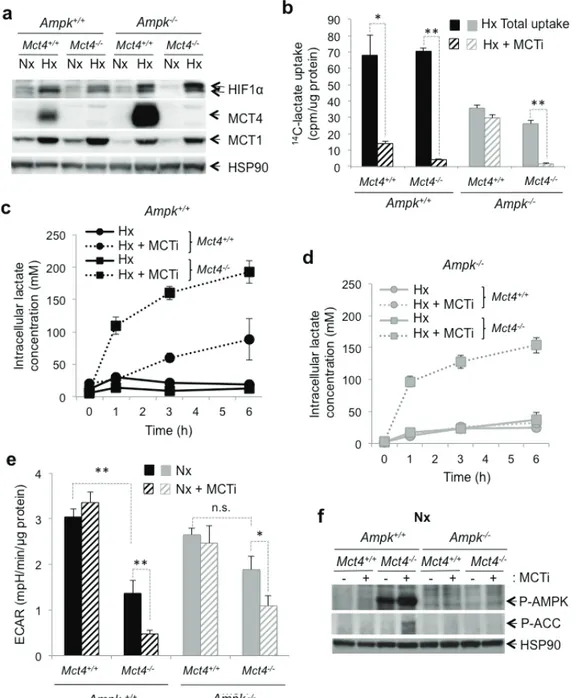
Figure 2: The MCT1 pharmacological inhibitor (MCTi) reduced lactate transport and the glycolytic rate in Ampk
+/+and Ampk
-/-MEFs.
(a) Cell lysates of Ampk+/+ (Ampk+/+) and Ampk-/- MEFs (Ampk-/-) expressing (Mct4+/+) or not (Mct4-/-) MCT4incubated in normoxia (Nx) or hypoxia 1% O2 (Hx). Whole-cell lysates were analyzed by immunoblotting for HIF-1α, MCT4, and MCT1.
Detection of HSP90 was used as a loading control. (b) Lactate uptake in Ampk+/+ (Ampk+/+, black bars) and Ampk-/- (Ampk-/-, grey bars)
MEFs expressing (Mct4+/+) or not (Mct4-/-) MCT4 in the absence (total uptake, filled bars) or presence of 300 nM MCTi (MCTi, hatched
bars) in hypoxia 1% O2 (Hx) for 48 h. Uptake was conducted as 3-min time-points in duplicate. The mean±S.E.M. is representative of two independent experiments carried out in duplicate. * p < 0.005, ** p < 0.001. (c) Time-course of intracellular lactate concentration in response to glucose addition (25 mM) to Ampk+/+ (Ampk+/+) MEF expressing (Mct4+/+) or not (Mct4-/-) MCT4 in the presence of DMSO (solid
line) or MCTi (300 nM, dotted line) in hypoxia 1% O2 (Hx). The mean±S.E.M. is representative of three independent experiments carried
out in triplicate. (d) Time-course of intracellular lactate concentration in response to glucose addition (25mM) to Ampk-/- MEFs expressing
(Mct4+/+) or not (Mct4-/-) MCT4 in the presence of DMSO (solid line) or MCTi (300 nM, dotted line). The mean±S.E.M. is representative
of three independent experiments carried out in triplicate. (e) The extracellular acidification rate (ECAR) of Ampk+/+ (Ampk+/+, black bars)
and Ampk-/- MEFs (Ampk-/-, grey bars) expressing (Mct4+/+) or not (Mct4-/-) MCT4 with DMSO (filled bars) or MCTi (300nM, signs in
bars) in normoxia (Nx) were evaluated after injection of glucose (10 mM) with a Seahorse XF. The mean±S.E.M. is representative of three independent experiments carried out in quadruplicate. * p < 0.005, ** p < 0.001. (f) Cell lysates of Ampk+/+ (Ampk+/+) and Ampk-/- (Ampk-/-)
MEFs expressing (Mct4+/+) or not (Mct4-/-) MCT4 in normoxia (Nx) in the absence (-) or presence (+) of 300nM MCTi were analyzed by
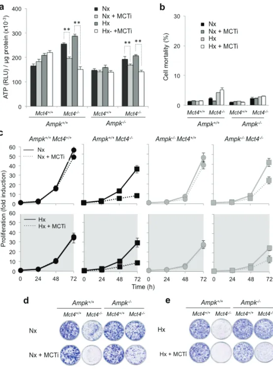
Figure 3: Pharmacological inhibition of MCT1 (MCTi) in combination with knockout of Mct4 (Ampk
+/+Mct4
-/-and
Ampk
-/-Mct4
-/-MEFs) decreased the AtP level and the proliferation independently of the presence or absence of AMPK
but did not alter cellular viability.
(a) Total cellular ATP level in Ampk+/+ (Ampk+/+) and Ampk-/- (Ampk-/-) MEFs expressing (Mct4+/+)or not (Mct4-/-) MCT4 incubated in normoxia (Nx) or in hypoxia 1% O
2 (Hx) for 48 h in the absence (DMSO) or presence (+ MCTi,
300 nM) of MCTi. ATP levels were measured in whole-cell lysates and standardized to the cell protein content for each condition. The mean±S.E.M. is representative of three independent experiments carried out in triplicate. The mean±S.E.M. is representative of three independent experiments carried out in quadruplicate. ** p < 0.001. (b) Ampk+/+ (Ampk+/+) and Ampk-/- MEFs (Ampk-/-) expressing (Mct4+/+)
or not (Mct4-/-) MCT4 incubated in normoxia (Nx) or hypoxia 1% O
2 (Hx) for 48h in the absence or the presence (+ MCTi, 300 nM) of MCT
inhibitor. Cell mortality (%) was evaluated by Trypan blue exclusion. The mean±S.E.M. is representative of three independent experiments carried out in duplicate. (c) In vitro exponential growth of Ampk+/+ (Ampk+/+) and Ampk-/- MEFs (Ampk-/-) expressing (Mct4+/+) or not (Mct4 -/-) MCT4 incubated up to 72 h in normoxia (Nx – top panel) or hypoxia 1% O
2 (Hx – bottom panel) in the absence or the presence of the
MCT inhibitor (+ MCTi, 300 nM). The mean±S.E.M. is representative of three independent experiments carried out in duplicate. (d) Clonal growth in normoxia (Nx) of Ampk+/+ (Ampk+/+) and Ampk-/- (Ampk-/-) MEFs expressing (Mct4+/+) or not (Mct4-/-) MCT4, in the absence or
presence of the MCT inhibitor (+ MCTi, 300 nM) for 8 days before staining and visualization of the colonies. (e) Clonal growth in hypoxia 1% O2 (Hx) of Ampk+/+ (Ampk+/+) and Ampk-/- (Ampk-/-) MEFs expressing (Mct4+/+) or not (Mct4-/-) MCT4, in the absence or presence of the
expressing
MCT4
(
Ampk
+/+Mct4
+/+et Ampk
-/-Mct4
+/+)
(
Figure
3c
).
When
Mct4
was knocked out
, inhibition of
MCT1
greatly reduced
proliferation
at
48h
(
Ampk
+/+Mct4
-/-ou Ampk
-/-Mct4
-/-)
but
less in MEFs
that did
not express
functional
AMPK
.
At 72h
,
both inhibition
of
MCT1
and
knockout of
MCT4
led to
a stronger
inhibition of
proliferation
in
Ampk
+/+in
normoxia
(76
%
±0.6
%) than
that in Ampk
-/-MEFs
(32%±3%).
In
hypoxia,
Ampk
+/+and Ampk
-/-MEFs
proliferated
more slowly than that
in
normoxia
and MCT1
inhibition
had no
significant effect
on
these cells (
Figure
3c
).
In contrast,
MCTi
inhibited
proliferation
of Mct4-
deficient cells
,
in
a manner
similar
to that of Ampk
+/+Mct4
-/-MEFs
(
75%
±0.6
%)
and
Ampk
+/+Mct4
-/-MEFs
(62
%
±0.8
%)
(
Figure
3c and
Supplementary
Fig
ure
3c
). Cell growth and/or mortality
over a long period of time in normoxia
(
Figure
3d
and
Supplementary
Fig
ure
3d
)
and
in hypoxia (Figure
3e
and
Supplementary
Figure 3e
) were determined by the
clonogenicity assay. P
roliferation was
strongly
reduced in
Ampk
+/+Mct4
-/-or
Ampk
-/-Mct4
+/+MEFs
in the presence of
MCTi
in both conditions
.
Although
small differences
between
Ampk
+/+and Ampk
-/-MEFs were found
,
all
these results suggest
that
after blockade
of lactate
export and inhibition of
glycolysis
,
MEFs did not require
the
presence
of AMPK
to maintain
a
viable
level
of ATP
in vitro.
AMPK did not confer a survival benefit following
the combined blockade of glycolysis and
respiration, but induced resistance to glucose
deprivation
Ampk
+/+and
Ampk
-/-MEFs
expressing
MCT4
(
Mct4
+/+) or not (
Mct4
-/-) were treated in hypoxia
with
MCTi
combined with
phenformin (MCTi/Phenf)
an
inhibitor of
mitochondrial
respiratory chain complex
1.
Combination of
MCTi/Phenf
did not alter
ATP levels
in MEF expressing
MCT4
(
Ampk
+/+Mct4
+/+et Ampk
-/-Mct4
+/+MEFs
) (
Figure
4a,
left panel)
.
In
Ampk
+/+Mct4
-/-MEFs,
while
phenformin
alone did not affect
the
level
of ATP (Supplementary
Fig
ure
4a
)
the
combination
of
MCTi/Phenf
gradually
reduced
the
ATP
pool
from
97%±1
,
60%
±1
and
25%±3 of its initial level
after
8, 24
and 48h of
treatment,
respectively
(
Figure
4a,
left panel)
. A r
eduction
in the ATP
level
in
the
Ampk
-/-Mct4
-/-MEFs was
similar to
that of
Ampk
+/+Mct4
-/-MEFs.
No difference in
mortality
was observed
between
Ampk
+/+Mct4
+/+or
Ampk
-/-Mct4
+/+MEFs
(
Figure
4a,
right panel).
However,
for
Ampk
+/+Mct4
-/-and Ampk
-/-Mct4
-/-MEFs
27
%±3% and
36
%
±4%
cell
mortality
was observed
, respectively.
Similar results
were
obtained
when
ATP
was
collapsed with the
MCTi/Oligo
combination
(
Supplementary
Figure 4b
). Phenformin
slowed cell proliferation in a similar manner: 34%±1%
(Ampk
+/+Mct4
+/+), 39%±4% (Ampk
+/+Mct4
-/-), 30%±6%
(Ampk
-/-Mct4
+/+) and 28%±3.8% (Ampk
-/-Mct4
-/-) (Figure
4b) without affecting cell viability (data not shown).
Combination of MCTi/Phenf decreased slightly the cell
number over 72 h in a similar manner in Ampk
+/+Mct4
+/+(28%±1) and Ampk
-/-Mct4
+/+(33%±2) MEFs (Figure 4B).
However, the cell number was significantly reduced by
94%±2% in Ampk
+/+Mct4
-/-MEFs and by 91%±2% in
Ampk
+/+Mct4
-/-MEFs after 72 h of MCTi/Phenf treatment.
The same effect was observed after 8 days of growth using
the clonogenicity assay (Supplementary Figure 4c).
Taken together these results showed that
MCT4 knockout sensitized MEFs to the MCTi/Phenf
combination, which reduced substantially the ATP level,
viability and proliferation. Knockout of AMPK was
neither beneficial nor unfavourable to these parameters.
The
question then was whether
AMPK
provides an
advantage
in cell survival
in response to a
rapid
decrease in
the
level of ATP
. The
MCTi/Phenf
combination
impacted
on
the
ATP
levels after 24h of treatment
(
Figure
4a)
.
As
oligomycin
rapidly
blocked production
of ATP
in contrast
to
phenformin
,
it
was added in the presence
of
decreasing
concentrations
of
glucose
(10 mM
,
1mM
or
0.
1 mM
)
(
Figure
4c
).
In the presence of
10mM glucose
oligomycin
dropped the
level
of ATP (
49%
±6
%)
in
10min either
in
the
Ampk
+/+or
Ampk
-/-MEFs,
.
This level
was
gradually
restored
,
and even
reached
values above the
basal
level
of ATP
1h
after treatment (
Figure
4c,
top panel)
.
When
oligomycin
was added
to medium containing
1mM
glucose
,
ATP levels
dropped
rapidly
to similar levels
to
those
in the presence
of
10 mM glucose
(
49%
±3
%
and
38%±2
% for Ampk
+/+or
Ampk
-/-MEFs
,
respectively)
(
Figure
4c,
middle panel
).
However, none
of these two
populations
of
MEFs completely
restored
their
basal
level
of ATP.
When oligomycin
was added
to medium
containing
0.1mM glucose
,
a
significant difference was
observed
between
levels
of ATP
for Ampk
+/+compared
to
Ampk
-/-MEFs,
28
%
±3% versus
9
%
±2%, respectively
(
Figure
4c,
bottom panel
).
This difference
was amplified
after
1h of treatment
as Ampk
+/+MEFs
had
an
ATP
level
of
63
%
±0.5%
compared to
Ampk
-/-MEFs (28%±3
%).
In the absence of
10 mM
glucose±2-deoxy-glucose
,
oligomycin
collapsed
ATP production
,
independently
of
the AMPK activity
(
Supplementary
Fig
ure
4d
and
4e
).
Little
difference in cell number was observed
between
Ampk
+/+and
Ampk
-/-MEFs in the presence of 25 mM
glucose (
Figure
4d
)
.
However
,
in the presence
of
0.1 or
1mM
glucose
, proliferation
of
Ampk
+/+MEFs slowed
while a decrease in the glucose concentration
dramatically
impacted
cell viability
of Ampk
-/-MEFs
.
Taken together
these
results
suggest
that AMPK
is
not required
to
maintain a
viable
level
of ATP
in response
to
inhibition of
glycolysis
via blockade of
MCTs or
metabolic
shock (
MCTi/Phenf). The
only differences
observed
were obtained
under conditions of low glucose
Figure 4: AMPK is not sufficient to guaranty viability in response to inhibition of MCT1 (MCTi) and MCT4 (Mct4
-/-)
inhibition combined with phenformin (Phenf), but is important to promote cell survival following a nutritive stress.
(a) Left panel - Total cellular ATP level in Ampk+/+ (Ampk+/+) and Ampk-/- (Ampk-/-) MEFs expressing (Mct4+/+) or not (Mct4-/-) MCT4 incubatedin hypoxia 1% O2 (Hx) for up to 48 h in the presence of a combination with MCTi (300 nM) and phenformine (50 µM) (+ MCTi/Phenf),
standardized to the protein content for each condition and normalized to initial cellular ATP level (%). The mean±S.E.M. is representative of three independent experiments carried out in quadruplicate. Right panel –Ampk+/+ (Ampk+/+) and Ampk-/- (Ampk-/-) MEFs in the presence
(black signs in bars) or absence (grey signs in bars) of Mct4 (± Mct4) were incubated in normoxia (Nx) for 48 h with the MCTi/Phenf combination, 300 nM and 50µM respectively. Cell mortality (%) was evaluated with the automatic cell counter ADAM. The mean±S.E.M. is representative of three independent experiments carried out in triplicate. (b) In vitro exponential growth of Ampk+/+ (Ampk+/+) and Ampk -/- MEFs (Ampk-/-) expressing (Mct4+/+) or not (Mct4-/-) MCT4 incubated for up to 72 h in hypoxia 1% O
2 (Hx) in the absence or presence of
phenformine (+ Phenf) or presence of the MCTi/Phenf combination (+ MCTi/Phenf), 300 nM and 50 µM respectively. The mean±S.E.M. is representative of three independent experiments carried out in duplicate. (c) Total cellular ATP level in Ampk+/+ (Ampk+/+) and Ampk -/- (Ampk-/-) MEFs expressing MCT4 (Mct4+/+) incubated in normoxia (Nx) in 10-, 1- and 0.1-mM glucose, in the presence of oligomycine
(Oligo, 1 µM) for up to 180min, standardized to the cell protein content for each condition and normalized to initial cellular ATP level (%). The mean±S.E.M. is representative of three independent experiments carried out in triplicate. The mean±S.E.M. is representative of two independent experiments carried out in quadruplicate. * p < 0.005, ** p < 0.001. (d) Clonal growth in normoxia (Nx) of Ampk+/+ (Ampk+/+)
and Ampk-/- (Ampk-/-) MEFs expressing MCT4 (Mct4+/+) in 10-, 1- and 0.1-mM glucose for 3 days then incubated for 10 days in 25 mM
Knockout of AMPK or Mct4 or both strongly
impact on the tumourigenicity
We then investigated the behaviour of these cells in
a tumour context. Ampk
+/+Mct4
+/+control MEFs
showed
rapid tumour
growth,
up to
200mm
3after
12 days
(
±
3
)
and
greater than 1000mm
3after
21 days
of
injection
(Figure
5a
). W
e observed a
high latency
in tumour growth
with
Ampk
-/-Mct4
+/+MEFs
as
the tumours reach
ed
200mm
3after
44 days
(
±
6) post-injection.
However,
after this period,
the
rate of tumour
growth
was identical
for
Ampk
+/+and
Ampk
-/-MEFs
(
Figure
5a).
Moreover,
Ampk
+/+Mct4
-/-MEF
showed
slowed
tumourigenicity
but
not
tumour
growth
as
Ampk
+/+Mct4
-/-MEFs
exhibited
a
tumour volume
of
200mm
3to 1000mm
3in
33 days
(
±
6
)
and
40 days
(
±
6
),
respectively
(
Figure
5a
). Ampk
-/-Mct4
-/-MEF-derived
tumour
s grew much slower (
Figure
5a
). The absence of
AMPK activity in MEF-derived tumuors was confirmed
(
Figure
5b)
.
These results
clearly show
that
independently
AMPK
and
MCT4
impact on tumour development
but
when taken
together the
in vivo consequence is reinforced.
DIscUssION
AMPK is widely recognized as the guardian
of the balance
in
energy and as a critical
regulator of
intracellular ATP level
. However, in the present study,
we
showed
for the first time,
that AMPK
was dispensable for
maintenance of the ATP level and viability of
fibroblasts
when glycolysis was limited by MCT blockade.
However
,
we confirmed that
Ampk
-/-MEFs
were more sensitive to
glucose deprivation or to acute energy stress than their
wild type counterpart.
While the
double
knockout
of
AMPK
and
MCT4
(
Ampk
-/-Mct4
-/-)
did
not
drastically
change
the
ATP level
or
the
characteristics
of
growth
in
vitro, it
showed a marked decrease in the tumour incidence.
These results
lead us to
ask the following
questions:
(
i)
What are the
differences,
from an energy point
of view,
between inhibiting
glycolysis
by
restricting lactic acid
export
versus
decreasing availability
of
glucose
?
(
ii)
Is
AMPK
a tumour suppressor
or
an
oncogene in our model
?
(
iii) Why did the
double
knockout
of
AMPK and
MCT4
reduce
substantially
tumourigenicity
while no
phenotypic
differences
were found in vitro?
Our approach
to reducing tumour growth by
blocking the last step of fermentative
glycolysis (export
of
lactic
acid) [15]
was recently confirmed in and extended to
other cancer cell lines [17, 25, 26, 47]
.
Although
blockade
of
glycolysis
was capable of restricting proliferation,
intracellular
ATP levels
and cell viability were maintained
due to re-activation of OXPHOS.
Indeed
discontinuation
of
MCTi treatment
restored
tumour growth
indicating
a
cytostatic effect [15, 8]
.
The aim of this
study was
to test
the hypothesis that
combining inhibition of glycolysis
with
suppression of AMPK
should induce acute tumour
cell death from ‘ATP crisis’.
In the absence of
a
specific
inhibitor
of AMPK
, we used Ras-transformed
MEFs
knocked out
for
the
two
isoforms
of
AMPK, α1
and
α
2
[44].
Differences in blockade in energy.
We showed that
pharmacological inhibition
of
MCT1
in Mct4-/- MEFs
in
normoxia
rapidly led to
inhibition
of glycolysis,
which
induced a
stress in energy as observed from the
activation
of AMPK
in
Ampk
+/+MEFs
(
Figure
2f)
.
In this
circumstance,
deletion of
AMPK
did not affect
either
ATP
levels
(
Figure
3a
)
or
the rate of
proliferation
(
Figure
3c
) in
response to energy stress suggesting that
MEFs
adjust their
energetic homeostasis
independently
of AMPK
.
However
,
a beneficial effect
of AMPK in cell survival was
evident
when
MEFs were
subjected to
limiting
concentrations
of
glucose
.
Why such a difference?
Our main
hypothesis
to explain
this phenomenon lies in
the inhibitory effect
of pHi on
protein synthesis
and proliferation.
Indeed
,
inhibition
of
MCTs
is associated with intracellular
acidification [15, 17]
.
However,
intracellular
acidosis,
in addition to reducing
the
glycolytic flux
through the
inhibition of
hexokinase
or
phosphofructokinase [48-50]
leads to inhibition
of
mTORC [51-53]
,
one of the most
energy-demanding
anabolic
pathways [54]
.
Therefore,
we
proposed that intracellular
acidification
resulting from
our
anti-
MCT
strategy
mimicked the action
of AMPK
under
conditions of
energy
stress
:
inhibition
of
mTORC1
and
presumably other
anabolic
pathways.
Two recent
studies
showing that
mTORC1
may be inhibited
in response to
energy stress
through a mechanism that
does not involve
AMPK [55, 56],
support
our hypothesis.
In contrast, when
the glucose concentration was reduced from 10 to 1mM, a
significant decrease in the rate of glycolysis was observed.
Moreover, suppression of glycolysis was reached at
0.3mM glucose. Under these conditions cellular growth
stopped and MEFs maintained their ATP levels through
OXPHOS using glutamine and residual glucose. Thereby,
AMPK facilitated cell survival by supporting energy
reprogramming through blockade of anabolic pathways.
These more pathophysiological conditions of energy stress
obtain by a gradual deficiency in glucose allow a better
understanding of the beneficial effect on survival of cells
expressing AMPK.
It is now well
established that
glycolysis
is closely
linked to
cell proliferation [2, 57]
.
Differentiated cells
and cells
with a
low
proliferative rate depend almost
exclusively on
cellular respiration
to produce
cellular
ATP
.
To counteract
this metabolic
reprogramming
,
we combined
inhibition
of mitochondrial respiration
(
oligomycin
or
phenformin
)
with inhibition of
glycolysis.
The conclusion
regarding
the benefit
of AMPK
in these
conditions
seems to depend on
the intensity/kinetics
of
the imposed
“metabolic
block”.
Indeed,
the
MCTi/Phenf
combination
,
by gradually
decreasing
the
ATP
pool
,
However, the MCTi/Oligo combination
, which affected
more rapidly
ATP
levels
than the previous
combination,
had a
stronger effect
on
ATP levels
in Ampk
-/-MEFs
after
24
or 48 h of treatment
(Supplementary
Figure
4b
).
In
conclusion, the
apparent
role
of AMPK
in regulating the
level
of ATP
is
mainly
highlighted
when
glycolysis
is
inhibited by
a
gradual
reduction
in the
concentration of its
critical
substrate
, glucose
(Fig
ure
4c
).
AMPK
and
tumour development.
The
role of
AMPK
in cancer is
controversial [58]
.
AMPK
is both
a
tumour suppressor,
responsible for a
cytostatic effect
when
activated, and
a
protector of
tumour cells
, allowing them
to
survive in a hostile
environment for
extended periods
of time
, which occurrs
during development of
solid
tumours [59, 60]
.
In our study
, we observed
a strong
delay
in tumour
establishment
of Ampk
-/-MEFs
compared to
Ampk
+/+MEFs
.
This tumour growth delay
,
also described
by
Laderoute
et al.
[44]
,
confered to AMPK a protective
or a pro-tumoural role
.
However
,
when
tumour growth
was initiated
, the rates of
tumour growth
of Ampk
+/+and
Ampk
-/-MEFs were
similar
, which is not in favour of an
“
oncogene” function.
We propose
that the delay
observed
at the time
of tumour
establishment
would reflect
a
much higher sensitivity
of
Ampk
-/-MEFs
to the poorly
vascularised
microenvironment, which is
low in oxygen
and glucose.
Results reported
in figure
4d
reinforce this
hypothesis. Indeed, they showed
that
Ampk
-/-MEFs
are
particularly sensitive to
a decrease
in glucose availability
,
Figure 5: Dual knockout of Ampk (Ampk
-/-) and Mct4 (Mct4
-/-) dramatically decreased xenograft tumour development.
(a) In vivo xenograft assays were performed by injecting s.c. into the back of athymic nude mice 1x10⁶ viable and individual tumour Ampk+/+ (Ampk+/+) or Ampk-/- (Ampk-/-) MEFs expressing (Mct4+/+) or not (Mct4-/-) MCT4. Xenograft growth was determined by measuring
the tumour volume. In vivo experiments were repeated twice. Five mice were studied per condition. (b) Immunohistological confirmation of the expression of Phospho-AMPK (P-AMPK) and Phospho-ACC (P-ACC) in the corresponding Ampk+/+ and Ampk-/- tumour xenografts