HAL Id: hal-02863650
https://hal.archives-ouvertes.fr/hal-02863650
Submitted on 3 Dec 2020HAL is a multi-disciplinary open access archive for the deposit and dissemination of sci-entific research documents, whether they are pub-lished or not. The documents may come from teaching and research institutions in France or abroad, or from public or private research centers.
L’archive ouverte pluridisciplinaire HAL, est destinée au dépôt et à la diffusion de documents scientifiques de niveau recherche, publiés ou non, émanant des établissements d’enseignement et de recherche français ou étrangers, des laboratoires publics ou privés.
Design and synthesis of thiourea-based derivatives as
Mycobacterium tuberculosis growth and enoyl acyl
carrier protein reductase (InhA) inhibitors
Şengül Dilem Doğan, Miyase Gözde Gündüz, Hilal Doğan, Vagolu Siva
Krishna, Christian Lherbet, Dharmarajan Sriram
To cite this version:
Şengül Dilem Doğan, Miyase Gözde Gündüz, Hilal Doğan, Vagolu Siva Krishna, Christian Lherbet, et al.. Design and synthesis of thiourea-based derivatives as Mycobacterium tuberculosis growth and enoyl acyl carrier protein reductase (InhA) inhibitors. European Journal of Medicinal Chemistry, Elsevier, 2020, 199, pp.112402. �10.1016/j.ejmech.2020.112402�. �hal-02863650�
1 Design and synthesis of thiourea-based derivatives as Mycobacterium tuberculosis growth and enoyl acyl carrier protein reductase (InhA) inhibitors
Şengül Dilem Doğana*
, Miyase Gözde Gündüzb, Hilal Doğana, Vagolu Siva Krishnac, Christian Lherbetd, Dharmarajan Sriramc
a
Department of Basic Sciences, Faculty of Pharmacy, Erciyes University, 38039, Kayseri, Turkey
b
Department of Pharmaceutical Chemistry, Faculty of Pharmacy, Hacettepe University, Sıhhiye, 06100, Ankara, Turkey
c
Department of Pharmacy, Birla Institute of Technology and Science-Pilani, 500078, Hyderabad, India
d
LSPCMIB, UMR-CNRS 5068, Université Paul Sabatier-Toulouse III, 118, route de Narbonne, 236 Cours Eugene Cosserat, 31062, Toulouse Cedex, France
*Corresponding author
Dr. Şengül Dilem Doğan
Address: Erciyes University Faculty of Pharmacy
Department of Basic Sciences 38039
Kayseri, TURKEY
E-mail address: dogandilem@gmail.com Phone number: +90 352 2076666-28032
2 Abstract
Tuberculosis remains the most deadly infectious disease worldwide due to the emergence of drug-resistant strains of Mycobacterium tuberculosis. Hence, there is a great need for more efficient treatment regimens. Herein, we carried out rational molecular modifications on the chemical structure of the urea-based co-crystallized ligand of enoyl acyl carrier protein reductase (InhA) (PDB code:5OIL). Although this compound fulfills all structural requirements to interact with InhA, it does not inhibit the enzyme effectively. With the aim of improving the inhibition value, we synthesized thiourea-based derivatives by one-pot reaction of the amines with corresponding isothiocyanates. After the structural characterization using 1H NMR, 13C NMR, FTIR and HRMS, the obtained compounds were initially tested for their abilities to inhibit
Mycobacterium tuberculosis growth. The results revealed that some compounds exhibited
promising antitubercular activity, MIC values at 0.78 and 1.56 µg/mL, combined with low cytotoxicity. Moreover, the most active compounds were tested against latent as well as dormant forms of the bacteria utilizing nutrient starvation model and Mycobacterium tuberculosis infected macrophage assay. Enzyme inhibition assay against enoyl-acyl carrier protein reductase identified InhA as the important target of some compounds. Molecular docking studies were performed to correlate InhA inhibition data with in silico results. Finally, theoretical calculations were established to predict the physicochemical properties of the most active compounds.
Keywords: Antitubercular; antimycobacterial; enoyl-ACP reductase; molecular modeling;
3 1. Introduction
Tuberculosis (TB) is an infectious pulmonary disease caused by Mycobacterium tuberculosis (M.
tuberculosis) and still remains a major global health-threatening problem although existed for
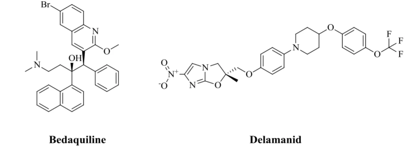
centuries [1]. According to the World Health Organization (WHO), ten million people developed TB in 2018 revealing this disease as the foremost reason of life lost by the cause of a single infectious agent [2]. Despite the approval of new anti-TB drugs such as bedaquiline and delamanid (Figure 1) in addition to the existing traditional ones [3,4], TB treatment continues to be a formidable challenge worldwide.
Figure 1. Chemical structures of bedaquiline and delamanid.
The main obstacle to the success of the current TB chemotherapy regime is the emerging extensively drug-resistant and multidrug-resistant tuberculosis as well as the toxicity of the drugs resulting in adverse side effects [5]. Hence, to overcome these problems, the development of new therapeutics with safer toxicity profiles is mandatory.
Thiourea is the classic bioisostere of urea functionality, in which oxygen is replaced by sulfur atom and represents one of the most important core structures incorporated into medicinal chemistry [6,7]. This scaffold serves as an excellent building block in the discovery of new drug candidates with diverse therapeutic applications. There are many commercial examples (Figure 2) that carry thiourea group integrated into their structures such as thiocarlide, thioacetazone (antitubercular), noxytiolin (antibacterial), and thiopental (general anesthetic) [8].
4 Figure 2. Examples for the drugs carrying thiourea moiety.
Among the above-mentioned commercial ones, thiocarlide (or isoxyl) and thioacetazone are thiourea containing compounds known as effective antitubercular agents against multidrug-resistant strains of M. tuberculosis. They interfere with the synthesis of oleic acid, consequently tuberculostearic acid in M. tuberculosis affecting mycolic acid synthesis, as well [9]. Both drugs were recently identified to show their mechanism of action by covalently inhibiting HadA component of M. tuberculosis fatty acid synthase type II (FAS-II) dehydratase through sulfur atom of thioketone functionality [10].
The enoyl-acyl carrier protein (ACP) reductase, InhA, is an important enzyme employed in the mycolic acid biosynthesis pathway of M. tuberculosis. Mycolic acids are long-chain fatty acids and are major components of mycobacterial cell envelope [11]. Therefore, InhA is essential for the growth of M. tuberculosis and stands as an attractive target for the discovery of novel antitubercular agents [12].
Although various structurally diverse inhibitors of InhA have been reported (Figure 3), in almost every case, the inhibitors bind to InhA forming interactions with cofactor (NADH), tyrosine amino acid and lipophilic binding pockets of the enzyme [13].
The binding pocket of InhA includes there key regions occupied by the inhibitors. The catalytic site, Site I, contains a tyrosine amino acid and the ribose group of the nicotinamide adenine dinucleotide (NAD) cofactor. The inhibitors interact with the enzyme through hydrogen bonds with the hydroxyl group of Tyr and the ribose ring of NAD. Depending on the chemical structure of the inhibitor, additional hydrogen bond is possible with methionine amino acid. Site II is formed by the hydrophobic pocket near the side chain of Tyr. This site enables the hydrophobic moieties of its inhibitors to bind to InhA via hydrophobic interactions. Site III is comparatively unexplored but the hydrophobic rings of the inhibitors remain close to the phosphate groups of NAD as well as Gly96 and Phe97 at van der Waals distance [14]. These reported interactions of
5
various ligands with InhA enzyme provide possibilities for the rational design of new inhibitors that can bind to this region.
Figure 3. Chemical structures of inhibitors co-crystallized in InhA enzyme. Parts of the
molecules participating in InhA binding are highlighted: Hydrogen bonds and hydrophobic interactions are represented as red dotted lines and yellow spheres, respectively.
In 2018, Prati et al. reported several crystal structures of novel InhA–NADH–ligand complexes [15]. One of them contains a urea-based co-crystallized ligand, which is deposited in Protein Data Bank under PDB code 5OIL (Figure 4). Although this compound fulfills all structural requirements to interact with InhA, it is a weak inhibitor at the same time. This situation encouraged us to carry out different modifications on this compound to achieve novel inhibitors capable of targeting InhA.
6 Figure 4. Molecular surface representation of InhA with the co-crystallized ligand and NAD
(PDB code:5OIL) shown as green and orange sticks, respectively (A). Expanded view of InhA active pocket: the ligand is represented as green stick, residues providing ligand binding are displayed as lines, hydrogen bonds are shown as green dotted lines (B).
As both urea and thiourea groups possess near-equal molecular volumes and shapes, swapping urea with thiourea is considered to be a rational approach in the drug design process [16,17]. In light of these considerations, we designed and synthesized thiourea derivatives (Figure 5) carrying necessary structural moieties to be M. tuberculosis growth and InhA inhibitors. We checked the validity of our hypotheses by various assays on M. tuberculosis as well as enzyme inhibition and molecular modeling studies.
7 Figure 5. Design strategy for the synthesized compounds
2. Results and discussion
2.1. Synthesis
In the present study, we synthesized two subseries of compounds. Initially, we synthesized N-pyridine-N′-aryl thiourea derivatives introducing (substituted) phenyl/naphthyl rings into the structure of the compounds (TU1-TU10). Subsequently, we obtained benzyl and phenyl substituted thiourea derivatives as the second subseries (TU11-TU14). As our aim was to carry out different modifications on the week inhibitor urea-based ligand of 5OIL to reach more active ones, we swapped the urea functionality with thiourea group in these compounds. To check the validity of this modification, we also added two representative urea derivatives in this subseries (U1-U2). The fundamental synthetic path was applied for the synthesis of target compounds
TU1-T14 and U1-U2, shown in Figure 6. All compounds were successfully synthesized in a
simple one-step method over the reaction of amine with equivalent amount of various isothiocyanate/isocyanate derivatives in toluene at 80 °C. The structures of the target compounds were elucidated by 1H NMR, 13C NMR, HRMS and FTIR. 1H NMR and 13C NMR spectra of the synthesized compounds are provided as supplementary materials.
8 Figure 6. Synthetic pathway for the preparation of target compounds
2.2. Mycobacterium tuberculosis growth inhibition and cytotoxicity determination
The minimum inhibitory concentration (MIC) of the synthesized compounds against M.
tuberculosis H37Rv was determined using Microplate Alamar Blue Assay (MABA) method.
Isoniazid, rifampicin, and ethambutol were employed as standard drugs. As cytotoxicity to the healthy cells can emerge a serious problem during the discovery of new antitubercular agents, the most active compounds, possessing MIC values ≤1.56 µg/mL, were evaluated for the cytotoxic effects using MTT assay. The structures as well as MIC values (µg/mL and µM) of the compounds and the cytotoxicity of the most active derivatives are presented in Table 2.
Table 2. Chemical structures, antitubercular activity and cytotoxicity data of the compounds
Compound R MIC (µg/mL) MIC (µM) Cytotoxicity
(% inhibition at 50 µg/mL) TU1 H >25 >102.88 TU2 4-OCH3 12.5±1.4 45.79 TU3 2,4-diCl 0.78±0.45 2.50 34.05 TU4 4-Br 0.78±0.55 2.42 30.8 TU5 2-CF3 >25 >80.39 TU6 4-CH3 25±0 97.28 TU7 4-F 12.5±1.4 47.89
9
TU8 4-I 0.78±0.27 2.11 31.19
TU9 3,5-diCF3 >25 >65.96
TU10 >25 >85.32
Compound X R1 R2 MIC (µg/mL) MIC (µM) Cytotoxicity
(% inhibition at 50 µg/mL) TU11 S Cl H >25 >90.25 TU12 S Cl NO2 0.78±0.59 2.42 29.86 TU13 S Br H >25 >77.88 TU14 S Br NO2 1.56±0.82 4.26 24.93 U1 O Cl H >25 >95.79 U2 O Br H >25 >81.97 Isoniazid 0.05±0.02 0.36 Rifampicin 0.1±0.07 0.12 Ethambutol 1.56±0.9 7.6
According to the obtained results, five compounds displayed MIC values at ≤ 1.56 µg/mL and appeared as exceptional antimycobacterial agents. TU3, TU4, TU8 and TU12 stand out with MIC value of 0.78 µg/mL among other derivatives. It is important to emphasize that these compounds are more active than ethambutol, which is among the first-line drugs for tuberculosis treatment. Furthermore, TU14 showed MIC value at 1.56 µg/mL likewise ethambutol. In the first subseries (TU1-TU10) changing the substituents of the phenyl ring played an important role in the mentioned activity. Introducing the chlorine, bromine and iodine atoms led to a great decrease in the MIC values. However, keeping the phenyl/naphthyl ring unsubstituted destroyed the antitubercular effect of the compounds. In the second subseries, it is obvious that nitro group on the phenyl ring made a great contribution to mentioned activity. On the other hand, changing the substituent on the benzyl functionality as bromine or chlorine did not play a role in either urea or thiourea derivatives. Consequently, thiourea derivatives with nitro groups appeared as the most active ones in this subseries. With these results, we also observed that swapping the urea group with thiourea functionality was a logical modification to obtain more active antitubercular agents. As toxicity, particularly hepatic and ocular toxicity, is one of the most important limiting factors of TB treatment with first-line drugs such as isoniazid, rifampicin and ethambutol, it is of utmost
10
significance to discover new agents with low cytotoxicity [18,19]. In this study, five compounds (TU3, TU4, TU8, TU12 and TU14), which have MIC values ≤ 1.56 µg/mL, were found to be non-toxic with <50% inhibition in accordance with cytotoxicity data obtained from MTT assay.
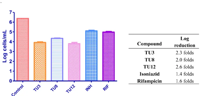
2.3. Nutrient starvation model of Mycobacterium tuberculosis H37Rv
While determining the antitubercular activities of the new drug candidates, it is critical to perform the biological experiments on inactive or persistent form of M. tuberculosis, which can endure oxygen and supplement deprived conditions. With this aim, a nutrient starvation model was applied on the bacteria by starving the culture in phosphate buffer saline (PBS) for six weeks. Afterward, TU3, TU8 and TU12, the representative compounds with the lowest MIC values obtained from MABA, were treated with the culture. The obtained results are provided in Figure 7.
Figure 7. Activity profile of the selected compounds against M. tuberculosis in the nutrient starvation model. Bacterial count estimation (Mean ± S.D., n = 3) was conducted using the MPN (most probable number) assay. Significance plot was generated using two way ANOVA (p < 0.0001, GraphPad Prism Software).
The tested compounds, TU3, TU8 and TU12 showed significant inhibition in the growth with a log reduction of 2.3, 2.0 and 2.6, respectively. These results confirm that tested compounds significantly inhibited the growth of M. tuberculosis in the nutrient starvation model.
11
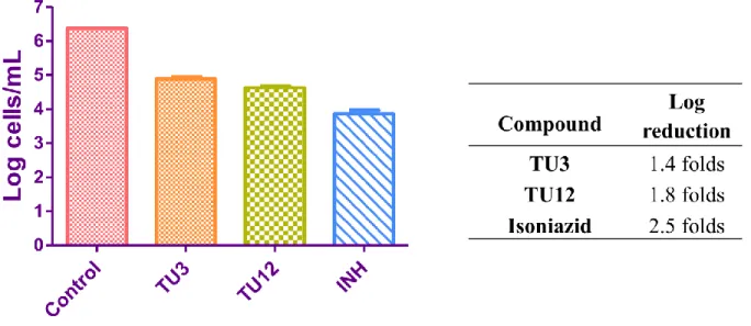
2.4. Mycobacterium tuberculosis infected macrophage assay
When M. tuberculosis is ingested by macrophages, most of the bacteria are found in fused phagolysosomes, but many escapes. Thus, determining the efficiency of the molecules in this model help us to understand how the drug is inhibiting M. tuberculosis in the macrophages. With this purpose, macrophages were initially infected with M. tuberculosis. Afterward, the cells were treated with TU3 and TU12, the most active structurally different derivatives obtained from MABA and nutrient starvation model, at 10 µM concentrations for two days. Finally, the bacterial count was performed using MPN assay. Figure 8 represents the obtained values from M.
tuberculosis infected macrophage assay.
Figure 8. Comparative M. tuberculosis infected macrophage inhibitory activity plots. Bacterial count estimation (Mean ± S.D., n = 4) was conducted using MPN assay.
The obtained results revealed 1.4 and 1.8 log reductions in growth, for compounds TU3 and
TU12 respectively, compared to isoniazid (2.5 log reduction).
2.5. InhA inhibition assay and IC50 determination
The synthesized compounds carry all structural moieties required to interact with InhA enzyme; hydrogen bond participating atoms to form hydrogen bonds to NAD and Tyr and two hydrophobic rings. Hence, they were tested to determine their inhibition values for InhA from M.
12
Triclosan was used as positive control during the experiments. The compounds possessing more than 70% inhibition were also tested at 5 µM concentration. Enzyme inhibition results are presented in Table 2.
Table 2. Enzyme inhibition values for the compounds, expressed as a percentage of InhA
inhibition Compound % Inhibition at 50 µM of compounds (in bracket at 5 µM) TU1 35 TU2 20 TU3 47 TU4 25 TU5 32 TU6 23 TU7 17 TU8 8 TU9 22 TU10 38 TU11 31 TU12 72 (25) IC50 = 17.7 ± 1.3 μM TU13 23 TU14 78 (29) IC50 = 15.6 ± 1.6 μM U1 14 U2 NI TCL >99 (>95) NI: no inhibition
According to the obtained inhibition values, TU3, one of the compounds with the lowest MIC value in the first series, showed moderate activity with 47% inhibition. Lack of efficient InhA inhibition for the active compounds from MABA assay, TU3, TU4 and TU8, suggests a different target for these compounds to show their antitubercular effects. Because of their strong structural similarity to thiocarlide [20], we surmise that TU3, TU4 and TU8 are likely to exhibit their antituberular activities by targeting type II fatty acid synthase pathway via binding to HadA
13
through thiocarbonyl functionality in the same way as thiocarlide. Further studies must be conducted to verify this approach.
On the other side, TU12 and TU14 from the second subseries appeared as clearly better inhibitors of InhA with 72% and 78% inhibition values, respectively.
IC50 values were also calculated for the most active InhA inhibitors, TU12 and TU14, and found
to be 17.7 ± 1.3 μM and 15.6 ± 1.6 μM, respectively (Figure 9).
Figure 9. Determination of IC50 for TU12 (A) and TU14 (B) against InhA enzyme
Considering the enzyme inhibition values are consistent with the results obtained from MABA assay for TU12 and TU14, it can be concluded that InhA is an important target for these compounds that exhibit antimycobacterial activities.
2.6. Molecular docking
In order to support experimental InhA inhibition data with computational methods, we docked all compounds into the active pocket of InhA and illuminated their binding modes to the enzyme. The binding affinity as well as interacting amino acid residues of the enzyme with the compounds are provided as supplementary data. Here, we focus on TU12 and TU14, which were found to be the most effective inhibitors of InhA. The spatial orientation and interaction patterns of TU12 are shown in Figure 10. TU14 has exactly the same orientation in the active pocket of InhA and 3D
14
interaction figure is given in supplementary material. As TU12 and TU14 differ from each other with only the type of the halogen they carry, molecular docking studies help us to explain their similar enzyme inhibition values. Both bromine and chlorine atoms interact with the enzyme through hydrophobic interactions.
Figure 10. Proposed binding mode of TU12 (pink stick) in the binding pocket of InhA enzyme.
(A). 2D depiction of enzyme-ligand interactions: Hydrogen bond acceptor and donor interactions are represented as red and green arrows, respectively. Hydrophobic interactions are shown by yellow spheres (B).
According to the obtained interactions, TU12 forms three key hydrogen bonds with the enzyme via the thiourea functionality. Sulfur atom of the thioketone moiety served as hydrogen bond acceptor with NAD301 and Tyr158. In thioureas, electron delocalization involving C=S and lone pairs of nitrogen atom induces an important Cδ+=Sδ- dipole, which enables the formation resonance-induced of C=S…H bonds in this functional group [21]. Additionally, TU12 also interacted with the enzyme by forming one more hydrogen bond with the sulfur atom of Met199 through one of the nitrogen atoms of thiourea group. Benzyl moiety provided more flexibity to this compound comparing to phenyl ring and in this way, TU12 completely occupied the
15
hydrophobic pocket formed by Phe149, Pro193, Ile215, Leu218, and Glu219. The chlorine atom also participated in these hydrophobic interactions. Nitrophenyl group positioned towards the phosphate groups of NAD as well as Gly96 and Phe97 at van der Waals distance. It is noteworthy that the oxygen atom of the nitro group formed non-classical hydrogen bond (C-H…O) with Phe97 (represented by gray dotted lines) in this region. Although it is a weak interaction type, it can be helpful to explain why nitro derivatives are better InhA inhibitors. Consequently, TU12 satisfies all structural requirements of well-known inhibitors of InhA enzyme. Interaction details such as bond lengths, angle and contributing amino acids for TU12 as well as the co-crystallized ligand are summarized in Table 3.
Table 3. Interaction characteristics of TU12 and the co-crystallized ligand of 5OIL with InhA
Compound Interactions with amino acid residues of 5OIL
Bond Angle (°) (Tyr158, C=O, NAD301) Hydrogen Bonds (Bond Length-Å) Hydrophobic Interactions Co-crystallized Ligand Tyr158, (2.74), Met199 (3.66), NAD301 (2.75)
Phe149, Tyr158, Met199, Ile215, Leu218
98.06
TU12 Gly96 (3.29), Tyr158 (2.84),
Met199 (2.65), NAD301 (2.81),
Phe149, Tyr158, Met199, Pro193, Ile215, Leu218,
Glu219
94.79
When we analyze the interaction characteristics of original ligand of 5OIL and TU12 with InhA, it is clear that TU12 interacts with the enzyme through more amino acids. Compared to the co-crystallized ligand, TU12 forms one extra hydrogen bond with InhA via Gly96. Also, the length of the hydrogen bond to Met199 of TU12 is obviously shorter than the co-crystallized ligand’s interaction. These are the determining factors that TU12 inhibits InhA more efficiently. Additionally, the increased number of amino acids of the hydrophobic pocket interacting with
16
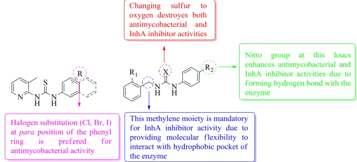
2.7. Structure-activity relationships
According to the data obtained from biological activity determination experiments, we summarized the structural requirements of the compounds which present antimycobacterial and InhA inhibitor activities in Figure 11.
Figure 11. Structure-activity relationships of the synthesized compounds
2.8. In silico prediction of physicochemical properties
Some bioactive molecules may be unsuccessful to turn into good drug molecules because of low bioavailability. For this reason, molecular properties prediction can be considered as a useful tool to initially determine novel drug molecules. Herein, we applied computational methods to predict the parameters of the compounds to determine physicochemical properties. We provide the parameters of the most active compounds in Table 4. The complete table reporting about all compounds is given as supplementary data.
17 Table 4. Calculated physicochemical properties data of the most active compounds
a
LogP: logarithm of n-octanol-water partition coefficient
b
M.W: molecular weight
c
HBA: number of hydrogen bond acceptors
d
HBD: number of hydrogen bond donors
e
TPSA: topological polar surface area
f
NROTB: number of rotatable bonds
g
S: aqueous solubility
Lipinski’s rule of five is used to evaluate drug-likeness and explains the main molecular descriptors which are employed to forecast the oral bioavailability and cell permeability of novel drug candidates [22]. The newly discovered molecules must comply with at least three of these following rules: molecular weight (M.W) ≤ 500 Da, n-octanol-water partition coefficient (log P) ≤ 5, number of hydrogen bond acceptors (HBA) ≤ 10, number of hydrogen bond donors (HBD) ≤ 5. In addition to these descriptors, we also computed rotatable bonds (NROTB) and topological polar surface area (TPSA), which are respected to be significant parameters to determine the oral bioavailability of drug molecules [23].
According to the calculated parameters, all compounds completely adhere to Lipinski’s rule of five. The active compounds are considered to be lipophilic with their calculated LogP values. Among the first-line drugs (isoniazid, ethambutol, rifampicin and pyrazinamide), only rifampicin is lipophilic. However, newly approved drugs such as bedaquiline and delamanid, additionally the molecules under clinical trials like sutezolid, clofazimine, SQ-109 and AZD-5847 are highly lipophilic [24]. This situation supports the idea that lipophilic compounds can easily penetrate through the cell wall of M. tuberculosis, thus lipophilic molecules have great therapeutic value as future anti-tubercular agents. TPSA values of the most active compounds were found to be 36.95 and 69.88, which are acceptable values considering that this value is below 140-150 Å2 for the
Compound LogP[a] M.W.[b] HBA[c] HBD[d] TPSA[e] nROTB[f] Lipinski’s Violation S[g] (mg/mL) TU3 3.83 312.23 3 2 36.95 4 0 4.97 TU5 3.40 311.33 3 2 36.95 5 0 11.60 TU8 3.63 369.23 3 2 36.95 4 0 17.34 TU12 3.34 321.79 5 2 69.88 6 0 21.43 TU14 3.47 366.24 5 2 69.88 6 0 29.90
18
most known drug molecules. Number of rotatable bonds is an important parameter which indicates molecular flexibility. In order to restrict the conformational changes while interaction with the biological target, this value should be ≤ 10 [23]. All of our compounds have less than ten rotatable bonds.
Consequently, these compounds exhibit excellent physicochemical properties to serve as antitubercular drug candidates.
3. Conclusions
Tuberculosis still causes a massive amount of deaths despite the availability of many commercial anti-TB drugs. Thus, the development of more effective and safer therapeutics is of extreme importance. With this purpose, we presented herein the rational synthesis of thiourea derivatives as antitubercular agents. We achieved effective Mycobacterium tuberculosis growth inhibitors, which are also capable of acting on dormant or latent forms of the bacteria. Moreover, we demonstrated that some compounds exhibit their antimycobacterial activities by inhibiting InhA enzyme, which plays a vital role in the fatty acid biosynthesis pathway in M. tuberculosis. Overall, thiourea can serve as an important pharmacophore in designing future therapeutics against Mycobacterium tuberculosis for the improvement of the treatment success rates.
4. Experimental
4.1. Chemistry
4.1.1. Materials and methods
Toluene was distilled from sodium-benzophenone just prior to use. All reagents were purchased and used as received. All volatiles were removed under the reduced pressure. Melting points were measured using open glass capillaries and are uncorrected. Infrared (IR) spectra were recorded in the range 4000–600 cm−1 via ATR diamond. 1H-(400 MHz) and 13C-NMR spectra (100 MHz) were obtained on a Bruker AM 400 spectrometer in (D6) DMSO solution. Coupling constants, J, are reported in hertz. MS spectra were carried out on an LC/MS High-Resolution Time of Flight (TOF) Agilent 1200/6530 instrument at the Atatürk University-East Anatolian High Technology Research and Application Center (DAYTAM).
19
4.1.2. General procedure for the synthesis of the compounds
These compounds were synthesized according to the modified literature procedure [25,26]. 3-Methylpyridin-2-amine (TU1-TU10) or benzylamine derivative (TU11-TU14, U1-U2) was added to equimolar amount of corresponding phenyl/naphthyl isothiocyanate (TU1-TU14) or phenyl isocyanate (U1-U2) solutions in 10 mL dry toluene. The mixture was stirred at 80 °C and then cooled to room temperature after completion of the reaction. The precipitate was filtered off and washed with toluene. The resulting residue was purified by crystallization from ethanol to afford the target compounds.
4.1.2.1. 1-(3-Methylpyridin-2-yl)-3-phenylthiourea (TU1) [27]
Yellow solid, yield: 72%. Mp 123-124 ºC; Rf(EtOAc:Hexane=4:6): 0.66. IR (ATR) 3424, 3268,
1634, 1596, 1559, 1505, 1226, 739. 1H NMR (400 MHz, DMSO-d6) 13.67 (s, 1H), 9.07 (s, 1H),
8.22 (d, J = 3.9 Hz, 1H), 7.72 (d, J = 7.3 Hz, 1H), 7.67 (d, J = 7.6 Hz, 2H), 7.39 (t, J = 7.7 Hz, 2H), 7.21 (t, J = 7.2 Hz, 1H), 7.14 – 7.07 (m, 1H), 2.35 (s, 3H). 13C NMR (100 MHz, DMSO-d6)
δ 178.78 (C=S), 151.82, 143.83, 140.77, 139.26, 129.01, 126.02, 124.63, 122.35, 119.52 (aromatic carbons) and 17.01 (-CH3). HRMS (EI): [M+H]+, found 244.0887. C13H14N3S requires
244.0908.
4.1.2.2. 1-(4-Methoxyphenyl)-3-(3-methylpyridin-2-yl)thiourea (TU2)
Yellow solid, yield: 32%. Mp 83-84 ºC; Rf(EtOAc:Hexane=4:6): 0.45. IR (ATR) 3424, 3268,
1634, 1596, 1559, 1505, 1226, 739. 1H NMR (400 MHz, DMSO-d6) 13.47 (s, 1H), 8.96 (s, 1H),
8.20 (d, J = 4.6 Hz, 1H), 7.70 (d, J = 7.3 Hz, 1H), 7.52 (d, J = 8.8 Hz, 2H), 7.08 (dd, J = 7.3, 5.1 Hz, 1H), 6.94 (d, J = 8.8 Hz, 2H), 3.76 (s, 3H), 2.35 (s, 3H). 13C NMR (100 MHz, DMSO-d6) δ
179.01 (C=S), 157.47, 151.90, 143.80, 140.63, 132.20, 126.41, 122.10, 119.29, 114.14 (aromatic carbons) and 55.73 (-OCH3), 17.02 (-CH3). HRMS (EI): [M+H]+, found 274.0983. C14H15N3OS
20
4.1.2.3. 1-(2,4-Dichlorophenyl)-3-(3-methylpyridin-2-yl)thiourea (TU3)
White solid, yield: 15%. Mp 94-96 ºC; Rf (EtOAc:Hexane=4:6): 0.79. IR (ATR) 3416, 2676,
1606, 1585, 1506, 1338, 1138. 1H NMR (400 MHz, DMSO-d6) δ 14.03 (s, 1H), 9.40 (s, 1H), 8.32
– 8.14 (m, 2H), 7.73 (d, J = 7.0 Hz, 1H), 7.69 (d, J = 2.3 Hz, 1H), 7.44 (dd, J = 8.7, 2.3 Hz, 1H), 7.24 – 7.02 (m, 1H), 2.37 (s, 3H). 13C NMR (100 MHz, DMSO) δ 179.54 (C=S), 151.51, 143.60, 141.03, 135.99, 130.70, 129.23, 129.04, 128.90, 127.60, 122.48, 119.82 (aromatic carbons) and 17.03 (-CH3). HRMS (EI): [M+H]+, found 312.0095. C13H12Cl2N3S requires 312.0129.
4.1.2.4. 1-(4-Bromophenyl)-3-(3-methylpyridin-2-yl)thiourea (TU4)
White solid, yield: 59%. Mp 136-138 ºC; Rf(EtOAc:Hexane=4:6): 0.28. IR (ATR) 3437, 2761,
1608, 1582, 1505, 1352, 1152. 1H NMR (400 MHz, DMSO-d6) δ 13.64 (s, 1H), 9.23 (s, 1H), 8.23
(d, J = 4.3 Hz, 1H), 7.73 (d, J = 7.2 Hz, 1H), 7.68 (d, J = 8.8 Hz, 2H), 7.56 (d, J = 8.7 Hz, 2H), 7.16 – 7.06 (m, 1H), 2.35 (s, 3H). 13C NMR (100 MHz, DMSO) δ 178.86 (C=S), 151.72, 143.90, 140.84, 138.73, 131.78, 126.56, 122.69, 119.69, 117.98 (aromatic carbons) and 17.06 (-CH3).
HRMS (EI): [M+H]+, found 321.9975. C13H12BrN3S requires 322.0014.
4.1.2.5. 1-(3-Methylpyridin-2-yl)-3-(2-(trifluoromethyl)phenyl)thiourea (TU5)
White solid, yield: 81%. Mp 154-156 ºC; Rf(EtOAc:Hexane=4:6): 0.10. IR (ATR) 3425, 3406,
3082, 1634, 1593, 1506, 1381, 1270, 1123. 1H NMR (400 MHz, DMSO-d6) δ 14.02 (s, 1H), 9.38
(s, 1H), 8.12 (d, J = 4.4 Hz, 1H), 7.90 (d, J = 8.0 Hz, 1H), 7.81 – 7.66 (m, 3H), 7.48 (t, J = 7.6 Hz, 1H), 7.11 (dd, J = 7.3, 5.1 Hz, 1H), 2.38 (s, 3H). 13C NMR (100 MHz, DMSO) δ 180.76 (C=S), 151.72, 143.30, 141.00, 137.21, 132.97, 131.02, 127.24, 126.35, 124.36 (aromatic carbons), 122.74 (CF3), 122.19, 119.63 (aromatic carbons), 16.98 (-CH3). HRMS (EI): [M+H]+,
found 312.0777. C13H12F3N3S requires 312.0782.
4.1.2.6. 1-(3-Methylpyridin-2-yl)-3-(p-tolyl)thiourea (TU6) [28]
White solid, yield: 41%. Mp 128-129 ºC; Rf(EtOAc:Hexane=4:6): 0.28. IR (ATR) 3379, 2712,
1634, 1593, 1506, 1362, 1251, 1150. 1H NMR (400 MHz, DMSO-d6) δ 13.58 (s, 1H), 9.02 (s,
1H), 8.22 (s, 1H), 7.72 (d, J = 7.5 Hz, 1H), 7.54 (d, J = 8.2 Hz, 2H), 7.19 (d, J = 7.9 Hz, 2H), 7.13 – 7.07 (m, 1H), 2.35 (s, 3H), 2.30 (s, 3H). 13C NMR (100 MHz, DMSO) δ 178.68 (C=S), 151.87, 143.78, 140.67, 136.75, 135.19, 129.41, 124.52, 122.18, 119.34 (aromatic carbons) and 21.04, 17.02 (2x-CH3). HRMS (EI): [M+H]+, found 258.1067. C14H16N3S requires 258.1065.
21
4.1.2.7. 1-(4-Fluorophenyl)-3-(3-methylpyridin-2-yl)thiourea (TU7)
White solid, yield: 39%. Mp 90-91 ºC; Rf (EtOAc:Hexane=4:6): 0.72. IR (ATR) 3377, 2707,
1629, 1589, 1504, 1447, 1362, 1227, 1151. 1H NMR (400 MHz, DMSO-d6) δ 13.54 (s, 1H), 9.15 (s, 1H), 8.22 (s, 1H), 7.72 (d, J = 7.1 Hz, 1H), 7.69 – 7.60 (m, 2H), 7.22 (t, J = 8.7 Hz, 2H), 7.16 – 7.07 (m, 1H), 2.35 (s, 3H). 13C NMR (100 MHz, DMSO) δ 179.30 (C=S), 160.08 (d, 1 JF-C = 242.6 Hz), 151.80, 143.88, 140.77, 135.66 (d, 4JF-C = 2.6 Hz), 127.10 (d, 3JF-C = 8.3 Hz), 122.47, 119.56, 115.64 (d, 2JF-C = 22.5 Hz), 17.04 (-CH3). HRMS (EI): [M+H]+, found 262.0784. C13H13FN3S requires 262.0814. 4.1.2.8. 1-(4-Iodophenyl)-3-(3-methylpyridin-2-yl)thiourea (TU8)
White solid, yield: 55%. Mp 126-128 ºC; Rf(EtOAc:Hexane=4:6): 0.76. IR (ATR) 3374, 2734,
1614, 1583, 1506, 1445, 1346, 1147. 1H NMR (400 MHz, DMSO-d6) δ 13.63 (s, 1H), 9.21 (s,
1H), 8.22 (d, J = 3.8 Hz, 1H), 7.72 (d, J = 8.2 Hz, 3H), 7.55 (d, J = 8.4 Hz, 2H), 7.17 – 7.06 (m, 1H), 2.35 (s, 3H). 13C NMR (100 MHz, DMSO) δ 178.75 (C=S), 151.74, 143.91, 140.86, 139.21, 137.66, 126.67, 122.70, 119.71, 90.42 (aromatic carbons) and 17.09 (-CH3). HRMS (EI):
[M+H]+, found 369.9871. C13H13IN3S requires 369.9875.
4.1.2.9. 1-(3,5-Bis(trifluoromethyl)phenyl)-3-(3-methylpyridin-2-yl)thiourea (TU9)
White solid, yield: 28%. Mp 139-141 ºC; Rf(EtOAc:Hexane=4:6): 0.79. IR (ATR) 3445, 2249,
1622, 1052, 1024, 1005, 819.1H NMR (400 MHz, DMSO-d6) δ 13.70 (s, 1H), 9.64 (s, 1H), 8.48
(s, 2H), 8.27 (s, 1H), 7.86 (s, 1H), 7.77 (s, 1H), 7.25 – 7.05 (m, 1H), 2.36 (s, 3H). 13C NMR (100 MHz, DMSO) δ 179.77 (C=S), 151.36, 144.28, 141.47, 140.91, 130.78, 124.99, 123.50 (aromatic carbons), 122.28 (CF3), 120.20, 118.58 (aromatic carbonss), 16.99 (-CH3). HRMS (EI): [M+H]+,
found 380.0659. C15H12F6N3S requires 380.0656.
4.1.2.10. 1-(3-Methylpyridin-2-yl)-3-(naphthalen-2-yl)thiourea (TU10)
Pale yellow solid, yield: 75%. Mp 151-153 ºC; Rf(EtOAc:Hexane=4:6): 0.69. IR (ATR) 3402,
2856, 1637, 1567, 1505, 1338, 1150. 1H NMR (400 MHz, DMSO-d6) δ 13.75 (s, 1H), 9.29 (s,
1H), 8.23 (d, J = 4.9 Hz, 1H), 8.00 (dd, J = 6.4, 2.7 Hz, 1H), 7.96 (d, J = 9.1 Hz, 1H), 7.89 (d, J = 8.1 Hz, 1H), 7.82 (d, J = 7.2 Hz, 1H), 7.77 (d, J = 7.4 Hz, 1H), 7.65 – 7.50 (m, 3H), 7.13 (dd, J =
22
7.4, 5.0 Hz, 1H), 2.41 (s, 3H). 13C NMR (100 MHz, DMSO) δ 180.69 (C=S), 152.08, 143.88, 140.87, 135.45, 134.24, 130.19, 129.50, 128.83, 127.16, 126.70, 125.98, 124.99, 122.72, 122.45, 119.58 (aromatic carbons) and 17.16 (-CH3). HRMS (EI): [M+H]+, found 294.1036. C17H16N3S
requires 294.1065.
4.1.2.11. 1-(2-Chlorobenzyl)-3-phenylthiourea (TU11) [29]
White solid, yield: 69%. Mp 178-180 ºC; Rf(EtOAc:Hexane=4:6): 0.81. IR (ATR) 3663, 2987,
1592, 1410, 1243, 1066. 1H NMR (400 MHz, DMSO-d6) δ 9.77 (s, 1H), 8.14 (s, 1H), 7.50 – 7.41
(m, 3H), 7.40 – 7.25 (m, 5H), 7.13 (t, J = 7.3 Hz, 1H), 4.80 (d, J = 5.4 Hz, 2H). 13C NMR (100 MHz, DMSO) δ 181.56 (C=S), 139.46, 136.55, 132.35, 129.56, 129.29, 129.17, 129.06, 127.55, 124.99, 123.91 (aromatic carbons) and 45.53 (-CH2-). HRMS (EI): [M+H]+, found 277.0570.
C14H14ClN2S requires 277.0566.
4.1.2.12. 1-(2-Chlorobenzyl)-3-(4-nitrophenyl)thiourea (TU12)
Pale yellow solid, yield: 76%. Mp 148-150 ºC; Rf(EtOAc:Hexane=4:6): 0.74. IR (ATR) 3674,
2987, 1594, 1529, 1508, 1407, 1302, 1240, 1066. 1H NMR (400 MHz, DMSO-d6) δ 10.38 (s,
1H), 8.65 (s, 1H), 8.19 (d, J = 9.2 Hz, 2H), 7.89 (d, J = 9.0 Hz, 2H), 7.51 – 7.44 (m, 1H), 7.44 – 7.39 (m, 1H), 7.39 – 7.26 (m, 2H), 4.82 (d, J = 5.1 Hz, 2H). 13C NMR (100 MHz, DMSO) δ 181.13 (C=S), 146.67, 142.50, 135.80, 132.60, 129.67, 129.36, 127.63, 124.93, 121.22 (aromatic carbons, one carbon signal was overlapped) and 45.52 (-CH2-). HRMS (EI): [M+H]+, found
322.0435. C14H13ClN3O2S requires 322.0417.
4.1.2.13. 1-(2-Bromobenzyl)-3-phenylthiourea (TU13)
Pale yellow solid, yield: 72%. Mp 187-189 ºC; Rf(EtOAc:Hexane=4:6): 0.81. IR (ATR) 3377,
3135, 1610, 1531, 1504, 1297, 1242. 1H NMR (400 MHz, DMSO-d6) δ 9.80 (s, 1H), 8.14 (s, 1H),
7.61 (d, J = 7.9 Hz, 1H), 7.46 (d, J = 7.7 Hz, 2H), 7.43 – 7.28 (m, 4H), 7.27 – 7.19 (m, 1H), 7.20 – 7.09 (m, 1H), 4.75 (d, J = 4.7 Hz, 2H). 13C NMR (100 MHz, DMSO) δ 181.55 (C=S), 139.45,
138.08, 132.80, 129.35, 129.29, 129.18, 128.11, 125.00, 123.90, 122.65 (aromatic carbons) and 48.01 (-CH2-). HRMS (EI): [M+H]+, found 321.0045. C14H14BrN2S requires 321.0061.
4.1.2.14. 1-(2-Bromobenzyl)-3-(4-nitrophenyl)thiourea (TU14)
White solid, yield: 65%. Mp 185-187 ºC; Rf(EtOAc:Hexane=4:6): 0.74. IR (ATR) 3350, 3025,
23
(s, 1H), 8.19 (d, J = 9.2 Hz, 2H), 7.89 (d, J = 8.9 Hz, 2H), 7.64 (d, J = 8.2 Hz, 1H), 7.48 – 7.34 (m, 2H), 7.27 – 7.20 (m, 1H), 4.78 (d, J = 4.9 Hz, 2H). 13C NMR (100 MHz, DMSO) δ 181.14 (C=S), 146.66, 142.51, 137.35, 132.92, 129.63, 128.19, 124.94, 122.92, 121.23 (aromatic carbons one carbon signal was overlapped) and 47.98 (-CH2-). HRMS (EI): [M+H]+, found 365.9937.
C14H13BrN3O2S requires 365.9912.
4.1.2.15. 1-(2-Chlorobenzyl)-3-phenylurea (U1) [30]
White solid, yield: 85%. Mp 180-182 ºC; Rf(EtOAc:Hexane=4:6): 0.74. IR (ATR) 3315, 2971,
1633, 1531, 1439, 1241, 1039. 1H NMR (400 MHz, DMSO-d6) δ 8.67 (s, 1H), 7.44 (dd, J = 7.6,
1.4 Hz, 1H), 7.42 – 7.36 (m, 3H), 7.38 – 7.25 (m, 2H), 7.22 (t, J = 7.9 Hz, 2H), 6.89 (t, J = 7.3 Hz, 1H), 6.65 (t, J = 5.9 Hz, 1H), 4.36 (d, J = 6.0 Hz, 2H). 13C NMR (100 MHz, DMSO) δ 155.73 (C=O), 140.43, 137.51, 132.49, 129.59, 129.39, 129.20, 129.17, 127.68, 122.00, 118.34 (aromatic carbons) and 41.19 (-CH2-). HRMS (EI): [M+H]+, found 261.0799. C14H14ClN2O
requires 261.0795.
4.1.2.16. 1-(2-Bromobenzyl)-3-phenylurea (U2) [31]
White solid, yield: 89%. Mp 189-191 ºC; Rf(EtOAc:Hexane=4:6): 0.71. IR (ATR) 3662, 2987,
1633, 1410, 1241, 1066. 1H NMR (400 MHz, DMSO-d6) δ 8.70 (s, 1H), 7.61 (d, J = 7.8 Hz, 1H),
7.39 (t, J = 6.1 Hz, 4H), 7.28 – 7.17 (m, 3H), 6.89 (t, J = 7.3 Hz, 1H), 6.66 (t, J = 5.8 Hz, 1H), 4.32 (d, J = 5.9 Hz, 2H). 13C NMR (100 MHz, DMSO) δ 155.59 (C=O), 140.70, 139.24, 132.81, 129.40, 129.36, 129.15, 128.23, 122.82, 121.75, 118.20 (aromatic carbons) and 43.67 (-CH2-).
HRMS (EI): [M+H]+, found 305.0298. C14H14BrN2O requires 305.0290.
4.2. Microplate Alamar Blue Assay for Mycobacterium tuberculosis
In vitro antitubercular activities of the synthesized compounds were determined using Microplate
Alamar Blue Assay (MABA) method. The results are stated as MIC values against M.
tuberculosis H37Rv [32,33].
24
The in vitro cytotoxicity of the active compounds from MABA assay with MIC values ≤ 1,56 μg/mL were evaluated by 3-(4,5-dimethylthiazol-2-yl)-2,5-diphenyltetrazolium bromide (MTT) assay against growth inhibition of RAW 264.7 cells at 50 μg/mL concentration [34].
4.4. Nutrient starvation model
A nutrient starvation model was applied on the bacteria by starving the culture in phosphate buffer saline (PBS) and incubating at 37 °C for six weeks. Afterward, the cultures were treated with isoniazid, rifampicin and selected compounds (10µg/ml concentration) for seven days. The frequency of persistors was enumerated by MPN assay [35].
4.5. Mycobacterium tuberculosis infected macrophage assay
The selected compounds were evaluated for their activities in M. tuberculosis infected macrophage model at 10 µM final concentration. Bacteria were suspended in 7H9 broth. Serial dilutions were performed, the plates were incubated at 37 ºC and growth was observed after three weeks [33,36].
4.6. InhA inhibition and IC50 determination
The InhA inhibition values of the compounds were evaluated according to the previously reported method using triclosan as a positive control. All activity assays were carried out in triplicate. IC50 values were determined using the 4-parameter curve-fitting software XLFit
(IDBS) with at least seven points [37,38].
Detailed experimental procedures for the biological activity evaluation studies are provided as supplementary materials.
4.7. Molecular docking
The crystal structure of Mycobacterium tuberculosis enoyl-ACP reductase (InhA) with the co-crystallized ligand 1-cyclohexyl-3-(pyridin-3-ylmethyl)urea was obtained from Protein Data Bank under the PDB code 5OIL [15]. All synthesized compounds were docked into the active pocket of the enzyme using AutoDock Vina 1.1,[39] integrated into LigandScout 4.2,[40] with default parameters. The obtained results were visually analyzed using LigandScout 4.2. The
25
molecular docking figures were generated by Maestro[41], DiscoveryStudio [42] and LigandScout 4.2 in this study.
4.8. In silico prediction of physicochemical properties
The chemical formulae of the compounds were drawn in ChemDraw Ultra 12.0 and saved as Simplified Molecule Input Entry System (SMILES) file. The calculation of physicochemical properties was accomplished by Molinspiration (https://www.molinspiration.com)[43], and Molsoft drug-likeness and molecular property prediction tool [44].
Author Statements
ŞDD and HD obtained the compounds and evaluated their structures. VSK and DS carried out MABA, nutrition starvation model, M. tuberculosis infected macrophage assay and cytotoxicity experiments. CL accomplished InhA inhibition assay and IC50 determination. MGG was
responsible for the computational studies. MGG and ŞDD wrote the manuscript.
Conflict of Interest
None of the authors declared any potential conflicts of interest.
Acknowledgements
Faculty of Pharmacy at Erciyes University is gratefully acknowledged. MGG is grateful to Prof. Dr. Gerhard Wolber, Freie Universitat Berlin, for providing the license for LigandScout 4.2.
References
[1] S. Tiberi, M. Muñoz-Torrico, R. Duarte, M. Dalcolmo, L. D’Ambrosio, G.-B. Migliori, New drugs and perspectives for new anti-tuberculosis regimens, Pulmonology. 24 (2018) 86–98. doi:10.1016/J.RPPNEN.2017.10.009.
[2] WHO | Global tuberculosis report 2019, WHO. (2019). https://www.who.int/tb/publications/global_report/en/.
[3] D.T. Hoagland, J. Liu, R.B. Lee, R.E. Lee, New agents for the treatment of drug-resistant Mycobacterium tuberculosis, Adv. Drug Deliv. Rev. 102 (2016) 55–72.
26
[4] K. Pyta, K. Klich, J. Domagalska, P. Przybylski, Structure and evaluation of antibacterial and antitubercular properties of new basic and heterocyclic 3-formylrifamycin SV
derivatives obtained via “click chemistry” approach, Eur. J. Med. Chem. 84 (2014) 651– 676. doi:10.1016/j.ejmech.2014.07.066.
[5] A. Campaniço, R. Moreira, F. Lopes, Drug discovery in tuberculosis. New drug targets and antimycobacterial agents, Eur. J. Med. Chem. 150 (2018) 525–545.
doi:10.1016/J.EJMECH.2018.03.020.
[6] N.A. Meanwell, Synopsis of Some Recent Tactical Application of Bioisosteres in Drug Design, J. Med. Chem. 54 (2011) 2529–2591. doi:10.1021/jm1013693.
[7] V. Kumar, S. Chimni, Recent Developments on Thiourea Based Anticancer Chemotherapeutics, Anticancer. Agents Med. Chem. 15 (2015) 163–175. doi:10.2174/1871520614666140407123526.
[8] A. Shakeel, Thiourea Derivatives in Drug Design and Medicinal Chemistry: A Short Review, J. Drug Des. Med. Chem. 2 (2016) 10. doi:10.11648/j.jddmc.20160201.12. [9] B. Phetsuksiri, M. Jackson, H. Scherman, M. McNeil, G.S. Besra, A.R. Baulard, R.A.
Slayden, A.E. DeBarber, C.E. Barry, M.S. Baird, D.C. Crick, P.J. Brennan, Unique
Mechanism of Action of the Thiourea Drug Isoxyl on Mycobacterium tuberculosis, J. Biol. Chem. 278 (2003) 53123–53130. doi:10.1074/jbc.M311209200.
[10] A.E. Grzegorzewicz, N. Eynard, A. Quémard, E.J. North, A. Margolis, J.J. Lindenberger, V. Jones, J. Korduláková, P.J. Brennan, R.E. Lee, D.R. Ronning, M.R. McNeil, M. Jackson, Covalent Modification of the Mycobacterium tuberculosis FAS-II Dehydratase by Isoxyl and Thiacetazone, ACS Infect. Dis. 1 (2016) 91–97. doi:10.1021/id500032q. [11] I. Pauli, R.N. dos Santos, D.C. Rostirolla, L.K. Martinelli, R.G. Ducati, L.F.S.M.
Timmers, L.A. Basso, D.S. Santos, R.V.C. Guido, A.D. Andricopulo, O. Norberto de Souza, Discovery of New Inhibitors of Mycobacterium tuberculosis InhA Enzyme Using Virtual Screening and a 3D-Pharmacophore-Based Approach, J. Chem. Inf. Model. 53 (2013) 2390–2401. doi:10.1021/ci400202t.
[12] S.D. Joshi, U.A. More, D. Koli, M.S. Kulkarni, M.N. Nadagouda, T.M. Aminabhavi, Synthesis, evaluation and in silico molecular modeling of pyrroyl-1,3,4-thiadiazole inhibitors of InhA, Bioorg. Chem. 59 (2015) 151–167. doi:10.1016/j.bioorg.2015.03.001. [13] K. Rožman, I. Sosič, R. Fernandez, R.J. Young, A. Mendoza, S. Gobec, L. Encinas, A new
‘golden age’ for the antitubercular target InhA, Drug Discov. Today. 22 (2017) 492–502. doi:10.1016/J.DRUDIS.2016.09.009.
[14] A. Chollet, L. Maveyraud, C. Lherbet, V. Bernardes-Génisson, An overview on crystal structures of InhA protein: Apo-form, in complex with its natural ligands and inhibitors, Eur. J. Med. Chem. 146 (2018) 318–343. doi:10.1016/J.EJMECH.2018.01.047.
[15] F. Prati, F. Zuccotto, D. Fletcher, M.A. Convery, R. Fernandez-Menendez, R. Bates, L. Encinas, J. Zeng, C. Chung, P. De Dios Anton, A. Mendoza-Losana, C. Mackenzie, S.R. Green, M. Huggett, D. Barros, P.G. Wyatt, P.C. Ray, Screening of a Novel Fragment
27
Library with Functional Complexity against Mycobacterium tuberculosis InhA, ChemMedChem. 13 (2018) 672–677. doi:10.1002/cmdc.201700774.
[16] G.P. Suresha, R. Suhas, W. Kapfo, D. Channe Gowda, Urea/thiourea derivatives of quinazolinone-lysine conjugates: Synthesis and structure-activity relationships of a new series of antimicrobials, Eur. J. Med. Chem. 46 (2011) 2530–2540.
doi:10.1016/j.ejmech.2011.03.041.
[17] J.M. Vega-Pérez, I. Periñán, M. Argandoña, M. Vega-Holm, C. Palo-Nieto, E. Burgos-Morón, M. López-Lázaro, C. Vargas, J.J. Nieto, F. Iglesias-Guerra, Isoprenyl-thiourea and urea derivatives as new farnesyl diphosphate analogues: Synthesis and in vitro
antimicrobial and cytotoxic activities, Eur. J. Med. Chem. 58 (2012) 591–612. doi:10.1016/j.ejmech.2012.10.042.
[18] M. Singh, P. Sasi, G. Rai, V.H. Gupta, D. Amarapurkar, P.P. Wangikar, Studies on toxicity of antitubercular drugs namely isoniazid, rifampicin, and pyrazinamide in an in vitro model of HepG2 cell line, in: Med. Chem. Res., Springer, 2011: pp. 1611–1615. doi:10.1007/s00044-010-9405-3.
[19] A. Melamud, G.S. Kosmorsky, M.S. Lee, Ocular Ethambutol Toxicity, Mayo Clin. Proc. 78 (2003) 1409–1411. doi:10.4065/78.11.1409.
[20] A.E. Grzegorzewicz, J. Korduláková, V. Jones, S.E.M. Born, J.M. Belardinelli, A. Vaquié, V.A.K.B. Gundi, J. Madacki, N. Slama, F. Laval, J. Vaubourgeix, R.M. Crew, B. Gicquel, M. Daffé, H.R. Morbidoni, P.J. Brennan, A. Quémard, M.R. McNeil, M. Jackson, A common mechanism of inhibition of the Mycobacterium tuberculosis mycolic acid
biosynthetic pathway by isoxyl and thiacetazone, J. Biol. Chem. 287 (2012) 38434–38441. doi:10.1074/jbc.M112.400994.
[21] P.A. Wood, E. Pidcock, F.H. Allen, Interaction geometries and energies of hydrogen bonds to C=O and C=S acceptors: A comparative study, Acta Crystallogr. Sect. B Struct. Sci. 64 (2008) 491–496. doi:10.1107/S0108768108015437.
[22] C.A. Lipinski, F. Lombardo, B.W. Dominy, P.J. Feeney, Experimental and computational approaches to estimate solubility and permeability in drug discovery and development settings, Adv. Drug Deliv. Rev. 46 (2001) 3–26. doi:10.1016/S0169-409X(00)00129-0. [23] D.F. Veber, S.R. Johnson, H.Y. Cheng, B.R. Smith, K.W. Ward, K.D. Kopple, Molecular
properties that influence the oral bioavailability of drug candidates, J. Med. Chem. 45 (2002) 2615–2623. doi:10.1021/jm020017n.
[24] G. Piccaro, G. Poce, M. Biava, F. Giannoni, L. Fattorini, Activity of lipophilic and hydrophilic drugs against dormant and replicating Mycobacterium tuberculosis, J. Antibiot. (Tokyo). 68 (2015) 711–714. doi:10.1038/ja.2015.52.
[25] Ş.D. Doğan, Copper-catalyzed NH/SH functionalization: A strategy for the synthesis of benzothiadiazine derivatives, Tetrahedron. 73 (2017) 2217–2224.
doi:10.1016/J.TET.2017.02.063.
28
Sarıpınar, Discovery of hydrazone containing thiadiazoles as Mycobacterium tuberculosis growth and enoyl acyl carrier protein reductase (InhA) inhibitors, Eur. J. Med. Chem. (2020) 112035. doi:10.1016/j.ejmech.2020.112035.
[27] V.A. Mamedov, A.A. Kalinin, E.A. Gorbunova, W.D. Habicher, Fused polycyclic nitrogen-containing heterocycles: IX. Oxidative fusion of imidazole ring to 3-benzoylquinoxalin-2-ones, Russ. J. Org. Chem. 40 (2004) 1041–1046. doi:10.1023/B:RUJO.0000045201.90930.ff.
[28] J. Valdés-Martínez, S. Hernández-Ortega, A.K. Hermetet, L.J. Ackerman, C.A. Presto, J.K. Swearingen, D.R. Kelman, K.I. Goldberg, W. Kaminsky, D.X. West, Structural studies of N-2-(3-picolyl)- and N-2-(4-picolyl)-N′ -tolylthioureas, J. Chem. Crystallogr. 32 (2002) 431–438. doi:10.1023/A:1021136508168.
[29] M. Mushtaque, F. Avecilla, Z. Bin Hafeez, M.M.A. Rizvi, Synthesis, Characterization, Molecular Docking, and Anticancer Evaluation of 4‐Thiazolidinone Analogues, J. Heterocycl. Chem. 56 (2019) 1794–1805. doi:10.1002/jhet.3549.
[30] F. Li, C. Sun, H. Shan, X. Zou, J. Xie, From Regioselective Condensation to
Regioselective N-Alkylation: A Novel and Environmentally Benign Strategy for the Synthesis of N,N′-Alkyl Aryl Ureas and N,N′-Dialkyl Ureas, ChemCatChem. 5 (2013) 1543–1552. doi:10.1002/cctc.201200648.
[31] X. Ran, Y. Long, S. Yang, C. Peng, Y. Zhang, S. Qian, Z. Jiang, X. Zhang, L. Yang, Z. Wang, X. Yu, A novel route to unsymmetrical disubstituted ureas and thioureas by HMPA catalyzed reductive alkylation with trichlorosilane, Org. Chem. Front. 7 (2020) 472–481. doi:10.1039/c9qo01321k.
[32] L. Collins, S.G. Franzblau, Microplate alamar blue assay versus BACTEC 460 system for high-throughput screening of compounds against Mycobacterium tuberculosis and
Mycobacterium avium., Antimicrob. Agents Chemother. 41 (1997) 1004–1009. doi:10.1128/AAC.41.5.1004.
[33] V.S. Krishna, S. Zheng, E.M. Rekha, L.W. Guddat, D. Sriram, Discovery and evaluation of novel Mycobacterium tuberculosis ketol-acid reductoisomerase inhibitors as therapeutic drug leads, J. Comput. Aided. Mol. Des. 33 (2019) 357–366. doi:10.1007/s10822-019-00184-1.
[34] J. van Meerloo, G.J.L. Kaspers, J. Cloos, Cell Sensitivity Assays: The MTT Assay, in: Humana Press, 2011: pp. 237–245. doi:10.1007/978-1-61779-080-5_20.
[35] J.C. Betts, P.T. Lukey, L.C. Robb, R.A. McAdam, K. Duncan, Evaluation of a nutrient starvation model of Mycobacterium tuberculosis persistence by gene and protein expression profiling, Mol. Microbiol. 43 (2002) 717–731. doi:10.1046/j.1365-2958.2002.02779.x.
[36] S. Biketov, G. V. Mukamolova, V. Potapov, E. Gilenkov, G. Vostroknutova, D.B. Kell, M. Young, A.S. Kaprelyants, Culturability of Mycobacterium tuberculosis cells isolated from murine macrophages: a bacterial growth factor promotes recovery , FEMS Immunol. Med.
29
Microbiol. 29 (2000) 233–240. doi:10.1111/j.1574-695x.2000.tb01528.x.
[37] T. Matviiuk, J. Madacki, G. Mori, B.S. Orena, C. Menendez, A. Kysil, C. André-Barrès, F. Rodriguez, J. Korduláková, S. Mallet-Ladeira, Z. Voitenko, M.R. Pasca, C. Lherbet, M. Baltas, Pyrrolidinone and pyrrolidine derivatives: Evaluation as inhibitors of InhA and Mycobacterium tuberculosis, Eur. J. Med. Chem. 123 (2016) 462–475.
doi:10.1016/J.EJMECH.2016.07.028.
[38] P. Oliveira, B. Guidetti, A. Chamayou, C. André-Barrès, J. Madacki, J. Korduláková, G. Mori, B. Orena, L. Chiarelli, M. Pasca, C. Lherbet, C. Carayon, S. Massou, M. Baron, M. Baltas, Mechanochemical Synthesis and Biological Evaluation of Novel Isoniazid
Derivatives with Potent Antitubercular Activity, Molecules. 22 (2017) 1457. doi:10.3390/molecules22091457.
[39] O. Trott, A.J. Olson, AutoDock Vina: Improving the speed and accuracy of docking with a new scoring function, efficient optimization, and multithreading, J. Comput. Chem. 31 (2009) 455–461. doi:10.1002/jcc.21334.
[40] G. Wolber, T. Langer, LigandScout: 3-D pharmacophores derived from protein-bound ligands and their use as virtual screening filters., J. Chem. Inf. Model. 45 (2005) 160–9. doi:10.1021/ci049885e.
[41] Schrödinger Release 2018-3: Maestro, Schrödinger, LLC, New York, NY, 2018., (2018). [42] Dassault Systèmes BIOVIA, Discovery Studio Visuliazer, Version 4.1.34., (2016). [43] Molinspiration, https://www.molinspiration.com/cgi-bin/properties.