HAL Id: hal-02317924
https://hal.archives-ouvertes.fr/hal-02317924
Submitted on 15 Oct 2020HAL is a multi-disciplinary open access archive for the deposit and dissemination of sci-entific research documents, whether they are pub-lished or not. The documents may come from teaching and research institutions in France or abroad, or from public or private research centers.
L’archive ouverte pluridisciplinaire HAL, est destinée au dépôt et à la diffusion de documents scientifiques de niveau recherche, publiés ou non, émanant des établissements d’enseignement et de recherche français ou étrangers, des laboratoires publics ou privés.
The lipid metabolism in thraustochytrids
Christian Morabito, Caroline Bournaud, Cécile Maës, Martin Schuler,
Riccardo Aiese Cigliano, Younes Dellero, Eric Maréchal, Alberto Amato,
Fabrice Rébeillé
To cite this version:
Christian Morabito, Caroline Bournaud, Cécile Maës, Martin Schuler, Riccardo Aiese Cigliano, et al.. The lipid metabolism in thraustochytrids. Progress in Lipid Research, Elsevier, 2019, 76, pp.101007. �10.1016/j.plipres.2019.101007�. �hal-02317924�
THE LIPID METABOLISM IN THRAUSTOCHYTRIDS
Christian Morabito1, Caroline Bournaud1, Cécile Maës1, Martin Schuler1, Riccardo Aiese Cigliano2, Younès Dellero3, Eric Maréchal1, Alberto Amato1* and Fabrice Rébeillé1*
1 Laboratoire de Physiologie Cellulaire et Végétale, Université Grenoble Alpes, CNRS, CEA, INRA; 38054, Grenoble Cedex 9, France
2 Sequentia Biotech Campus UAB, Edifici Eureka Av. de Can Domènech s/n 08193 Bellaterra (Cerdanyola del Vallès), Spain
3 Institute of Genetic, Environment and Plant Protection, UMR 1349 IGEPP INRA/Agrocampus Ouest Rennes/Université Rennes 1, Domaine de la Motte, BP35327, 35653 Le Rheu cedex, France
*Corresponding authors
Email adresses: [email protected] (C. Morabito), [email protected] (C. Bournaud),
[email protected] (C. Maës), [email protected] (M. Schuler),
[email protected] (R.A. Cigliano), [email protected] (Y. Dellero),
[email protected] (E. Maréchal), [email protected] (A. Amato), [email protected] (F. Rébeillé).
ABSTRACT
Thraustochytrids are unicellular heterotrophic marine protists of the Stramenopile group, often considered as non-photosynthetic microalgae. They have been isolated from a wide range of habitats including deep sea, but are mostly present in waters rich in sediments and organic materials. They are abundant in mangrove forests where they are major colonizers, feeding on decaying leaves and initiating the mangrove food web. Discovered 80 years ago, they have recently attracted considerable attention due to their biotechnological potential. This interest arises from their fast growth, their specific lipid metabolism and the improvement of the genetic tools and transformation techniques. These organisms are particularly rich in ω3-docosahexaenoic acid (DHA), an ‘essential’ fatty acid poorly encountered in land plants and animals but required for human health. To produce their DHA they use a complex system different from the classical fatty acid synthase system. They are also a potential source of squalene and carotenoids. Here we review our current knowledge about the life cycle, ecophysiology, and metabolism of these organisms, with a particular focus on lipid dynamics. We describe the different pathways involved in lipid and fatty acid syntheses, emphasizing their specificity, and we report on the recent efforts aimed to engineer their lipid metabolism.
Key words: Triacylglycerol (TAG); Carotenoids;; PUFA synthase; Squalene; Thraustochytrids; ω3-docosahexaenoic acid (DHA); Lipid metabolism; Nitrogen deficiency.
List of abbreviations: FA: fatty acid; PUFA: polyunsaturated fatty acid; VLCPUFA: very long chain polyunsaturated fatty acid; FAS: fatty acid synthase; PUFAS: polyunsaturated fatty acid synthase; PKS: polyketide synthase; DHA: docosahexaenoic acid (22:6); DPA: docosapentaenoic acid (22:5); EPA: eicosapentaenoic acid (20:5); ARA: arachidonic acid (20:4); PC: phosphatidylcholine; PE: phosphatidylethanolamine; PI: phosphatidylinositol; PG: phosphatidylglycerol; PS: phosphatidylserine; PA: phosphatidic acid; DPG: diphosphatidylglycerol (cardiolipin); DAG: diacylglycerol; TAG: triacylglycerol; KS: β-ketoacyl synthase; MAT: malonyl-CoA:ACP transacylase; ACP
acyl-carrier protein; KR: β-ketoreductase; DH: dehydratase; CLF: chain length factor; AT: acyl transferase; ER: enoyl-reductase; DH/I: dehydratase/isomerase.
1. Introduction
Thraustochytrids were first described 80 years ago [1]. They are marine unicellular protists and obligate heterotrophic organisms, requiring the presence of organic matter to grow and develop. They were first classified as Phycomycetes (fungi) because of their ability to produce zoospores and to develop ‘rhizoid-like’ structures called the ectoplasmic nets [2]. At the end of the last century, Cavalier-Smith and collaborators pointed out that thraustochytrids are not true fungi but chromists, and belong to the Stramenopile (also named Heterokonta) phylum, class Labyrinthulomycetes [3]. They are not Oomycetes as previously thought (Oomycetes or Pseudofungi constitute another class in the Heterokonta group), but they cluster within a distinctive clade grouping labyrinthulids and aplanochytrids [4-6]. The most recent phylogenomic analyses robustly place thraustochytrids within the SAR super-group, among the Stramenopiles [7]. It is not yet clearly established if all the lineages belonging to the Stramenopile group are the result of a secondary endosymbiosis [8], but if the secondary endosymbiosis theory involving an eukaryote and a red alga [9] holds true for thraustochytrids then thraustochytrids have lost their plastids during the course of evolution [5]. Thraustochytrids are present almost everywhere in the oceans, from tropical to Antarctic waters, from the surface down to 2000 m depth [10-13]. In the bathypelagic zone, they have been found associated with ‘marine snow’ which represents a potential nutrient-rich substrate able to sustain their heterotrophic life-style [14]. Because they are obligate heterotrophic organisms, thraustochytrids are more abundant in habitats containing decaying biological material, such as superficial sediment layers, mangroves or river effluents [12]. Indeed, many of the strains isolated so far and used as research or biotechnology models were obtained from different mangrove forests [6, 15-19]. Thraustochytrids are rarely associated with living marine plants [12] and there is no evidence that thraustochytrids are plant pathogens, in contrast with oomycetes [20]. However, they may associate with invertebrates presenting either benefic [21] or parasitic [22, 23] relationships. Most of the time, thraustochytrids grow on decaying biological materials, playing therefore an important ecological role for organic matter decomposition and carbon recycling. Mangrove forests occupy a small surface (0.6% of total tropical forests) but represent a high biomass (1.6% of the same forests) because of their prominent net carbon fixation [24, 25]. The leaf litterfall in mangroves has been estimated to several kilograms dry weight per square meter and per year [26]. Thraustochytrids are well equipped to grow and develop within such areas. Probably, the ectoplasmic net system associated with most thraustochytrid species contribute to the colonization of the litterfall, facilitating the movement of the cells along the ectoplasmic threads and allowing their attachment to the leaf surfaces [12, 27]. The ectoplasmic net system also contains hydrolytic enzymes such as cellulases, amylases, lipases, phosphatases or proteases that are localized at the outer surface of the plasma membrane or excreted to digest organic materials [12, 15, 28, 29]. The presence of these enzymes is essential to these saprophytic organisms that play a main ecological role by recycling nutrients in marine and coastal ecosystems [5]. In their natural environment thraustochytrids are considered, together with bacteria, as major remineralizers and decomposers [13] within the mangrove food web.
Besides their ecological relevance, thraustochytrids have attracted biotechnological interest because they naturally accumulate high levels of triacylglycerols (TAGs), like many other microalgae [30]. The peculiarity of thraustochytrids is their very high content in very long chains of polyunsaturated fatty acids (VLCPUFA), mainly ω3-docosahexaenoic acid (DHA, 22:6) [6, 31-35]. DHA and other ω3-PUFAs such as eicosapentaenoic acid (EPA, 20:5) are synthesized at extremely low levels in animals and are therefore considered ‘conditionally essential’ fatty acids (FAs) [36], which means they must be obtained from the diet. Multiple benefits for human health have been attributed to these
ω3-VLCPUFAs [37], including anti-inflammatory properties, cardiovascular protection, a proper development of neural tissues and decrease risks of depression, Alzheimer’s and Parkinson’s diseases [38-40]. In humans, DHA accumulates in the brain and is required for the good visual and neural development in infants [38]. The DHA status of the newborn and breast-fed infants depends on the maternal intake, and a low intake increases the risk of poor child neural development. Since our western diet is often low in ω3-FAs and often displays a high ratio of ω6/ω3 PUFAs, there is a need for supplementation in DHA and ω3-FAs [39]. Today, the most widely and naturally available diet source of ω3-VLCPUFAs is fish oil. However, overexploitation of fish stocks and their contamination by toxic substances such as heavy metals impose to find alternative and more sustainable sources [39]. Since fish obtain their ω3-FAs from zooplankton that consume phytoplankton, microalgae and marine protists appear to be a promising direct source of ω3 VLCPUFAs [41, 42]. Another interesting feature of some thraustochytrids, in particular species belonging to the genus Aurantiochytrium, is their ability to produce relatively high amounts of squalene [31, 43, 44]. Squalene is a natural antioxidant and a key precursor of sterols that play a role in membrane organization [45]. Squalene is extensively used in the pharmaceutical industry either in cosmetics or in emulsion for the delivery of drugs. Shark liver oil has been the traditional source of squalene, but other sources have to be found because of the uncontrolled killing of these animals and environmental concerns.
Thraustochytrids are emerging new models for fundamental research, especially (but not only) to elucidate the complex pathways involved in the synthesis of VLCPUFAs. They are also promising models for biotechnological applications in several fields, such as human health and green chemistry. Their potential in green chemistry arises from their unsaturated oil, the high level of double bonds allowing interesting epoxidation reactions. For example, unsaturated oils could serve for manufacturing biosourced polyurethane-modified foams, useful for thermal insulation (patent N° US 2018/0030196, https://patents.google.com/patent/US20180030196A1/). Here, we describe some of our recent knowledge concerning the life cycle of thraustochytrids, the synthesis of their lipids and the role these lipids play within this complex life cycle. The various attempts to engineer these organisms for potential biotechnological innovations are also reviewed.
2. The phylogeny of thraustochytrids
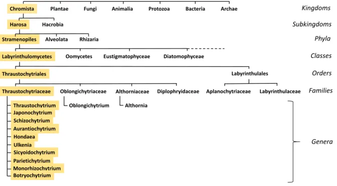
The filiation that links thraustochytrids to the tree of life is presented in Fig. 1. This classification is now generally accepted [46, 47], but the number of genera present in the Thraustochytrid family is still a matter of debate and subject to changes since recent and accurate phylogenetic analyses of various Labyrinthulomycetes species have already led to significant taxonomic rearrangements. The delineation of species boundary in protists is a difficult task [48] and the criteria chosen to discriminate species are often arbitrary. Taxonomy and systematics are usually based on a combination of the phylogenetic, morphological and ‘chemotaxonomic’ species concepts [49, 50]. Nevertheless, numerous thraustochytrid taxa show ambiguous polyphyly [6]. It is likely that a number of DNA sequences have been uploaded in databases with questionable identifications or with fragmentary taxonomic descriptions. This, together with nomenclatural errors [51], made the phylogenic reconstructions of thraustochytrids confusing. Morphological differences such as the size of the zoospores (the motile cells with a whiplash and a tinsel flagellum), the development of the ectoplasmic net, and the presence or absence of amoeboid-like cells were also used to discriminate species (reviewed in [52]). Although the abovementioned features might present discrete and measurable differences among genera or species, they can also be rather subtle between two genera and cannot always be taken as absolute discriminating criteria. For example, the genus Ulkenia is characterized by an amoeboid stage, but amoeboid cells can also be found in species belonging to the genus
within thraustochytrids are a non-cellulosic cell wall (in contrast with oomycetes [54]), the presence of an ectoplasmic net and the production of motile cells (zoospores) with a long and a short flagellum. This last point is not however a characteristic trait of thraustochytrids and some other features can be very discrete even in the Thaustochytrid family, like the ectoplasmic net which is not always observed in Aurantiochytrium [6]. Carotenoid and PUFA profiles, were also proposed to discriminate species [50, 55], but growth conditions (temperature, composition of the media) might differently affect their synthesis and accumulation [32]. For this reason, biochemical profiles alone would not be enough to identify species boundaries. Plausibly, a certain level of cryptic diversity can be present within
Thraustochytriaceae. By definition, two entities can be considered as 'cryptic' if they are
morphologically undistinguishable but genetically different. The number of cryptic species in unicellular organisms has increased exponentially in the last two decades because re-examination of taxa previously defined on morphological traits revealed a wider diversity than expected when molecular criteria were coupled with morphology [56]. From this point of view, coupling genomic sequence information with morphological and physiological observations allowed taxonomic rearrangement of the previous genus Ulkenia into now four genera, Ulkenia, Botryochytrium,
Parietichytrium and Sicyoidochytrium [50]. Likewise, it was shown that the previous genus Schizochytrium was not a natural taxon, as primarily thought, but forms three monophyletic groups,
i.e. Schizochytrium, Aurantiochytrium and Oblongichytrium [49]. Then, further phylogenetic considerations lead to the separation of the genera Oblongichytrium and Althornia from the Thraustochytrid family and create two new families [47]. Eventually, this combination of approaches also led to the emergence of the new genus Hondaea in the Thraustochytriaceae family [6, 57] (Fig. 1).
2.1. Phylogenomic analysis of thraustochytrids and positioning in the Stramenopile phylum
Phylogenomics can be considered as the next generation of single-locus or multiple concatenated loci phylogenies. A very elegant omni-comprehensive review article was recently published on this topic, hence we will refer to it for the main conceptual items [58]. As delineated above (Fig. 1), thraustochytrids belong to the Stramenopile group. Stramenopiles are now included in one supergroup in the eukaryotic tree of life, the so-called SAR (Stramenopiles Alveolates Rhizaria) supergroup. This supergroup assembles Stramenopiles (such as diatoms, thraustochytrids, oomycetes, etc), Alveolates (dinoflagellates, apicomplexans, ciliates, etc) and Rhizaria (foraminifera, radiolarian, etc). SAR gathers highly diverse organisms, both photosynthetic and heterotrophic protists with very divergent morphological, physiological and ecological features [59].
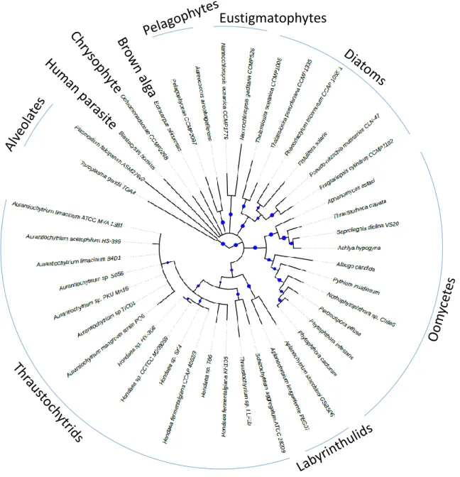
To obtain the highest representation of the Stramenopile group, we included species with no available reference genome or without publicly available genome annotations. In the former case, we downloaded RNA-seq data and performed de novo assembly and in silico translation to produce protein sequences. About 249000 protein sequences were predicted, with an average of 22655 proteins per species. For Aurantiochytrium sp. T66 and Aurantiochytrium acetophilum available genomes no official annotation nor proteome sequence were retrievable. Therefore, an ab initio annotation was performed. About 20700 and 5021 proteins were predicted for Aurantiochytrium sp. T66 and Aurantiochytrium acetophilum, respectively. A phylogenomic analysis was then performed on 184 informative orthogroups identified by the software Orthofinder [60]. In the final dataset, the apicomplexans Toxoplasma and Plasmodium (Alveolates) were chosen as outgroups to root the tree. As Stramenopile ingroups, we chose Diatoms, Pelagophytes, the uncharacterized Chrysophyte strain CCMP2298, Eustigmatophytes, the brown macroalga Ectocarpus siliculosus, Oomycetes, the human parasite Blastocystis hominis, Labyrinthulids and Thraustochytrids (see the legend of Figure 2 for the name of the different species used in this study).
The phylogenomic tree (Fig. 2) shows that diatoms, pelagophytes, eustigmatophytes and oomycetes form distinct and well identified clusters in variably supported end clades. Thraustochytrids and Labyrinthulids, instead, cluster together in relatively weakly supported clades, confirming that they are close relatives and possibly revealing a fast evolution rate of the sequences chosen for the analysis. 3. The life cycle of thraustochytrids and their lipid dynamics
3.1. The life cycle
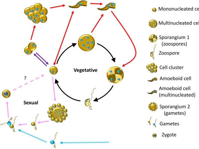
The life cycle of thraustochytrids is quite complex and may vary from one genus to another. Among the ten accepted genera described to date, the life cycle was only investigated for a few of them. Several types of cells are involved, i.e. mononucleated cells, multinucleated cells, zoosporangia (multinucleated cells with cytokinesis), amoeboid-shape cells and zoospores [17, 18, 61]. The relationships between these different cell types have been studied in more detail in Aurantiochytrium
limacinum (formerly Schizochytrium limacinum), Thraustochytrium gaertnerium and
Aurantiochytrium acetophilum [17, 18, 61]. The common feature to all the genera is a core cycle where
vegetative cells produce multinucleated cells then sporangia that contain zoospores. Upon release, zoospores colonize new areas, settle and start the cycle over again (Fig. 3). To this general scheme, a number of more complex side loops can be added, depending on the considered strain. Mononucleated cells are able to perform binary divisions in Ulkenia, as well as in Schizochytrium,
Hondaea and Aurantiochytrium (red arrows, Fig. 3) [52]. In the latter genus, binary divisions may give
rise to cell clusters (purple arrows, Fig. 3), from which amoeboid cells emerge [53]. Amoeboid cells, though rarely observed in Thraustochytrium [17, 18, 61], are also characteristic to Ulkenia (and the sister genera), in which they emerge from either mono- or multinucleated cells (red arrows, Fig. 3) [52]. In all the aforementioned genera, amoeboid cells settle and transform into a zoosporangium, but they also have been seen to divide into mononucleated cells in A. acetophilum [61]. Amoeboid cells are able to move slowly with elongating and curving movements, and it is possible that they represent a form of propagation of the colony over solid object, such as a leaf [17].
Available data concerning thraustochytrid zoospores are quite scarce. Their ploidy remains an open question, but sequence analyses of the genome and its heterozygosity suggest that vegetative cells are diploid [61]. It is likely that most of the time these zoospores constitute a form of vegetative reproduction, able to disperse and to propagate the species by settling new colonies [13]. However, a recent report presents evidence for a sexual reproduction in Aurantiochytrium acetophilum [61]. In A.
acetophilum, the life cycle involves two types of sporangia, one giving birth to zoospores and the other
one to round flagellated cells (interpreted as gametes, pink arrows, Fig. 3) [61]. Unique to date is the observation of two flagellated cells (gametes?) fusing and giving rise to a zygote. Unfortunately, the fate of the zygote was not followed (pink broken arrow, Fig. 3) and the ploidy of gametes not directly measured. The sexual reproduction in A. acetophilum does not seem to be homothallic, i.e. occurring within one monoclonal culture. Rather, only upon crossing two different strains (blue arrows, Fig. 3), was nuclear fusion observed in the pairing cells [61].
3.2. Lipids and the life cycle
The life stages of thraustochytrids are highly nutritional dependent. When cultivated in artificial media containing yeast extract or peptone (for a review see [52]), thraustochytrids can accumulate large amounts of lipids as well as produce high biomass [53], which makes them very attractive for biotechnological applications. Indeed, when grown in the appropriate conditions, thraustochytrids
from the genera Aurantiochytrium and Schizochytrium may accumulate lipids up to 30-50% of their dry weight, with DHA representing about half of the total FAs [6, 19, 35, 62, 63].
Lipid production depends on the composition of the medium [31, 64], and thus varies along the life cycle [53]. These cells display extraordinary lipid dynamics along the growth period. Such variations are correlated with an important environmental/cultivation parameter, the C:N ratio. Indeed, a carbon source is required for these heterotrophic cells to synthesize FAs and lipids. Furthermore, it is now well established that nitrogen deprived media trigger lipid accumulation in higher plant cell cultures [65], microalga species such as Chlamydomonas reinhardtii [66, 67], Phaeodactylum
tricornutum [30, 68], Nannochloropsis gaditana [69, 70], and also thraustochytrids [71]. The
physiological meanings of these metabolic changes are not yet fully understood. Likely, in the absence of cell division caused by nitrogen limitation the carbon normally used for protein and membrane syntheses is partly redirected toward TAG production [69]. The amounts of carbon and nitrogen in the culture medium are highly fluctuating during a growth period. It has been shown [53] that when
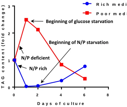
Aurantiochytrium limacinum are transferred in a new rich medium containing glucose and yeast
extract (the source of nitrogen and phosphorus), almost all the TAGs accumulated during the previous culture are consumed within the first day (Fig. 4). Then, after two days, nitrogen become limiting and cells start to accumulate lipids (TAGs) again, as long as glucose is present, to reach at the end of the culture period a lipid weight of about 30% of the dry weight. It is more the absolute nitrogen concentration rather than the C:N ratio that controls the flux of carbon towards or from TAGs. Indeed, when cells are transferred in a poor medium containing only 1/40 of the nutrients but displaying the same C:N ratio, the nitrogen level is already limiting and cells immediately accumulate lipids at the expense of glucose [53]. After one day, all the glucose present in the medium is consumed and lipids are used to provide energy to the swimming zoospores (Fig. 4), with zoospores constituting the main cell type in this situation [53]. These data indicate that lipid degradation (β-oxidation) is a main and quickly available source of energy.
Although these experiments do not reproduce the real nutritional conditions of decaying mangrove leaves, they might be suggestive of what happens in a natural environment. Indeed, in such an ecosystem, the rate of leaf decomposition is inversely correlated with the C:N ratio of the litter [72]. C:N ratio may fluctuate from about 30-70 in fresh leaves to 90-200 in senescent leaves [16], suggesting that nitrogen could indeed become a limiting factor over time for the proliferation of the cells involved in the leaf degradation. In this limiting situation, TAGs accumulate and more zoosporangia are produced. Then, zoosporangia may release zoospores, which swim in search of a new territory to colonise and consume the TAGs previously accumulated to respond to the energy demand. Thus, the zoospore lifetime will depend on the amount of TAGs stored during the biomass accumulation. In contact with a new leaf, the presence of organic carbon and adequate nutrients trigger zoospore maturation into mononucleated cells, and a new cycle starts.
4. Lipid composition and synthesis
4.1. Glycerolipid composition
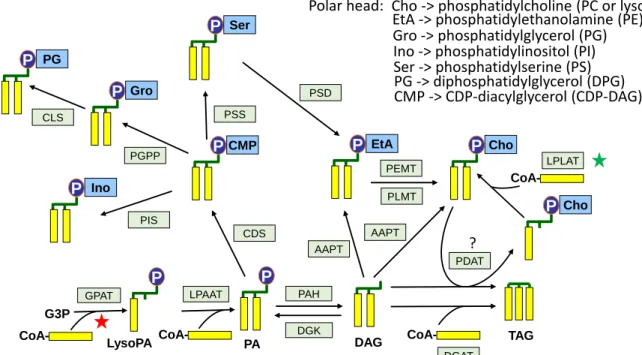
In almost all organisms glycerolipids are essential components of the membrane architecture and the main form of lipid storage. Membrane glycerolipids are diesters of FA and glycerol, the third hydroxyl function of the glycerol backbone being linked to a polar group. Storage glycerolipids are triesters of FA and glycerol, i.e TAG, and they represent a highly reduced form of carbon that serves as an energy stock for cell functions, or as a reservoir of FAs for the synthesis of new membranes. The membrane glycerolipid composition has been determined in a few thraustochytrid [6, 33, 73, 74]. In all these species, membranes are essentially constituted of phosphoglycerids (Table 1), which are dominated
by phosphatidylcholine (PC), followed by phosphatidylethanolamine (PE), then phosphatidylinositol (PI) and phosphatidylglycerol (PG). Other phosphoglycerids such as phosphatidylserine (PS), phosphatidic acid (PA) and diphosphatidylglycerol (DPG) are minor representatives of the phosphoglycerolipidome. The presence of lysoPC (a PC with only one FA) is also reported in
Aurantiochytrium and Schizochytrium [6, 33]. Low levels of glycoglycerolipids are also observed in Aurantiochytrium mangrovei [33], but the nature of the sugar constituting the polar head is not
known. Aurantiochytrium does not contain any galactolipid (plastid-specific glycerolipid), which is consistent with the absence of plastids in thraustochytrids. The presence of an unknown glycerolipid, which is not a phosphoglycerid nor a betaine lipid, is also reported in Aurantiochytrium, but the developed formula of the polar head is still uncharacterized [6]. Among non-polar lipids, diacylglycerol (DAG) represents less than 1% of total lipids whereas TAG may fluctuate widely during the growth period and can reach up to 90% of total lipids (Table 1). Mining the available sequencing data of
Aurantiochytrium limacinum genome, and using reciprocal blast approach to identify orthologous
genes in H. fermentalgiana, all the genes involved in the synthesis of glycerolipids have now been identified [6, 75, 76], enabling the reconstitution of the pathway (Fig. 5).
4.2. Fatty acid composition
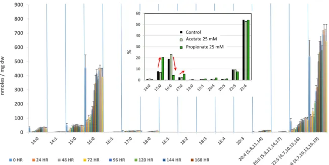
FAs are mostly present in glycerolipids. The FA compositions of numerous thraustochytrid species have been determined [6, 29, 55, 77, 78] to assess their DHA content and biotechnological relevance (Table 2). Analyses of thraustochytrid FAs were often performed using different culture conditions (varying the source of carbon, oxygen concentration, salinity, temperature, nitrogen availability etc..) with the aim to improve either the yield of FA production or to modify the nature of FAs (length of the carbon chain and level of unsaturation) [62-64, 71, 79, 80], reviewed in [52]. To summarize, most thraustochytrid species have a FA composition dominated by palmitic acid (16:0) and DHA (22:6), these two FAs representing often more than 65% of total FAs. When compared with other important PUFA producers (Table 2), thraustochytrids display the highest relative DHA content. The total amount of FAs [6, 77] and the proportion of DHA [55, 78] in thraustochytrids may vary from one species to another, and species from the genera Aurantiochytrium, Thraustochytrium and Schizochytrium appear to be among the most productive [52]. The medium composition is an important parameter, the presence of glucose or glycerol as a carbon source and yeast extract or peptone as a nitrogen source allowing the highest yield [31]. The composition of the medium varies during growth, and thus the synthesis and accumulation of FAs can be affected, mirroring TAG fluctuations as shown for
Aurantiochytrium limacinum (Fig. 6 and [53]). Clearly, when nitrogen is not limiting TAGs are
consumed first and their FAs are degraded through β-oxidation to provide energy for growth. In nitrogen-limiting conditions, FAs and TAGs are synthesized from the organic carbon present in the external medium and accumulate in lipid bodies. This behavior led to recommend a two stage growth method to increase the yield of DHA production [81]: the first stage with a low C:N ratio (high nitrogen) allows a rapid growth and a high biomass, and the second stage with a high C:N ratio (low nitrogen) triggers the production of FAs and DHA. Interestingly, odd-numbered FAs (mainly 15:0) can be observed in specific growth conditions. They are produced when the growth medium contains high levels of branched-chain amino acids as a source of nitrogen (those amino acids are present in the yeast extract) [82] because the degradation of these amino acids produces propionyl-CoA. Propionyl-CoA, a 3C unit, can be used as a starting unit for FA synthesis instead of the classical substrate acetyl-CoA (Fig. 6, insert).
Thus, thraustochytrids appear as major and relatively specific DHA producers. Why such an amount of this particular FA? Reasons could be linked to their physiology and their environment or to specific biochemical features linked to the presence of a PUFA synthase (see Section 4.3). There is no clear
answer to this question, but it is interesting to note that not all the organisms displaying a PUFA synthase accumulate DHA (Table 2). The presence of DHA in phospholipids can possibly affect the physical properties of membranes (for a review, see [83]), by affecting the structure and permeability, and by enhancing flip-flop activities, which might be an advantage in particular environments. Also, DHA might play an antioxidant role in membranes, and perhaps might also affect the buoyancy [84].
4.3 Fatty acid synthesis
FA synthesis in thraustochytrids is more complex than in many other marine protists because it involves two different pathways that operate independently. The first pathway requires a type I Fatty Acid Synthase (FAS) system similar to the one found in mammals, and produces FA chain length of 16C. The second pathway involves a Polyketide Synthase-like (PKS-like) machinery, or PUFA synthase, to synthesize VLCPUFAs of 20C and 22C [85-88]. PKS are sophisticated molecular machines responsible for the synthesis of polyketides, natural products of the secondary metabolism that share similarities with FAs (similar chemistry, similar structure, common precursors) [89, 90]. There are two types of PKS classified in a way resembling the classification of FAS. Like type I FAS, type I PKS displays a multidomain architecture with active sites arranged on large modules, whereas type II PKS consists of monofunctional enzymes with each catalytic site on a separate protein. PUFA synthases are often referred to as PKS, but PKS-like is a more appropriate term since they are not synthesizing polyketides and show features that are found in PKS I, PKS II and FAS [91, 92]. So far, PUFA synthases were found in some bacteria (γ-proteobacteria, myxobacteria, sphingobacteria, flavobacteria), mostly (but not only) marine [93-96], and in a few eukaryotes (thraustochytrids and another type of microalga,
Emiliana huxleyi, [97]). The origin of the PUFA synthase in thraustochytrids is unclear, but a lateral
transfer from marine bacteria was hypothesized [88].
The FAS system is sometimes called ‘aerobic’ pathway and the PUFA synthase ‘anaerobic’ because the former unlike the latter requires oxygen-dependent desaturations to introduce double bonds. As illustrated in Figure 7, the domains of the FAS complex catalyze a set of strictly ordered and iterative reactions where two carbons are added at each cycle, eventually leading to the formation of a saturated 16C FA (16:0). From 16:0, various elongases and desaturases are then required to produce VLCPUFA such as EPA (20:5), docosapentaenoic acid (DPA, 22:5) or DHA (22:6) [98-100]. A distinct organization is described for PUFA synthase. The multifunctional complex PUFA synthase is made of different subunits, the number varying from one (E. huxleyi) to four (γ-proteobacteria). There are three subunits in thraustochytrids, each harbouring different domains (Fig. 7). These domains have been determined for Schizochytrium sp. [91, 92] and Thraustochytrium sp. 26185 [87]. Subunit A contains one ketoacyl synthase (KS), one malonyl-CoA:ACP acyl transferase(MAT), a variable number of acyl-carrier proteins (ACP), one ketoreductase (KR) and one dehydratase (DH) domains. Subunit B displays one KS, one chain length factor (CLF), one acyl transferase (AT) and one enoyl reductase (ER) domains. The domains in subunit C are two DHs and one ER. In the subunit A of Schizochytrium and
Thraustochytrium, the ACP domains are repeated respectively 9 and 8 times and it was shown that
the number of these tandemly repeated domains is important for PUFA productivity [101]. To be fully functional, PUFA synthase complex requires a phosphopantetheine transferase activity that activates the ACPs. The linear organization of PUFA synthase subunits are reminiscent of that in type I FAS, but KS domains show higher homology to bacterial type I PKS than to those of FAS [88, 92]. The CLF domain shows homology with KS but lacks active site residues and is thus assumed to be inactive, a situation similar to the one found in type II PKS [102]. This domain has been functionally characterized in association with its KS neighbour domain [103] and plays an important role in the carbon chain elongation, especially for the elongation from C20 to C22 (EPA to DHA). In thraustochytrids it also controls the final yield of PUFA production [104]. Two types of DH domains are likely required in PUFA
synthase to introduce either saturation or cis double bonds on growing acyl chains [105]. The first DH domain in subunit A is similar to the one found in PKS and only catalyses the dehydration reaction. The DH domains associated with subunit C present high homology with FabA [103], one of the two acyl-ACP dehydratases involved in the biosynthesis of FAs in E. coli. Besides catalysing the dehydration reaction, FabA also isomerizes trans-2-decenoyl-ACP into cis-3-decenoyl-ACP, an essential step to produce PUFAs in these bacteria. Likely, the homologous DH domains of thraustochytrids share this property (DH/I) [92, 106]. To achieve the final pattern of double bonds observed in DHA (or EPA), not only trans-2,3-cis but also trans-2,2-cis isomerizations are likely to occur at several rounds of elongation during the FA construction process [92]. When such a DH/I domain is disrupted in
Schizochytrium sp., the total PUFA yield decreases by more than half, highlighting its key role [104]. A
putative scheme showing the sequence of reactions catalysed by the PUFA synthase is shown in Figure 8. In a first step acetyl-CoA and malonyl-CoA are condensed to provide a starter acyl-ACP unit. Then, two carbons from another malonyl-CoA are added to this starter unit to form an acetoacyl-ACP. This is followed by reduction and dehydration steps to give an acyl chain with a 2-trans double bond. Depending on the type of DH (DH or DH/I) that was involved, the next reaction may differ from the one catalysed by FAS. Indeed, either the acyl chain is reduced (as it is the case with FAS) before entering a second round of elongation or an isomerization reaction switch the double bond to a 3-cis or a 2-cis position. How these isomerization reactions are controlled? By which mechanism the trans double bond formed by the DH activity is selected for either reduction or cis-isomerization? These remain important and intriguing questions since the final design of the PUFA will depend on how these reactions are ordered. As recently shown [105], the length of the growing acyl chain is possibly a key factor to determine the type of DH that needs to be involved. To summarize, PUFA synthase, like FAS, catalyzes a series of iterative and sequential reactions that add two carbons per cycle, but the way these two carbons are processed is not yet fully understood. The ability of PUFA synthase to directly release VLCPUFAs suggests a more modular system than the FAS system, where the ER step is not systematically involved.
The need of two independent pathways for the synthesis of FAs in thraustochytrids is unclear, but mutants devoid of PUFA synthase activity cannot grow and are auxotroph for PUFAs [85], thus indicating that these two systems are not redundant. Actually, the FAS route seems to be incomplete, genomic analyses suggesting that Δ5 desaturase (involved in the synthesis of DHA from 20:4∆8,11,14,17)
is missing in A. limacinum [6, 75] and H. fermentalgiana [6]. However, this might not be a general case in thraustochytrids since Δ5 desaturases from several Thraustochytrium and Schizochytrium species have been identified [107] and functionally characterized [108]. Furthermore, the addition of free 20:4∆8,11,14,17 and 20:3∆8,11,14 in the culture medium of A. limacinum does not increase the DHA
production, but DHA is produced when these cells overexpressed the Δ5 desaturase from
Thraustochytrium aureum. This clearly indicates that a Δ5 desaturase is indeed absent in the A. limacinum strain [108]. Likewise, a Schizochytrium mutant devoid of the PUFA synthase subunit A
displays a lethal phenotype which is partially-to-fully recovered by addition of free PUFAs in the culture medium [85]. However, the added labelled free PUFAs (20:4∆5,8,11,14, 20:5∆5,8,11,14,17,
22:5∆4,7,10,13,16) are not converted into DHA (22:6∆4,7,10,13,16,19) in the mutant, indicating that some
elongase and desaturase activities independent on the PUFA synthase system are either absent in this strain or not able to drive DHA biosynthesis from the adequate precursors. Altogether, these results show that the PUFA synthase activity is essential for thraustochytrids and cannot be compensated by the FAS pathway. Since the FA profile displays two major FAs, 16:0 and 22:6, it is tempting to say that the FAS and the PUFA synthase pathways are respectively involved in 16:0 and 22:6 syntheses. The situation might be however more complex since it was shown that an elongase presumably associated with the FAS pathway is also essential for the growth of Aurantiochytrium sp. [109].
4.4 Phylogeny of PUFA synthases
The PUFA synthase organization may vary from one organism to another, as illustrated in Figure 9A, but the domains in the N-terminal part of subunits A are however similarly arranged in most species. A Bayesian phylogenetic analysis was performed with the KS domain (Fig. 9B) amino acid sequences from thraustochytrids, a Haptophyte (Emiliania huxleyi) and several bacteria. The unrooted tree clearly shows two main well-supported clades, one containing eukaryotic organisms (thraustochytrids) and the other bacteria. Emiliana huxleyi, which displays a unique organization in PUFA synthase, appears closer to thraustochytrids than to bacteria but possibly represents another distantly related group. Among bacteria, γ-proteobacteria in one hand and mixobacteria and Bacteroidetes in the other hand are segregated into two well-supported clades. The types of PUFA synthase present in the eukaryote clades are DHA (E. huxleyi) and DHA/DPA (thraustochytrids) synthases. They are more diverse in the bacterial clades where DHA, EPA, ARA (arachidonic acid, 20:4), EPA/DHA, EPA/ARA synthases can be found. It is difficult from this tree to correlate a specific clade with a particular type of PUFA synthase. For example, in the γ-proteobacteria clade there are genera that are DHA producers (such as Colwellia or Moritella) whereas others are EPA producers (Shewanella) (see also Table 2). Thus, the organization alone of the PUFA synthase does not appear as a sufficient criterion to predict the type of FA that is synthesized. The specificity of the various PUFA synthases relies probably more on specific or structural details than just its global organization. From this point of view, it was recently shown that the type of PUFA produced in bacteria was determined by the subunit C of these organisms [103]. Indeed, when the KS-CLF domains of an EPA synthase is replaced by the KS-CLF domains of a DHA synthase, the resulting chimeric EPA synthase is able to produce DHA. The opposite situation is true as well. Thus, the KS-CLF domains appear as essential to determine the final products of PUFA synthases. It was postulated that a lateral transfer from marine bacteria is at the origin of the PUFA synthase in thraustochytrids [88]. If this holds true, it is tempting to think that the bacteria at the origin of this transfer was also a DHA producer, such as a γ-proteobacterium or a mixobacterium.
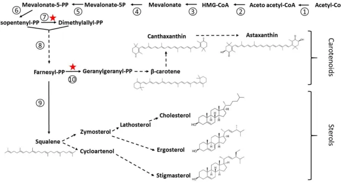
4.5 Carotenoids, sterols and squalene
Carotenoids and sterols are synthesized from the mevalonate pathway, and the branch point for these two lipid classes is at the level of farnesyl pyrophosphate (Fig. 10). All the genes involved in the mevalonate pathway from acetyl-CoA downstream to farnesyl pyrophosphate have been mapped in
Aurantiochytrium [6].
Thraustochytrids are often pigmented cells due to their ability to synthesize and accumulate carotenoids [78], and are generally considered as a valuable source of carotenoids and xanthophylls (oxygenated carotenoids) [31]. Carotenoids are important natural antioxidants that have many human health benefits, reacting with and scavenging reactive oxygen species [110]. Astaxanthin is also used as a natural food colorant and feed supplement in fish and poultry industries. The main carotenoids found in thraustochytrids are β-carotene and the two xanthophylls astaxanthin and canthaxanthin (Table 3), although the amount of each of these compounds may vary with the period of the culture and depends on the considered strain [111]. In general, carotenoids accumulate during growth and there are more carotenoids at the late than at the early exponential phase of growth. In addition, β-carotene is not always observed or is present in variable amounts reflecting the efficiency of its conversion into xantophylls, which efficiency may vary from one strain to another (Table 3). For example, there is four times more carotenoids in A. limacinum than in H. fermentalgiana, canthaxanthin being the dominant species in the former, whereas astaxanthin, canthaxanthin and β-carotene showed an equal distribution in the latter [6]. All the genes coding for the synthesis of
astaxanthin from farnesyl pyrophosphate are identified in Aurantiochytrium, opening the route for metabolic engineering [112].
The synthesis of sterols from farnesyl pyrophosphate results in a very complex pathway because the various paths leading to the different classes of sterols often share common enzymatic steps and are therefore tightly interconnected. The complete pathway has been tentatively reconstituted from the genome of A. limacinum and H. fermentalgiana [6], and suggests that these strains are theoretically apt to produce stigmasterol, cholesterol and ergosterol, i.e. the three main classes of sterols often classified as plant, animal and fungi sterols, respectively (Fig. 10). The free sterol composition has been determined in only a few thraustochytrid species, the strain ACEM 6063 [113], A. limacinum CCAP 4062/1 and H. fermentalgiana CCAP 4062/3 [6]. In all cases, the dominant sterols are cholesterol, followed by stigmasterol or Δ7-stigmasterol (Table 3). Lathosterol, a precursor of cholesterol, is also found in the three strains, but ergosterol is either absent or only present in trace amounts, as seen in
A. limacinum [6]. The amount of sterols fluctuates within the growth period, often being the lowest
at the end of the exponential phase of growth.
Squalene is the precursor of all sterols and significant amounts are observed in the aforementioned strains, especially in the exponential phase of growth (Table 3). It is synthesized from farnesyl pyrophosphate in one step catalyzed by squalene synthase [43]. Squalene is of great commercial interest. It is extensively used as an excipient for the delivery of drugs and in the cosmetic industry as an emollient and moisturizing agent. It also displays interesting human health properties, as an antioxidant protecting cells from oxidative stresses, and as an agent stimulating non-specific immune functions and having protective actions in cancer treatments [114, 115]. The major commercial source is the shark liver oil, but its availability in the future is questionable due to ecological concerns, the decrease of the shark population and the increase of the societal demand. Today, thraustochytrids appear as sustainable and promising sources of squalene, more sustainable than shark livers and easier to process than plants or bacteria that have generally lower amount of squalene. Indeed, and depending on the growth medium, the squalene content of thraustochytrids can be higher than 10 mg/g dry weight [116], which may represent about 30-50 mg/g of oil, assuming that oil can represent up to 30-50% of the dry weight (see above section 3.2). By comparison, olive oil, one of the most squalene-rich vegetable oil, contains about 1.5-1.7 mg/g [117]. Therefore, many thraustochytrids have been analyzed for their squalene content, which may strongly vary from one strain to another. In many studies, the genus Aurantiochytrium appears as the most promising [6, 31, 44, 116, 118, 119]. For example, A. limacinum displays five times more squalene than its close relative H. fermentalgiana [6]. However, a recent study identified new highly producing strains, forming a distinct clade on a phylogenetic tree and branching out of the Aurantiochytrium cluster [120]. These new thraustochytrid members are potential novel candidates for the commercial production of squalene.
5. Engineering the metabolism of lipids
The potential of thraustochytrids in the market prospects for DHA, carotenoids and squalene is reviewed in [31]. Although thraustochytrids are clearly a very attractive and promising material for a sustainable production of such compounds, improvements are required to meet the demand. Thus, many efforts are currently undertaken to improve and engineer the lipid production of thraustochytrids, using different strategies. Indeed, the literature is profuse about the impact of culture conditions, including the composition of growth media. Mutagenesis approaches such as gamma-irradiation were also used and mutants with five times more carotenoids were successfully obtained [121]. Other methods such as metabolic engineering by genetic modifications appear very promising to improve the lipid production, and we will focus on them below.
Molecular approaches were limited until recently because genomic data were scarce and methods for genetic transformation in thraustochytrids poorly efficient. Although never mentioned, potential difficulties may also arise from the multinuclear stage of the cells, leading to poor transformation efficiency or rapid loss of the transgene if the nuclei are not all transformed. Nevertheless, these approaches are now more widely used, and with obvious successes, due to the improvement of knowledge and techniques. Today, seven genomes are available (Hondaea fermentalgiana strain CCAP 4062/3, Schizochytrium sp. CCTCC M209059, Aurantiochytrium sp. T66, Thraustochytrium sp. ATCC 26185, Aurantiochytrium limacinum ATCC MYA-1381, Aurantiochytrium acetophilum strain HS-399) and these data, together with the design of molecular tools adapted to the transformation of Thraustochytrids [122, 123], lead now to an exponential increase of the number of publications in the field. The main techniques used for transformations are biolistic and electroporation. Both methods appear valuable in the case of thraustochytrids, although electroporation is generally preferred. Most of the time, homologous recombination approaches are involved, using disruption vectors containing a cassette associated with the Sh Ble resistance gene and surrounded by homologous regions of the target gene [85, 124]. The Sh Ble gene confers resistance to glycopeptides such as the traditionally used zeocin, and allows selection of the transformed cells in the appropriate medium. The promoter sequences in the cassette are often those of ubiquitin, α-tubulin or the translation elongation factor EF1α genes. The transferred genetic material can be either a circular [125] or a linearized plasmid [126], or just the amplified cassette with the flanking regions for the homologous recombination [127]. With these methods, overexpressing [128], knocked-in [129], and disrupted [104] mutants have been obtained. To improve the efficiency of transformation, the cell wall can be weakened by DTT treatments [130], by vortexing cells with zircon beads before resuspension with the amplified DNA construct [127], or by incubation for few hours with a cocktail of various cell-wall degrading enzymes [126]. Some authors also obtained high yield of transformation using commercial electroporation buffers (either Lonza’s Nucleofector Solution or Bio-rad’s Electroporation Buffer) rather than the ‘classical’ sorbitol medium [123].
In the following paragraphs, we report on the main attempts to modify genetically thraustochytrids in order to improve their lipid contents. Stars in Figures 5, 7 and 10, indicate the various metabolic steps that were targeted in these experiments.
5.1. Squalene sterols and carotenoids
To our knowledge, there is no publication yet aiming to improve the squalene and sterol contents of thraustochytrids by genetic modifications, and there are only a few for carotenoids. In order to help cells to adapt to hypoxic environments resulting from a high cell density, a hemoglobin gene from
Vitreoscilla was expressed in Aurantiochytrium sp. The transformed strain produces nine times more
astaxanthin and 44% more FAs, suggesting that the biosynthesis of carotenoids and FAs are coordinated [125]. Likewise, the combined heterologous expression of a geranylgeranyl diphosphate synthase from Archaeoglobus, together with an isopentenyl pyrophosphate isomerase from E. coli and a hemoglobin from Vitreoscilla results in a two-time increase of total carotenoids, with a 5-fold increase of astaxanthin, and a 2.5-3 fold increase of total FAs and DHA [131]. Interestingly, the amount of squalene is strongly reduced in this mutant, indicating that sterol synthesis in one hand and carotenoid and FA syntheses in the other hand are in competition, as previously observed in other microalgae [132].
More data are available about genetic modifications aimed to improve the VLCPUFA production. Most of these attempts target genes directly connected with lipid and FA metabolisms, and a few are more indirect.
Several of these engineering experiments focused on the FAS pathway. A four times increase of EPA (20:5) is observed in a mutant of A. limacinum expressing a Δ5 desaturase from Thraustochytrium
aureum, but only when the transformed cells are grown in the presence of 20:4, the substrate of the
desaturase [108]. In the same species, disrupting a Δ12 desaturase converting 18:1 into 18:2 and presumably associated with the FAS/elongase/desaturase system results in a small but significant (35%) increase of DHA [133]. These results suggest that part of the carbon flux could be diverted from the FAS to the PUFA synthase, and that the two pathways could work independently. Also, overexpression in Schizochytrium sp of a malonyl-CoA:ACP transacylase, an essential activity to elongate FAs (Fig. 7), resulted in a 1.5-fold increase of total lipids with two times more DHA and DPA [134]. To increase the rate of acetyl-CoA production, an acetyl-CoA synthetase from E. coli was overexpressed in Schizochytrium, resulting in a modest (11%) increase of FA [135].
In an attempt to modify the PUFA synthase activity in Aurantiochytrium limacinum, the KR and DH domains of the subunit A (Fig. 7) were separately overexpressed by knock-in strategies [129]. In these mutants, the total content of PUFAs is only marginally increased, but the amount of 20:4 (ARA), a minor FA in A. limacinum, is nearly doubled. Other interesting and promising works indicate that disruption of the CLF domain of the subunit B of the PUFA synthase and disruption of the second DH/I domain of the subunit C (Fig. 7) strongly affect the FA profile and DHA content [104] in Schizochytrium sp., decreasing both the total FA content and the unsaturated/saturated FA ratio. In the same species, disruption of the AT domain of the PUFA synthase subunit B [136] affects the growth rate and the size of the cells. The FA profile is also strongly modified, with 1.5 times less DHA and 2 times more 16:0 than in the wild type, illustrating again the competition between the two systems involved in FA synthesis.
Works aimed to engineer the pathway involved in glycerolipid synthesis were also reported. For example, overexpression of a glycerol-3-phosphate acyltransferase, the first enzyme in this pathway, responsible for the attachment of the first FA on the glycerol backbone (Fig. 5) results in a modest increase of the TAG content [76]. Also, a lysophosphatidyl acyltransferase involved in the transfer of 16:0 to lysoPC and lysoPE was identified [73]. A knock-out mutant displays much lower amounts of lyso-PC and lyso-PE and less PC and PE, suggesting that the turnover of FAs on the glycerol backbone might play an important role in the overall glycerolipid dynamics, especially for these two major phospholipids often considered as metabolic hubs [65, 137]. However, the impact of this mutation on the level of TAGs was not investigated.
Less direct but promising approaches were also undertaken. For example, it was shown that disrupting a thraustochytrid-specific lipid droplet protein impacts TAG accumulation in A. limacinum [138]. Because of their high number of double bonds, PUFAs are highly sensitive to reactive oxygen species, which presumably increase their rate of degradation. Alleviation of reactive oxygen species by overexpression of a superoxide dismutase in Schizochytrium sp. results in a down regulation of genes potentially involved in β-oxidation and in a modest but significant (30%) increase of total FAs, including DHA [126]. Another indirect approach attempted to increase the rate of NADPH turnover. Indeed, the synthesis of FAs requires the contribution of NADPH, as many other anabolic pathways, and the availability of NADPH could be a limiting factor. In non-photosynthetic cells, the main source of NADPH is linked to the pentose phosphate cycle in the cytosol, a pathway initiated by the glucose-6-phosphate dehydrogenase. The glucose-6-phosphate dehydrogenase was overexpressed in Aurantiochytrium sp.SD116 [128], resulting in a slight increase of the proportion of DHA versus 16:0, but also and surprisingly in a slight decrease of total FAs.
The interesting feature characterizing thraustochytrids, i.e. the high productivity of VLCPUFAs, was tentatively transferred to higher plants. The three subunits of the PUFA synthase from various strains of thraustochytrids were overexpressed in canola [139]. The goal of this work was to produce a vegetable oil with commercially relevant amounts of DHA. As a matter of fact, the engineered canola oil obtained from field-grown grains can provide more than 600 mg of combined DHA and EPA in one 14 g serving, which fully meet the dietary recommendations of 250-500 mg of ω-3 VLCPUFAs per day [139]. Using a similar approach, a diacylglycerol acyltransferase (DGAT, the last step in TAG synthesis, see Fig. 5) from Thraustochytrium aureum was overexpressed in Arabidopsis with the purpose to increase the lipid production. However, the seed-specific expression of TaDGAT2 resulted in no detectable increase in the oil content of the transformed Arabidopsis seeds [140].
All these recent experiments indicate that functional genomics in thraustochytrids is an emerging and promising area. Our knowledge of their lipid metabolism is still fragmentary but will certainly progress rapidly in the coming years, allowing a better understanding of how this metabolism is adjusted as a function of the life cycle and environmental conditions. These results also show that, despite the already high level of lipids and FAs found in natural strains, it is possible to increase further their oil content by genetic engineering, thus opening the way for industrial applications and the sustainable production of interesting compounds.
6. Conclusion
Marine protists are able to synthesize relatively large amounts of ω-3 VLCPUFA. These FAs are important from a nutritional point of view and required for optimal human health. These ω-3 VLCPUFA are however not fully equivalent when health benefits have to be considered. For example, EPA and DHA have both anti-inflammatory properties, but DHA is also specifically required in the development of retina and brain in infants [38, 40]. Because most photosynthetic microalgae do not accumulate large amounts of DHA, the specific features displayed by thraustochytrids are interesting and not redundant compared to other microalgae. Indeed, they not only accumulate large amounts of lipids but also high quantities of DHA which may represent up to 20-25% of the dry weight, making these cells one of the highest DHA producers. The FA composition in thraustochytrids is relatively simple, dominated by two main species: a saturated and relatively short chain FA, 16:0, and DHA. Whereas 16:0 is an interesting product for biofuel production, DHA has applications in the field of human health. Since these two FAs are likely produced by two different enzymatic systems, it might be possible through genetic engineering to favour one pathway over another. Other compounds produced by several strains of thraustochytrids are also of valuable interest from a societal point of view. Carotenoids and squalene are surely good examples, but they could be only the tip of the iceberg since the metabolism of these protists is largely unknown, especially when considering the secondary metabolism.
A second important reason for considering thraustochytrids as an economically interesting material to produce lipids is linked to their very rapid growth rate and to the resulting high biomass that can be collected in only a few days. Thraustochytrids are obligatory heterotrophic organisms, a life status that could be analyzed as a disadvantage in terms of energy invested value compared to photosynthetic microalgae. However, thraustochytrids are able to grow on various sources of carbon such as molasses with an energy return that is higher than the exploitation of fossil diesel [141]. Thus, the use of low-cost carbon sources derived from agro-industrial wastes could be a promising solution to reduce the effective cost of lipid production from thraustochytrids.
Thraustochytrids are emerging models not only to produce compounds that would benefit our societies, but also to improve our knowledge on marine life biology and diversity. For example, little is known about the life cycle of thraustochytrids. What are the role, physiology and metabolism of
zoospores or amoeboid-type cells? What triggers sexual reproduction and the formation of gametes? What elicits the formation of zoospores and what induces their maturation, or how the population size of a colony is controlled [53] are also unanswered questions. Another open and intriguing question is why marine microalgae and marine protists contain such high quantities of VLCPUFAs, a feature not or rarely found in photosynthetic eukaryotes of the green lineage (Viridiplantae, first endosymbiosis) nor in cyanobacteria which are supposed to be at the origin of the first endosymbiosis, as shown in marine Synechococcus [142]. This characteristic may result from a specific adaption to the marine environment. In addition, VLCPUFAs are extremely sensitive to oxidative stress, and variations in salt level in the environment are known to trigger such a cellular response. Consistently, an important feature reported here in thaustochytrids living in mangrove forests, coping with highly variable levels in salt throughout the day, is their high level in antioxidant carotenoids. More studies are needed to address the interplay between oxidative stress responses and the production of VLCPUFAs in these organisms. Clearly, the structural, metabolic and physiological role of these VLCPUFA need to be assessed in the particular environmental context in which these organisms evolve. The recent genetic and genomic progresses made to transform thraustochytrids should allow addressing these fundamental questions through functional genomic approaches. A better knowledge of the metabolism and biological functions of thraustochytrids will, in turn, allow developing targeted strategies to modify and adapt these cells for commercial and societal purposes.
Acknowledgments: Authors were supported by the French National Research Agency (ANR-10-LABEX-04, GRAL Labex; ANR-11-BTBR-0008, Océanomics; ANR-17-EURE-0003, EUR CBS) and by the Trans’Alg Bpifrance PSPC partnership.
Conflicts of Interest: The authors declare no conflict of interest. References
[1] Sparrow FK, JR. Biological observations on the marine fungi of woods hole waters. The Biological Bulletin. 1936;70:236-63.
[2] Ellenbogen BB, Aaronson S, Goldstein S, Belsky M. Polyunsaturated fatty acids of aquatic fungi: Possible phylogenetic significance. Comparative Biochemistry and Physiology. 1969;29:805-11. [3] Cavalier-Smith T, Allsopp Mtep, Chao Ee. Thraustochytrids are chromists, not fungi: 18s rrna signatures of heterokonta. Phil Trans R Soc Lond B. 1994;346:387-97.
[4] Honda D, Yokochi T, Nakahara T, Raghukumar S, Nakagiri A, Schaumann K, et al. Molecular phylogeny of labyrinthulids and thraustochytrids based on the sequencing of 18s ribosomal rna gene. Journal of Eukaryotic Microbiology. 1999;46:637-47.
[5] Tsui CKM, Marshall W, Yokoyama R, Honda D, Lippmeier JC, Craven KD, et al. Labyrinthulomycetes phylogeny and its implications for the evolutionary loss of chloroplasts and gain of ectoplasmic gliding. Molecular Phylogenetics and Evolution. 2009;50:129-40.
[6] Dellero Y, Cagnac O, Rose S, Seddiki K, Cussac M, Morabito C, et al. Proposal of a new thraustochytrid genus hondaea gen. Nov and comparison of its lipid dynamics with the closely related pseudo-cryptic genus aurantiochytrium. Algal Research-Biomass Biofuels and Bioproducts. 2018;35:125-41.
[7] Strassert JFH, Jamy M, Burki F, Mylnikov AP, Tikhonenkov DV. New phylogenomic analysis of the enigmatic phylum telonemia further resolves the eukaryote tree of life. 2019.
[8] Leyland B, Leu S, Boussiba S. Are thraustochytrids algae? Fungal Biology. 2017;121:835-40. [9] Petroutsos D, Amiar S, Abida H, Dolch LJ, Bastien O, Rebeille F, et al. Evolution of galactoglycerolipid biosynthetic pathways--from cyanobacteria to primary plastids and from primary to secondary plastids. Prog Lipid Res. 2014;54:68-85.
[10] Massana R, Pernice M, Bunge JA, del Campo J. Sequence diversity and novelty of natural assemblages of picoeukaryotes from the indian ocean. Isme Journal. 2011;5:184-95.
[11] Li Q, Wang X, Liu XH, Jiao NZ, Wang GY. Abundance and novel lineages of thraustochytrids in hawaiian waters. Microbial Ecology. 2013;66:823-30.
[12] Raghukumar S. Ecology of the marine protists, the labyrinthulomycetes (thraustochytrids and labyrinthulids). European Journal of Protistology. 2002;38:127-45.
[13] Raghukumar S, Damare VS. Increasing evidence for the important role of labyrinthulomycetes in marine ecosystems. Botanica Marina. 2011;54:3-11.
[14] Bochdansky AB, Clouse MA, Herndl GJ. Eukaryotic microbes, principally fungi and labyrinthulomycetes, dominate biomass on bathypelagic marine snow. ISME J. 2017;11:362-73. [15] Raghukumar S, Sharma S, Raghukumar C, Sathepathak V, Chandramohan D. Thraustochytrid and fungal component of marine detritus .4. Laboratory studies on decomposition of leaves of the mangrove rhizophora-apiculata blume. Journal of Experimental Marine Biology and Ecology. 1994;183:113-31.
[16] Rao RG, Woitchik AF, Goeyens L, van Riet A, Kazungu J, Dehairs F. Carbon, nitrogen contents and stable carbon isotope abundance in mangrove leaves from an east african coastal lagoon (kenya). Aquatic Botany. 1994;47:175-83.
[17] Honda D, Yokochi T, Nakahara T, Erata M, Higashihara T. Schizochytrium limacinum sp. Nov., a new thraustochytrid from a mangrove area in the west pacific ocean. Mycological Research. 1998;102:439-48.
[18] Bongiorni L, Jain R, Raghukumar S, Aggarwal RK. Thraustochytrium gaertnerium sp nov.: A new thraustochytrid stramenopilan protist from mangroves of goa, india. Protist. 2005;156:303-15. [19] Gao M, Song XJ, Feng YG, Li WL, Cui Q. Isolation and characterization of aurantiochytrium species: High docosahexaenoic acid (dha) production by the newly isolated microalga, aurantiochytrium sp sd116. Journal of Oleo Science. 2013;62:143-51.
[20] Hardham AR. Cell biology of plant–oomycete interactions. Cellular Microbiology. 2007;9:31-9. [21] Harel M, Ben-Dov E, Rasoulouniriana D, Siboni N, Kramarsky-Winter E, Loya Y, et al. A new thraustochytrid, strain fng1, isolated from the surface mucus of the hermatypic coral fungia granulosa. FEMS Microbiology Ecology. 2008;64:378-87.
[22] Polglase Jane L. A preliminary report on the thraustochytrid(s) and labyrinthulid(s) associated with a pathological condition in the lesser octopus eledone cirrhosa. Botanica Marina1980. p. 699. [23] Garcia-Vedrenne AE, Groner M, Page-Karjian A, Siegmund G-F, Singhal S, Sziklay J, et al. Development of genomic resources for a thraustochytrid pathogen and investigation of temperature influences on gene expression. PLoS One. 2013;8:e74196.
[24] Clough B. Mangrove forest productivity and biomass accumulation in hinchinbrook channel, australia. Mangroves and Salt Marshes. 1998;2:191-8.
[25] Hutchison J, Manica A, Swetnam R, Balmford A, Spalding M. Predicting global patterns in mangrove forest biomass. Conservation Letters. 2014;7:233-40.
[26] Hoque MM, Mustafa Kamal AH, Idris MH, Haruna Ahmed O, Rafiqul Hoque ATM, Masum Billah M. Litterfall production in a tropical mangrove of sarawak, malaysia. Zoology and Ecology. 2015;25:157-65.
[27] Iwata I, Kimura K, Tomaru Y, Motomura T, Koike K, Honda D. Bothrosome formation in schizochytrium aggregatum (labyrinthulomycetes, stramenopiles) during zoospore settlement. Protist. 2017;168:206-19.
[28] Nagano N, Matsui S, Kuramura T, Taoka Y, Honda D, Hayashi M. The distribution of extracellular cellulase activity in marine eukaryotes, thraustochytrids. Marine Biotechnology. 2011;13:133-6. [29] Liu Y, Singh P, Sun Y, Luan S, Wang G. Culturable diversity and biochemical features of thraustochytrids from coastal waters of southern china. Applied Microbiology and Biotechnology. 2014;98:3241-55.
[30] Abida H, Dolch L-J, Mei C, Villanova V, Conte M, Block MA, et al. Membrane glycerolipid remodeling triggered by nitrogen and phosphorus starvation in phaeodactylum tricornutum. Plant Physiology. 2015;167:118-36.
[31] Aasen IM, Ertesvåg H, Heggeset TMB, Liu B, Brautaset T, Vadstein O, et al. Thraustochytrids as production organisms for docosahexaenoic acid (dha), squalene, and carotenoids. Applied Microbiology and Biotechnology. 2016;100:4309-21.
[32] Bowles RD, Hunt AE, Bremer GB, Duchars MG, Eaton RA. Long-chain n-3 polyunsaturated fatty acid production by members of the marine protistan group the thraustochytrids: Screening of isolates and optimisation of docosahexaenoic acid production. Journal of Biotechnology. 1999;70:193-202. [33] Fan KW, Jiang Y, Faan YW, Chen F. Lipid characterization of mangrove thraustochytrid - schizochytrium mangrovei. Journal of Agricultural and Food Chemistry. 2007;55:2906-10.
[34] Raghukumar S. Thraustochytrid marine protists: Production of pufas and other emerging technologies. Marine Biotechnology. 2008;10:631-40.
[35] Manikan V, Nazir MYM, Kalil MS, Isa MHM, Kader AJA, Yusoff WMW, et al. A new strain of docosahexaenoic acid producing microalga from malaysian coastal waters. Algal Research. 2015;9:40-7.
[36] Cunnane SC. Problems with essential fatty acids: Time for a new paradigm? Prog Lipid Res. 2003;42:544-68.
[37] Simopoulos AP. The importance of the omega-6/omega-3 fatty acid ratio in cardiovascular disease and other chronic diseases. Experimental Biology and Medicine. 2008;233:674-88.
[38] Innis SM. Dietary omega 3 fatty acids and the developing brain. Brain Research. 2008;1237:35-43.
[39] Byreddy AR. Thraustochytrids as an alternative source of omega-3 fatty acids, carotenoids and enzymes. Lipid Technology. 2016;28:68-70.
[40] Calder PC. Marine omega-3 fatty acids and inflammatory processes: Effects, mechanisms and clinical relevance. Biochimica Et Biophysica Acta-Molecular and Cell Biology of Lipids. 2015;1851:469-84.
[41] Ward OP, Singh A. Omega-3/6 fatty acids: Alternative sources of production. Process Biochemistry. 2005;40:3627-52.
[42] Armenta RE, Valentine MC. Single-cell oils as a source of omega-3 fatty acids: An overview of recent advances. J Am Oil Chem Soc. 2013;90:167-82.
[43] Hong WK, Heo SY, Park HM, Kim CH, Sohn JH, Kondo A, et al. Characterization of a squalene synthase from the thraustochytrid microalga aurantiochytrium sp krs101. Journal of Microbiology and Biotechnology. 2013;23:759-65.
[44] Fan KW, Aki T, Chen F, Jiang Y. Enhanced production of squalene in the thraustochytrid aurantiochytrium mangrovei by medium optimization and treatment with terbinafine. World Journal of Microbiology & Biotechnology. 2010;26:1303-9.
[45] Spanova M, Zweytick D, Lohner K, Klug L, Leitner E, Hermetter A, et al. Influence of squalene on lipid particle/droplet and membrane organization in the yeast saccharomyces cerevisiae. Biochimica Et Biophysica Acta-Molecular and Cell Biology of Lipids. 2012;1821:647-53.
[46] Ruggiero MA, Gordon DP, Orrell TM, Bailly N, Bourgoin T, Brusca RC, et al. A higher level classification of all living organisms. PLoS One. 2015;10:e0119248-e.
[47] Anderson OR, Cavalier-Smith T. Ultrastructure of diplophrys parva, a new small freshwater species, and a revised analysis of labyrinthulea (heterokonta). Volume 51 Issue 4. 2013;2012.
[48] Amato A. Species concepts and definitions: Reproductive isolation as a tool to reveal species boundaries. The International Journal of Plant Reproductive Biology. 2010;2:114-26.
[49] Yokoyama R, Honda D. Taxonomic rearrangement of the genus schizochytrium sensu lato based on morphology, chemotaxonomic characteristics, and 18s rrna gene phylogeny (thraustochytriaceae, labyrinthulomycetes): Emendation for schizochytrium and erection of aurantiochytrium and oblongichytrium gen. Nov. 2007;48:199-211.

![Fig. 8. Putative scheme illustrating the reactions catalyzed by the PUFA synthase. This scheme relies on previous reports describing the functions of the different domains [103-105]](https://thumb-eu.123doks.com/thumbv2/123doknet/13143800.388818/37.892.122.779.105.483/putative-illustrating-reactions-catalyzed-synthase-describing-functions-different.webp)