HAL Id: hal-00824822
https://hal.archives-ouvertes.fr/hal-00824822
Submitted on 13 Jun 2018
HAL is a multi-disciplinary open access
archive for the deposit and dissemination of
sci-entific research documents, whether they are
pub-lished or not. The documents may come from
teaching and research institutions in France or
abroad, or from public or private research centers.
L’archive ouverte pluridisciplinaire HAL, est
destinée au dépôt et à la diffusion de documents
scientifiques de niveau recherche, publiés ou non,
émanant des établissements d’enseignement et de
recherche français ou étrangers, des laboratoires
publics ou privés.
Ligand-binding properties of a juvenile hormone
receptor, Methoprene-tolerant.
Jean-Philippe Charles, Thomas Iwema, V Chandana Epa, Keiko Takaki, Jan
Rynes, Marek Jindra
To cite this version:
Jean-Philippe Charles, Thomas Iwema, V Chandana Epa, Keiko Takaki, Jan Rynes, et al..
Ligand-binding properties of a juvenile hormone receptor, Methoprene-tolerant.. Proceedings of the National
Academy of Sciences of the United States of America , National Academy of Sciences, 2011, 108 (52),
pp.21128-21133. �10.1073/pnas.1116123109�. �hal-00824822�
Ligand-binding properties of a juvenile hormone
receptor, Methoprene-tolerant
Jean-Philippe Charlesa, Thomas Iwemab,c, V. Chandana Epad, Keiko Takakie, Jan Rynesf, and Marek Jindrae,1
aCentre des Sciences du Gout et de l’Alimentation, Centre National de la Recherche Scientifique Unité Mixte de Recherche 6265, Institut National de la Recherche Agronomique Unité Mixte de Recherche 1324, Université de Bourgogne, 21000 Dijon, France;bLaboratoire de Cristallographie et Cristallogenèse des Protéines, Institut de Biologie Structurale Jean-Pierre Ebel, Commissariat à l’Energie Atomique, Centre National de la Recherche Scientifique Unité Mixte de Recherche 5075, Université Joseph Fourier Grenoble 1, 38027 Grenoble, France;cGroupe de Recherche Immunopathologies et Maladies Infectieuses, University of La Réunion, Cyclotron Réunion Océan Indien, 97491 Ste Clotilde, Réunion, France;dDivision of Materials Science and
Engineering, Commonwealth Scientific and Industrial Research Organization, Parkville, Victoria 3052, Australia;eBiology Center, Academy of Sciences of the Czech Republic, Ceske Budejovice 37005, Czech Republic; andfDepartment of Molecular Biology, University of South Bohemia, Ceske Budejovice 37005, Czech Republic
Edited by Lynn M. Riddiford, Howard Hughes Medical Institute, Janelia Farm Research Campus, Ashburn, VA, and approved November 17, 2011 (received for review September 30, 2011)
Juvenile hormone (JH) is a sesquiterpenoid of vital importance for insect development, yet the molecular basis of JH signaling remains obscure, mainly because a bonafide JH receptor has not been identified. Mounting evidence points to the basic helix–loop– helix (bHLH)/Per-Arnt-Sim (PAS) domain protein Methoprene-tol-erant (Met) as the best JH receptor candidate. However, details of how Met transduces the hormonal signal are missing. Here, we demonstrate that Met specifically binds JH III and its biologically active mimics, methoprene and pyriproxyfen, through its C-termi-nal PAS domain. Substitution of individual amino acids, predicted to form a ligand-binding pocket, with residues possessing bulkier side chains reduces JH III binding likely because of steric hindrance. Although a mutation that abolishes JH III binding does not affect a Met–Met complex that forms in the absence of methoprene, it prevents both the ligand-dependent dissociation of the Met–Met dimer and the ligand-dependent interaction of Met with its part-ner bHLH-PAS protein Taiman. These results show that Met can sense the JH signal through direct, specific binding, thus establish-ing a unique class of intracellular hormone receptors.
structure modeling
|
insecticide action|
metamorphosis|
Tribolium|
DrosophilaJ
uvenile hormone (JH) prevents adult transition (metamorpho-sis) of insect larvae until they have attained an appropriate stage (1, 2), and it typically stimulates oogenesis in adult females (3). How JH achieves its function remains unclear, mainly be-cause a JH receptor has long eluded identification (4). The lipo-philic nature of the sesquiterpene JH suggests an intracellular receptor, yet none of the known insect nuclear hormone receptors have been linked with the biological function of JH. A screen for Drosophila mutants resistant to methoprene (5), a JH mimic and a widely used insecticide (6), uncovered the Methoprene-tolerant (Met) protein containing a basic helix–loop–helix (bHLH) motif followed by two Per-Arnt-Sim (PAS) domains (7). Recombinant Drosophila Met was shown to bind JH at physiological (nano-molar) concentrations and to mediate a weak JH- and metho-prene-dependent transcriptional activation in vitro (8). However, Met-null mutantflies were viable and fertile (5), leaving the notion that Met is a putative JH receptor unsupported with an antici-pated developmental phenotype. Latest reports show that, in Drosophila, Met might functionally overlap with its paralog, encoded by the germ cell-expressed (gce) gene. Gce can increase sensitivity of Met-null mutants to methoprene (9), and only si-multaneous loss of both Met and Gce is lethal (10). However, the actual mode of interaction between JH/methoprene and Met or Gce still remains unclear.Knockdown of the single Met gene in theflour beetle Tribo-lium castaneum induced beetle larvae to pupate before reaching their final instar (11), producing a precocious metamorphosis phenotype similar to that caused by loss of JH itself (12).
Conversely, removal of Met precluded inhibition of adult de-velopment by exogenous JH (11, 13). The studies in Tribolium have thus provided the missing evidence that Met is an essential, JH-dependent repressor of insect metamorphosis. Recently, premature degeneration of the fat body was observed in Dro-sophila larvae that either were deprived of JH or lacked both Met and Gce, and addition of a JH mimic (pyriproxyfen) could remedy only deficiency of JH but not the loss of Met and Gce (10). Nevertheless, Met and Gce may not always act redundantly be-cause precocious metamorphic development occurring within the nervous system of either JH-deficient or Met-null (gce+)
Dro-sophila prepupae was only suppressed to a minor degree in Met mutants treated with pyriproxyfen (14). Met exerts its anti-metamorphic effect at least in part via JH-inducible activation of the Krüppel homolog 1 (Kr-h1) gene (13).
bHLH-PAS proteins typically form heterodimeric transcrip-tion factors. The vertebrate aryl hydrocarbon receptor (AhR) requires activation by a ligand bound to its C-terminal PAS domain (PAS-B) to combine with the AhR nuclear translocator (Arnt) and to activate transcription (15). Similarly, Met has been recently shown to form a JH-dependent transcriptionally active complex with another member of the bHLH-PAS family, termed FISC (16) or SRC (17), the latter name reflecting its homology with the mammalian steroid receptor coactivator 1 (SRC-1)/ NCoA-1/p160 (18). Following the FlyBase nomenclature, we will refer to this protein as Taiman.
Despite the recent progress, Met has not yet been generally recognized as a bonafide JH receptor. The high-affinity binding of JH by Met (8) has neither been verified nor extended to other species, and a ligand-binding domain of Met has not been char-acterized. Consequently, it could not be ascertained whether the JH-dependent interaction between Met and Taiman requires the hormone to be bound to a specific ligand-binding site. Here, we show that Tribolium Met binds JH and its mimics with high affinity through a well-conserved hydrophobic pocket within its PAS-B domain. We identify specific amino acid residues responsible for JH binding and demonstrate that the ligand-binding capacity is necessary for interaction of Met with its partner Taiman.
Author contributions: J.-P.C., T.I., and M.J. designed research; J.-P.C., T.I., V.C.E., K.T., J.R., and M.J. performed research; J.-P.C., T.I., and V.C.E. contributed new reagents/analytic tools; J.-P.C., T.I., V.C.E., and M.J. analyzed data; and M.J. wrote the paper.
The authors declare no conflict of interest. This article is a PNAS Direct Submission.
Freely available online through the PNAS open access option.
Data deposition: The sequences reported in this paper have been deposited in the Gen-Bank database (accession nos.JN416984,JN416985, andJN416986).
1To whom correspondence should be addressed. E-mail: [email protected].
This article contains supporting information online atwww.pnas.org/lookup/suppl/doi:10. 1073/pnas.1116123109/-/DCSupplemental.
Results and Discussion
Binding of JH III to Met Homologs.The in vitro-translated product of the Drosophila Met gene was shown to bind JH III with a Kdof
5.3 nM (8). Ourfirst goal was to reproduce this result and see whether it applied to homologous proteins in Drosophila mela-nogaster and other species. Met from the beetle Tribolium was our primary interest because we have defined Met as an essential transducer of the JH signal during metamorphosis in this model (11). In all hormone-binding assays, we have been using proteins with an N-terminal Myc epitope tag, which were translated from DNA optimized for mammalian codon use in rabbit reticulocyte lysates. This system allowed us to (i) ensure standard expression of different proteins, (ii) track the amount and stability of each protein in every assay, and (iii) eliminate endogenous JH binders occurring in insect systems.
In agreement with the previous report (8), we detected low but reproducible binding of [3H]JH III to the Drosophila Met
(DmMet) protein (Fig. 1A). Unexpectedly, the activity of DmMet appeared much weaker compared with its paralog DmGce (Fig. 1A), which had not been previously tested. The capacity of both Drosophila Met and Gce to bind JH III agrees with the ability of Gce to restore methoprene sensitivity in Met-nullflies (9) and supports the view that Met and Gce functionally overlap (10). The full-length Met protein from the beetle Tribolium showed robust binding of JH III (Fig. 1A). Saturation experiments determined the Kdvalue of 2.94± 0.68 nM (Fig. 1B). Therefore, Tribolium
Met binds JH III with an affinity comparable to that reported for Drosophila Met (8).
To test whether Met might have the JH-binding capacity in evolutionarily distant insects, we chose the firebrat, Thermobia domestica (Zygentoma), which represents a basal wingless insect lineage and is known to possess endogenous JH III (19). We have cloned an ortholog of Met from thefirebrat (20). The in vitro-translated PAS-B domain of Thermobia Met proved to be a potent JH III binder (Fig. 1A), suggesting that the capacity of Met to sense the JH signal resides in the conserved C-terminal PAS domain and that it predates the evolution of insect metamorphosis.
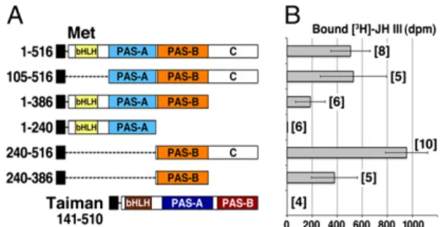
PAS-B Domain of Tribolium Met Specifically Binds JH III and Its Mimics.To determine which part of the Tribolium Met protein is responsible for binding JH III, we systematically deleted the conserved domains (Fig. 2A andFig. S1). Testing the truncated proteins in the ligand-binding assay revealed that the N-terminal half of Met, including the bHLH and PAS-A domains (amino acids 1–240), was not required and had no JH-binding activity alone (Fig. 2B). Only constructs containing the PAS-B domain were capable of binding the hormone. The C-terminal region, which shows poor sequence conservation (residues 387–516), was dispensable for hormone binding, but its removal lowered the protein yield (Fig. S1), likely causing lower JH binding in con-structs Met(1–386) and Met(240–386) (Fig. 2B). Because the C-terminal region improved protein expression, we performed further analyses either on full-length Met(1–516) or on Met (240–516), which includes the PAS-B domain and the native protein end. Taiman, the bHLH-PAS dimerization partner of Met (16, 17), did not bind JH III (Fig. 2B). To verify the ligand-binding capacity of Met PAS-B in another type of assay, we subjected Met to equilibrium dialysis in the presence of radio-labeled JH III. The hormone specifically accumulated in the dialysis compartment containing Met(1–516) and Met(240–516) but not Met(1–240), which lacks the PAS-B domain (Fig. S2).
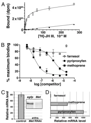
Saturation assays determined that Met(240–516) bound JH III with an average Kd of 12.3 ± 0.62 nM (Fig. 3A), an affinity
slightly lower than that of the entire protein (Fig. 1B). Metho-prene and pyriproxyfen, known to be effective juvenoids in Tri-bolium (11, 13, 21), competed against JH III in binding to Met(240–516), whereas the inactive JH precursor farnesol did not (Fig. 3B). Kivalues of 388± 52 nM for methoprene and 4.75 ±
0.86 nM for pyriproxyfen suggested an affinity ranking of pyr-iproxyfen> JH III > methoprene for Met(240–516). The above data show that the PAS-B domain of Met is necessary and suf-ficient for specific, high-affinity ligand binding and discriminates between biologically active and inert compounds.
Pyriproxyfen Is a Potent Met Agonist. Unlike methoprene, pyr-iproxyfen has a chemical structure unrelated to natural JH (6). Because pyriproxyfen is both a potent ligand of Met (Fig. 3B) and more effective than methoprene as insecticide against Tri-bolium (21), we tested whether pyriproxyfen exerted a specific biological effect in vivo through Met. Indeed, pyriproxyfen in-duced expression of a well-characterized Met target gene, Kr-h1, in Tribolium pupae. This induction required Met because it was abolished in animals lacking the Met protein (Fig. 3C). Consis-tent with its higher affinity to Met(240–516), pyriproxyfen
Fig. 1. Tribolium Met and its orthologs bind JH III. (A) Myc-tagged full-length proteins from D. melanogaster (Dm) and T. castaneum (Tc) and the PAS-B region from T. domestica (Td) were translated in vitro (Inset, immu-noblot) and incubated with 0.5 pmol of [3H]JH III. Total binding is compared against reticulocyte lysate without DNA (mock). The value obtained for DmMet is significantly higher than mock (Mann–Whitney test, P < 0.05; n = 4). (B) Full-length Tribolium Met was incubated with increasing concentrations of [3H]JH III in the absence (○, total binding) or presence (●, nonspecific binding) of a 100-fold molar excess of unlabeled JH III. Each data point is mean± SD of two to four assays, and the saturation curve shown is average of four independent experiments. The calculated Kdis 2.94± 0.68 nM.
Fig. 2. The PAS-B domain of Met is necessary and sufficient for JH III binding. (A) Deletion constructs representing the individual domains of Tribolium Met and Taiman proteins tagged with the Myc epitope (black boxes) were translated in vitro (for immunoblot, seeFig. S1). Numbers in-dicate amino acid positions; 1–516 is the entire Met protein. (B) JH III-binding activities are plotted next to the respective proteins as total radioactivity bound. Values are mean± SD of several independent repeats (n numbers are in brackets).
Charles et al. PNAS | December 27, 2011 | vol. 108 | no. 52 | 21129
DEVELOPM
ENTAL
BIO
appeared to be more potent than methoprene in inducing Kr-h1 transcription (Fig. 3D). Therefore, like JH III or its structural analog methoprene, pyriproxyfen activates JH-dependent gene expression through the same receptor protein. These data
establish pyriproxyfen as a Met agonist and Met as a target of chemically diverse insecticides that mimic the effect of JH.
Structural Models Predict a JH-Binding Pocket Within the PAS-B Domain of Met.To understand how the structure of Met might accommodate the hormonal ligand, we modeled the Tribolium Met PAS-B domain based on the crystal structure of hypoxia-inducible factor 2α (HIF2α) PAS-B (22) as a homologous tem-plate (Fig. S3). The obtained model contained an elongated in-ternal cavity of 625 Å3that extended from helix Cα to Fα and presented an opening between the Fα-helix and the β-sheet (Fig. 4A). Secondary structure elements of the PAS-B domain con-tributed to this pocket with mainly hydrophobic residues (Fig. 4B). Both the size and the hydrophobic nature of the cavityfit the expected role of binding small hydrophobic ligands.
Computational docking of JH III to the Met PAS-B model led to several solutions, which allfilled the bottom part of the pocket near the Cα-helix (Fig. S4A). The best docking result corre-sponded to a theoretical affinity of −7.4 kcal/mol and showed JH III forming a single hydrogen bond with the Tyr-252 side-chain hydroxyl group (N-terminal β-strand Aβ) through its epoxide moiety (Fig. 4C). Docking of the chemically disparate but bi-ologically active pyriproxyfen molecule into the Met PAS-B domain produced a single best solution with a theoretical affinity of−9.2 kcal/mol. Pyriproxyfen in its position overlapped with JH III and contacted the side chains of Tyr-252 and Lys-311 through hydrogen bonds involving two of its ether groups (Fig. S4B). The goodfit of pyriproxyfen to the PAS-B model corresponded with its ability to effectively compete with JH III for binding Met (240–516) (Fig. 3B).
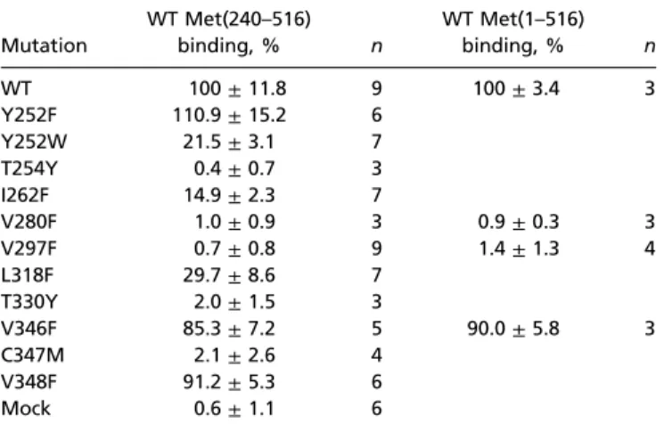
Mutations Within the Ligand-Binding Pocket Disrupt JH III Binding.To test the model of JH binding to Met, we changed several of the residues whose side chains point toward the ligand (Fig. 4C). These mutations (Fig. S5) were to amino acids with larger side chains but similar physicochemical properties to block ligand binding by steric hindrance. All versions of Met(240–516) har-boring the individual mutations were expressed to the same ex-tent and remained stable throughout the JH III-binding assay (Fig. S6). Mutations T254Y, V280F, V297F, T330Y, and C347M resulted in total or nearly total loss of detectable JH III binding; I262F and L318F reduced it to 15% and 30%, respectively, of WT PAS-B activity (Table 1). In contrast, substituting phenyl-alanine for the conserved valine residues at positions 346 and 348, whose side chains point away from the ligand-binding pocket, only reduced JH III binding to 85% and 91% of the WT, respectively (Table 1). To verify whether mutations disrupting JH III binding of Met PAS-B had the same effect in the context of the entire Met protein, we introduced mutations of two
Fig. 3. Met selectively binds JH III and its mimics and mediates the effect of pyriproxyfen in vivo. (A) In vitro-translated Met PAS-B was incubated with [3H]JH III in the absence (○, total binding) or presence (●, nonspecific binding) of a 100-fold molar excess of cold JH III. The saturation curve shown is average of six independent experiments. The calculated Kdis 12.3± 0.62 nM. (B) Met PAS-B was incubated with 2 pmol of [3H]JH III in the presence of increasing concentrations of the indicated compounds. The competition curves shown are average of three independent experiments. The calculated dissociation constants (Ki) are 388± 52 nM for methoprene and 4.75 ± 0.86 nM for pyriproxyfen; farnesol did not significantly compete for binding. (C) At 3 d after injection with egfp (control) or Met dsRNA, Tribolium pupae were treated with pyriproxyfen and tested for Kr-h1 mRNA expression 12 h later. Inset shows RNAi knockdown of the Met protein in these pupae. Data are mean± SD from six animals. (D) Kr-h1 mRNA levels were assessed in Tri-bolium pupae at 8 h after treatment with 0.1 mM solutions of the indicated compounds. Data are mean± SD from n = 4 pupae; the difference between pyriproxyfen and methoprene is significant at P = 0.03 (Student’s t test).
Fig. 4. Model of the ligand-binding cavity of the Tribolium Met PAS-B domain. (A) Overall structure of Met PAS-B (blue, N terminus; red, C terminus) with the cavity. Position of a hypothetical heterodimeric partner, here represented by Arnt as in the HIF2α–Arnt crystal structure PDB ID 3F1P (22), is shown in gray. (B) A closer view of the pocket relative to the amino acid residues mutated in this study. (C) Docking model of a Met–JH III complex. A hydrogen bond (dotted line) is predicted between the hydroxyl group of Tyr-252 and the epoxide moiety of JH III. Orientation is the same in all models.
selected amino acids residing in opposite corners of the ligand-binding cavity, V280F and V297F (Fig. 4B), into Met(1–516). Both mutations (but not V346F) abolished JH III binding (Table 1) by the full-length Met protein. The mutant and WT Met(1–516) proteins showed equal expression levels and stability (Fig. S6).
Interestingly, some of the amino acids critical for JH binding in Met occur in positions corresponding to residues within the ligand-binding pocket of AhR PAS-B, which are important for binding of the dioxin (TCDD) ligand (23, 24). Mutations of T283, homologous to T254 in Met (Fig. S7), abolish TCDD binding. AhR mutations P291F and C327A, which affect residues at positions occupied by I262 and V297, respectively, in
Tribo-lium Met (Fig. 4C), strongly reduce TCDD binding. Finally, substitution of A375 in the center of the AhR ligand-binding pocket with bulkier residues prevents TCDD binding because of steric hindrance (23). A375 corresponds to C347, which is in-variant in Met proteins (Figs. S5andS7).
Although docking solutions for both JH III and pyriproxyfen involved a hydrogen bond with Tyr-252, contribution of this H bond was not critical because mutation Y252F did not reduce JH binding (Table 1). However, tryptophan occurs in place of Tyr-252 in the true bugs, Pyrrhocoris apterus (20) and Rhodnius pro-lixus (Hemiptera) (Fig. S5), and the Y252W substitution lowered JH III binding to 21.5% (Table 1). Interestingly, a closely related bug species, Plautia stali, possesses a skipped bisepoxide type of JH (25), suggesting that the tryptophan residue might reflect the structural difference of the bug JH.
Ligand-Dependent Protein–Protein Interactions.Met proteins from Drosophila and the Aedes mosquito form homodimers (and a heterodimer with Gce in Drosophila) in the absence of JH III or methoprene; addition of either compound leads to dissocia-tion of the complexes (16, 26). Using immunoprecipitadissocia-tion in transfected human cells, we found that Tribolium Met also formed a dimer that dissociated upon methoprene addition (Fig. 5A). However, this methoprene-induced dissociation was par-tially prevented if one of the Met monomers carried the V297F mutation that abolished JH binding, and, when both monomers were mutated, the complex became resistant to methoprene (Fig. 5A). Immunoprecipitation of Met(240–516) proteins showed that the PAS-B domain alone was sufficient for dimer formation and that the JH-dependent inhibition of Met dimerization re-sided within the PAS-B domain itself (Fig. 5B). Although Met
Table 1. Effect of mutations within the PAS-B domain of Tribolium Met on JH III binding
Mutation WT Met(240–516) binding, % n WT Met(1–516) binding, % n WT 100± 11.8 9 100± 3.4 3 Y252F 110.9± 15.2 6 Y252W 21.5± 3.1 7 T254Y 0.4± 0.7 3 I262F 14.9± 2.3 7 V280F 1.0± 0.9 3 0.9± 0.3 3 V297F 0.7± 0.8 9 1.4± 1.3 4 L318F 29.7± 8.6 7 T330Y 2.0± 1.5 3 V346F 85.3± 7.2 5 90.0± 5.8 3 C347M 2.1± 2.6 4 V348F 91.2± 5.3 6 Mock 0.6± 1.1 6
Fig. 5. Ligand-dependent protein interactions of Tribolium Met. Proteins N-terminally tagged with either EGFP (shaded) or the Myc epitope were coex-pressed in human HEK293 cells. Methoprene (1μM) or ethanol was added to cell cultures at 1 h before lysis. Cell lysates were subjected to immunopre-cipitation (IP, outlined) with an anti-EGFP serum, and interacting proteins were detected on Western blots (WB) with an anti-Myc antibody. Input panels represent 10% of the initial material. Methoprene disrupted homophilic complexes of full-length Met(1–516) (A) or PAS-B Met(240–516) (B) proteins but not of their V297F mutant versions lacking the JH binding capability. Binding of Met(1–240) bHLH and PAS-A domains to full-length Met was insensitive to methoprene (C). Interaction of full-length Met (D) or its PAS-B domain (E) with Taiman required methoprene and was prevented by mutations that abolish binding of JH.
Charles et al. PNAS | December 27, 2011 | vol. 108 | no. 52 | 21131
DEVELOPM
ENTAL
BIO
(1–240) harboring the bHLH and PAS-A regions also dimerized, it did so in a methoprene-insensitive manner (Fig. 5C).
The fact that the single point mutation V297F renders the Met–Met complex resistant to the effect of methoprene shows that the dissociation is induced by methoprene through the specific ligand-binding pocket within PAS-B. This process might involve a ligand-induced conformational change of Met that is incompatible with formation of a homophilic complex. The ability of Met V297F to dimerize in the absence of methoprene also confirms that the failure of this mutant to bind JH III is not attributable to a compromised integrity of the protein.
bHLH-PAS proteins typically act as heterodimers (15), and Met proteins from Aedes and Tribolium have been recently shown to activate gene expression upon JH-dependent physical interaction with Taiman (16, 17). These results have suggested that the Met–Taiman dimer might be an active JH receptor, but the mechanism through which JH induces Met to bind Taiman remains unclear. We addressed this question by using Met mutants incapable of binding JH. The WT Tribolium Met bound Taiman only in the presence of methoprene (Fig. 5D), con-firming the published data (16, 17). By contrast, two mutations that abolished JH III binding, V297F and V280F (Table 1 and Fig. 4C), severely reduced the capacity of Met to respond to methoprene by binding Taiman, whereas the neutral V346F substitution left the interaction unaffected (Fig. 5D). The same effect of V297F was observed with the truncated PAS-B domain (Fig. 5E). These data demonstrate that the interaction between Met and Taiman specifically depends on methoprene binding to the PAS-B domain of Met. Therefore, the critical role of PAS-B in regulating heterodimer formation is common to insect Met and mammalian bHLH-PAS proteins such as HIF2α and Arnt (27).
Together with other recent advances (16, 17, 26), our results support a model in which unliganded Met occurs as a presumably inactive homodimer. Upon JH binding to the PAS-B domain, Met undergoes a conformational change that (i) liberates Met from the homophilic complex and (ii) allows it to bind Taiman. By sensing JH and forming a ligand-dependent complex with a partner of its own bHLH-PAS family, Met establishes a unique class of intracellular hormone receptors. Its action resembles type II nuclear receptors, whereby a ligand-specific sensor (such as a thyroid hormone or retinoic acid receptor) combines with a versatile heterodimer partner, the retinoid X receptor RXR (28). Although the exact mechanism of JH-dependent activation of Met has yet to be determined, the present study sheds light on the long-elusive problem of JH reception, including the action of insecticidal JH mimics.
Methods
DNA Constructs and Protein Expression. DNA sequences encoding all Met and Gce proteins or their truncated and mutant versions were synthesized for optimal mammalian codon use (GenScript); taiman cDNA was cloned from Tribolium larval RNA by RT-PCR. Appropriate DNA regions were cloned into
the pK-Myc-C2 vector containing a T7 promoter and an N-terminal Myc epitope tag (29). Proteins were produced with the TnT Quick Coupled in vitro transcription/translation system (Promega) with 400 ng of template plasmid per 50-μL reaction. The efficiency of translation was assessed on immunoblots with the mouse anti-Myc antibody 9E10 (Roche).
Ligand-Binding Assays. Racemic (RS)-tritiated JH III (10–20 Ci·mMol−1) was from Perkin-Elmer and racemic JH III, pyriproxyfen, trans,trans-farnesol, and methoprene were from Sigma-Aldrich. Dextran-coated charcoal (DCC) assays were performed as described (8, 30), and integrity of radiolabeled JH III throughout the assay was verified (SI Methods andFig. S8). Nonspecific binding was determined in DCC assays with a 100-fold molar excess of un-labeled ligand in addition to [3H]JH III. Dissociation constants (Kd) were de-termined by nonlinear regression from total and nonspecific binding data of saturation experiments by using GraphPad Prism 5.00 (GraphPad Software) on the assumption that JH III binds to a single site and that both the 10R-and 10S-JH III isomers bind equally. For competition assays, the Kiof JH mimics was calculated by using the“one site fit Ki” model.
Immunoprecipitation. Tribolium Met and Taiman proteins were expressed with N-terminal EGFP or Myc epitope tags from pEGFP-C2 (Clontech) or pK-Myc-C2 vectors, respectively, by transient transfection in the HEK293 cells. At 1 h before cell harvesting, cells were treated with 1μM methoprene (or ethanol for control) and then lysed (SI Methods). The lysate was applied (with or without methoprene) to Dynabeads Protein G (Invitrogen) pre-bound with rabbit anti-EGFP antiserum. Input and pre-bound proteins were detected with mouse anti-GFP (Sigma-Aldrich) and anti-Myc 9E10 (Roche) antibodies on immunoblots.
Protein Structure Modeling and Ligand Docking. The Tribolium Met PAS-B domain (Leu-240–Leu-358) structure was modeled with Modeler version 9.9 software (31) with HIF2α PAS-B crystal structure (PDB ID 3F1P) (22) as a ho-mologous template. Among 10 models generated, the one with the lowest objective function was retained for ligand docking with AutoDock Vina (32) after the receptor and ligandfiles were prepared with AutoDock Tools (33). The volume of the cavity of the modeled PAS-B was calculated by using CASTp with default parameters (34). Figures were prepared with Pymol version 0.99 (DeLano Scientific).
RNAi, Hormonal Treatments, and mRNA Expression Analysis. Within 12 h after ecdysis, Tribolium pupae were injected with Met or control (egfp) dsRNA and after 3 d were treated with acetone-diluted 0.1 mM farnesol, metho-prene, or pyriproxyfen as described (11). Total RNA was isolated from in-dividual pupae and subjected to quantitative RT-PCR with Kr-h1 primers (13) by using the iQ SYBR Green Supermix kit and the C1000 Thermal Cycler (Bio-Rad). Data were normalized to the relative levels of ribosomal protein (Rp49) mRNA.
ACKNOWLEDGMENTS. We thank Dr. Bruce Hammock for 3-octylthio-1,1,1-trifluoro-2-propanone (OTFP), Dr. Michel Narce for access to the radioactive facility, François Bonneton and Franck Borel for helpful discussions, and Jordan Ward for reading the manuscript. This work was supported by Acad-emy of Sciences Grant 500960906 and Ministry of Education of the Czech Republic Grant 6007665801 and was partly carried out during a pre-second-ment phase of Marie Curie Fellowship Award 276569 (to M.J.). This work was also supported by Czech Science Foundation Projects 204/09/H058 (to J.R.) and 204/07/1032 (to K.T.).
1. Wigglesworth VB (1934) The physiology of ecdysis in Rhodnius prolixus (Hemi-ptera). II. Factors controlling moulting and“metamorphosis”. Q J Microsc Sci 77: 191–222.
2. Riddiford LM (1994) Cellular and molecular actions of juvenile hormone. I. General considerations and premetamorphic actions. Adv Insect Physiol 24:213–274. 3. Wyatt G, Davey K (1996) Cellular and molecular actions of juvenile hormone. II roles
of juvenile hormones in adult insects. Adv Insect Physiol 26:1–155.
4. Riddiford LM (2008) Juvenile hormone action: A 2007 perspective. J Insect Physiol 54: 895–901.
5. Wilson TG, Fabian J (1986) A Drosophila melanogaster mutant resistant to a chemical analog of juvenile hormone. Dev Biol 118:190–201.
6. Minakuchi C, Riddiford LM (2006) Insect juvenile hormone action as a potential target of pest management. J Pestic Sci 31:77–84.
7. Ashok M, Turner C, Wilson TG (1998) Insect juvenile hormone resistance gene ho-mology with the bHLH-PAS family of transcriptional regulators. Proc Natl Acad Sci USA 95:2761–2766.
8. Miura K, Oda M, Makita S, Chinzei Y (2005) Characterization of the Drosophila Me-thoprene-tolerant gene product. Juvenile hormone binding and ligand-dependent gene regulation. FEBS J 272:1169–1178.
9. Baumann A, Barry J, Wang S, Fujiwara Y, Wilson TG (2010) Paralogous genes involved in juvenile hormone action in Drosophila melanogaster. Genetics 185:1327–1336. 10. Abdou MA, et al. (2011) Drosophila Met and Gce are partially redundant in
trans-ducing juvenile hormone action. Insect Biochem Mol Biol 41:938–945.
11. Konopova B, Jindra M (2007) Juvenile hormone resistance gene Methoprene-tolerant controls entry into metamorphosis in the beetle Tribolium castaneum. Proc Natl Acad Sci USA 104:10488–10493.
12. Minakuchi C, Namiki T, Yoshiyama M, Shinoda T (2008) RNAi-mediated knockdown of juvenile hormone acid O-methyltransferase gene causes precocious metamorphosis in the redflour beetle Tribolium castaneum. FEBS J 275:2919–2931.
13. Minakuchi C, Namiki T, Shinoda T (2009) Krüppel homolog 1, an early juvenile hor-mone-response gene downstream of Methoprene-tolerant, mediates its anti-meta-morphic action in the redflour beetle Tribolium castaneum. Dev Biol 325:341–350.
14. Riddiford LM, Truman JW, Mirth CK, Shen YC (2010) A role for juvenile hormone in the prepupal development of Drosophila melanogaster. Development 137:1117– 1126.
15. Kewley RJ, Whitelaw ML, Chapman-Smith A (2004) The mammalian basic helix-loop-helix/PAS family of transcriptional regulators. Int J Biochem Cell Biol 36: 189–204.
16. Li M, Mead EA, Zhu J (2011) Heterodimer of two bHLH-PAS proteins mediates juvenile hormone-induced gene expression. Proc Natl Acad Sci USA 108:638–643. 17. Zhang Z, Xu J, Sheng Z, Sui Y, Palli SR (2011) Steroid receptor co-activator is required
for juvenile hormone signal transduction through a bHLH-PAS transcription factor, methoprene tolerant. J Biol Chem 286:8437–8447.
18. Beischlag TV, et al. (2002) Recruitment of the NCoA/SRC-1/p160 family of transcrip-tional coactivators by the aryl hydrocarbon receptor/aryl hydrocarbon receptor nu-clear translocator complex. Mol Cell Biol 22:4319–4333.
19. Baker FC, et al. (1984) Detection of only JH III in several life-stages of Nauphoeta cinerea and Thermobia domestica. Life Sci 35:1553–1560.
20. Konopova B, Smykal V, Jindra M (2011) Common and distinct roles of juvenile hor-mone signaling genes in metamorphosis of holometabolous and hemimetabolous insects. PLoS ONE 6(12):e28728.
21. Kostyukovsky M, Chen B, Atsmi S, Shaaya E (2000) Biological activity of two juvenoids and two ecdysteroids against three stored product insects. Insect Biochem Mol Biol 30:891–897.
22. Scheuermann TH, et al. (2009) Artificial ligand binding within the HIF2α PAS-B do-main of the HIF2 transcription factor. Proc Natl Acad Sci USA 106:450–455. 23. Pandini A, Denison MS, Song Y, Soshilov AA, Bonati L (2007) Structural and functional
characterization of the aryl hydrocarbon receptor ligand binding domain by ho-mology modeling and mutational analysis. Biochemistry 46:696–708.
24. Pandini A, et al. (2009) Detection of the TCDD binding-fingerprint within the Ah receptor ligand binding domain by structurally driven mutagenesis and functional analysis. Biochemistry 48:5972–5983.
25. Kotaki T, Shinada T, Kaihara K, Ohfune Y, Numata H (2009) Structure determination of a new juvenile hormone from a heteropteran insect. Org Lett 11:5234–5237. 26. Godlewski J, Wang S, Wilson TG (2006) Interaction of bHLH-PAS proteins involved in
juvenile hormone reception in Drosophila. Biochem Biophys Res Commun 342: 1305–1311.
27. Erbel PJA, Card PB, Karakuzu O, Bruick RK, Gardner KH (2003) Structural basis for PAS domain heterodimerization in the basic helix–loop–helix-PAS transcription factor hypoxia-inducible factor. Proc Natl Acad Sci USA 100:15504–15509.
28. Mangelsdorf DJ, Evans RM (1995) The RXR heterodimers and orphan receptors. Cell 83:841–850.
29. Valenta T, Lukas J, Korinek V (2003) HMG box transcription factor TCF-4’s interaction with CtBP1 controls the expression of the Wnt target Axin2/Conductin in human embryonic kidney cells. Nucleic Acids Res 31:2369–2380.
30. Touhara K, Lerro KA, Bonning BC, Hammock BD, Prestwich GD (1993) Ligand binding by a recombinant insect juvenile hormone binding protein. Biochemistry 32:2068–2075. 31. Sali A, Blundell TL (1993) Comparative protein modelling by satisfaction of spatial
restraints. J Mol Biol 234:779–815.
32. Trott O, Olson AJ (2010) AutoDock Vina: Improving the speed and accuracy of docking with a new scoring function, efficient optimization, and multithreading. J Comput Chem 31:455–461.
33. Sanner MF (1999) Python: A programming language for software integration and development. J Mol Graph Model 17:57–61.
34. Dundas J, et al. (2006) CASTp: Computed atlas of surface topography of proteins with structural and topographical mapping of functionally annotated residues. Nucleic Acids Res 34(Web Server issue):W116–W118.
Charles et al. PNAS | December 27, 2011 | vol. 108 | no. 52 | 21133
DEVELOPM
ENTAL
BIO