HAL Id: hal-00814442
https://hal.archives-ouvertes.fr/hal-00814442
Submitted on 29 May 2020
HAL is a multi-disciplinary open access
archive for the deposit and dissemination of
sci-entific research documents, whether they are
pub-lished or not. The documents may come from
teaching and research institutions in France or
abroad, or from public or private research centers.
L’archive ouverte pluridisciplinaire HAL, est
destinée au dépôt et à la diffusion de documents
scientifiques de niveau recherche, publiés ou non,
émanant des établissements d’enseignement et de
recherche français ou étrangers, des laboratoires
publics ou privés.
Copyright
copper homeostasis in Chlamydomonas reinhardtii.
Davin Malasarn, Janette Kropat, Scott I Hsieh, Giovanni Finazzi, David
Casero, Joseph A Loo, Matteo Pellegrini, Francis-André Wollman, Sabeeha S
Merchant
To cite this version:
Davin Malasarn, Janette Kropat, Scott I Hsieh, Giovanni Finazzi, David Casero, et al.. Zinc deficiency
impacts CO2 assimilation and disrupts copper homeostasis in Chlamydomonas reinhardtii.. Journal
of Biological Chemistry, American Society for Biochemistry and Molecular Biology, 2013, 288 (15),
pp.10672-83. �10.1074/jbc.M113.455105�. �hal-00814442�
Zinc Deficiency Impacts CO
2
Assimilation and Disrupts
Copper Homeostasis in Chlamydomonas reinhardtii
*
□SReceived for publication, January 21, 2013, and in revised form, February 19, 2013Published, JBC Papers in Press, February 25, 2013, DOI 10.1074/jbc.M113.455105
Davin Malasarn‡1, Janette Kropat‡, Scott I. Hsieh‡2, Giovanni Finazzi§¶, David Casero储, Joseph A. Loo‡储, Matteo Pellegrini储**, Francis-André Wollman§, and Sabeeha S. Merchant‡储3
From the‡Department of Chemistry and Biochemistry, the储UCLA/Department of Energy Institute for Genomics and Proteomics,
and the **Department of Molecular, Cell and Developmental Biology, UCLA, Los Angeles, California 90095,§CNRS, Université Paris
6, Institut de Biologie Physico-Chimique, 75005 Paris, France, and¶CNRS, Université Joseph Fourier, Commissariat à l’Energie
Atomique et aux Energies Alternatives, Institut National Recherche Agronomique, UMR 5168 Laboratoire de Physiologie Cellulaire et Végétale, Institut de Recherche en Sciences et Technologie du Vivant, Commissariat à l’Energie Atomique Grenoble, 17 Rue des Martyrs, 38054 Grenoble, France
Background:Zinc is required for catalysis and protein structure.
Results:Zinc-deficient Chlamydomonas lose carbonic anhydrases and cannot grow photoautotrophically in air. They also hyperaccumulate copper but are phenotypically copper-deficient and therefore require Crr1, the nutritional copper sensor. Conclusion:Zinc deficiency impacts the carbon-concentrating mechanism and disrupts copper homeostasis.
Significance:Cross-talk exists between zinc and copper homeostasis pathways.
Zinc is an essential nutrient because of its role in catalysis and in protein stabilization, but excess zinc is deleterious. We dis-tinguished four nutritional zinc states in the alga Chlamydomo-nas reinhardtii: toxic, replete, deficient, and limited. Growth is inhibited in limited and toxic cells relative to zinc-replete cells, whereas zinc deficiency is visually asymptomatic but distinguished by the accumulation of transcripts encoding ZIP family transporters. To identify targets of zinc deficiency and mechanisms of zinc acclimation, we used RNA-seq to probe zinc nutrition-responsive changes in gene expression. We iden-tified genes encoding zinc-handling components, including ZIP family transporters and candidate chaperones. Additionally, we noted an impact on two other regulatory pathways, the carbon-concentrating mechanism (CCM) and the nutritional copper regulon. Targets of transcription factor Ccm1 and various CAH genes are up-regulated in zinc deficiency, probably due to reduced carbonic anhydrase activity, validated by quantitative proteomics and immunoblot analysis of Cah1, Cah3, and Cah4. Chlamydomonas is therefore not able to grow photoautotrophi-cally in zinc-limiting conditions, but supplementation with 1% CO2 restores growth to wild-type rates, suggesting that the inability to maintain CCM is a major consequence of zinc
limi-tation. The Crr1 regulon responds to copper limitation and is turned on in zinc deficiency, and Crr1 is required for growth in zinc-limiting conditions. Zinc-deficient cells are functionally copper-deficient, although they hyperaccumulate copper up to 50-fold over normal levels. We suggest that zinc-deficient cells sequester copper in a biounavailable form, perhaps to prevent mismetallation of critical zinc sites.
Zinc is an essential nutrient required in abundance by organ-isms ranging from bacteria to humans. Over 300 known enzymes utilize zinc as a cofactor, and whole genome surveys estimate that 4 –10% of all sequenced proteins from pro-karyotes and eupro-karyotes contain zinc-binding domains (1). Excess zinc is believed to be toxic because it can compete for metal binding sites in other proteins and can indirectly generate damaging reactive oxygen species. Thus, intracellular zinc con-tent must be regulated to ensure that zinc-containing proteins can function while excess zinc is avoided. The first level of con-trol is at the step of assimilation, where intracellular zinc status controls the expression and presentation of low and high affin-ity transporters at the plasma membrane (2–7). Another level of control is by compartmentalization, where transporters can sequester zinc in the vacuole (in fungi), lysosome-related com-partments (in Caenorhabditis elegans), or in zincosomes (in mammalian cells) in a situation of excess (8 –10). Sequestered zinc can be mobilized by efflux transporters (11). The expres-sion of each type of transporter is therefore critical for homeo-stasis, and there are multiple levels of control from transcrip-tion to trafficking to protein degradatranscrip-tion (2, 12).
Transporters in two families are important for zinc metabo-lism, the ZIP (Zrt-, Irt-like protein) family, whose members function primarily to move either zinc or iron into the cyto-plasm, and the CDF (cation diffusion facilitator) family, whose members function primarily to move zinc out of the cytoplasm (13) (reviewed in Ref. 14). In plants, members of the CDF family
*This work was supported, in whole or in part, by National Institutes of Health Grant GM42143 (to S. M.) for work on the zinc-deficiency transcriptome. The proteomic analyses and expression of proteins for use as antigens were supported by the Institute of Genomics and Proteomics at UCLA (funded by the Office of Science (Biological and Environmental Research), United States Department of Energy, through Cooperative Agreement DE-FC02-02ER63421).
□S This article containssupplemental Fig. S1 and Data Sets 1–3.
1Supported in part by National Institutes of Health Ruth L. Kirschstein
National Research Service Award F32GM083562 and a Chateaubriand Fel-lowship for collaborative experiments with G. F. and F. A. W.
2Supported in part by National Institutes of Health Ruth L. Kirschstein
National Research Service Award T32 GM07185 for the UCLA predoctoral Cellular and Molecular Biology Training Program.
3To whom correspondence may be addressed: 607 Charles E. Young Dr. E.,
Los Angeles, CA 90095-1569. Fax: 310-206-1035; E-mail: merchant@ chem.ucla.edu.
at INRA Institut National de la Recherche Agronomique on June 13, 2018
http://www.jbc.org/
are also called MTPs (metal tolerance proteins) because their function in moving metals out of the cytoplasm is important for handling toxicity (15, 16). The pattern of expression of individ-ual members of these families in response to the concentration and type of metal nutrient provides a clue to their physiological functions.
Photosynthetic microorganisms rely on the carbon-con-centrating mechanism (CCM)4 to concentrate CO
2 at the
active site of ribulose-bisphosphate carboxylase/oxygenase, the enzyme that catalyzes the first step in the CO2fixation
path-way (reviewed in Refs. 17–20). Carbonic anhydrases are inte-gral components in this pathway and contribute to the high productivity of some of these organisms. These enzymes gen-erally rely on a zinc cofactor (to activate water) to catalyze the interconversion of bicarbonate and CO2. In a zinc-deficient
marine environment, cobalt can replace zinc, or an alternate enzyme that uses a cadmium cofactor can substitute (21–23). The occurrence of these zinc-sparing mechanisms is indicative of the contribution of carbonic anhydrases to the cellular zinc quota, meaning the optimally desired zinc content of the cell in a zinc-replete medium (24).
We have developed Chlamydomonas reinhardtii as a refer-ence organism for understanding pathways of trace metal metabolism and homeostasis in algae and in the plant lineage, especially with respect to the impact of deficiency on bioener-getic pathways in the mitochondria and chloroplast (14, 25).
Chlamydomonasspecies have been found in various environ-mental niches distinguished by metal content, pH, and oxygen availability, suggesting that this genus has developed adaptive systems to deal with changing environmental conditions (26). In the laboratory, dilute Chlamydomonas cultures can reach stationary phase in 2–3 days in a simple salt-containing medium in which the trace elements are buffered by chelation with EDTA (27). The absence of serum or amino acid supple-mentation simplifies the provision of trace metal nutrients and the establishment of deficiency. We have been able to exploit this in previous work with Chlamydomonas cells experiencing iron, copper, or manganese deficiency, in which we noted that the assimilatory transporters are responsive to metal nutrition at the transcriptional level, including two genes encoding ZIP family members, which were named IRT1 and IRT2 because they responded to iron deficiency (28 –31). Nevertheless, there are as many as a dozen members of the ZIP family in
Chlamy-domonas, and some of them are likely to be involved in zinc assimilation.
The interaction of zinc and copper homeostasis pathways is also likely in Chlamydomonas. Cu(I) is taken up by the CTR (copper transporter) family of transporters, whose members are components of the nutritional copper regulon in
Chlamy-domonas. A zinc-containing SBP (squamosa-promoter binding protein) domain transcription factor named Crr1 (copper response regulator) controls the expression of the CTR genes
(28). Its functional homolog in Arabidopsis is SPL7 (32–34). Besides the CTR genes encoding the plasma membrane-local-ized assimilatory transporters, Crr1 regulates over 60 genes involved in acclimation to copper deficiency through associ-ated copper-responsive elements, which are the target sites for the SBP domain (28, 35–37). The best characterized of these target genes is CYC6, encoding cytochrome (Cyt) c6, which is a
heme-containing replacement of the usual copper-containing protein, plastocyanin, in the photosynthetic electron transfer chain. Therefore, although copper-deficient Chlamydomonas cells do not accumulate plastocyanin, they remain photosyn-thetically competent (38). Decreased plastocyanin abundance and transcriptional activation of CYC6 are classic markers for the copper deficiency state.
In this work, we use growth and the expression of a subset of genes for ZIP family transporters as sentinels of zinc status to establish zinc deficiency and zinc limitation in
Chlamydomo-nas, which are achieved by serial transfer of replete cells to medium lacking supplemental zinc. Transcriptome and pro-teome surveys identify the carbonic anhydrases and hence the carbon concentrating mechanism as a pathway impacted by poor zinc nutrition. We note also an impact on copper home-ostasis, and we suggest the existence of mechanisms that con-trol the ratio of intracellular metal ions.
EXPERIMENTAL PROCEDURES
Culturing and Strains—C. reinhardtii strains CC-4532 (wild-type, 2137), crr1–2 (referred to subsequently as crr1), and a rescued strain crr1–2::CRR1 (referred to subsequently as CRR1) were cultured under 50 –100mol m⫺2s⫺1continuous illumi-nation (2:1, cool white/warm white light) in Tris acetate-phos-phate (TAP) or Tris phosacetate-phos-phate (TP) medium with trace ele-ment suppleele-ments described by Kropat et al. (27). Briefly, stock solutions of 25 mMEDTA-Na2, 28.5M(NH4)6Mo7O24, 0.1 mM
Na2SeO3, 2.5 mMZnSO4in 2.75 mMEDTA, 6 mMMnCl2in 6
mMEDTA, 20 mMFeCl3in 22 mMEDTA, and 2 mMCuCl2in 2
mMEDTA were made individually in Milli-Q-purified water and diluted 1:1000 in the final growth medium. For metal-free studies, all glassware was triple washed in 6Nhydrochloric acid followed by at least six rinses in Milli-Q-purified (Millipore) water. All media were made using Milli-Q water (39). For experiments other than the proteomic studies, cells were grown in nutrient-replete medium, followed by one transfer into zinc medium with no supplemental zinc, before inoculation into the experimental conditions. For the proteomic studies, cells were grown in replete medium and directly inoculated into the experimental conditions. For experiments involving CO2
sup-plementation, cultures were bubbled with filtered air (control) or a mixture of 1% CO2with air. Cell density was measured by counting with a hemocytometer.
Fluorescence Rise and Decay Kinetics—For CC-4532, room temperature fluorescence rise and decay kinetics were analyzed using a FluorCam 700MF (Photon Systems Instruments). Approximately 50l of concentrated mid-log phase liquid cul-ture was spotted onto the lid of a plastic Petri dish and dark-adapted for 10 min prior to a flash of saturating light and meas-urement of the Kautsky effect in continuous red light at 150 mol m⫺2s⫺1PFD (photon flux density) with 100% actinic
4The abbreviations used are: CCM, carbon-concentrating mechanism; CA,
carbonic anhydrase; Cyt, cytochrome; TAP, Tris acetate-phosphate; TP, Tris phosphate; BisTris, 2-[bis(2-hydroxyethyl)amino]-2-(hydroxymethyl)pro-pane-1,3-diol; PSI and PSII, photosystem I and II, respectively; ICP, induc-tively coupled plasma; RPKM, reads per kilobase of mappable transcript length per million mapped reads.
at INRA Institut National de la Recherche Agronomique on June 13, 2018
http://www.jbc.org/
light and 60% activity. For measurements of crr1 and CRR1 cells in zinc-replete and zinc-limited conditions, room temperature kinetics were measured using a laboratory-built instrument as described by Joliot and Joliot (40).
Cell Size Determination—Cell size was determined with a Beckman Coulter laser diffraction particle size analyzer LS13 320. Approximately 100 ml of cultures in exponential phase were poured into the micro-liquid module with a magnetic stir bar included to keep the cells in suspension. If necessary, cul-tures were concentrated or diluted until the sample obscura-tion was between 8 and 12%.
Nucleic Acid Analysis—Total Chlamydomonas RNA was prepared as described by Quinn and Merchant (39). RNA qual-ity was assessed on an Agilent 2100 Bioanalyzer and by hybrid-ization to CBLP (also called RACK1) as described previously (39). A 915-bp EcoRI fragment from the cDNA cloned in
pcf8-13 was used as the probe (41).
Quantitative RT-PCR—Genomic DNA was removed from the total RNA preparation by treatment with Turbo DNase (Ambion) according to the manufacturer’s instructions with the following modifications. 3 units of enzyme was used per 10 g of nucleic acid, and incubation at 37 °C was increased to 90 min. Complementary DNA, primed with oligo(dT), was gener-ated with reverse transcriptase (Invitrogen) according to the manufacturer’s instructions. Amplification was carried out with reagents from the iQ SYBR Green Supermix qPCR kit (Bio-Rad). Each reaction contained the vendor’s master mix, a 0.3Mconcentration of each primer, and cDNA corresponding to 20 ng of input RNA in the reverse transcriptase reaction. The reaction conditions for the Opticon 2 from MJ Research were as follows: 95 °C for 5 min, followed by cycles of 95 °C for 10 s, 65 °C for 30 s, and 72 °C for 30 s, up to 40 cycles. The fluores-cence was measured at each cycle at 72 and 83 °C. The 2⫺⌬⌬CT
method was used to analyze the database on the fluorescence at 83 °C (42). Melting curves were performed after the PCR to assess the presence of a unique final product.
RNA-seq—RNAs were sequenced by Illumina on a GAIIx sys-tem for estimating transcript abundance. The reads were aligned using Bowtie (43) in single-end mode and with a maxi-mum tolerance of three mismatches to the Au10.2 transcript sequences (see the Phytozome Web site), corresponding to the version 4.0 assembly of the Chlamydomonas genome. Expres-sion estimates were obtained for each individual run in units of reads per kilobase of mappable transcript length per million mapped reads (RPKM; see Ref. 44) after normalization by the number of aligned reads and transcript mappable length. The transcript coverage across the genome was visualized on a local installation of the UCSC browser. Differential expression anal-ysis was performed in R with the DESeq package (45), and p values were adjusted to control for false discovery rate with the Benjamini-Hochberg method (46). Sequence files are publicly available in the NCBI Gene Expression Omnibus (accession numbers GSE25622 and GSE41096).
Protein Isolation for Immunoblot Analysis
—Chlamydomo-nascultures were collected by centrifugation (1000⫻ g, 5 min) and washed twice with 10 mMsodium phosphate, pH 7.0. The total protein fraction was further subfractionated into soluble and membrane components as described by Howe and
Mer-chant (47). Protein concentration of soluble fractions was determined using the Pierce BCA protein assay kit following the manufacturer’s instructions. Samples were diluted to 4 g/l. Membrane fractions were normalized by resuspending them to a volume that was equivalent to the final soluble frac-tion volume.
Immunoblot Analysis—Protein samples were denatured by the addition of 5%-mercaptoethanol and boiling for 10 min before separation on denaturing polyacrylamide gels and trans-ferring in a semidry blotter onto nitrocellulose membranes in transfer buffer (25 mM Tris, 192 mM glycine, 0.0004% SDS (w/v), and 20% (v/v) methanol). The membrane was blocked overnight with 1% dried milk in Tris-buffered saline (10 mM Tris-Cl, 150 mMNaCl, pH 7.5) plus Tween 20 (0.05% (w/v)) before incubation in primary antiserum for 4 –12 h. Gel con-centrations (acrylamide monomer) and dilutions for each pri-mary antibody were as follows: plastocyanin, 16%, 1:1000 dilu-tion; Cyt c6, 16%, 1:1000 dilution; Cah1, 12%, 1:2000 dilution; Cah3, 12%, 1:2000; Cah4, 12%, 1:15,000 dilution; ferredoxin 15%, 1:10,000 dilution; and chloroplast ATP synthase, 10%, 1:20,000 dilution. Secondary goat anti-rabbit horseradish per-oxidase (Pierce) was used at a 1:5000 dilution.
Sample Preparation and Analysis for Quantitative Proteomics—Soluble protein was extracted from CC-4532 cells growing in 2.5Mzinc and the first round of 0 supplemental zinc. For each condition, label-free, data-independent quanti-tative liquid chromatography-tandem mass spectrometry (LC-MS/MS or “LC-MSE”) was performed as previously described
with slight modifications (35, 48). In brief, cells were collected at late exponential phase from replete zinc conditions or from the first transfer to zinc-depleted medium by centrifugation. Cells were broken by slow freezing and thawing at⫺80 °C and 24 °C, respectively. Insoluble material was removed by centrif-ugation at 16,000⫻ g for 10 min at 4 °C followed by centrifuga-tion at 253,000⫻ g for 20 min at 4 °C. Approximately 30g of protein (per lane) was separated by gel electrophoresis on 4 –12% NuPage BisTris gels (Invitrogen) and visualized by staining with Coomassie Blue (Bio-Rad). Gel lanes were divided into⬃3-mm bands, and individual bands were subjected to in-gel trypsin digestion (sequencing grade modified trypsin; Promega). Digested peptides were extracted into a 50:50 water/ acetonitrile solution containing 2.5% formic acid and lyophi-lized. Peptides were then resuspended into a 25 fmol/l bovine serum albumin (BSA) digest and quantification standard (Waters). LC-MSEwas performed using a nanoAcquity UPLC
(Waters) system coupled to a quadrupole time-of-flight mass spectrometer (Waters Xevo QTOF). Protein Lynx Global Server (PLGS version 2.4; Waters) was used to process the LC-MS raw data and determine protein identification and quantification. The quantification of protein levels was achieved via the addition of an internal protein standard (BSA trypsin digest standard) to which the data set was normalized. Our criteria were that the difference in protein abundance between growth conditions must be statistically significant (p ⬍ 0.05 by Student’s t test) and at least 2-fold or greater in magnitude in order to define a change in protein abundance (48).
at INRA Institut National de la Recherche Agronomique on June 13, 2018
http://www.jbc.org/
Stoichiometric Measurements of Cytochrome/P700and Pho-tosystem I (PSI)/PSII—Spectroscopic measurements were per-formed using a JTS-10 spectrophotometer (Biologic, Claix, France). Light-induced absorption changes were measured as absorption of flashed light at discrete times. Changes in the amount of functional photosynthetic complexes were evalu-ated measuring the electrochromic shift spectral change, a shift in the pigment absorption bands that is linearly correlated to the number of light-induced charge separations within the reaction centers. Functional PSI and PSII content was esti-mated from changes in the amplitude of the fast phase of the electrochromic shift signal (at 520 –546 nm) upon excitation with a saturating laser flash (520 nm, 5-ns duration). PSII con-tribution was calculated from the decrease in the signal amplitude upon the addition of DCMU (3-(3 ⬘,4⬘-dichlorophenyl)-1,1-dimethylurea) (20M) and hydroxylamine (2 mM) to irrevers-ibly block PSII charge separation. Conversely, PSI was esti-mated as the fraction of the signal that was insensitive to these inhibitors (49). Cytochrome redox changes were calculated as the difference between the absorption at 554 nm and a base line drawn between 545 and 573 nm and corrected for the contri-bution of the electrochromic signal (50). P700(the primary elec-tron donor to PSI) was measured at 705 nm. To evaluate the relative cytochrome/P700 stoichiometry, measurements were
performed in continuous saturating light (1100mol of pho-tons m⫺2s⫺1) and in the presence of saturating concentrations of DBMIB (2,5-dibromo-3-methyl-6-isopropyl-p-benzoqui-none) (10M). This is required to ensure full inhibition of cyto-chrome b6f reduction by light-generated plastoquinol and
therefore a maximum oxidation of P700and of the cytochrome.
Measurement of Intracellular Metal Content—Cells were collected by centrifugation at 1700⫻ g for 5 min. Pellets were washed once in 1 mMEDTA to remove cell surface-associated metals and once in Milli-Q water. The washed cell paste was overlaid with nitric acid corresponding to a final concentration of 24% in 1 ml and digested at 65 °C. To obtain a corresponding blank, the volume of the cell paste was replaced by deionized water and treated as described above. Total metal and phospho-rous content was measured by inductively coupled plasma-MS (ICP-MS; Agilent 7500).
RESULTS
Identification of Four Distinct Zinc Nutrition States
—Chla-mydomonasgrows in media containing a wide range of supple-mental zinc concentrations (51). Elesupple-mental analysis of wild-type Chlamydomonas cells grown in standard TAP medium supplemented with Hutner’s trace elements, which contains about 80Mof EDTA-chelated zinc, indicated a zinc quota of ⬃2–3 ⫻ 107atoms/cell. We calculated that a minimum
con-centration of 0.85Mzinc ions would be necessary to support zinc-replete growth to stationary phase (⬃2 ⫻ 107cells/ml). After allowing for variation in zinc quota in situations of altered physiology (e.g. low CO2partial pressure when carbonic
anhy-drases would be induced), we chose 2.5Mzinc ions in a revised micronutrient solution (27, 51, 52). Indeed, this concentration of zinc is more than sufficient to support normal growth and a typical intracellular trace metal (copper, iron, manganese) quo-ta; when the zinc content of the medium is reduced to 0.25M,
there is no impact on growth (Fig. 1A). Therefore, 2.5Mzinc ions is considered metal replete for laboratory growth of
Chlamydomonas.
To generate zinc limitation, we inoculated cells (to a density of 105cells/ml) from a replete culture into growth medium
without any zinc supplementation (labeled as 0 in the figures). Although all medium constituents were prepared using high purity salts and all glassware was freshly acid-washed (39), there remained residual zinc in the medium, which we estimated at ⬃10 nMbased on ICP-MS analysis. When this culture (referred to as the first round in zinc-depleted medium) reached early stationary phase (⬃1 ⫻ 107cells/ml), the cells were used to reinoculate fresh “zinc-free” medium or medium with various amounts of supplemented zinc (Fig. 1A). A growth phenotype was identified in medium supplemented with no (0) or very low (10 –25 nM; data not shown) zinc, and this correlated with a reduced intracellular zinc content of these cultures (varying from 25 to 50% of the zinc in the replete situation in individual experiments) (supplemental Fig. S1). The zinc content of cells transferred twice to the 0 zinc medium was only slightly less than or nearly the same as that of cultures transferred once to 0 zinc medium. Further subculturing was therefore not neces-sary, and we routinely used two transfers to 0 zinc medium as the base line for poor zinc nutrition. We also tried to generate zinc deficiency by reducing zinc bioavailability with the intro-duction of Zn(II)-specific chelators, like N,N,N ⬘,N⬘-tetrakis(2-pyridylmethyl)ethylenediamine into the medium, but this did not enhance the phenotype of cells in the second round of growth in 0 zinc medium. N,N,N ⬘,N⬘-Tetrakis(2-pyridylmeth-yl)ethylenediamine was also not effective in generating a zinc-deficient (see below) or zinc-limited condition when it was added at higher concentrations to zinc-replete medium. There-fore, we concluded that, in the absence of a transporter-defec-tive mutant, sequential transfer into defined medium with 0 supplemental Zn(II) is the only effective way to generate zinc
FIGURE 1. Phenotypes of zinc-deficient Chlamydomonas. Strain CC-4532 was grown in TAP medium with different amounts of supplemental zinc: 0M (black), 0.25M(blue), 2.5M(green), 80M(purple), and 250M(red). A, growth measured by counting cells; B, Kautsky fluorescence rise and decay kinetics; C, diameters of cells growing in medium containing 2.5Mzinc (left) and 0 supplemental zinc (right). Growth curves represent the averages of biological triplicates from three separate inocula (S.D. values not shown for clarity). For fluorescence measurements and cell size determination, repre-sentative data from experimental triplicates are shown.
at INRA Institut National de la Recherche Agronomique on June 13, 2018
http://www.jbc.org/
limitation in Chlamydomonas. Analysis of the fluorescence induction curves shows that PSII activity is retained by cells growing in all concentrations of supplemental zinc, including those from cultures with no supplemental zinc, and that elec-tron transfer between the two photosystems still operates under all of these conditions (Fig. 1B). Light microscopy indi-cated that zinc-limited cells are larger than the replete ones, and this is confirmed by measuring the size distribution of cells in batch culture. The average diameter of zinc-limited cells is 11 m compared with the 9-m size of zinc-replete cells. In addi-tion, we noted the occurrence of smaller bodies of size⬃2.5m in the zinc-limited culture. Their identity is not known, but they may represent stress-induced cell fragmentation (Fig. 1C).
At the other extreme, zinc excess is defined as high zinc ion concentrations at which zinc starts to become toxic and the growth rate is inhibited relative to the maximum observed growth rates. In TAP medium, growth is inhibited when the medium contains more than 125–250 M chelated (EDTA) zinc ions (Fig. 1A, red curve), and this defines the zinc excess situation.
In previous work on iron nutrition, we distinguished the defi-ciency state from the limitation state. The iron defidefi-ciency state is characterized by the absence of a growth phenotype or other visual symptoms but the presence of a molecular signature, which in the case of iron nutrition is the expression of the iron assimilation pathway (31). Therefore, we sought to identify bio-markers for zinc deficiency. As putative zinc transporters, members of the ZIP family are excellent candidates for zinc assimilation proteins. Previously, 14 members of this family were identified in Chlamydomonas based on homology to
Ara-bidopsis, yeast, and human sequences (14, 53). We assessed the patterns of mRNA accumulation for each of these as a function of zinc nutrition status to distinguish (a) which of these might respond to zinc nutrition and (b) whether their expression might be an early gauge or a sentinel of the zinc status. RNAs were isolated from cells transferred to medium containing the indicated amounts of zinc (after 1 round in 0 supplemental zinc) and analyzed by real-time RT-PCR for the expression of each of the ZIP genes. Among these, five zinc nutrition-respon-sive genes were identified and named ZRT1, ZRT2, ZRT3,
ZRT4, and ZRT5 (for zinc-responsive transporter (Fig. 2, A and
B). Based on the magnitude of the change, we chose ZRT1 and
ZRT3as sentinel genes and defined 25 nM as the boundary between zinc deficiency and zinc-limiting laboratory condi-tions (Fig. 2C). Thus, we established growth condicondi-tions to gen-erate each of four states of zinc nutrition: 0 –10 nMzinc (no added zinc) for zinc-limiting conditions, 25 nMzinc for defi-cient conditions, 2.5Mzinc for replete conditions, and 250M for zinc-toxic conditions.
Transcriptome Analysis of Cells Growing in Limited Versus Replete Zinc Conditions—To understand the basis for growth inhibition and to discover new zinc homeostasis factors, we used RNA-Seq for an exploratory transcriptome of zinc-limited CC-4532 (wild-type) cells. The cells were generated by transfer from the first round of growth in medium with no supplemental zinc into TAP medium supplemented or not with 2.5M Zn-EDTA. Cells were collected from all cultures at mid-exponen-tial phase. RNA was prepared and validated for physiology by
RT-PCR for sentinel gene expression and analyzed by sequenc-ing of cDNA libraries on the Illumina platform. The reads were aligned to the Augustus 10.2 gene models on the version 4 assembly of the Chlamydomonas genome and were analyzed to quantify abundance of transcripts as described previously. Using a cut-off of 4-fold change and a p value of⬍0.05, we identified 533 genes that showed increased transcript dance and 119 genes that showed decreased transcript abun-dance in zinc-limiting conditions relative to zinc-replete con-ditions (supplemental Data Set 1).
Total transcript abundance for the ZRT genes dramatically increased in zinc-limited conditions relative to zinc-replete conditions (Fig. 3). However, the proportion of each individual
ZRTtranscript in the total pool is different in the zinc-limited
versus replete state. For instance, ZRT1 and ZRT3 are not prominent in replete cells but become more prominent in zinc-limited conditions, suggesting that they may encode the pri-mary high affinity assimilative zinc transporters in a situation of deficiency. ZRT5 may encode a lower affinity form that can function with higher extracellular zinc availability.
Evident in the data set are genes belonging to three physio-logical categories: transcripts that were shown previously to increase in CO2-limited cells (54), transcripts that increase in
copper-deficient cells (35), and transcripts encoding candidate zinc transporters and zinc homeostasis factors. The CO2
-re-sponsive genes HAP3 (Cre03.g177250.t1.1) and a gene encod-ing an unknown hypothetical protein (Cre10.g436450.t1.1) are among those whose transcripts increase most dramatically (⬃103-fold), and the same is true for genes in the copper
regu-lon, like CYC6 (Cre16.g651050.t1.1) encoding Cyt c6and a gene encoding a hypothetical protein (Cre07.g352000.t1.1) (Table
FIGURE 2. Relative expression of ZIP family transporter genes identifies a
subset responsive to zinc nutrition. RNA was isolated from cells grown in
TAP medium containing various amounts of zinc or copper supplementation as described below. RNA abundance was assessed by real-time RT-PCR using the 2⫺⌬⌬CTmethod. RNA abundance was normalized to CBLP abundance,
and average CTvalues were calculated from technical triplicates. Each data
point represents an independent RNA sample. A, RNA abundance in 0 zinc relative to 2.5Mzinc supplementation. B, RNA abundance in 0 copper rela-tive to 2Mcopper supplementation. C, ZRT1 (left) and ZRT3 (right) mRNA abundances in cells grown in medium containing 0, 0.025, 0.25, 2.5, 25, 80, 125, and 250Msupplemental zinc. The zinc-deficient state was identified as the concentration of supplemental zinc at which ZRT1 (left) and ZRT3 (right) transcripts increased relative to 2.5 M zinc supplementation (replete conditions).
at INRA Institut National de la Recherche Agronomique on June 13, 2018
http://www.jbc.org/
1). The degree of regulation is as dramatic as that for the puta-tive zinc transporters ZRT1 (Cre07.g351950.t1.2) and ZRT3 (Cre13.g573950.t1.2) and two genes encoding COG0523 domain-containing proteins that are implicated in zinc home-ostasis (55). We named the two COG0523-domain proteins Zcp1 and Zcp2 (for zinc-responsive COG0523 domain-con-taining protein). Zcp2 corresponds to Cre02.g118400.t1.2, but Zcp1 is missing in the version 4 assembly of the
Chlamydomo-nasgenome. Nevertheless, when the reads are aligned to the version 3 assembly, the increase in transcript abundance for Zcp1 (Protein ID 117458) is dramatically evident.
The zinc responsiveness of two previously identified limiting CO2-inducible genes prompted us to compare the genes
regu-lated by zinc starvation with those reguregu-lated by low CO2,
including targets of the transcription factor Ccm1 (54) ( supple-mental Data Set 2). We found that 82 genes were similarly zinc-responsive and CO2-responsive, with 54 of them being
predicted Ccm1 targets. Among extensively studied low CO2
-induced genes that are also zinc-responsive are CAH4, CAH5,
CCP1, HLA3, and LCIA. When we compared the genes regu-lated by zinc starvation with those reguregu-lated by copper starva-tion, we found a similar situation. There are 23 instances of genes responding similarly to deprivation of either micronutri-ent (supplemental Data Set 3), with 16 increasing in transcript abundance and 7 decreasing, and of the 23, 11 (or about half) are Crr1 targets, which is the same fraction as that of Crr1 targets among genes that respond to copper nutrition (35). The reason that we pick up only a fraction of the copper regulon and Crr1 targets is that the copper regulon is not as highly activated in zinc deficiency as it is in copper deficiency, and some of the transcript changes did not meet our criteria for inclusion. A few (eight) genes respond in an opposite fashion to zinc versus cop-per nutrition, and two of these are Crr1 targets, pointing to additional Crr1-independent controls at the transcriptional level for these two genes. When we checked the reproducibility of a subset of the changes in RNA abundance by real-time RT-PCR on RNAs isolated from triplicate cultures, we confirmed the findings (data not shown).
Therefore, we conclude that (a) there is a zinc nutrition-sensing regulatory pathway in Chlamydomonas that operates in part to control the abundance of transcripts for zinc transport-ers of the ZIP family and zinc homeostasis factors, and (b) there may be an impact of zinc starvation on copper homeostasis and CO2assimilation.
When we used the Algal Functional Annotation Tool (10), with selected gene ontology terms from Arabidopsis thaliana to overcome the limitation of available annotations for
Chla-mydomonas, to identify metabolic functions or pathways that might be associated with the zinc-responsive genes identified from transcriptome profiling, we noted three meaningful (p value⬍0.01) biological processes for transcripts whose abun-dance increases: zinc and other ion transport (17 hits, scores range from 2⫻ 10⫺3to 5⫻ 10⫺4), zinc and other ion homeo-stasis (4 hits, scores range from 8⫻ 10⫺3to 2⫻ 10⫺4), and the response to arsenic (2 hits, score⫽ 1 ⫻ 10⫺3). For transcripts that decrease, we found genes related to organelle and peroxi-some organization, organic acid metabolism, fatty acid metab-olism, and regulation of cell division and DNA replication, probably a consequence of the role of zinc ions as a structural cofactor in proteins involved in nucleic acid transactions.
Proteomic Analysis—To determine if changes in the tran-script abundance of some genes were recapitulated at the level of the soluble proteome, we analyzed soluble protein samples by quantitative LC-MSEfrom cells growing in medium
supple-mented with 2.5Mzinc or in an initial round of medium with no supplemental zinc (see also Ref. 48). We chose to use only one round of growth in medium lacking zinc in an attempt to distinguish between the primary effects of zinc deficiency
ver-susmore general stress markers, which are likely to be more prevalent after sustained growth (i.e. two transfers) in medium with 0 supplemental zinc. Among the proteins identified as the most abundant and differentially regulated in zinc-limiting conditions are the hypothetical protein Cre07.g352000.t1.1 and both Zcp1 and Zcp2. In addition, we identified and showed an increase in the abundance of Cgl78/Ycf54 and Fea1, whose transcripts increase in both zinc limitation and copper defi-ciency (supplemental Data Set 3).
Carbonic Anhydrases and CO2 Requirements during
Pho-toautotrophic Growth—The carbon-concentrating mechanism allows algae and cyanobacteria to grow phototrophically at air levels of CO2, and carbonic anhydrases (CAs) are key enzymes
in this mechanism (56 –58). In Chlamydomonas, there are 12 genes predicted to encode zinc-containing carbonic anhydrases (59). The up-regulation of the CCM genes suggested to us that the activities of one or more of these enzymes might be com-promised in zinc limitation, leading to a functional low CO2
phenotype despite the presence of acetate, which normally sup-presses the low CO2-activated genes. Therefore, we surveyed
the expression of the CAH genes that contribute to the bulk of the CA activity or that are known to be required for operation of the CCM. Cah1 is the major contributor to carbonic anhy-drase activity in whole cell extracts in the presence of light and low (i.e. atmospheric levels) CO2, although no phenotype has been associated with its absence. Differences in transcript accu-mulation for CAH1 in response to zinc ion concentration were small but suggested a slight decrease in expression in response to decreasing zinc (supplemental Data Set 1). Immunoblot analysis revealed a more dramatic decrease in Cah1 accumula-tion in zinc-limiting medium (Fig. 4), and this result was reca-pitulated in proteomic studies (48). Cah3 is localized to the chloroplast and is believed to be the primary carbonic anhy-drase required for maintenance of the CCM. Transcripts for
FIGURE 3. Abundance of Chlamydomonas ZRT transcripts. Pie charts show the contribution of each ZRT transcript during growth in zinc-limited (no sup-plemental zinc) and zinc-replete (2.5Mzinc) media. The size of each pie chart represents the sum of all ZRT transcripts in that condition relative to the other.
at INRA Institut National de la Recherche Agronomique on June 13, 2018
http://www.jbc.org/
CAH3increase in zinc limitation, but again immunoblot anal-yses showed that Cah3 abundance is decreased in zinc defi-ciency. Two other carbonic anhydrases, Cah4 and Cah5 (nearly identical in sequence), are located in the mitochondria. The transcripts encoding these enzymes increase under zinc limita-tion, but immunoblot analysis revealed the decreasing abun-dance of Cah4 in zinc limitation as well. We conclude that zinc limitation has a major impact on carbonic anhydrase activity, as documented already for diatoms, which led to the hypothesis that the decreased growth rate under phototrophic conditions might result from impaired CCM.
To test this hypothesis, we grew zinc-limited and zinc-re-plete cultures in TP medium, which lacks acetate as a carbon source, and bubbled the flasks with either air, representing lim-iting CO2conditions, or air with 1% CO2, representing high
CO2conditions, where CA activity is not required. In
air-bub-bled flasks, zinc-limited cultures displayed limited growth rel-ative to zinc-replete cultures (Fig. 5). When flasks were bubbled with 1% CO2, growth in zinc-limited cultures was comparable
with growth in zinc-replete cultures, although overall growth in both cultures decreased slightly. We attribute the slight change
in both cultures to a change in the pH of the medium caused by the introduction of 1% CO2. The zinc limitation phenotype in phototrophic medium is therefore rescued by high CO2,
estab-lishing a causal connection between CCM and the zinc limita-tion phenotype resulting from impaired CA activity. The importance of back-up CAs in diatoms for productivity in low zinc environments is evident.
Copper Deficiency and Crr1 Function in Zinc-limited Cells— The up-regulation of the Crr1 regulon and copper deficiency responses suggests that the zinc-deficient cells may be func-tionally copper-deficient, reminiscent of the connection between iron and copper, where copper-deficient cells are sec-ondarily iron-deficient (60). To test this idea, we monitored the abundance of plastocyanin and Cyt c6as a function of zinc con-TABLE 1
Subsets of CO2-, Cu-, and Zn-responsive genes are highly up-regulated in Zn-limitation
Comparison of mRNA abundances among genes that are the most highly up-regulated in Zn-limited relative to Zn-replete photoheterotrophic wild-type cells identifies genes known previously to be regulated by CO2, Cu, and Zn. Protein ID Augustus 10.2, loci identified in the Augustus update 10.2 annotation of the JGI assembly version 4 for each gene. -Fold change is presented as the ratio of⫺Zn/⫹Zn. Differential expression statistics are provided in terms of p values.
Protein ID Augustus 10.2 Gene name Defline ⴚZinc ⴙZinc Change p value
RPKM RPKM -fold
Cre02.g118400.t1.2 ZCP2 Epressed hypothetical protein 614 0.08 7⫻103 2⫻10⫺41 Cre16.g651050.t1.1 CYC6 Cytochrome c6 1825 0.25 7⫻10
3 6⫻10⫺41 Cre10.g436450.t1.1 Expressed hypothetical protein 114 0.03 4⫻103 5⫻10⫺33 Cre03.g177250.t1.1 HAP3 Haloperoxidase-like protein 304 0.18 1⫻103 2⫻10⫺32 Cre07.g351950.t1.2 ZRT1 Zinc nutrition-responsive transporter 563 0.40 1⫻103 7⫻10⫺38 Cre07.g352000.t1.1 Expressed hypothetical protein 4364 4.3 1⫻103 1⫻10⫺37 Cre13.g573950.t1.2 ZRT3 Zinc nutrition-responsive transporter 171 0.19 1⫻103 2⫻10⫺29
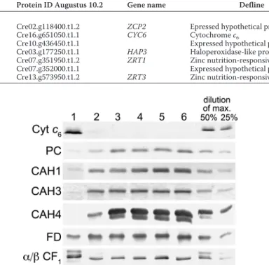
FIGURE 4. Impact of zinc nutrition on abundance of select proteins in the
photosynthetic apparatus. Total soluble and resuspended particulate
frac-tions from Chlamydomonas cultures grown in TAP medium with the various supplemental zinc concentrations were separated on polyacrylamide gels under denaturing conditions. Lane 1, 0 zinc; lane 2, 0.25Mzinc; lane 3, 2.5M zinc; lane 4, 25Mzinc; lane 5, 80Mzinc; lane 6, 125Mzinc. The separated proteins were transferred to nitrocellulose membranes. Proteins of interest were detected by immunoblot analysis with antisera raised against Cyt c6,
plastocyanin (PC), carbonic anhydrase 1 (CAH1), carbonic anhydrase 3 (CAH3), carbonic anhydrase 4 (CAH4), ferredoxin (FD), and the␣ and  subunits of the chloroplast ATP synthase (␣/ CF1). For Cyt c6and the␣ and  subunits of the
chloroplast ATP synthase, dilutions of maximum (dilutions of max) are based on lane 1. For all other proteins, they are based on lane 6.
FIGURE 5. High CO2suppresses the growth phenotype of zinc-limited photoautotrophically grown cells. Chlamydomonas cells (strain CC-4532)
were preconditioned in TP medium and inoculated to a density of 105cells/ml
into TP medium bubbled with filtered air (top) or a mixture of 1% CO2and air
(bottom). Growth was monitored by counting with a hemocytometer. Dupli-cates of zinc-replete (closed symbols) and zinc-limited (open symbols) cultures are shown and represent one of two experimental duplicates.
at INRA Institut National de la Recherche Agronomique on June 13, 2018
http://www.jbc.org/
centration by immunoblotting (Fig. 4). Indeed, plastocyanin is decreased in zinc-deficient cells, and a concomitant increase in cytochrome c6is evident. If excess copper is provided, we can
restore plastocyanin content in proportion to the added copper, but much greater amounts of copper are required to maintain plastocyanin in zinc-limited cells compared with zinc-replete ones (Fig. 6). This supports the idea that zinc-limited cells are functionally copper-deficient. Therefore, we tested whether Crr1 might be required for Chlamydomonas to acclimate to zinc limitation. In zinc-limiting and toxic conditions, growth of
crr1 mutant cultures was lower than growth of the comple-mented strain, CRR1 (Fig. 7A). Growth of the mutant was also slightly decreased in replete conditions, consistent with the role of Crr1 in all states of copper nutrition.
Measurement of fluorescence rise and decay kinetics in crr1
versus CRR1cells revealed that the mutant maintains a high steady state level of fluorescence emission in zinc limitation (Fig. 7B). This is probably due to the inability of the mutant to transfer electrons downstream of PSII to PSI in the photosyn-thetic electron transport chain, most probably as a result of the inability to up-regulate Cyt c6. To support this, we estimated
the stoichiometry of Cyt c6to P700(PSI) in zinc-limited crr1 and
CRR1cultures by quantifying the ratio of redox-active Cyt/P700
using the electrochromic shift assay (Fig. 7C). For the comple-mented strain, Cyt/P700ratios increased from 0.15 in zinc-re-plete conditions to 0.25 in zinc limitation, whereas the mutant showed a slight decrease in the Cyt/P700ratio from 0.15 in
zinc-replete conditions to⬃0.05 in zinc limitation (Fig. 7D). PSI/ PSII ratios were⬃1:1 for both strains in both zinc-replete and zinc-limited conditions (Fig. 7D). These results support the idea that the increase in Cyt c6is necessary to maintain
photo-synthesis in zinc-limited Chlamydomonas cells, even when copper is available in concentrations that should be sufficient to support plastocyanin maintenance.
Trace Metal Quota—To validate the impact of zinc nutrition on copper content, we measured the metal content of
Chlamy-domonascells over a range of supplemental zinc concentrations
(Fig. 8). In the range of concentrations from 25 to 250M, the zinc quota is maintained at ⬃3 ⫻ 107atoms/cell with some
variation that tracked changing external zinc concentrations. When zinc in the medium was less than 25 nM, the quota decreased to⬃1 ⫻ 107atoms/cell, consistent with symptoms of
zinc deficiency and limitation. In contrast, the abundance of iron and copper ions (and possibly also manganese ions) increased with decreasing zinc ion supplementation. This might be attributed to up-regulation of ZIP transporters and the relatively high concentrations of these divalent cations in the medium. In fact, we note that sentinel genes for iron assim-ilation are down-regulated in zinc deficiency, presumably because of higher intracellular iron content. Unexpectedly, given the molecular phenotype of the copper regulon (see above), the copper quota was increased most dramatically. Its intracellular abundance increased by over an order of magni-tude from⬃1–2 ⫻ 107atoms/cell in zinc-replete conditions to
as much as 4⫻ 108atoms/cell in zinc-limiting conditions. This FIGURE 6. Supplemental copper can restore plastocyanin abundance in
zinc-limited cultures. Total soluble and particulate fractions were separated
on a polyacrylamide gel and transferred as described in the legend to Fig. 5. Top, zinc-replete cultures express a constant level of plastocyanin (PC) over the range of 2– 80Msupplemental copper ion concentration. Bottom, zinc-limited cultures show a steady increase in plastocyanin with the increase in supplemental copper. Lane 1, 0 copper; lane 2, 2Mcopper; lane 3, 5M copper; lane 4, 10Mcopper; lane 5, 20Mcopper; lane 6, 40Mcopper; lane 7, 80Mcopper. Note that at 80Mcopper ion concentration, plastocyanin levels in limited cultures are still lower than plastocyanin levels in zinc-replete cultures growing with 2Msupplemental copper.
FIGURE 7. Crr1 is required for acclimation to zinc deficiency. A, crr1 and CRR1 strains grown in TAP medium supplemented with 0, 1, and 250Mzinc. Cells were inoculated at 105cells/ml and photographed after 3 days of
growth. B, Kautsky fluorescence rise and decay kinetics of strains growing in zinc-replete or zinc-limiting conditions normalized to maximum fluorescence intensity measured upon exposure to a saturating light pulse (1100mol of photons m⫺2s⫺1). Squares, CRR1 in replete medium; circles, CRR1 in limited medium; diamonds, crr1 in limited medium; triangles, crr1 in zinc-replete medium. C, Cyt (solid symbols) and P700(open symbols) redox changes
of dark-adapted cells as measured by absorbance changes (same symbols as above); r.u., relative units. D, cytochrome/P700and PSI/PSII ratios. In A–C,
rep-resentative data are shown. For D, the error bars represent S.D. of three inde-pendent measurements.
at INRA Institut National de la Recherche Agronomique on June 13, 2018
http://www.jbc.org/
contrasts strikingly with the tight regulation of copper content observed in previous work (28) and presents a conundrum with respect to the molecular phenotype. We conclude that normal copper homeostasis requires zinc.
DISCUSSION
Although studies investigating acclimation strategies to zinc deficiency have been performed in yeast and other organisms (61), the impact of zinc deficiency on photosynthetic organisms has been underinvestigated. Here, we have defined and charac-terized distinct stages of zinc nutrition in Chlamydomonas because of an excellent infrastructure for understanding trace element homeostasis developed through our prior studies (25). We have distinguished a stage where growth is inhibited, and we refer to that as zinc limitation versus a stage where the zinc-sensing signal transduction pathway is operational to turn on assimilation genes but growth is not affected, and we refer to that as deficiency. As is the case for other organisms, excess zinc results in toxicity, which is also distinguished by growth inhibi-tion (Fig. 1).
In Saccharomyces cerevisiae and Arabidopsis, a primary strategy for the regulation of zinc homeostasis involves the zinc-responsive expression of low and high affinity zinc trans-porters (62, 63). Therefore, these seemed like excellent candi-dates for targets of nutritional zinc signaling as well. A genome survey of ZIP and CDF family transporters (14) led to the iden-tification of five putative Chlamydomonas zinc transporters whose transcripts accumulate in zinc-limiting conditions (Fig. 2). The regulation of these genes was used to identify zinc
con-centrations that induce a change in the abundance of the cor-responding RNAs, and this corresponds to concentrations at which the zinc content is less than⬃2 ⫻ 107atoms/cell, which
defines the typical zinc quota for Chlamydomonas. Very low zinc content in the medium (zinc limitation) results in physio-logical stress, as suggested by the change in cell size (Fig. 1) as well as by the accumulation of Nile Red-staining bodies under zinc-limiting conditions (27).
Based on fluorescence measurements of photosynthetic abil-ity, the impact of zinc limitation did not affect the photosyn-thetic apparatus per se (Fig. 1B). However, the known role of zinc in DNA-binding proteins and CAs participating in the CCM suggested that photosynthetic growth might be affected indirectly as a result of zinc deficiency. RNA-seq technology has been employed and rigorously evaluated in Chlamydomonas, showing this technology to be as quantitative as real-time PCR to evaluate changes in transcript abundance in response to external conditions (35). Therefore, we used it as an exploratory tool to discover pathways that might be impacted by poor zinc nutrition. We identified nearly 700 genes whose expression was significantly changed in zinc-limited versus -replete cells ( sup-plemental Data Set 1).
Photoautotrophic Growth at Air Levels of CO2and Carbonic
Anhydrases—The finding that genes described/discovered in previous studies as being targets of the CCM pathway or Crr1 pointed to inorganic carbon assimilation and copper homeo-stasis, both of which affect photosynthetic performance, as being affected by poor zinc nutrition. In both cases, we
substan-FIGURE 8. Zinc-deficient Chlamydomonas cells accumulate other metal ions. Intracellular metal ion concentrations of zinc (A), copper (B), manganese (C), and iron (D) were quantified as a function of supplemental zinc. Chlamydomonas strain CC-4532 was grown in TAP medium supplemented with 0, 0.025, orM 0.25 zinc (Experiment 1) or with 2.5 25, 80, 125, or 250Mzinc (Experiment 2). For analysis, cells were collected by centrifugation. The cell pellet was washed with 1 mMEDTA to remove extracellular bound metal and dissolved in 30% nitric acid by incubation at 65 °C. The metal content was measured by ICP-MS. Data points represent the average of experimental triplicates.
at INRA Institut National de la Recherche Agronomique on June 13, 2018
http://www.jbc.org/
tiated this by rescuing the zinc starvation phenotype by provi-sion of high CO2or excess copper (Figs. 5 and 6, respectively).
Consistent with an impact on the CCM pathway, we found that photoautotrophic growth of zinc-limited cells was inhibited relative to zinc-replete cells at air levels of CO2(low CO2
con-ditions), but provision of 1% CO2increased photoautotrophic growth rates in zinc-limited cells. The LHCSR1, LHCSR3.1, and
LHCSR3.2genes encoding a subtype of light-harvesting chloro-phyll-binding protein are regulated by CO2as well, but they are inversely regulated in response to zinc limitation relative to CO2limitation (64). This suggests that these genes are under
the control of multiple regulatory factors because they respond differently to low CO2than they do to other nutrient
depriva-tion regimes, including nitrogen and sulfur starvadepriva-tion (65). The most obvious targets of poor zinc nutrition are the car-bonic anhydrases, and we characterized their expression pro-files relative to zinc nutrition. Transcripts for some carbonic anhydrases are increased in zinc limitation; however, immuno-blots for Cah1, Cah3, and Cah4/5 revealed that these proteins decreased in zinc-limiting conditions. We could not detect Cah2 because its abundance is quite low (5%) relative to that of Cah1, and indeed it is not detected in cells grown in uninduced conditions (i.e. high CO2) (59). The discrepancy between
tran-script and protein accumulation may suggest post-transcrip-tional regulation of these genes or a compensatory up-regula-tion of the genes in response to decreased activity. It is possible that the proteins are indeed synthesized, but they might be degraded in the absence of the essential zinc cofactor, or they might be actively degraded as part of a zinc-sparing program (24, 48). Carbonic anhydrases are important for photosynthesis under low CO2conditions based on the effect of enzyme
inhib-itors (acetazolamide and DBS) and based on the phenotype of a mutant lacking lumenal carbonic anhydrase Cah3 (66), and the expression of several CAH genes is increased in cells grown at air levels of CO2as part of the CCM (59). The conditional CO2
-repressed phenotype of zinc-deficient Chlamydomonas cells establishes the carbonic anhydrases as key targets of zinc defi-ciency and therefore, in an environmental context, emphasizes the importance of the ability of diatoms to substitute cobalt or cadmium in the active site of CAs (22, 67) to maintain photo-synthetic growth in face of zinc deficiency.
Influence of Zinc on Copper Nutrition—The similar regula-tion of 23 copper-responsive genes, including 11 Crr1 targets, suggested a connection between copper and zinc metabolism. The fraction of Crr1 targets (50%) is about the same as the fraction of the responding genes in copper-deficient cells (35). Because most of these are genes that are highly responsive and highly expressed, we concluded that zinc-limited cells are sig-naling a slightly copper-deficient status. Nevertheless, when we measured intracellular metal content, we noted the opposite (i.e. severalfold higher copper content in zinc-deficient cells) (Fig. 8). We conclude therefore that cell-associated copper is somehow not accessible to the copper sensor. Further, the cop-per also seems to be inaccessible to plastocyanin. The biosyn-thetic pathway for plastocyanin is normal because when excess copper is added to zinc-deficient cells, this copper can be used for holoplastocyanin synthesis, and the expression of CYC6 is decreased. Therefore, cell-associated copper is generally not
accessible for cuproprotein biosynthesis. The biosynthesis of Cyt c6would, in this case, become essential, which may explain
the non-photosynthetic phenotype of zinc-deficient crr1 cells (Fig. 7).
The advantage, if any, of copper sequestration is not known. The effects of zinc nutrition on organisms is known to be com-plicated by the fact that interactions exist between zinc and copper homeostasis pathways. In vivo, Drosophila metallothio-nein genes are induced by both copper and zinc, suggesting that metallothioneins bind both metals (68). Among humans, serum copper/zinc ratios were significantly higher in patients with malignant lung tumors than in healthy patients and may serve as a diagnostic test in lung cancer patients (69). The copper/ zinc ratio is also significantly increased in disabled elderly patients relative to healthy elderly people and was associated with levels of systemic oxidative stress (70). In the case of patients with Wilson disease, zinc supplementation has been used to counteract copper hyperaccumulation. Mechanistic explanations for this effect are not readily available.
Crr1 and Ccm1 are both zinc-dependent transcription fac-tors (37, 71). The maintenance of transcription facfac-tors Crr1 and Ccm1 in zinc limitation indicates that zinc remains available for their function although carbonic anhydrase function is com-promised, indicative of hierarchical rules governing the alloca-tion of zinc ions. This mechanism has already been docu-mented previously for copper, manganese, and iron in
Chlamydomonas(24, 72). It is tempting to speculate that the COG0523 domain proteins, Zcp1 and Zcp2, may play a role in metal allocation and sparing. The corresponding genes are as highly up-regulated (⬃103-fold and ⬃7 ⫻ 103-fold,
respec-tively) and as highly expressed (⬃6 ⫻ 102RPKM) as are some of
the genes encoding ZRT proteins (e.g. ZRT1 and ZRT3). These are excellent candidates for discovery of the mechanism of reg-ulation or the zinc sensor in Chlamydomonas. There are also a number of proteins of unknown function that are also highly regulated by zinc deficiency, indicative of room for new discov-ery in zinc homeostasis.
Acknowledgments—We thank Rey Martin, Kara Velez and Esper-anza del Rio for assistance during the early phases of the project; Joan Valentine at UCLA for use of the ICP-MS for metal measurements; Dr. James V. Moroney for providing antibodies to Cah1 and Cah4; Dr. Martin Spalding for providing an additional antibody for Cah1; and Dr. Joanna Porankiewicz for providing an antibody to Cah3.
REFERENCES
1. Andreini, C., Banci, L., Bertini, I., and Rosato, A. (2006) Zinc through the three domains of life. J. Proteome Res. 5, 3173–3178
2. Ehrensberger, K. M., and Bird, A. J. (2011) Hammering out details. Regu-lating metal levels in eukaryotes. Trends Biochem. Sci. 36, 524 –531 3. Eide, D. J. (2009) Homeostatic and adaptive responses to zinc deficiency in
Saccharomyces cerevisiae. J. Biol. Chem. 284, 18565–18569
4. Ryu, M. S., Lichten, L. A., Liuzzi, J. P., and Cousins, R. J. (2008) Zinc transporters ZnT1 (Slc30a1), Zip8 (Slc39a8), and Zip10 (Slc39a10) in mouse red blood cells are differentially regulated during erythroid devel-opment and by dietary zinc deficiency. J. Nutr. 138, 2076 –2083 5. Dainty, S. J., Kennedy, C. A., Watt, S., Ba¨hler, J., and Whitehall, S. K. (2008)
Response of Schizosaccharomyces pombe to zinc deficiency. Eukaryot Cell
7,454 – 464
at INRA Institut National de la Recherche Agronomique on June 13, 2018
http://www.jbc.org/
6. Zhao, H., and Eide, D. (1996) The ZRT2 gene encodes the low affinity zinc transporter in Saccharomyces cerevisiae. J. Biol. Chem. 271, 23203–23210 7. Zhao, H., and Eide, D. (1996) The yeast ZRT1 gene encodes the zinc transporter protein of a high affinity uptake system induced by zinc limi-tation. Proc. Natl. Acad. Sci. U.S.A. 93, 2454 –2458
8. Simm, C., Lahner, B., Salt, D., LeFurgey, A., Ingram, P., Yandell, B., and Eide, D. J. (2007) Saccharomyces cerevisiae vacuole in zinc storage and intracellular zinc distribution. Eukaryot. Cell 6, 1166 –1177
9. Roh, H. C., Collier, S., Guthrie, J., Robertson, J. D., and Kornfeld, K. (2012) Lysosome-related organelles in intestinal cells are a zinc storage site in C. elegans. Cell Metab. 15, 88 –99
10. Ballestı´n, R., Molowny, A., Marı´n, M. P., Esteban-Pretel, G., Romero, A. M., Lopez-Garcia, C., Renau-Piqueras, J., and Ponsoda, X. (2011) Eth-anol reduces zincosome formation in cultured astrocytes. Alcohol. Alco-hol. 46,17–25
11. Williams, L. E., Pittman, J. K., and Hall, J. L. (2000) Emerging mechanisms for heavy metal transport in plants. Biochim. Biophys. Acta 1465, 104 –126 12. Kambe, T., and Andrews, G. K. (2009) Novel proteolytic processing of the ectodomain of the zinc transporter ZIP4 (SLC39A4) during zinc defi-ciency is inhibited by acrodermatitis enteropathica mutations. Mol. Cell Biol. 29,129 –139
13. Davis, D. E., Roh, H. C., Deshmukh, K., Bruinsma, J. J., Schneider, D. L., Guthrie, J., Robertson, J. D., and Kornfeld, K. (2009) The cation diffusion facilitator gene cdf-2 mediates zinc metabolism in Caenorhabditis elegans. Genetics 182,1015–1033
14. Blaby-Haas, C. E., and Merchant, S. S. (2012) The ins and outs of algal metal transport. Biochim. Biophys. Acta 1823, 1531–1552
15. Kim, D., Gustin, J. L., Lahner, B., Persans, M. W., Baek, D., Yun, D. J., and Salt, D. E. (2004) The plant CDF family member TgMTP1 from the Ni/Zn hyperaccumulator Thlaspi goesingense acts to enhance efflux of Zn at the plasma membrane when expressed in Saccharomyces cerevisiae. Plant J.
39,237–251
16. Podar, D., Scherer, J., Noordally, Z., Herzyk, P., Nies, D., and Sanders, D. (2012) Metal selectivity determinants in a family of transition metal trans-porters. J. Biol. Chem. 287, 3185–3196
17. Badger, M. R., and Price, G. D. (2003) CO2concentrating mechanisms in
cyanobacteria. Molecular components, their diversity and evolution. J. Exp. Bot. 54,609 – 622
18. Giordano, M., Beardall, J., and Raven, J. A. (2005) CO2concentrating
mechanisms in algae. Mechanisms, environmental modulation, and evo-lution. Annu. Rev. Plant Biol. 56, 99 –131
19. Roberts, K., Granum, E., Leegood, R. C., and Raven, J. A. (2007) Carbon acquisition by diatoms. Photosynth. Res. 93, 79 – 88
20. Wang, Y., Duanmu, D., and Spalding, M. H. (2011) Carbon dioxide con-centrating mechanism in Chlamydomonas reinhardtii. Inorganic carbon transport and CO2recapture. Photosynth. Res. 109, 115–122
21. Lane, T. W., and Morel, F. M. (2000) A biological function for cadmium in marine diatoms. Proc. Natl. Acad. Sci. U.S.A. 97, 4627– 4631
22. Lane, T. W., and Morel, F. M. (2000) Regulation of carbonic anhydrase expression by zinc, cobalt, and carbon dioxide in the marine diatom Thalassiosira weissflogii. Plant Physiol. 123, 345–352
23. Yee, D., and Morel, F. M. M. (1996) In vivo substitution of zinc by cobalt in carbonic anhydrase of a marine diatom. Limnol. Oceanogr. 41, 573–577 24. Merchant, S. S., and Helmann, J. D. (2012) Elemental economy. Microbial
strategies for optimizing growth in the face of nutrient limitation. Adv. Microb. Physiol. 60,91–210
25. Merchant, S. S., Allen, M. D., Kropat, J., Moseley, J. L., Long, J. C., Tottey, S., and Terauchi, A. M. (2006) Between a rock and a hard place. Trace element nutrition in Chlamydomonas. Biochim. Biophys. Acta 1763, 578 –594
26. Harris, E. H. (1989) The Chlamydomonas Sourcebook: A Comprehensive Guide to Biology and Laboratory Use, Academic Press, Inc., San Diego, CA 27. Kropat, J., Hong-Hermesdorf, A., Casero, D., Ent, P., Castruita, M., Pel-legrini, M., Merchant, S. S., and Malasarn, D. (2011) A revised mineral nutrient supplement increases biomass and growth rate in Chlamydomo-nas reinhardtii. Plant J. 66, 770 –780
28. Page, M. D., Kropat, J., Hamel, P. P., and Merchant, S. S. (2009) Two ChlamydomonasCTR copper transporters with a novel Cys-Met motif
are localized to the plasma membrane and function in copper assimilation. Plant Cell 21,928 –943
29. Allen, M. D., del Campo, J. A., Kropat, J., and Merchant, S. S. (2007) FEA1, FEA2, and FRE1, encoding two homologous secreted proteins and a can-didate ferrireductase, are expressed coordinately with FOX1 and FTR1 in iron-deficient Chlamydomonas reinhardtii. Eukaryot. Cell 6, 1841–1852 30. Allen, M. D., Kropat, J., Tottey, S., Del Campo, J. A., and Merchant, S. S. (2007) Manganese deficiency in Chlamydomonas results in loss of photo-system II and MnSOD function, sensitivity to peroxides, and secondary phosphorus and iron deficiency. Plant Physiol. 143, 263–277
31. La Fontaine, S., Quinn, J. M., Nakamoto, S. S., Page, M. D., Göhre, V., Moseley, J. L., Kropat, J., and Merchant, S. (2002) Copper-dependent iron assimilation pathway in the model photosynthetic eukaryote Chlamy-domonas reinhardtii. Eukaryot. Cell 1, 736 –757
32. Kropat, J., Tottey, S., Birkenbihl, R. P., Depège, N., Huijser, P., and Mer-chant, S. (2005) A regulator of nutritional copper signaling in Chlamy-domonasis an SBP domain protein that recognizes the GTAC core of copper response element. Proc. Natl. Acad. Sci. U.S.A. 102, 18730 –18735 33. Yamasaki, H., Hayashi, M., Fukazawa, M., Kobayashi, Y., and Shikanai, T. (2009) SQUAMOSA promoter binding protein-like7 is a central regulator for copper homeostasis in Arabidopsis. Plant Cell 21, 347–361 34. Bernal, M., Casero, D., Singh, V., Wilson, G. T., Grande, A., Yang, H.,
Dodani, S. C., Pellegrini, M., Huijser, P., Connolly, E. L., Merchant, S. S., and Kra¨mer, U. (2012) Transcriptome sequencing identifies SPL7-regu-lated copper acquisition genes FRO4/FRO5 and the copper dependence of iron homeostasis in Arabidopsis. Plant Cell 24, 738 –761
35. Castruita, M., Casero, D., Karpowicz, S. J., Kropat, J., Vieler, A., Hsieh, S. I., Yan, W., Cokus, S., Loo, J. A., Benning, C., Pellegrini, M., and Merchant, S. S. (2011) Systems biology approach in Chlamydomonas reveals connec-tions between copper nutrition and multiple metabolic steps. Plant Cell
23,1273–1292
36. Quinn, J. M., Barraco, P., Eriksson, M., and Merchant, S. (2000) Coordi-nate copper- and oxygen-responsive Cyc6 and Cpx1 expression in Chla-mydomonas is mediated by the same element. J. Biol. Chem. 275, 6080 – 6089
37. Sommer, F., Kropat, J., Malasarn, D., Grossoehme, N. E., Chen, X., Giedroc, D. P., and Merchant, S. S. (2010) The CRR1 nutritional copper sensor in Chlamydomonas contains two distinct metal-responsive do-mains. Plant Cell 22, 4098 – 4113
38. Merchant, S., and Bogorad, L. (1986) Regulation by copper of the expres-sion of plastocyanin and cytochrome c552in Chlamydomonas reinhardtii.
Mol. Cell Biol. 6,462– 469
39. Quinn, J. M., and Merchant, S. (1998) Copper-responsive gene expression during adaptation to copper deficiency. Methods Enzymol. 297, 263–279 40. Joliot, P., and Joliot, A. (2002) Cyclic electron transfer in plant leaf. Proc.
Natl. Acad. Sci. U.S.A. 99,10209 –10214
41. Schloss, J. A. (1990) A Chlamydomonas gene encodes a G protein sub-unit-like polypeptide. Mol. Gen. Genet. 221, 443– 452
42. Livak, K. J., and Schmittgen, T. D. (2001) Analysis of relative gene expres-sion data using real-time quantitative PCR and the 2⫺⌬⌬CT method.
Methods 25,402– 408
43. Langmead, B., Trapnell, C., Pop, M., and Salzberg, S. L. (2009) Ultrafast and memory-efficient alignment of short DNA sequences to the human genome. Genome Biol. 10, R25
44. Mortazavi, A., Williams, B. A., McCue, K., Schaeffer, L., and Wold, B. (2008) Mapping and quantifying mammalian transcriptomes by RNA-Seq. Nat. Methods 5, 621– 628
45. Anders, S., and Huber, W. (2010) Differential expression analysis for se-quence count data. Genome Biol. 11, R106
46. Benjamini, Y., and Hochberg, Y. (1995) Controlling the false discovery rate. A practical and powerful approach to multiple testing. J. R. Stat. Soc. Ser. B 57,289 –300
47. Howe, G., and Merchant, S. (1992) The biosynthesis of membrane and soluble plastidic c-type cytochromes of Chlamydomonas reinhardtii is dependent on multiple common gene products. EMBO J. 11, 2789 –2801 48. Hsieh, S. I., Castruita, M., Malasarn, D., Urzica, E., Erde, J., Page, M. D., Yamasaki, H., Casero, D., Pellegrini, M., Merchant, S. S., and Loo, J. A. (2013) The proteome of copper, iron, zinc, and manganese micronutrient
at INRA Institut National de la Recherche Agronomique on June 13, 2018
http://www.jbc.org/