Relevance of intraocular involvement in the management of primary central nervous system lymphomas
8
0
0
Texte intégral
(2) 532 a non-specific uveitis refractory to topical or systemic corticosteroids, associated with floaters or campimeter deficit, which precedes cerebral symptoms by months or years [4–6, 8]. The use of slit-lamp examination, indirect ophthalmoscopy and ophthalmic ultrasonography during the staging of PCNSL patients can aid in detection of asymptomatic ocular localisation in >5% of cases [9, 10]. The optimal treatment for IOL remains to be defined. With the use of radiotherapy (RT) alone in symptomatic patients, almost all subjects developed early CNS progression and died [11–13]. Promising anecdotal results in a few case reports and small retrospective series treated with chemotherapy (CHT) have been reported [6, 11, 12, 14–17]. A small number of cases of IOL have also been included in prospective trials evaluating new strategies in PCNSL, with conflicting and disappointing outcomes [3, 18]. Ultimately, the efficacy of CHT is dependent on intraocular pharmacokinetics, which are not well understood [19]. This paper reports the clinical features, behaviour and therapeutic results of a series of 22 patients with IOL. These patients were identified in an international multicentre series of 378 immunocompetent patients with PCNSL. The analysis focuses on the clinical and therapeutic variables related to ocular disease control and the impact of ocular control on survival.. Patients and methods Study group Clinical and therapeutic data were reviewed in patients with IOL who were included in a retrospective series of 378 PCNSL patients (1980– 1999) from 23 cancer centres of five different countries. A questionnaire requesting information about patient characteristics, clinical presentation, diagnosis, staging, planned and performed treatment, objective response, site and date of relapse, second-line treatment, neurotoxicity and survival was sent to each centre participating in the International Extranodal Lymphoma Study Group. Selection criteria of that series were: (i) histological or cytological diagnosis of lymphoma; (ii) disease localised exclusively in the brain, cranial nerves, meninges or eyes and (iii) no evidence of HIV-1 infection or other immunodeficiencies. Any age, performance status, histotype and treatment modality were considered. Clinical staging work-up included at least a total-body computed tomography scan and bone marrow biopsy. Staging procedures included slit-lamp examination of the eyes in 170 cases (45%) (Table 1). Twenty-two patients (13%) had positive results to the slit-lamp examination and these patients constitute the study group. Twenty-one patients had concomitant ocular and brain lesions, while one patient had only ocular disease. Histotype was defined according to the Working Formulation Classification [20]. Histological sample was obtained by stereotactic biopsy in 17 cases, subtotal resection of brain lesions in two, cerebrospinal fluid (CSF) cytological examination in two and vitrectomy in one case.. Therapeutic management Planned treatment (Table 2) included CHT followed by RT (CHT→RT) in 13 patients, CHT alone in three and primary RT in five (RT→CHT in two, RT alone in three). One patient did not receive any treatment. CHT. included high-dose (HD) methotrexate (HD-MTX) in 12 cases (dose ≥3 g/m2/course in 10 cases) and CHOP (cyclophosphamide/adriamycin/ vincristine/prednisone) regimen in six. HD-MTX was combined with (i) HD-cytarabine, doxorubicin, cyclophosphamide and vincristine in six patients; (ii) procarbazine and vincristine in three cases; (iii) doxorubicin, cyclophosphamide and vincristine in two and (iv) thiotepa, nitrosourea and etoposide in one. Intrathecal CHT was used in 10 cases, MTX and cytarabine in eight, MTX alone in two. All patients received steroid therapy with a daily dose of 8–24 mg dexamethasone. Planned RT for patients with brain lesions consisted of whole brain irradiation (33 ± 9 Gy, range 20–50 Gy), followed or not followed by a tumour bed boost, with a mean tumour-bed dose of 45 ± 5 Gy (range 30– 60 Gy); one patient without brain lesions received irradiation to the orbits only (50 Gy). The irradiation of the posterior two-thirds of the eyes was a part of the planned first-line treatment in 15 patients (37 ± 6 Gy). All patients were treated with 4–6 MeV photons and standard daily fractionation (1.8–2 Gy). All but four patients (case Nos 10–13) completed the planned treatment (Table 2). It had been planned that these patients were to receive CHT→RT, but they were not irradiated due to early progression, toxic death (n = 2) or refusal. It had been planned for all four of these patients to receive ocular irradiation.. Statistical considerations Response rates and clinical characteristics of the different groups of patients were compared using the χ2 test or Fisher’s exact test for categorical variables, according to the sample size. Survival curves were generated by the Kaplan–Meier method. Overall survival (OS) was calculated from the date of histological diagnosis to death or last date of followup, while failure-free survival (FFS) was calculated from the first day of treatment to relapse, progression, death or to last date of follow-up. The time to ocular relapse was analysed with ocular failure as the event. Ocular failure was defined as residual disease, relapse or progression, associated or not with brain recurrence. Cerebral relapse or progression without concomitant ocular disease was considered censored for this analysis. Survival curves were compared through the log-rank test. All probability values were two-sided.. Results Patient characteristics The main IOL patients’ characteristics and the comparison of these patients with patients who had negative slit-lamp examination results are summarised in Table 1. The median age of the 22 IOL patients was 54 years (range 16–74 years), with a male/female ratio of 1.5. Two patients had stage B symptoms. Ten patients complained of symptomatic ocular disease, while eye involvement was asymptomatic in 12 cases. One patient had a prior malignancy (melanoma). The duration period between symptomatic onset and histological diagnosis ranged between 1 month and 12 months, with a median of 3 months. In 20 cases, an intermediate or high-grade lymphoma was diagnosed, with diffuse large B-cell lymphoma being the most common histological subtype (n = 17); the other histotypes were B-lymphoblastic (n = 1), Burkitt-like (n =1) and unclassifiable (n = 3). Eight patients (36%) had multiple lesions. Deep structures of the brain, that is basal ganglia,.
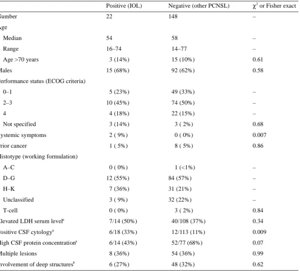
(3) 533 Table 1. Patients’ characteristics according to the result of the slit-lamp examination Positive (IOL). Negative (other PCNSL). χ2 or Fisher exact. 22. 148. –. Median. 54. 58. –. Range. 16–74. 14–77. –. Number Age. Age >70 years. 3 (14%). 15 (10%). 0.61. 15 (68%). 92 (62%). 0.58. 0–1. 5 (23%). 49 (33%). –. 2–3. Males Performance status (ECOG criteria). 10 (45%). 74 (50%). –. 4. 4 (18%). 22 (15%). –. Not specified. 3 (14%). 3 ( 2%). 0.68. Systemic symptoms. 2 ( 9%). 0 ( 0%). 0.007. Prior cancer. 1 ( 5%). 8 ( 5%). 0.86. 1 (<1%). –. Histotype (working formulation) A–C. 0 ( 0%). D–G. 12 (55%). 84 (57%). –. H–K. 7 (36%). 31 (21%). –. Unclassified. 3 ( 9%). 32 (22%). –. T-cell. 0 ( 0%). Elevated LDH serum levela. 3 ( 2%). 0.84. 7/14 (50%). 40/108 (37%). 0.34. Positive CSF cytology. 6/18 (33%). 12/113 (11%). 0.009. High CSF protein concentrationa. 6/14 (43%). 52/77 (68%). 0.07. Multiple lesions. 8 (36%). 54 (36%). 0.99. Involvement of deep structuresb. 6 (27%). 48 (32%). 0.62. a. IOL, intraocular lymphoma; PCNSL, primary central nervous system lymphomas; LDH, lactate dehydrogenase; CSF, cerebrospinal fluid; ECOG, Eastern Cooperative Oncology Group. a Relationship between the number of positive cases and the total number of assessed patients. The cut-off of normal protein CSF level was 45 mg/dl in patients ≤60 years and 60 mg/dl in patients >60 years. b Cases of involvement of basal ganglia and/or brain stem and/or cerebellum.. corpus callosum, brain stem and/or cerebellum, were involved in six patients. CSF cytological examination was positive in six (33%) of the 18 assessed cases. A statistically significant correlation between ocular involvement and systemic symptoms and positive CSF cytological examination was observed (Table 1).. Objective response and relapse rates Fifteen of 21 treated patients (71%) achieved an objective response (Table 2), which was complete in 13 cases (complete remission rate 62%). Four patients experienced progression and two died of toxicity during primary CHT. The two patients who had partial remission (case Nos 5 and 7) had residual disease in the brain, but not in the eyes. They had been treated with CHT→RT, including ocular irradiation. Sixteen patients experienced a failure (Table 2): two toxic deaths. (case Nos 11 and 13), four cases of progressive disease during treatment and 10 relapses after an initial objective response. Lymphoma involved the primary sites of disease in all failed cases: the brain in seven cases, brain plus eyes in six (including cases of toxic death), eyes in two and brain plus meninges in one. No cases of systemic dissemination were observed. Table 3 summarises the relapse rates and sites in patients grouped according to the result of the slit-lamp examination. The ocular relapse rate is clearly higher in IOL patients with respect to patients who had negative slit-lamp examination at diagnosis. The median FFS for the entire series was 10 months, with a 2-year FFS of 34 ± 10%. Seven of the 14 patients who experienced progression or relapse received a salvage therapy: CHT alone in six cases and ocular enucleation in one. The salvage CHT regimen consisted of HD-cytarabine in three cases, and HD-MTX, temozolomide and HD-MTX plus HD-cytarabine.
(4) 534 Table 2. Planned and performed treatment, objective response, relapse and survival in IOL patients Case. Planned treatment. Performed CHT treatment regimen. MTX IT Ocular dosea CHT planned. RT (Gy). OR. Ocular TTF remission. Failure site. OS. Cause of death. performed. 1. CHT→RT CHT→RT MTX. 6. –. 30. 30. CR. yes. 39+. –. 39+. –. 2. CHT→RT CHT→RT CHOP. 0. +. 30. 30. CR. yes. 96+. –. 96+. –. 3. CHT→RT CHT→RT MTX. 6. –. 30. 30. CR. yes. 21. brain. 23. neurotoxicity. 4. CHT→RT CHT→RT MTX–araC 3. +. 40. 40. CR. yes. 62+. –. 62+. –. 5. CHT→RT CHT→RT CHOP. 0. –. 40. 40. PR. yes. 19. brain. 23. lymphoma. 6. CHT→RT CHT→RT MTX. 3. –. 40. 40. CR. yes. 17. eyes. 90+. –. 7. CHT→RT CHT→RT MTX–araC 3. +. 40. 40. PR. yes. 54. brain. 55. lymphoma. 8. CHT→RT CHT→RT MTX. +. 0. 0. PD. no. 7. 7. lymphoma. 9. CHT→RT CHT→RT MTX–araC 2. +. 0. 0. CR. yes. 26. brain. 30. lymphoma. 10. CHT→RT CHT. CHOP. 0. –. 30. 0. CR. yes. 5. brain. 7. lymphoma. 11. CHT→RT CHT. MTX-araC 3. +. 40. 0. NA. NA. 4. brain + eyes. 4. toxicity. 12. CHT→RT CHT. MTX. 3. +. 30. 0. PD. no. 3. brain + eyes. 3. lymphoma. 13. CHT→RT CHT. MTX-araC 3. +. 40. 0. NA. NA. 1. brain + eyes. 1. toxicity. 14. CHT. CHT. CHOP. 0. –. 0. 0. PD. no. 1. brain + eyes. 14. lymphoma. 15. CHT. CHT. CHOP. 0. –. 0. 0. CR. yes. 18. brain. 21. lymphoma. 16. CHT. CHT. MTX. 8. +. 0. 0. CR. yes. 12+. –. 12+. –. 17. RT→CHT RT→CHT MTX–araC 3. –. 30. 30. CR. yes. 13. brain + meninges 15. lymphoma. 18. RT→CHT RT→CHT CHOP. 0. +. 0. 0. CR. yes. 40. brain. 53+. –. 19b. RT. RT. –. –. –. 50. 50. CR. yes. 14+. –. 14+. –. 20. RT. RT. –. –. –. 40. 40. CR. yes. 4. eyes. 10. lymphoma. 21. RT. RT. –. –. –. 40. 40. PD. no. 1. brain + eyes. 1. lymphoma. 22. None. none. –. –. –. –. –. –. –. 1. brain + eyes. 1. lymphoma. 1.5. brain + eyes. –, not applicable; ara-C, high-dose cytarabine; CHOP, cyclophosphamide/adriamycin/vincristine/prednisone; CHT, chemotherapy; CR,complete remission; IT CHT, intrathecal chemotherapy; MTX, high-dose methotrexate; NA, not assessed; OR, objective response; OS, overall survival; PD, progressive disease; PR, partial response; RT, radiotherapy; TTF, time to treatment failure. a MTX dose is expressed in g/m2. b Patient with exclusive ocular disease.. in the other three cases, respectively. Three of these retreated patients achieved a second objective response (case Nos 3, 6 and 18). Median survival after progression was 2 months for the 14 patients with relapsed or progressive disease, and 4 months for retreated patients. Case No. 3 died of treatment-related neurotoxicity while in second complete remission.. Ocular disease control and OS The patient with isolated IOL (case No. 19) was blind at diagnosis. He achieved a complete remission following the irradiation of the orbits with 50 Gy but without symptomatic improvement, and was relapse-free after 14 months from RT. Ocular failure was detected among eight patients with concomitant brain and ocular lymphoma, with a 2-year time to ocular relapse of 59 ± 11%. Excluding the cases of toxic death, ocular failure was observed in one of the eight patients treated with combined CHT and RT with a field that included the. eyes, in three of the eight patients treated with CHT with or without RT with a field that excluded the eyes and in both patients treated with RT with a field that included the eyes but without CHT (χ2; P = 0.09). The median time to ocular failure for these groups was 20, 15 and 2.5 months, respectively. The poor disease control observed in patients treated with RT alone does not seem to be attributable to bias selection since both patients were younger than 65 years and had ECOG performance status <2. Depending on which drug was used, ocular failure was observed in three of the six patients receiving HD-MTX and in one of the six patients treated with CHOP regimen, while none of the patients treated with a HD-MTX– HD-cytarabine combination experienced an ocular relapse (χ2; P = 0.09). Seven patients are alive [six with no evidence of disease (NED)] with a median follow-up of 53 months (range 12–96 months). Five of them had been treated with RT including ocular irradiation. Actuarial survival of the entire series was.
(5) 535 Table 3. Comparison of relapse rate and sites in patients grouped according to the result of the slit-lamp examination. Relapse/progression. Positive (n = 22). Negative (n = 148). Not assessed (n = 208). 14 (64%). 85 (46%). 137 (66%). 7 (50%). 75 (88%). 118 (86%). a. Site of relapse Brain. a. Brain + meninges. 1 (7%). 5 (6%). 7 (5%). Brain + systemic. 0 (0%). 3 (4%). 1 (1%). Brain + eyes. 4 (29%). 1 (1%). 1 (1%). Meninges. 0 (0%). 1 (1%). 1 (1%). Eyes. 2 (14%). 0 (0%). 1 (1%). Systemic relapse. 0 (0%). 0 (0%). 8 (5%). Toxic deaths. 2 (9%). 9 (6%). 9 (4%). Untreated patients. 1 (5%). 7 (5%). 11 (5%). Percentages with regard to the proportion of the total number of relapses.. similar to those of PCNSL patients with negative slit-lamp examination results (2-year OS: 39 ± 11% compared with 42 ± 4%; P = 0.31; Figure 1). The detection of ocular relapse was associated with a shorter survival (2-year OS: 12 ± 11% compared with 55 ± 14%; P= 0.01). Excluding toxic deaths, five of the six patients with ocular relapse and seven of the 13 patients without ocular relapse died (2-year OS: 16: ± 15% and 55 ± 14%; P = 0.03). According to the therapeutic strategy, median OS was 47+, 9.5+ and 10 months, respectively for. Figure 1. Overall survival curves for patients grouped according to the results of the slit-lamp examination. Twenty-two intraocular lymphoma patients (solid line) were compared with 148 patients without ocular disease (broken line); P = 0.31.. the eight patients treated with combined CHT and RT with a field that included the eyes, the 10 patients treated with CHT with or without RT with a field that excluded the eyes and the three patients treated with RT alone with a field that included the eyes.. Discussion Intraocular lymphoma (IOL) is a rare malignancy with variable clinical presentation and behaviour considering that ocular involvement can be detected in different phases of PCNSL. Only a few small series of IOLs exist, the largest of which [7, 21] described a heterogeneous group of patients, including cases with exclusive or ‘pure’ IOL, ocular presentation followed by brain dissemination, concomitant oculo-cerebral lymphoma, or brain lymphoma followed by eye failure. In the present series, only cases with ocular disease at diagnosis were considered. Among 170 PCNSL patients examined with a slitlamp during staging, ocular involvement was detected in 22 (13%). The study of the relevance of ocular involvement in the management of PCNSL is strongly limited by some difficulties in diagnosis, staging and assessment of therapeutic response and failure. Diagnosis of IOL is not easy even when immunohistochemical techniques are used [22], and efforts to improve the diagnostic yield of vitrectomy include techniques of molecular biology, determination of intraocular levels of interleukin-10 and flow cytometry [23]. However, these are not standardised procedures currently used in PCNSL staging, and slit-lamp examination, even if not completely accurate, remains the most commonly used procedure for ocular evaluation in these malignancies. In our patients, ocular involvement was nearly always associated with brain lesions; only one patient had disease exclusively located in the eyes. IOL patients had similar clinical characteristics with respect to the.
(6) 536 remaining PCNSL cases with negative slit-lamp examination, while a significantly positive association between IOL and systemic symptoms and CSF cytological status was observed. However, this finding should be taken into account with caution considering the small number of cases with systemic symptoms observed in this series. From a prognostic standpoint, no significant difference in survival was observed between patients with or without ocular involvement at diagnosis. This seems to confirm that, with the presently available therapeutic strategies, brain disease control is the main prognostic-determining factor in PCNSL patients. The optimal treatment for IOL has not yet been defined. In the past, patients with symptomatic disease were treated with RT alone, but almost all patients developed early CNS progression and died, with a median OS of 20 months and a 2-year OS lower than 20% [11–13]. A few case reports and small retrospective series alluded to the efficacy of CHT, and promising anecdotal results using HD-cytarabine, HD-MTX, procarbazine and nitrosoureas have been reported [6, 11, 12, 14–17]. In six patients with IOL, including one case of ocular relapse from a systemic non-Hodgkin’s lymphoma, HDcytarabine produced one complete and four partial responses [17]. Therapeutic levels in intraocular fluids and prolonged objective tumour regression following systemic high doses of this drug have been documented in a case of relapsed IOL [16]. The use of this drug in combination with ocular irradiation has been recommended in IOL patients [16]. In the present series, none of the four patients who completed the planned treatment, including HD-cytarabine-containing CHT, experienced ocular relapse, with a near significantly better ocular disease control with respect to the other drugs. However, no definitive conclusions could be drawn because these patients also received ocular irradiation as part of the first-line treatment. Data suggesting the efficacy of HD-cytarabine in IOL patients should be confirmed in larger series. Some prospective trials evaluating new strategies in PCNSL included cases of IOL [18, 21, 24–26]. In those series, IOL were managed like other PCNSLs, with only minor differences in the extent of RT field and dose. However, IOL cases have been analysed separately from the rest of PCNSLs cases in just a few series. The efficacy of CHT seems to be strongly conditioned by intraocular pharmacokinetics, which are not well understood. The intraocular concentrations of MTX and cytarabine after intravenous administration have been analysed in a few case reports, with conflicting results [16, 17, 27]. Four hours after an intravenous infusion of MTX 8 g/m2, the vitreous concentration of MTX was 100-fold lower than in the serum [19]. These difficulties in achieving therapeutic concentrations of MTX in the vitreous humour seem to be confirmed by clinical data. In fact, a 27% response rate and a 90% treatment failure rate after first-line CHT containing HD-MTX and/or cytarabine were reported [3]. In five patients with cerebral and ocular lymphoma treated with HD-MTXcontaining CHT, despite the fact that brain lesions completely disappeared in all patients, persistent ocular disease was. observed in two cases, and relapses involving the eyes were observed in the other two [18]. The poor results obtained with conventional CHT have prompted investigators to search for alternative approaches for IOL patients. HD-CHT followed by autologous bone marrow transplantation [3, 21] and intravitreal injections of MTX, with or without thiotepa [28], have been proposed. Intriguing results have been reported in five patients with relapsed IOL treated with HD-CHT with thiotepa, busulfan and cyclophosphamide and autologous bone marrow transplantation [3]. All five patients achieved a complete remission and were alive (three NED) after a median follow-up of 26 months. More recently, a median survival after relapse of 53+ months was reported in 11 patients with relapsed or refractory IOL treated with the same strategy [21]. On the other hand, promising results with reduced morbidity were reported in four patients treated with repeated intravitreal injections of MTX 400 µg [29]. The addition of RT, including the irradiation of both orbits with 30 Gy, to HD-MTX-based CHT has been associated with a higher response rate [26, 30]. Although most of these patients experienced an aggressive relapse involving brain and meninges, no cases of ocular recurrence were reported. In another series, 11 of 12 cases of IOL treated with different strategies developed brain disease with a median time between onset of IOL and onset of brain disease of 14 months (range 1–85 months) [7]. Five of these patients had been treated with ocular irradiation as a part of first-line treatment: one of them also received prophylactic whole brain RT and was relapse-free at 18 months; two patients relapsed in the eyes and, after 17 and 29 months, in the brain; and two had relapses only in the brain at 12 and 85 months from treatment. Median survival of patients treated with ocular irradiation at diagnosis was 19+ months compared with 37 months for the rest [7]. Ocular irradiation as exclusive salvage therapy also seems to be an effective approach in patients with recurrent IOL, with a median post-failure survival of 20+ months for 13 previously reported cases [7, 18, 31, 32]. Considering this and the above-mentioned difficulties in achieving intraocular therapeutic levels of the most commonly used drugs, RT has been proposed as the first-line therapy for IOL [19]. The irradiation of the posterior two-thirds of the ocular globes with a dose between 35 and 45 Gy has been recommended [5, 6]. Higher doses should not been used for treating the full bilateral orbits considering the potential risk of severe toxicity. More recent experiences suggest irradiation of the entire orbit up to 20 Gy, followed by an additional 10 Gy after shielding the anterior chambers of the eyes [26, 30]. Considering the high frequency of bilateral involvement, the irradiation of both eyes has been recommended, even in the presence of monolateral disease. In the present series, ocular irradiation was associated with a better ocular disease control. In particular, the combination of MTX–cytarabine-containing CHT with ocular irradiation was associated with a lower ocular relapse rate, and the positive effect on ocular disease control of ocular irradiation suggests.
(7) 537 that it has a potential role in survival. In effect, the detection of ocular relapse was significantly associated with shorter survival compared with those patients without ocular failure. Considering the retrospective nature of the present study, no reliable data regarding ocular toxicity are available. However, in the authors’ experience [33], the irradiation of the posterior two-thirds of the eyes with 30 Gy has not been associated with a significant ocular toxicity in PCNSL patients. Considering the presence of concomitant brain lesions, at either diagnosis or relapse, in the vast majority of IOL cases, and the similarity in clinical characteristics and survival between IOL patients and the rest of PCNSL cases, therapeutic management for IOL patients should follow the same guidelines used for other PCNSL patients. Despite the retrospective nature and the relatively small number of patients included in the present series, our results seem to suggest that the combination of CHT and RT with a field including the eyes could have a positive effect on ocular disease control, which is a limiting factor significantly associated with survival. It is noteworthy that the survival benefit related to ocular irradiation may be biased by the high incidence of concomitant brain failure, which is the most important recognised prognostic event in PCNSL. In conclusion, IOL patients should be treated with a HD-MTX-based CHT followed by irradiation of the whole brain with 36–40 Gy and the posterior twothirds of both eyes with 30 Gy and a standard fractionation. Future advances in primary CHT improving brain disease control may permit a more objective evaluation of the potential role of irradiation of the eyes in PCNSL patients. This issue may gain major importance in modern phase II trials designed to assess the efficacy of CHT as exclusive treatment, to reduce the incidence of treatment-related neurotoxicity. In view of the difficulties of systemic CHT in obtaining remission in vitreous humor and the potential role of ocular irradiation, the use of CHT as exclusive treatment could be critically reconsidered in PCNSL patients with ocular disease.. Acknowledgements Other participants (in order of contribution): Pierre Biron, Centre Léon Bérard, Lyon, France; Achille Ambrosetti, Ospedale Policlinico di Borgo Roma, Verona, Italy; Paolo Iuzzolino, Ospedale Civile Maggiore Az. Ospedaliera, Verona, Italy; Christine Chevreau, Centre Claudius Regaud, Toulouse, France; Vittorio Vavassori and Michele Cerati, Ospedale di Circolo Fondazione Macchi, Varese, Italy; Houchingue Eghbali, Institut Bergonié, Bordeaux, France; Reda Bouabdallah, Institut Paoli Calmette, Marseille, France; Thierry Conroy, Centre Alexis Vautrin, Nancy, France; Michele Spina and Antonino Carbone, Centro di Riferimento Oncologico, National Cancer Institute, Aviano, Italy; Joachim Weis, Pathologisches Institut der Universität Bern, Bern, Switzerland; Dominique Dramais-Marcel, Centre G-F Leclerc, Dijon, France; Andrea Rossi, Tiziano Barbui and Teresio Motta,. Ospedali Riuniti di Bergamo, Bergamo, Italy; Alexander Pivnik, Haematological Centre of Russian Academy of Medical Sciences, Moscow, Russia; Annarita Conconi, Istituto Oncologico della Svizzera Italiana, Osp. San Giovanni, Bellinzona, Switzerland; Ennio Pedrinis, Istituto Cantonale di Patologia, Locarno, Switzerland; Gian Marco Aondio and Mario Milani, Azienda Ospedaliera di Lecco, Lecco, Italy; Jean Philippe Wagner, Centre Paul Strauss, Strasbourg, France; Anne Marie Le Mevel, Centre René Gauducheau, Nantes, France; Enrico Orvieto and Fabio Ferrarese, Ospedale Regionale di Treviso, Treviso, Italy; Henry Gomez and Juvenal Sanchez, Instituto de Enfermedades Neoplásicas, Lima, Peru; Anne Jouvet, Hopital Neurologique, Lyon, France; Bertrand Coiffier, Centre Hospitalier Lyon-Sud, Pierre Bénite, France; Elisabeth Baumelou, Hopital Foch, Suresne, France; Antoine Thyss, Centre Antoine Lacassagne, Nice, France; Liliana Devizzi, Istituto Nazionale dei Tumori, Milan, Italy; Stefania Dell’Oro, San Raffaele H Scientific Institute, Milan, Italy.. References 1. Deangelis LM, Yahalom J, Heinemann MH et al. Primary CNS lymphoma: combined treatment with chemotherapy and radiotherapy. Neurology 1990; 40: 80–86. 2. Ferreri AJ, Reni M, Villa E. Primary central nervous system lymphoma in immunocompetent patients. Cancer Treat Rev 1995; 21: 415–446. 3. Soussain C, Merle-Beral H, Reux I et al. A single-center study of 11 patients with intraocular lymphoma treated with conventional chemotherapy followed by high-dose chemotherapy and autologous bone marrow transplantation in five cases. Leuk Lymphoma 1996; 23: 339–345. 4. Akpek EK, Ahmed I, Hochberg FH et al. Intraocular–central nervous system lymphoma: clinical features, diagnosis, and outcomes. Ophthalmology 1999; 106: 1805–1810. 5. Rockwood EJ, Zakov ZN, Bay JW. Combined malignant lymphoma of the eye and CNS (reticulum-cell sarcoma). Report of three cases. J Neurosurg 1984; 61: 369–374. 6. Qualman SJ, Mendelsohn G, Mann RB, Green WR. Intraocular lymphomas. Natural history based on a clinicopathologic study of eight cases and review of the literature. Cancer 1983; 52: 878–886. 7. Peterson K, Gordon KB, Heinemann MH, Deangelis LM. The clinical spectrum of ocular lymphoma. Cancer 1993; 72: 843–849. 8. Cassoux N, Merle-Beral H, Leblond V et al. Ocular and central nervous system lymphoma: clinical features and diagnosis. Ocul Immunol Inflamm 2000; 8: 243–250. 9. Ursea R, Heinemann MH, Silverman RH et al. Ophthalmic, ultrasonographic findings in primary central nervous system lymphoma with ocular involvement. Retina 1997; 17: 118–123. 10. O’Neill BP, Illig JJ. Primary central nervous system lymphoma. Mayo Clin Proc 1989; 64: 1005–1020. 11. Char DH, Ljung BM, Deschenes J, Miller TR. Intraocular lymphoma: immunological and cytological analysis. Br J Ophthalmol 1988; 72: 905–911. 12. Freeman LN, Schachat AP, Knox DL et al. Clinical features, laboratory investigations, and survival in ocular reticulum cell sarcoma. Ophthalmology 1987; 94: 1631–1639..
(8) 538 13. Fine HA, Mayer RJ. Primary central nervous system lymphoma. Ann Intern Med 1993; 119: 1093–1104. 14. Whitcup SM, de Smet MD, Rubin BI et al. Intraocular lymphoma. Clinical and histopathologic diagnosis. Ophthalmology 1993; 100: 1399–1406. 15. Siegel MJ, Dalton J, Friedman AH et al. Ten-year experience with primary ocular ‘reticulum cell sarcoma’ (large cell non-Hodgkin’s lymphoma). Br J Ophthalmol 1989; 73: 342–346. 16. Baumann MA, Ritch PS, Hande KR et al. Treatment of intraocular lymphoma with high-dose Ara-C. Cancer 1986; 57: 1273–1275. 17. Strauchen JA, Dalton J, Friedman AH. Chemotherapy in the management of intraocular lymphoma. Cancer 1989; 63: 1918–1921. 18. Sandor V, Stark-Vancs V, Pearson D et al. Phase II trial of chemotherapy alone for primary CNS and intraocular lymphoma. J Clin Oncol 1998; 16: 3000–3006. 19. Henson JW, Yang J, Batchelor T. Intraocular methotrexate level after high-dose intravenous infusion. J Clin Oncol 1999; 17: 1329. 20. National Cancer Institute sponsored study of classifications of non-Hodgkin’s lymphomas: summary and description of a working formulation for clinical usage. The Non-Hodgkin’s Lymphoma Pathologic Classification Project. Cancer 1982; 49: 2112–2135. 21. Soussain C, Suzan F, Hoang-Xuan K et al. Results of intensive chemotherapy followed by hematopoietic stem-cell rescue in 22 patients with refractory or recurrent primary CNS lymphoma or intraocular lymphoma. J Clin Oncol 2001; 19: 742–749. 22. Wilson DJ, Braziel R, Rosenbaum JT. Intraocular lymphoma. Immunopathologic analysis of vitreous biopsy specimens. Arch Ophthalmol 1992; 110: 1455–1458. 23. Whitcup SM, Chan CC, Buggage RR et al. Improving the diagnostic yield of vitrectomy for intraocular lymphoma. Arch Ophthalmol 2000; 118: 446. 24. Schultz C, Scott C, Sherman W et al. Preirradiation chemotherapy with cyclophosphamide, doxorubicin, vincristine, and dexamethasone for primary CNS lymphomas: initial report of Radiation. Therapy Oncology Group Protocol 88-06. J Clin Oncol 1996; 14: 556–564. 25. Blay JY, Bouhour D, Carrie C et al. The C5R protocol: a regimen of high-dose chemotherapy and radiotherapy in primary cerebral nonHodgkin’s lymphoma of patients with no known cause of immunosuppression. Blood 1995; 86: 2922–2929. 26. Glass J, Gruber ML, Cher L, Hochberg FH. Preirradiation methotrexate chemotherapy of primary central nervous system lymphoma: long-term outcome. J Neurosurg 1994; 81: 188–195. 27. Stark-Vancs V, Setser A, Kohler D et al. Combination chemotherapy in the treatment of primary central nervous system and intraocular lymphoma. Proc Am Soc Clin Oncol 1995; 14: A306 (Abstr). 28. de Smet M, Vancs VS, Kohler D et al. Intravitreal chemotherapy for the treatment of recurrent intraocular lymphoma. Br J Ophthalmol 1999; 83: 448–451. 29. Fishburne BC, Wilson DJ, Rosenbaum JT, Neuwelt EA. Intravitreal methotrexate as an adjunctive treatment of intraocular lymphoma. Arch Ophthalmol 1997; 115: 1152–1156. 30. Gabbai AA, Hochberg FH, Linggood RM et al. High-dose methotrexate for non-AIDS primary central nervous system lymphoma. Report of 13 cases. J Neurosurg 1989; 70: 190–194. 31. Sarazin M, Ameri A, Monjour A et al. Primary central nervous system lymphoma: treatment with chemotherapy and radiotherapy. Eur J Cancer 1995; 31A: 2003–2007. 32. Shibamoto Y, Tsutsui K, Dodo Y et al. Improved survival rate in primary intracranial lymphoma treated by high-dose radiation and systemic vincristine–doxorubicin–cyclophosphamide–prednisolone chemotherapy. Cancer 1990; 65: 1907–1912. 33. Ferreri AJM, Reni M, Dell’Oro S et al. Combined treatment with high-dose methotrexate, vincristine and procarbazine, without intrathecal chemotherapy, followed by consolidation radiotherapy for primary central nervous system lymphoma in immunocompetent patients. Oncology 2001; 60: 134–140..
(9)
Figure


Documents relatifs
2539 Phys. 7, July 2004 An iterative study of induction effects.. midplane of the flow—note that the currents are confined, so that near the walls of the cylinder they curl to
In this article, I share some of my own stories of using narrative inquiry as a means of (re)interpreting and (re)connecting my understanding of my own shifts in consciousness as
Hence, the input to the AirClim model are 3-D aircraft emission data, precal- culated atmospheric data and some parameters describing the overall evolution of air traffic and
Therefore, curvature radiation is better seen as the mathematical zero-perpendicular- momentum limit of the so-called synchro-curvature radi- ation [5] that describes the
« L’acte de juger des valeurs que présentent les documents d’archives (valeur primaire et valeur secondaire) et de décider des périodes de temps pendant lesquelles ces
Ils le rappelèrent effectivement quelques jours plus tard pour lui dire que Miral avait enfin répondu à l’un des nombreux messages qu’ils lui avaient adressé mais qu’elle
By contrast, in amor- phous CMA1, the carbazole triplet cannot be accessed from the relaxed CT state, and no structured emission is observed even at low
The rheological properties of bladder cancer cells of different invasivities have been investigated using a microrheological technique well adapted in the range [1–300 Hz] of interest
