High-Dose (80 Gy) Intensity-Modulated Radiation
Therapy with Daily Image-Guidance as Primary
Treat-ment for Localized Prostate Cancer
Pirus Ghadjar, Nicole Gwerder, Peter Manser, Jacqueline Vock, Axel Madlung, Roberto Mini,
Daniel M. Aebersold
11Department of Radiation Oncology with Division of Medical Radiation Physics, Inselspital, Bern University Hospital, and University of Bern, Bern, Switzerland.
Received: May 25, 2010; accepted: September 16, 2010 Published Online: November 30, 2010
Purpose: To report acute and late toxicity in prostate cancer patients treated by high-dose intensity-modulated radiation
therapy (IMRT) with daily image-guidance.
Patients and Methods: From 06/2004–03/2008, 102 men were treated with 80 Gy IMRT with daily image-guidance. The risk
groups were as follows: low, intermediate, and high risk in 21%, 27%, and 52% of patients, respectively. Hormone therapy was given to 65% of patients. Toxicity was scored according to the CTC scale version 3.0.
Results: Median age was 69 years and median follow-up was 39 months (range, 16–61 months). Acute and late grade 2
gas-trointestinal (GI) toxicity occurred in 2% and 5% of patients, respectively, while acute and late grade 3 GI toxicity was absent. Grade 2 and 3 pretreatment genitourinary (GU) morbidity (PGUM) were 15% and 2%, respectively. Acute grade 2 and 3 GU toxic-ity were 43% and 5% and late grade 2 and 3 GU toxictoxic-ity were 21% and 1%, respectively. After multiple Cox regression analysis, PGUM was an independent predictor of decreased late ≥ grade 2 GU toxicity-free survival (hazard ratio = 9.4 (95% confidence interval: 4.1, 22.0), p < 0.001). At the end of follow-up, the incidence of late grade 2 and 3 GU toxicity decreased to 7% and 1%, respectively.
Conclusion: GI toxicity rates after IMRT with daily image-guidance were excellent. GU toxicity rates were acceptable and
strongly related to PGUM.
Key Words: High dose · IMRT · Daily image-guidance · Prostate cancer Strahlenther Onkol 2010;186:687–92
DOI 10.1007/s00066-010-2180-8
Intensitätsmodulierte Hochdosis-Radiotherapie (80 Gy) mit täglicher Image Guidance als Primärtherapie beim lokalisierten Prostatakarzinom
Ziel: Beschreibung der Akut- und Spätnebenwirkungen bei Patienten mit Prostatakarzinom, die mit intensitätsmodulierter
Hoch-dosis-Radiotherapie (IMRT) und täglicher Image Guidance behandelt wurden.
Patienten und Methodik: Von 06/2004 bis 03/2008 wurden 102 Männer mit 80 Gy IMRT behandelt. Die Zuordnung zu
Risi-kogruppen ergab: niedrig bei 21%, mittelhoch bei 27% und hoch bei 52% der Patienten. Hormontherapie erhielten 65% der Patienten. Die Toxizität wurde anhand der CTC Kriterien Version 3.0 bestimmt.
Ergebnisse: Das mediane Alter betrug 69 Jahre und die mediane Nachbeobachtungszeit war 39 Monate (Spannbreite, 16–61
Monate). Akute und späte Grad-2-gastrointestinale (GI) Nebenwirkungen traten in 2% und 5% der Fälle auf; akute oder späte Grad-3-GI Nebenwirkungen wurden nicht beobachtet. Die Grad-2- und -3-urogenitale (GU) Morbidität, welche bereits vor IMRT bestand (PGUM), betrug 15% und 2%. Akute Grad-2- und -3-GU Nebenwirkungen lagen bei 43% und 5% und späte Grad-2- und -3-GU Nebenwirkungen bei 21% und 1%. In der multiplen Cox Regressionsanalyse erwies sich PGUM als unabhängiger Prädiktor für ein verkürztes (spät) ≥Grad-2-GU nebenwirkungsfreies Überleben (hazard ratio = 9.4 [95% confidence interval: 4.1, 22.0], p < 0.001). Am Ende des Nachbeobachtungszeitraumes sank die Inzidenz von späten Grad-2 und -3-GU Nebenwirkungen auf 7% und 1%.
Schlussfolgerung: Die GI Nebenwirkungsrate nach IMRT mit täglicher Image Guidance war exzellent. GU Nebenwirkungsraten
waren akzeptabel und standen in engem Zusammenhang den urogenitalen Beschwerden vor Radiotherapie.
Introduction
Significantly increased gastrointestinal (GI) toxicity was the major drawback of three-dimensional conformal radiotherapy (3D-CRT)-based dose-escalation in the treatment of prostate cancer (PCA) [1, 4, 9, 15, 22, 31]. The development of intensi-ty-modulated radiation therapy (IMRT) allowed better pres-ervation of the organs at risk and significantly decreased GI toxicity compared to 3D-CRT [6, 18, 30]. Thus, today IMRT is the gold-standard treatment for radiotherapy (RT) of PCA [10, 28]. To account for the interfractional and intrafractional movement of the prostate [13, 21, 22, 29], image guidance was shown to be essential to avoid large safety margins around the clinical target volume (CTV). This report presents the pre-treatment GI and genitourinary (GU) morbidity (PGUM) rates as well as acute and late GI/GU toxicity and early bio-chemical control data of 102 patients treated using high-dose IMRT with daily image-guidance.
Patients and Methods
Patient Selection and Characteristics
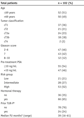
A total of 108 consecutive men with histologically proven PCA and cN0 cM0 status were treated by IMRT with daily im-age guidance after obtaining informed consent in accordance with the standards of the local ethics committee. The pretreat-ment staging included a digital rectal examination, transrectal ultrasound (TRUS), biopsy with specification of the Gleason score, prostate-specific antigen (PSA), computed tomography (CT) and/or magnetic resonance imaging (MRI), a total-body bone scan, and was defined according to the 2002 American Joint Committee on Cancer tumor, lymph nodes, and metas-tasis system [27]. Risk of recurrence was assessed according to the National Comprehensive Cancer Network practice guide-lines in oncology (www.nccn.org). A total of 6 patients were excluded from the study, 1 with a change in the treatment plan during the treatment, 1 because of interruption of the RT for 3 weeks, 1 with different dose constraints than described below and 3 patients because of death in the absence of PCA recur-rence < 9 months after RT (insufficient follow-up). The study population, thus, consisted of 102 patients, treated between 06/2004 and 03/2008. Patients were retrospectively analyzed. Median age of the patients was 69 years (range, 50–81 years), median follow-up was 39 months (range, 16–61 months). No patient was lost to follow-up, 87/102 (85%) patients had a min-imal follow-up of 24 months. The risk groups were as follows: low, intermediate, and high risk in 21 (21%), 28 (27%), and 53 (52%) patients, respectively. A total of 24/102 (24%) had undergone a transurethral resection of the prostate (TUR-P) a median of 42 months prior to IMRT (range, 1–199 months). In 9 of these patients, the time between TUR-P and IMRT was < 12 months. The clinical characteristics of the patients are shown in Table 1.
Hormonal Therapy
A total of 66 patients (65%) received concomitant and ei-ther neoadjuvant or adjuvant hormonal ei-therapy (HT), with a median duration of 6 months (range, 1–34 months). Indica-tions for HT were size reduction of the prostate, or interme-diate or high-risk features. A total of 65 patients received combined androgen blockade consisting of antiandrogen and gonadotropin-releasing hormone analogue. One patient re-ceived antiandrogen monotherapy.
External Beam Radiotherapy
All patients were treated with 80 Gy IMRT in standard frac-tionation with daily image-guidance as previously described [8]. Briefly, prior to the treatment, three gold markers were implanted into the prostate of every patient under TRUS
Table 1. Patient characteristics. PSA: prostate-specific antigen, FU:
fol-low-up, TUR-P: transurethral resection of the prostate. aMedian time
between TUR-P and IMRT was 42 months (range, 1–199 months), in 9 patients the time interval was <12 months; bperiod after completion
of treatment.
Tabelle 1. Patientencharakteristika. PSA: Prostata-spezifisches Antigen,
FU: follow-up, TUR-P: transurethrale Prostataresektion. aDie mediane
Zeit zwischen TUR-P und IMRT betrug 42 Monate (1–199 Monate), bei 9 Patienten war das Intervall <12 Monate; bZeitraum nach Abschluss
der Behandlung. Total patients n = 102 (%) Age ≤69 years 52 (51) >69 years 50 (49) Tumor classification cT1 37 (36) cT2 21 (21) cT3a 24 (23) cT3b 18 (18) cT4 2 (2) Gleason score 2–6 47 (46) 7 43 (42) 8–10 12 (12) Pre-treatment PSA ≤10 ng/mL 55 (54) >10 ng/mL 47 (46) Risk group Low 21 (21) Intermediate 28 (27) High 53 (52) Hormonal therapy no 36 (35) yes 66 (65) Prior TUR-Pa no 78 (76) yes 24 (24)
guidance. 94 of the 102 patients (92%) underwent an MRI of the pelvis, and the digital data were used for image fusion with the planning CT. To estimate the risk for seminal vesicle (SV) invasion, the Roach formula [23] was calculated for each patient. If the risk for SV invasion was > 15%, as it was the case in 59 patients, the proximal third of the SV was included in the CTV electively. If invasion of the base of the SV was seen in the MRI (cT3b) image, the whole SV were electively included in the CTV. The planning target volume (PTV) was then delineated by encompassing the prostate with 5 mm safety margins in all directions except dorsally, where a 3 mm margin was added. A set of dose constraints were used and lo-calization of the prostate prior to every RT fraction (daily im-age-guidance) was performed as previously described [8, 29]. The duration of IMRT reached a median of 56 days (range, 52–63 days).
Follow-Up Protocol
Patients were seen by a radiation oncologist at least weekly during the RT. Follow-up visits were arranged 2–4 weeks af-ter completion of IMRT and every 3–6 months for the first 2 years and annually thereafter with a digital rectal examina-tion and a serum PSA level obtained at each visit. Patients alternated follow-up visits between their urologist and radia-tion oncologist.
Toxicity Scoring and Biochemical Failure
Pretreatment morbidity and acute and late toxicity were graded using the common terminology criteria for adverse events (CTC AE) version 3.0 (http://ctep.cancer.gov/forms/ CTCAEv3.pdf) from the National Cancer Institute as previ-ously described [8].
Biochemical no evidence of disease (bNED) was assessed according to the Phoenix criteria, defining a biochemical fail-ure as a PSA rise of 2 ng/ml or more above the nadir PSA [24].
Statistical Analysis
Descriptives included absolute and relative frequencies for categorical variables, median and range for quantitative vari-ables. The primary objective was the occurrence of acute and late ≥ grade 2 GI and GU toxicity. For statistical analysis, age (≤ vs. > 69 years), PTV size (≤ vs. > 94 cm3), PTVD
max (≤ 83.9
Gy vs. > 83.9 Gy), rectal V75 (≤ 4% vs. > 4%) and rectal V47 (≤ 17% vs. > 17%) were grouped according to the median. PGUM (grade 0–1 vs. grade 2–3), acute and late GI toxic-ity (grade 0 vs. grade 1–2), and acute and late GU toxictoxic-ity (grade 0–1 vs. grade 2–3) were grouped. Categorical variables were compared using Fisher’s exact test. Estimation of actu-arial rates were calculated using the Kaplan–Meier product limit methodology and compared using the log-rank test [11]. Univariate Cox regression and a multiple Cox regres-sion model with forward and backward selection was used to determine independent prognostic factors for decreased late
≥ grade 2 GU toxicity-free survival [3]. Variables were includ-ed in the model if the univariate p value was < 0.1. Statisti-cal significance was considered on a 2-sided level of α = 0.05. Statistical analysis was performed with SPSS version 16.0 (SPSS Inc., Chicago, IL).
Results
Acute and Late Gastrointestinal Toxicity
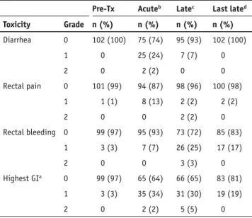
Acute grade 1 GI toxicity was experienced in 35 patients (34%), while grade 2 toxicity was observed in 2 patients (2%) with diarrhea being the most commonly observed symptom. Acute grade 3 GI toxicity was absent (Table 2).
Late grade 1 and 2 GI toxicity occurred in 31 (30%) and 5 (5%) patients, respectively, and rectal bleeding was the most commonly observed symptom. Late grade 3 GI toxicity was absent. No patient reported experiencing fecal incontinence. The median time from the completion of IMRT to the oc-currence of late ≥ grade 2 GI toxicity was 25 months (range, 12–41 months). The actuarial likelihood at 5 years for the de-velopment of late ≥ grade 2 GI toxicity was 7%. The incidence of late GI toxicity decreased as time from treatment elapsed. At the last follow-up visit, late grade 1 toxicity was present in 19 (19%) patients, and no patient suffered of late grade 2 GI
Table 2. Pre-treatment gastrointestinal morbidity and
gastrointesti-nal acute and late toxicity. Pre-Tx: pre-treatment; athe highest toxicity
in a patient was counted as a single event; b during therapy and until 3
months after completion; c maximal late toxicity >3 months after
com-pletion of therapy; d incidence of late toxicity at last follow-up visit.
There was no grade ≥ grade 3 GI toxicity observed.
Tabelle 2. Gastrointestinale Morbidität vor Behandlung und akut und
spät gastrointestinale Nebenwirkungen. Pre-Tx: vor Behandlung;.
adie maximal erreichte Toxizitäts eines Patienten wurde als 1 Ereignis
gewertet; bwährend und bis 3 Monate nach Abschluss der Behandlung; c maximale Spättoxizität >3 Monate nach Abschluss der Behandlung; dInzidenz von Spättoxizität beim letzten Nachuntersuchungstermin
(≥ Grad-3-GI Toxizität wurde nicht beobachtet).
Pre-Tx Acuteb Latec Last lated
Toxicity Grade n (%) n (%) n (%) n (%) Diarrhea 0 102 (100) 75 (74) 95 (93) 102 (100) 1 0 25 (24) 7 (7) 0 2 0 2 (2) 0 0 Rectal pain 0 101 (99) 94 (87) 98 (96) 100 (98) 1 1 (1) 8 (13) 2 (2) 2 (2) 2 0 0 2 (2) 0 Rectal bleeding 0 99 (97) 95 (93) 73 (72) 85 (83) 1 3 (3) 7 (7) 26 (25) 17 (17) 2 0 0 3 (3) 0 Highest GIa 0 99 (97) 65 (64) 66 (65) 83 (81) 1 3 (3) 35 (34) 31 (30) 19 (19) 2 0 2 (2) 5 (5) 0
toxicity anymore at that time. Acute and late GI toxicity was not associated with any DVH parameter tested.
Acute and Late Genitourinary Toxicity
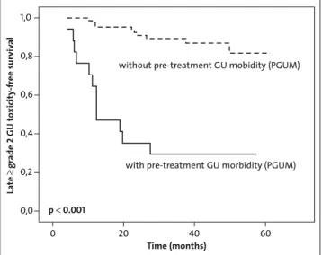
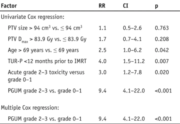
Prior to treatment, 15 patients (15%) had grade 2 and 2 pa-tients (2%) suffered of grade 3 PGUM (Table 3). The most commonly observed PGUM symptoms were increased fre-quency/urgency and retention. PGUM was not associated with a history of prior TUR-P (p = 1.0). After treatment acute grade 2 and 3 GU toxicity occurred in 44 (43%) and 5 (5%) patients, respectively. Acute ≥ grade 2 GU toxicity was sig-nificantly more common in patients who already suffered of PGUM (p < 0.001). Interestingly, acute toxicity was decreased in patients with history of TUR-P (n = 24, p = 0.039); how-ever, if only patients with TUR-P < 12 months prior to IMRT were analyzed (n = 9), acute GU toxicity was not significantly different (p = 0.50). Acute GU symptoms were often amelio-rated with α-blockers or anti-inflammatory medications. Late grade 2 and 3 GU toxicity occurred in 21 (21%) and 1 (1%) of patients, respectively. There was no acute or late grade 4 GU toxicity. The median time from the completion of IMRT to the occurrence of late ≥ grade 2 GU toxicity was 34 months (range, 4–61 months). The actuarial incidence of late ≥ grade 2 GU toxicity was 27% at 5 years. Late ≥ grade 2 GU toxicity-free survival was significantly associated with the presence of PGUM (p < 0.001) (Figure 1), presence of TUR-P < 12 months prior to IMRT (p = 0.003), presence of acute GU toxicity (p = 0.01), older age (p = 0.03) but not with HT (p = 0.66), PTV size (p = 0.76), or PTV Dmax (p = 0.2). After multiple Cox regression
analysis with forward and backward selection, PGUM was an independent positive predictor of late ≥ grade 2 GU toxicity-free survival (hazard ratio = 9.4 (95% confidence interval: 4.1, 22.0), p < 0.001) (Table 4). When all patients with a history of TUR-P were analyzed (n = 24), late ≥ grade 2 GU toxicity-free survival was not significantly decreased (p = 0.32). Interesting-ly, late GU toxicity decreased as time from treatment elapsed. At last follow-up visit, only 7 (7%) or 1 (1%) still complained of late grade 2 or 3 GU toxicity, respectively, being less than
Table 3. Pre-treatment genitourinary morbidity and genitourinary
acute and late toxicity. Pre-Tx: pre-treatment; athe highest toxicity
in a patient was counted as a single event; bduring therapy and until
3 months after completion; c maximal late toxicity >3 months after
completion of therapy; dincidence of late toxicity at last follow-up
visit. There was no grade ≥ grade 4 GU toxicity observed.
Tabelle 3. Urogenitale Morbidität vor Behandlung und akut und spät
urogenitale Nebenwirkungen. Pre-Tx: vor Behandlung; adie maximal
erreichte Toxizitäts eines Patienten wurde als 1 Ereignis gewertet;
bwährend und bis 3 Monate nach Abschluss der Behandlung; c
maxi-male Spättoxizität >3 Monate nach Abschluss der Behandlung; d
In-zidenz von Spättoxizität beim letzten Nachuntersuchungstermin (≥ Grad-4-GU Toxizität wurde nicht beobachtet).
Pre-Tx Acuteb Latec Last lated
Toxicity Grade n (%) n (%) n (%) n (%) Dysuria 0 95 (93) 63 (62) 93 (91) 98 (96) 1 7 (7) 30 (30) 8 (8) 4 (4) 2 0 7 (7) 1 (1) 0 3 0 2 (2) 0 0 Incontinence 0 94 (92) 92 (90) 88 (86) 92 (90) 1 7 (7) 9 (9) 12 (12) 9 (9) 2 1 (1) 1 (1) 2 (2) 1 (1) Retention 0 57 (56) 52 (51) 71 (70) 91 (89) 1 38 (37) 25 (25) 25 (24) 10 (10) 2 6 (6) 23 (22) 5 (5) 0 3 1 (1) 2 (2) 1 (1) 1 (1) Frequency/ur-gency 0 65 (64) 23 (23) 51 (50) 71 (70) 1 28 (27) 46 (45) 34 (33) 24 (23) 2 8 (8) 30 (29) 17 (17) 7 (7) 3 1 (1) 3 (3) 0 0 Hematuria 0 102 (100) 102 (100) 93 (91) 99 (97) 1 0 0 7 (7) 3 (3) 2 0 0 2 (2) 0 Highest GUa 0 39 (38) 11 (11) 37 (36) 59 (58) 1 46 (45) 42 (41) 43 (42) 35 (34) 2 15 (15) 44 (43) 21 (21) 7 (7) 3 2 (2) 5 (5) 1 (1) 1 (1) 1,0 0,8 0,6 0,4 0,2 0,0 0 20 40 60 Time (months) p< 0.001
without pre-treatment GU mobidity (PGUM)
with pre-treatment GU morbidity (PGUM)
La te gr ade 2 GU to xicity -fr ee sur viv al
Figure 1. Actuarial analysis of 3-year late ≥ grade 2 GU toxicity-free
sur-vival stratified by the presence of pretreatment genitourinary morbid-ity (PGUM).
Abbildung 1. Kaplan-Meier-Analyse des spät (≥ Grad 2) urogenital
nebenwirkungsfreien Überlebens nach 3 Jahren, stratifiziert nach Vorhandensein von urogenitaler Morbidität vor Behandlung (PGUM).
half of the patients who suffered of grade 2 or 3 PGUM prior to treatment.
Biochemical Control
With a median follow-up of 39 months, it is too early to re-port sound biochemical control rates, especially taking into account that 65% of the patients underwent HT. However, after IMRT, 92% (61/66) of patients with and 50% (18/36) of patients without HT reached a PSA nadir value ≤ 0.5 ng/mL. During follow-up, 12 patients (12%) experienced biochemical relapse, all of whom belonged to the high-risk of recurrence group. The actuarial 5-year biochemical relapse-free survival was 100%, 100%, and 68% for low-, intermediate- and high-risk patients, respectively (p = 0.003).
Discussion
Radical prostatectomy combined with pelvic lymphadenecto-my is known to produce comparable tumor control rates com-pared to conventional dose 3D-CRT in localized PCA [5, 16], and introduction of nerve-sparing operation technique could significantly reduce the risk of associated severe stress inconti-nence [14]. Thus, modern RT is required to achieve low toxic-ity rates and high long-term effectiveness to be recommended as a therapeutic alternative to radical prostatectomy.
After a median follow-up of 39 months, our results after high-dose IMRT daily image-guidance are encouraging with respect to observed treatment-related toxicity. Acute and late grade 3 GI toxicity was absent, while acute and late grade 2 GI
toxicity was observed in 2% and 5% of the patients, respective-ly.
Concerning GU toxicity, notably, our patients suffered of a significant PGUM, being of grade 2 and 3 in 15% and 2%, respectively, of the patients prior to IMRT. Acute grade 2 and 3 GU toxicity occurred in 43% and 5% and late grade 2 and 3 GU toxicity was observed in 21% and 1%, respectively.
Our acute toxicity rates compare favorably with those reported for 111 patients treated with IMRT 78 Gy within a prospective randomized trial, where the acute grade 2 and 3 GI toxicity was 41% and 8% and the acute grade 2 and 3 GU toxicity was 21% and 2% (CTC score), respectively [19].
Others have reported the toxicity profile (CTC score) after ultrahigh-dose IMRT with 86.4 Gy after a median fol-low-up of 53 months and acute grade 2 GI toxicity was 8%, acute grade 2 and 3 GU 22% and 1%, respectively, actuarial late ≥ grade 2 GI toxicity at 5 years was 4% and actuarial late ≥ grade 2 GU toxicity was 16% at 5 years [2]. Compared to this study, our GI rates seem comparably low; however, GU toxic-ity rates were higher in our patients despite the use of a lower total dose in our patients (80 Gy vs. 86.4 Gy). Importantly, one has to consider that in our treatment plans the dose coverage of the PTV was defined more strictly, thus, aiming to set the V95 to > 99% (volume of the PTV receiving at least 95% of the prescribed dose > 99%) [8]; however, the treatment plan cri-teria for 81 Gy or 86.4 Gy plans were V95 > 90% or V95 > 85%,
respectively [18], moreover for 81 Gy and 86.4 Gy, a lower single fraction dose (1.8 Gy) was used.
In this study, a significant association between acute GI/ GU toxicity and decreased late GI/GU toxicity-free survival could be demonstrated. This is in accordance with the findings of others [12, 26, 30]. In addition, we could also describe not only the PGUM rate of our patients and its significant associa-tion with acute GU toxicity, but also its role as an independent positive predictor for decreased late GU toxicity-free survival. A relative large proportion of our patients (24%) had under-gone TUR-P at different times prior to IMRT. We found that a history of TUR-P < 12 months before IMRT was associated with decreased late ≥ grade 2 GU toxicity-free survival. An as-sociation between previous TUR-P and GU toxicity has been previously described [20].
In our patients, HT did not influence the incidence of late GI or GU toxicity. A large meta-analysis [17] recently demon-strated that addition of short-term HT significantly decreased GI and GU late toxicities.
Late toxicity after RT is known to increase over time. However, during follow-up of our series of patients, the in-cidence of late GI and especially of late GU toxicity were decreasing. At the last follow-up visit, late GU toxicity rates dropped below half of the observed PGUM rates. A similar re-port was recently made by others [7], who showed that patients presenting with obstructive urinary symptoms experienced a clear symptom relieve after IMRT ± HT. Generally, the grade 2 GU toxicities observed in our patients consisted of increased
Table 4. Univariate and multiple Cox regression analysis of factors
associated with late ≥ grade 2 genitourinary toxicity-free survival. RR: relative risk, CI: 95% RR confidence intervals, TUR-P: transurethral resection of the prostate, IMRT: intensity modulated radiation therapy, PGUM: pre-treatment genitourinary morbidity.
Tabelle 4. Univariate und multiple Cox Regressionsanalyse von
Fak-toren welche mit spät-≥Grad-2-urogenital nebenwirkungsfreiem Überleben assoziiert sind. RR: relatives Risiko, CI: 95% RR Konfidenzin-tervalle, TUR-P: transurethrale Prostataresektion, IMRT: Intensitäts-modulierte Radiotherapie, PGUM: urogenitale Morbidität vor Behand-lungsbeginn.
Factor RR CI p
Univariate Cox regression:
PTV size > 94 cm3 vs. ≤ 94 cm3 1.1 0.5–2.6 0.763
PTV Dmax > 83.9 Gy vs. ≤ 83.9 Gy 1.7 0.7–4.1 0.208
Age > 69 years vs. ≤ 69 years 2.5 1.0–6.2 0.042
TUR-P <12 months prior to IMRT 4.0 1.5–11.2 0.007
Acute grade 2–3 toxicity versus grade 0–1
3.0 1.2–7.8 0.020
PGUM grade 2–3 vs. grade 0–1 9.4 4.1–22.0 <0.001
Multiple Cox regression:
frequency/urgency or retention and were often significantly improved after α-blocker or anti-inflammatory medications. These drugs were received by most of the symptomatic pa-tients during IMRT and during follow-up; thus, this drug therapy at least contributed to the observed decrease of late GU toxicity during time.
The follow-up of less than 5 years is too short to provide sound biochemical control data. However, all observed events of biochemical relapse (12%) occurred in patients with high-risk features and our actuarial 5-year biochemical relapse-free survival rates of 100%, 100%, and 68% are comparable with the 5-year rates of 98%, 85%, and 70% described after ultrahigh-dose IMRT for low-, intermediate-, and high-risk patients, respectively [2].
We are aware of the obvious limitations of our study, be-ing the retrospective nature of the study and limited follow-up. Nevertheless, this report provides strong evidence for the low toxicity profile of high-dose IMRT. In the decision mak-ing for appropriate local treatment of PCA, the patient should not be informed about “RT” but should be informed about “modern RT” and its associated low treatment-related acute and late toxicity.
References
1. Al-Mamgani A, van Putten WL, Heemsbergen WD, et al. Update of Dutch multi center dose-escalation trial of radiotherapy for localized prostate cancer. Int J Radiat Oncol Biol Phys 2008;72:980–8.
2. Cahlon O, Zelefsky MJ, Shippy A, et al. Ultra-high dose (86.4 Gy) IMRT for localized prostate cancer: toxicity and biochemical outcomes. Int J Radiat Oncol Biol Phys 2008;71:330–7.
3. Cox DR, Oakes D. Analysis of survival data. London, United Kingdom: Chap-man and Hall, 1984;110–20.
4. Dearnaley DP, Sydes MR, Graham JD, et al. Escalated-dose versus standard-dose conformal radiotherapy in prostate cancer: first results from the MRC RT01 randomised controlled trial. Lancet Oncol 2007;8:475–87.
5. D’Amico AV, Whittington R, Malkowitcz SB, et al. Biochemical outcome af-ter radical prostatectomy exaf-ternal beam radiation therapy, or inaf-terstitial radiation therapy for clinically localized prostate cancer. JAMA 1998;280: 969–74.
6. Dolezel M, Odrazka K, Vaculikova M, et al. Dose escalation in prostate ra-diotherapy up to 82 Gy using simultaneous integrated boost. Strahlenther Onkol 2009;186:197-202.
7. Fonteyne V, Villeirs G, Lumen N, et al. Urinary toxicity after high dose intensity modulated radiotherapy as primary therapy for prostate cancer. Radiother Oncol 2009;92:42–7.
8. Ghadjar P, Vock J, Vetterli D et al. Acute and late toxicity in prostate cancer patients treated by dose escalated intensity modulated radiation therapy and organ tracking. Radiat Oncol 2008;3:35.
9. Goldner G, Dimopoulos J, Kirisits C, et al. Moderate dose escalation in three-dimensional conformal localized prostate cancer radiotherapy. Strahlenther Onkol 2009;7:438–45.
10. Guckenberger M, Flentje M. Intensity-modulated radiotherapy (IMRT) of localized prostate cancer: a review and future perspectives. Strahlenther Onkol 2007;183:57–62.
11. Harris EK, Albert A. Survivorship analysis for clinical studies. New York, NY: Marcel Dekker, 1991;5–125.
12. Heemsbergen WD, Peeters STH, Koper PCM, et al. Acute and late gastroin-testinal toxicity after radiotherapy in prostate cancer patients: consequen-tial late damage. Int J Radiat Oncol Biol Phys 2006;66:3–10.
13. Kalz J, Sterzing F, Schubert K, et al. Dosimetric comparison of image guid-ance by megavoltage computed tomography versus bone alignment for prostate cancer radiotherapy. Strahlenther Onkol 2009;185:241–7.
14. Kessler TM, Burkhard FC, Studer UE. Nerve-sparing open radical retropubic prostatectomy. Eur Urol 2007;51:90–7.
15. Kuban DA, Tucker SL, Dong L, et al. Long-term results of of the M.D. Ander-son randomized dose-escalation trial for prostate cancer. Int J Radiat Oncol Biol Phys 2008;70:67–74.
16. Kupelian PA, Elshaikh M, Reddy CA, et al. Comparison of the efficacy of lo-cal therapies for lolo-calized prostate cancer in the prostate-specific antigen era: a large single-institution experience with radical prostatectomy and external-beam radiotherapy. J Clin Oncol 2002;20:3376–85.
17. Lawton CA, Bae K, Pilepich M, et al. Long-term treatment sequelae after ex-ternal beam irradiation with or without hormonal manipulation for adenocar-cinoma of the prostate: Analysis of Radiation Therapy Oncology Group studies 85-31, 86-10, and 92-02. Int J Radiat Oncol Biol Phys 2008;70:437–41. 18. Leibel SA, Fuks Z, Zelefsky MJ, et al. Technological advances in
external-beam radiation therapy for the treatment of localized prostate cancer. Semin Radiat Oncol 2003;30:596–615.
19. Matzinger O, Duclos F, van den Bergh A, et al. Acute toxicity of curative radiotherapy for intermediate- and high-risk localised prostate cancer in the EORTC trial 22991. Eur J Cancer 2009;45:2825–34.
20. Peeters ST, Heemsbergen WD, van Putten WL, et al. Acute and late com-plications after radiotherapy for prostate cancer: results of a multicenter randomized trial comparing 68 Gy to 78 Gy. Int J Radiat Oncol Biol Phys 2005;61:1019–34.
21. Pinkawa M, Pursch-Lee M, Asadpour B, et al. Image-guided radiotherapy for prostate cancer. Strahlenther Onkol 2008;12:679–85.
22. Polat B, Guenther I, Wilbert J. Intra-fractional uncertainties in im-age-guided intensity-modulated radiotherapy (IMRT) of prostate cancer. Strahlenther Onkol 2008;184:668–73.
23. Roach 3rd M. Re: The use of prostate specific antigen, clinical stage and
Gleason score to predict pathological stage in men with localized prostate cancer. J Urol 1993;150:1923–4.
24. Roach 3rd M, Hanks G, Thames Jr. H, et al. Defining biochemical failure
following radiotherapy with or without hormonal therapy in men with clini-cally localized prostate cancer: recommendations of the RTOG-ASTRO Phoe-nix consensus conference. Int J Radiat Oncol Biol Phys 2006;65:965–74. 25. Sathya JR, Davis IR, Julian JA, et al. Randomized trial comparing iridium
implant plus external-beam radiation therapy with external-beam radiation therapy alone in node-negative locally advanced cancer of the prostate. J Clin Oncol 2005;23:1192–9.
26. Schultheiss TE, Hanks GE, Hunt MA, et al. Incidence of and factors related to late complications in conformal and conventional radiation treatment of cancer of the prostate. Int J Radiat Oncol Biol Phys 2002;54:442–9. 27. Sobin LH, Wittekind CH. The prostate. In: TNM classification of malignant
tumors. New York: Wiley-Liss, 2002;184–7.
28. Treutwein M, Hipp M, Kölbl O, et al. IMRT of prostate cancer. Strahlenther Onkol 2009;185:379–83.
29. Vetterli D, Thalmann S, Behrensmeier Fet al. Daily organ tracking in inten-sity-modulated radiotherapy of prostate cancer using an electronic por-tal imaging device with a dose saving acquisition mode. Radiother Oncol 2006;79:101–8.
30. Zelefsky MJ, Levin EJ, Hunt M, et al. Incidence of late rectal and urinary toxicities after three-dimensional conformal radiotherapy and intensity-modulated radiotherapy for localized prostate cancer. Int J Radiat Oncol Biol Phys 2008;70:1124–9.
31. Zietman Al, DeSilvio ML, Slater JD, et al. Comparison of conventional-dose vs high-dose conformal radiation therapy in clinically localized adenocarcinoma of the prostate: a randomized controlled trial. JAMA 2005;294:1233–9.
Address for Correspondence
Prof. Dr. med. Daniel M. Aebersold University of Bern – Inselspital Department of Radiation Oncology Freiburgstr.
3011 Bern Switzerland
Phone (+41) 31 632-2431, Fax -2342 e-mail: daniel.aebersold@insel.ch