Characterization of the non-functional Fas
ligand of gld mice
Michael Hahne, Manuel C. Peitsch
2, Martin Irmler, Michael Schroter, Bente Lowin,
Marga Rousseau, Claude Bron, Toufic Renno
1, Lars French
3and JQrg Tschopp
Institute of Biochemistry, University of Lausanne and 1Ludwig Institute for Cancer Research, BIL Research Center, Chemin des Boveresses 155, CH-1066 Epalinges, Switzerland
^ l a x o Institute for Molecular Biology, Chemin des Aulx 14, CH-1228 Plan-les-Ouates, Switzerland 3Division of Dermatology, Geneva University Hospital, CH-1211 Geneva 14, Switzerland
Keywords: activity, Fas, gld, structure
Abstract
Mice homozygous for either the gld or Ipr mutation develop autoimmune diseases and progressive lymphadenopathy. The Ipr mutation Is characterized by the absence of functional Fas, whereas gld mice exhibit an Inactive FasL due to a point mutation proximal to the extracellular C-terminus. The structural repercussions of this amino acid substitution remain unknown. Here we report that FasL is expressed at similar levels on the surface of activated T lymphocytes from gld and wild-type mice. Using a polyclonal anti-FasL antibody, indistinguishable amounts of a 40 kDa protein are detected In both gld and wild-type splenocytes. The molecular model of FasL, based on the known structure of TNF-a, predicts that the Phe->Leu gld mutation is located at the protomer interface which is close to the FasR interaction site. We conclude that the gld mutation allows normal FasL biosynthesis, surface expression and oligomerlzatlon, but Induces structural alterations to the Fas binding region leading to the phenotyplc changes observed.
Introduction
FasL is a member of the type II membrane protein superfamily consisting of TNF-a and -p" (lymphotoxin-a), lymphotoxin-p, CD27L, CD30L, CD40L and 4-1BB (1). Despite the diversity of the biological activities elicited by these ligands, most of them are known to induce cellular differentiation or proliferation with two exceptions. FasL and, under some circumstances, TNF are responsible for rapid induction of apoptosis of receptor-bearing cells (2,3).
The importance of a functional Fas-Fas ligand (FasL) system is illustrated by the phenotype of Ipr (lymphoprolifera-tion)and gld (generalized disease) mutant mice. MLR mice homozygous for Ipr or gld develop a progressive autoimmune disorder, resembling systemic lupus erythematosis in human (4). These mice also suffer from a large accumulation of non-malignant CD4~CD8~ T cells in the spleen and in lymph nodes. Genetic analysis indicated that the gld phenotype results from a mutation in FasL (5,6). The Ipr mutation has been correlated with a defect in Fas (7).
The Fas system unquestionably plays a crucial role in the deletion of autoimmune cells. Fas-mediated elimination occurs in the periphery, since both negative and positive selection in the thymus proceed normally in gld (or Ipr) animals (8,9).
Fas also plays an important role in T cell cytotoxicity.
Cytolytic T cells use two major cytolytic pathways, one based on perforin-granzymes and one dependent on FasL (10-13). Cytotoxic T cells derived from perforin knock-out mice have considerable killing activity left (14) which is attributable to Fas (10-12). In turn, perforin-containing gld cytotoxic T cells still kill tumor cells, but with reduced efficiency (10).
FasL in gld/gld mice displays no killing activity upon stimulation with phorbol ester (PMA) and calcium ionophore (ionomycin) (5,6), in spite of normal mRNA levels present (5,6), Moreover, activated gld T cells fail to interact with a FasR.Fc hybrid protein (6,15). This functional loss is a result of a single point mutation changing Phe273 to Leu proximal to the extracellular C-terminus of this type II membrane protein (5,6). In the present study we report on the structural implications of this fatal single amino acid replacement.
Methods
Expression of bacterial recombinant FasL
cDNA coding for the extracellular domain of FasL was ampli-fied from 1 ng mouse FasL cDNA (GenBank accession no. MMU10984) using the following primers: primer 1, 5'-TTC Correspondence to. J. Tschopp
1382 Fas ligand in gld mice
GCT CGA GAA CTG GCA GAA CTC CGT GAG-3'; primer 2, 5'-AAG GAT CCT AGC TGA CCT GTT GGA CCT TGC-3'. The amplified product was cloned into the pCRII vector using the AT cloning kit (Invitrogen, Heidelberg, Germany) and subsequently subcloned into the page-KG expression vector (kindly provided by Dr A. Quest at our institute) at the EcoRI site. The glutathione-S-transferase (GST)-FasL fusion protein was expressed in bacteria upon induction with IPTG.
Antibody production
A peptide spanning amino acids 196-220 of the mouse Fas ligand (RGQSCNNQPLNHKVYMRNSKYPEDL), synthesized using the multiple antigen technology (16), was used as antigen. Spleen cells of an immunized rat were fused to NF1 mouse myeloma cells and hybridoma cells secreting peptide specific IgG were selected. Two clones, H11 and A11, were further analyzed. The same peptide construct was also injected into rabbits for polyclonal antibody production. The antiserum (PE62) was affinity purified on FasL peptide coupled to CNBr-Sepharose (Pharmacia, Zurich, Switzerland).
Splenocytes and their activation
B6.gld mice were purchased from Jackson Laboratories (Bar Harbor, ME). Splenocytes were isolated from 6-week-old mice and freed from red blood cells by 10 min incubation in an ice cold buffer containing 13 mM sodium bicarbonate, 156 mM ammonium chloride and 127 |iM EDTA, and then resuspended in complete medium. Cells were cultured for 4 h at ~1x10⁷ cells/ml in either the presence or the absence of PMA (5 ng/ ml) and ionomycin (500 ng/ml) (Sigma, Buchs, Switzerland).
Flow cytometry
Analysis was performed on a FACScan cytometer (Becton Dickinson, Mountain View, CA) using the Lysys II software Cells were stained with the anti-FasL antibody PE62 followed by donkey anti-rabbit coupled to fluorescein (Dianova, Hamburg, Germany) and the PE-labeled mAb H 129.19 for L3T3/CD4 and 53-6.7 for Ly-2/CD8 staining (Boehringer, Mannheim, Germany).
SDS-PAGE, Western blot and native electrophoresis
Splenocytes were washed in ice cold PBS and then boiled in sample buffer for 5 min. Cellular proteins of - 2 x 1 06 spleno-cytes per lane were electrophoretically separated on a 10% polyacrylamide gel in the presence of SDS under non-reducing conditions (17) and subsequently transferred to nitrocellulose. Immunoblot analysis was conducted using plain supernatant of the mAb H11 and A11, and 50 (ig/ml of affinity-purified PE62 antibody. First antibodies were detected using peroxidase-conjugated donkey rat and anti-rabbit antibodies (Dianova, Hamburg, Germany) followed by a chemiluminescence reaction using the ECL system (Amersham, Bucks, UK).
For native electrophoresis the Phast separation system of Pharmacia (Zurich, Switzerland) was employed. Approxi-mately 107 splenocytes were lysed in 50 ^J lysis buffer (100 mM NaCI, 1 % NP-40 in 50 mM Tris, pH 8.0) containing 1 mM PMSF. Cell lysate (1 \i\) was applied per lane and the proteins were electrophoretically separated on 4-15% gradient gels
and analyzed by immunoblot analysis as described above. Complement C8 and C9 components were used as molecular weight standards.
Results
Production of FasL specific antibodies
Polyclonal and monoclonal antibodies were generated against a peptide corresponding to residues 196-220 of the mouse FasL, predicted to lay on the loop forming the outer tip of the ligand (see below). Affinity-purified polyclonal antiserum (PE62) and two independent mAb (H11 and A11) were used throughout this study. To confirm that these anti-peptide antibodies specifically recognize FasL, the reactivity of the antibodies was tested against the recombinant extracellular domain of FasL. As shown in Fig. 1, both the monoclonal and polyclonal antibodies recognized bacterial lysates containing the GST-FasL fusion protein in Western blots, whereas no band was detectable in lysates transfected with the expression vector containing FasL insert in the inverse orientation. Flow cytometric analysis showed reactivity of the polyclonal anti-body PE62 with the native ligand on the cell surface (Fig. 2), while the use of the mAb was restricted to the analysis of denatured protein in Western blots .
Expression of FasL in T lymphocytes of wild-type and gld mutant mice
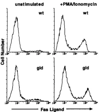
FasL mRNA has been detected in activated splenocytes (3,6). Flow cytometric analysis of activated T lymphocytes derived from spleens indeed showed high expression of FasL (Fig. 2). The CD4+ and the CD8+ subsets showed equally intense FasL staining (data not shown). FasL expression was critically dependent on lymphocyte activation. Only cells that had been previously treated with PMA/ionomycin were positive, while marginal staining of T lymphocytes was observed with non-activated T cells. Interestingly, only a subpopulation of activ-ated T lymphocytes was FasL positive. Approximately 70%
PE62 H11 D6
4 9
3 2 2 7-Fig. 1. Characterization of anti-mouse FasL antibodies. Western blot
analysis of a bacterial lysate containing recombinant mouse FasL-GST fusion protein (46 kDa) using the affinity-purified PE62 rabbit polyclonal antibody to FasL or rat monoclonal anti-mFasL H11. The low molecular weight bands most likely correspond to degradation products. Bacterial lysate expressing GST alone served as a negative control. The D6 mAb antibody detects the peptide 62 in ELISA, but does not react with the recombinant FasL (control lanes).
remained FasL low. We are currently investigating the pheno-type of the FasL positive lymphocyte subpopulation in greater detail.
The relative surface expression of FasL on splenic lympho-cytes isolated from wild-type B6 mice with that from gid B6 mice was compared Previously, Ramsdell (15) showed that the FasR.Fc hybrid protein detected the ligand only on wild-type lymphocytes, whereas the gid T lymphocytes were negative. Using FasL specific antibodies, however, staining on PMA/ionomycin treated gldl lymphocytes was comparable to that of wild-type T lymphocytes (Fig. 2). Only 30% of activated gid T lymphocytes expressed high levels of FasL.
This was confirmed by Western blot analysis An equal quantity of protein and an identical size was found in extracts from activated T lymphocytes of wild-type and of gid origin (Fig. 3). FasL exhibited a molecular weight of - 4 0 kDa, in agreement with the molecular weight of post-translationally modified FasL in transfected COS cells (18).
Structure of the gld-FasL
The inability of the FasR.Fc hybrid protein to detect gld-FasL points to structural dissimilarities in the Fas binding region of the ligand. Other members of the TNF family have been shown to be active only as trimers and not as monomers (19,20). Similarly, FasL, although present on the surface, could show impaired dimer or trimer formation on the surface of gid T cells. This would be compatible with the observed
unstimulated
+PMA/lonomycln
wt
Fas Ugand
Fig. 2. Expression of FasL on wild-type and gid lymphocytes. FACS
analysis of cell surface FasL expression on unstimulated (left panels) and PMA/lonomycin (4 h) stimulated (right panels) CD4+ or CD8+ splenocytes of C57BU6J (B6) (A) or B6-gld origin (B). The flow cytometric profiles show the combined staining of CD4+ and CD8+ splenocytes stained with the anti-FasL polyclonal antibody PE62. Unrelated antibody control is identical with the profiles of unstimulated splenocytes (upper left panel).
difference of FasL surface expression, which differs depending on whether a FasL antibody or the FasR.Fc hybrid protein is used as tool for detection. Whereas the antibody most likely detects multimers and monomers, the receptor.Fc hybrids are known to interact only with multimeric ligands with high affinity.
This hypothesis was supported by the spatial localization of FasL Phe273 to Leu mutation. Although the level of sequence identity between the individual members of the TNF super-family does not exceed 35%, their three-dimensional struc-tures are expected to be very similar as observed for TNF-a and -p for which X-ray structures are available. Both factors crystallize in the trimeric form and are detected as such in solution (20-22). Knowledge-based protein modeling of FasL was therefore performed (23). Figure 4(A and B) shows the predicted three-dimensional structure of the presumptive FasL trimer. The trimer interface is largely dominated by hydrophobic residues which are well conserved throughout the TNF family (23). In the gid mutation the bulky aromatic side chain of Phe273, which is part of the subunit interface, is replaced by the shorter leucine (Fig. 5A). Phe273 is posi-tioned at the center of a 12 A sphere where several of the best conserved residues of the TNF family are clustered (V144, A145, H146, L158, W160, Y190, V193, G244, A245, F247, F273, G274), although they are quite distant in the sequence. This suggests an important functional role for this particular region. Based on our model of murine FasL, Phe273 (located on strand H) interacts mainly with Ala245 (on strand F) of the adjacent subunit (Figs 4 and 5). It is responsible for -50 A2 of the hydrophobic contact surface between individual protomers. The gld-FasL model predicts that the contribution of amino acid 273 is reduced by >20 A2 when replaced by a leucine. This would in turn result in a decrease of hydrophobic interactions between the subunits and suggest that either trimer formation of FasL itself is impaired or that local distor-tions of the polypeptide backbone occur in the gid mutation. As in the gid mutation, this position is occupied by shorter hydrophobic residues, i.e. isoleucine and valine, in human and mouse CD30L respectively (Fig. 5A and B). The molecular model of the CD30L (23) shows that the consequent difference
80 49 32 27 A11 — — — — H11 %
-Fig. 3. FasL in splenocytes. Western blot analysis of PMA/ionomycin
(4 h) stimulated wild-type and gid splenocytes. FasL-detecting antibodies were H11 and A11. No staining is seen with an irrelevant mAb (data not shown).
1384 Fas ligand in gld mice
in subunit contact surface is, however, compensated by CD30LLeu199 which corresponds to FasL:Ala245 (Fig. 5B).
Oligomerization of FasL
To determine if there is a failure of trimerization of the gld-FasL, Western blot analysis was performed with the native FasL-recognizing polyclonal PE62 antibody. This antibody normally detected at least three bands under non-reducing conditions, the lowest of 40 kDa co-migrating with that detected by the mAb (Fig. 6A). The two larger bands had apparent molecular weights of -75 and 105 kDa respectively. Their molecular masses correlate with the predicted mass of FasL dimers and trimers, and most likely reflect non-dissoci-ated oligomeric forms of the ligand.
To obtain additional information with respect to the aggrega-tion state of FasL in the presence of non-denaturing deter-gents, activated splenocytes were disrupted with the mild detergent NP-40. The detergent extract was separated by native gradient gel electrophoresis. Subsequent immunoblot analysis revealed multiple FasL-antibody reactive bands. The lowest one had an apparent molecular size of -200 kDa, but two additional FasL antibody-reactive species with apparent molecular masses of -400 and 600 kDa were detected. These absolute values have to be interpreted with caution, since NP-40 forms large micellar structures. However, they corres-pond to dimeric and trimeric forms of the ligand. Importantly, the pattern of these bands was similar in PMA/ionomycin activated splenocytes of wild-type and gld origin.
Fig. 4. Molecular model of the murine FasL. Ribbons representation of the murine FasL. In the top view (A), all three subunits, their disulfide
bonds and Phe273 mutated in gld-fasL are represented, while in the side view (B) only the subunits A and C are shown for clarity. The receptor-binding loops are depicted in blue (darker blue for the loop linking the G and H strands) while the sequence corresponding to peptide 62 is colored in pink. The green sphere represents the side chain of Ala245 (see Fig. 5A), which is in contact with Phe273 (orange side chain). The residues at the beginning and end of the receptor-binding GH loop are indicated with light yellow and red circles. The N-and C-terminal ends of the extracellular FasL sequence which displays homology to the TNF molecule are indicated with white letters, N-and the p-strands are identified by black letters on the C subunit.
245 273
t
232- 1
231- 1
124- I
124- j
196- 1
196- 1
193- 1
193- 1
T
[TagiMAHaSYLOflVHJ™ L^BIIAHISYLHVBBJJI^PIVTAIYQBVJPP
jLQEP«LHmYHHA§Qp 3Y8SLBYI8VGF|OLAQESvrfelJklWGFloLVQ|fc
«YttJLSQFLLHYIQvfs| iVYBNLSQFLLDYlQVffrl FFFFFFFFFFF L !SA|HJK«WKK2LSLINHEEeH-ra|fvAMHH^BHybLSLINSIES-HB
SKmopTHTDGISHLH^pE-vB
}Q|BQ|STHTDGIPHLVLSPBT-vjD I S E R ^ ^ ^ H H P D M V D Y R R G H - HlRfeRPlHBlPDMVEiARG»-H
hsVRiDNFQYVDTNTFPLIMJVLSvj SlSVNfcTFQYIDTSTFPLENVLSJI ffiLYKL HLYKBkFA
HFAL
fflvMVG
HvMVG
E Y S S S D ILYSNS GGGGGGG HHHHHHHHg l d FasL
Mouse FasL
Rat FasL
Mouse TNFP Human TNFfi Mouse LTP Human LTp Mouse CD30L Human CD30LFig. 5. The interface of the g/d-FasL trimer. (A) Multiple amino acid
sequence alignment of TNF superfamily members that are most similar to FasL (>22% overall identity), i.e. TNF-f), lymphotoxin-p (LTp) and CD30L. Only the C-terminal region of FasL is shown. Residues identical in 50% of the sequences are indicated with a shaded background, while those involved in interface formation in the gld mutation are labeled in bold face and by arrows. The (}-strands are indicated in italics. (B) Representation of a section of the interface between the A (green) and C (blue) subunits of (A) moose FasL, (B) mouse gld-FasL and (C) human CD30L. The side chains of FasL:Phe273 and FasL.AIa245 are shown in orange and white respectively. The corresponding residues of murine CD30L are shown in the lower panel. The space left empty by the replacement of Phe by Ile228 in CD30L is filled by the compensatory replacement of Ala by Leu199. In gld-FasL, the mutation of Phe273 to Leu is not compensated by the replacement of Ala244 by a larger hydrophobic residue, leaving a large empty space in the structure. To fHI this cavity in the trimer structure, gld-FasL may undergo a distortion with dramatic effect on its FasR-binding activity.
A.
PE62 con
9 0 0
1 4 3
9 7
-49 —
3 2
-* -*
2 S2
1 4 0
7 0
-O)Fig. 6. Ohgomer formation by FasL. (A) Western blot analysis of PMA/
lonomycin activated splenocytes using the polyclonal affinity-purified PE62 antibody In the control lanes (con), the first antibody (PE62) was left out during the incubations Negative results were also obtained with irrelevant rabbit IgG. (B) Separation of splenocyte proteins by native gel electrophoresis. FasL antigen was detected by Western blot analysis using the PE62 antibody.
Discussion
Our results show that the single amino acid change in the
gld mutant FasL allows correct post-translational modification
and sorting of the ligand to the cellular surface. Furthermore, both the gld- and wild-type FasL seem to form SDS-resistant oligomers. As shown for soluble members of this ligand family (24), the membrane-associated form of FasL may require surface dimerization or trimerization to display functional activity. There are antibodies to Fas which have ligand-like activities and induce apoptosis in Fas-bearing cells. The two original antibodies which have led to the discovery of Fas (Apo-1) are aggregating antibodies, i.e. of either the IgM or lgG3 subtype (25,26). F(ab')2 fragments alone are inactive (27). Thus, it is very likely that FasL homotrimer formation is required to induce Fas aggregation and signal transduction. In the context that the apparent aggregation state of the
gld- and wild-type FasL does not differ in vitro, we favor the
conclusion that the g/d-FasL's inability to interact with soluble Fas dimers (FasR.Fc) and to transmit the signal via Fas results from a local distortion in FasL-FasR interaction site. Closer examination of the structure shown in Fig. 4(A) shows that the critical Phe273 is part of a loop involved in FasR contact (23). This loop links the strands G and H (the GH loop) and ends only four residues before Ftie273. The replacement of Phe273 by a leucine most likely aiters the structure of strand H in the FasL trimer. It may then be that the modified structure of strand H changes the conformation of the GH loop giving rise to a FasL with impaired FasR-binding activity.
Several mutations in the TNF family leading to distinct phenotypes have been reported. CD40L mutants were dis-covered in connection with the X-linked hyper-IgM syndrome (28). The spatial mapping of human CD40L mutants dis-covered in connection with the X-linked hyper-IgM syndrome (summarized in 28) show that several of the reported point
1386 Fas ligand in gld mice
mutations probably affect the folding of individual subunits or the interaction with the receptor (23). Two mutations, however, A123—>E and G227->V, are also found at the subunit interface. Interestingly, these two amino acids are located in the same three dimensional region as FasL:Phe273 (12 A sphere around Phe273) and correspond to two of the highly conserved residues throughout the TNF family Although more polar subunit interactions (CD40L:Ser256 instead of FasL:Phe273) replace the very hydrophobic ones in the other members of the TNF family, mutations in this region of CD40L lead to ligands with impaired receptor-binding function as evidenced by the appearance of the X-linked hyper-IgM syndrome in patients bearing such mutations (28). These mutations may lead to similar effects on the conformation of the GH loop (one of the receptor-binding loops) as in FasL and thus explain the lack of functionality of these mutants. In contrast to CD40L, no mutation affecting FasL activity has been described in humans to date. The pathological consequences of such a mutation, as seen in the gld mouse, may be too profound to allow its occurrence.
Acknowledgements
The authors gratefully acknowledge the technical assistance of H. Hasler, S. Hertig, T. Bornand and P Allegrini The helpful comments and suggestions of H. R. MacDonald and C. Kamel are also gratefully acknowledged. This work was supported by grants of the Swiss National Science Foundation
Abbreviations
FasL Fas ligand
GST glutathione-S-transferase
References
1 Smith, C. A., Farrah, T. and Goodwin, R. G. 1994. The TNF-receptor superfamily of cellular and viral proteins: activation, costimulation, and death. Cell 76:959
2 Aggarwal, B. B. and Eessaul, T. E. 1987. Human tumor necrosis factor and lymphotoxin: their structural and functional similarities In Tumor Necrosis Factor, p. 297. Alan R. Liss, New York. 3 Suda, T, Takahashi, T, Golstein, P. and Nagata, S. 1993. Molecular
cloning and expression of the Fas ligand, a novel member of the tumor necrosis factor family. Cell 75:1169.
4 Cohen, P. L. and Eisenberg, R. A 1991. Lpr and gld. single gene models for of systemic autoimmunity and lymphoproliferative disease. Annu. Rev. Immunol. 9:243.
5 Takahashi, T, Tanaka, M., Brannan, C. I , Jenkins, N. A., Copeland, N. G., Suda, T. and Nagata, S. 1994 Generalized lymphoproliferative disease in mice, caused by a point mutation in the Fas ligand. Cell 76:969.
6 Lynch, D. H , Watson, L. M., Alderson, M. R., Baum, P. R., Miller, R. E., Tough, T. W., Gibson, M., Davis-Smith, T, Smith, C. A., Hunter, G., Bhat, D., Din, W., Goodwin, R. G. and Seldin, M. F. 1994. The mouse Fas-ligand gene is mutated in gld mice and is part of a TNF family gene cluster. Immunity 1:131.
7 Watanabe-Fukunaga, R., Brannan, C. I., Itoh, N., Yonehara, S., Copeland, N. G., Jenkins, N. A. and Nagata, S. 1992. The cDNA structure, expression, and chromosomal assignment of the mouse Fas antigen. J. Immunol. 148:1274.
8 Singer, G. G. and Abbas, A K. 1994. Ttie Fas antigen is involved
in peripheral but not thymic deletion of T lymphocytes in T cell receptor transgenic mice. Immunity 1:365.
9 Sidman, C. L, Marshall, J. D and von Boehmer, H. 1992. Transgenic T cell receptor interactions in the lymphoproliferative and autoimunne syndromes of lpr and gld mutant mice. Eur. J. Immunol. 146:3340
10 Lowm, B., Hahne, M., Mattmann, C. and Tschopp, J. 1994. CytoJytic T-cell cytotoxicity is mediated through perforin and Fas lytic pathways. Nature370650.
11 Kojima, H., Shinohara, N., Hanaoka, S., Someya-Shirota, Y, Takagaki, Y, Ohno, H., Saito, T, Katayama, T, Yagita, H , Okumura, K., Shmkai, Y, Alt, F. W., Masutzawa, A., Yonehara, S. and Takayama, H 1994. Two distinct pathways of specific killing revealed by perforin mutant cytotoxic T cells Immunity 1:357 12 Kagi, D, Vignaux, F, Ledermann, B., Burki, K., Depraetere, V.,
Nagata, S., Hengartner, H. and Golstein, P. 1994. Fas and perforin pathways as major mechanisms of T cell-mediated cytotoxicity. Science. 265:528.
13 Heusel, J. W., Wesselschmidt, R. L, Shresta, S , Russell, J. H. and Ley, T. J. 1994. Cytotoxic lymphocytes require granzyme B for the rapid induction of DNA fragmentation and apoptosis in allogeneic target cells. Cell 76:977.
14 Lowin, B , Beermann, F, Schmidt, A. and Tschopp, J. 1994. A null mutation in the perforin gene impairs cytolytic T lymphocyte-and NK-mediated cytotoxicity. Proc. Natl Acad. Sci. USA 91 11151.
15 Ramsdell, F, Seaman, M. S., Miller, R. E., Tough, T. W., Alderson, M. R and Lynch, D H 1994. gldlgld mice are unable to express a functional ligand for Fas Eur J Immunol 24.928. 16 Francis, M. J., Hastings, G. Z , Brown, F, McDermed, J., Lu, Y. A.
and Tarn, J P 1991. Immunological evaluation of the multiple antigen peptide (MAP) system using the major immunogenic site of foot-and-mouth disease virus Immunology 73:249.
17 Laemmli, U. K 1970 Cleavage of structural proteins during the assembly of the head of Bactenophage T4. Nature 227:680. 18 Suda, T and Nagata, S. 1994 Purification and characterization
of the Fas-ligand that induces apoptosis J Exp. Mod 179873. 19 Smith, R A and Baglioni, C. 1987. The active form of tumor
necrosis factor is a trimer. J. Bid. Chem. 2626951.
20 Eck, M. J., Ultsch, M., Rinderknecht, E., de, V A. M. and Sprang, S. R. 1992 The structure of human lymphotoxin (tumor necrosis factor-beta) at 1.9-A resolution. J. Biol. Chem. 267:2119 21 Loetscher, H., Gentz, R., Zulauf, M., Lustig, A., Tabuchi, H.,
Schlaeger, E. J., Brockhaus, M , Gallati, H., Manneberg, M. and Lesslauer, W 1991 Recombinant 55-kDa tumor necrosis factor (TNF) receptor. Stoichiometry of binding to TNF alpha and TNF beta and inhibition of TNF activity. J. Biol. Chem. 266:18324. 22 Jones, E. Y, Stuart, D I. and Walker, N. P. 1989. Structure of
tumour necrosis factor. Nature 338:225.
23 Peitsch, M. C. and Tschopp, J. 1995. Comperative molecular modelling of the Fas-ligand and other members of the TNF family Mol. Immunol, in press.
24 Smith, R. A. and Baglioni, C 1989. Multimeric structure of the tumor necrosis factor receptor of HeLa cells. J. Biol. Chem. 264.14646.
25 Yonehara, S., Ishii, A. and Yonehara, M. 1989. A cell-killing monoclonal antibody (anti-Fas) to a cell surface antigen cc-downregulated with the receptor of tumor necrosis factor. J. Exp. Med. 169:1747.
26 Trauth, B C , Klas, C , Peters, A M., Matzku, S., Moller, P., Falk, W. and Krammer, P. H. 1989. Monoclonal antibody-mediated tumor regression by induction of apoptosis. Science 245:301.
27 Dhein, J., Daniel, P. T, Trauth, B. C , Oehm, A., Moller, P. and Krammer, P. H. 1992. Induction of apoptosis by monoclonal antibody anti-APO-1 class switch variants is dependent on cross-linking of APO-1 cell surface antigens. J. Immunol. 149:3166. 28 Callard, R. E., Armitage, R. J., Fanslow, W. C. and Spriggs, M. K.
1993. CD40 ligand and its role in X-linked hyper-IgM syndrome Immunol. Today 14:559.