© by Oldenbourg Wissenschaftsverlag, München
Uranyl complexes formed with a para-t-butylcalix[4]arene bearing
phosphinoyl pendant arms on the lower rim. Solid and solution
studies
By F. de M. Ram´ırez1,∗, S. Varbanov2, J.-C. G. Bünzli3, J. F. Rivas-Silva4, M. A. Ocaña-Bribiesca4, M. A. Cort´es-J´acome5 and J. A. Toledo-Antonio5
1
Instituto Nacional de Investigaciones Nucleares, Departamento de Qu´ımica, Carretera M´exico-Toluca S/N. La Marquesa, Ocoyoacac, C.P. 52750, M´exico
2
Institute of Organic Chemistry with Center of Phytochemistry, Bulgarian Academy of Sciences, 1113 Sofia, Bulgaria
3 Ecole polytechnique F´ed´erale de Lausanne (EPFL). Institute of Chemical Sciences and Engineering, BCH 1402, 1015 Lausanne, Switzerland´ 4 Instituto de F´ısica de la BUAP, Apdo. Postal J-48, Puebla, Puebla 72570, M´exico
5
Instituto Mexicano del Petr´oleo/Programa de Ingenier´ıa Molecular, Eje Central L´azaro C´ardenas 152, D.F., 7730, M´exico (Received May 11, 2011, accepted in revised form October 13, 2011)
(Published online February 27, 2012)
Actinides / Lanthanides / Calixarene /
Uranyl luminescence / XPS / Extraction / Separation / Density functional theory
Summary. The current interest in functionalized calixarenes with phosphorylated pendant arms resides in their coordi-nation ability towards f elements and capability towards actinide/rare earth separation. Uranyl cation forms 1 : 1 and 1: 2 (M : L) complexes with a tetra-phosphinoylated p-tert-butylcalix[4]arene, B4bL4: UO2(NO3)2(B4bL4)n·xH2O (n= 1, x= 2, 1; n = 2, x = 6, 2). Spectroscopic data point to the
inner coordination sphere of 1 containing one monodentate ni-trate anion, one water molecule and the four phosphinoylated arms bound to UO2
2+ while in 2, uranyl is only coordinated to calixarene ligands. In both cases the U(VI) ion is 8-coordinate. Uranyl complexes display enhanced metal-centred luminescence due to energy transfer from the calixarene ligands; the luminescence decays are bi-exponential with associated lifetimes in the ranges 220µs < τs< 250 µs and
630µs < τL< 640 µs, pointing to the presence of two species
with differently coordinated calixarene, as substantiated by a XPS study of U(4 f5/2,7/2), O(1s) and P(2 p) levels on solid
state samples. The extraction study of UO2
2+ cation and trivalent rare-earth (Y, La, Eu) ions from acidic nitrate media by B4bL4 in chloroform shows the uranyl cation being much
more extracted than rare earths.
1. Introduction
In general, the development of actinide (An) coordination chemistry has been somewhat limited in view of high ra-diotoxicity and the very small quantities available for some of the radioisotopes. Complexes with weaklyα-emitting, long half-life radio-elements such as uranium [1–7] and tetrava-lent thorium [1,6,7] have been the most studied while
lan-*Author for correspondence
(E-mail: [email protected], [email protected]).
thanide ions (Ln), in an indirect way, have afforded know-ledge on the chemical behaviour of trivalent actinides [7,8].
Physicochemical properties of simple uranyl compounds have been widely studied in aqueous solution [4,9,10], in organic solvents [6,10–12], and in the solid state [13,14]. Semi-empirical [15,16] and first-principles studies [17] have been essential to decipher the electronic and vibronic states, the nature of the chemical bonds in uranyl coordi-nation compounds [18–20], as well as to model extraction mechanisms [21]. For instance, DFT calculations of com-plexes with carboxylic [18], aromatic [19], and hydrox-amic acids [20] are in good agreement with experimental data such as stability constants, structural parameters de-rived from X-ray or EXAFS experiments, and spectroscopic properties.
The calixarene impact in different fields of science and technology has been reviewed very recently [22–24and ref-erences cited in].
Since the nineties, a large interest has developed for the interaction between f elements and properly functionalized calixarenes, in particular, in the hope of designing adequate systems from their selective extraction and for An/Ln sep-aration. In particular, calixarenes fitted with pendant arms containing groups such as phosphoryl and/or amide have proved to display powerful extraction ability and large se-lectivity [6,25–31]. Effectiveness of these macrocycles in the treatment of radioactive wastes containing lanthanides, actinides, alkaline, and alkaline earths has been practically demonstrated [22–24,28,31,32]. Calixarenes complexes with uranyl revealed to be quite stable [6,33] but few phos-phorylated calixarenes have been tested for uranyl extrac-tion [6,27,30,31].
With this in mind, we have been involved during the past years in the synthesis of two series of calix[n]arenes (n= 4, 6, 8; see Fig.1a) fitted with ether amide [34,35] and phos-phinoyl pendant arms [6,36–38] on the narrow rim. We have reported the structural and photophysical properties of their
Fig. 1. (a) Calixarenes with phosphinoyl
pen-dant arms in the lower rims, (b) tetra-phosphino-ylated p-tert-butylcalix[4]arene, B4bL4. Hydro-gen atoms are not shown for clarity.
lanthanide complexes [34–38]. Recently, our work has been expanded to the study of actinide complexes formed with the phosphinoyl-derivatized calix[6]arene B6bL6, as well as
to its extraction capability towards uranyl, thorium(IV), and representative trivalent rare earths (Y, La, Eu) [6]. The syn-ergistic effect of the phosphinoyl-derivatized calix[4]arene B4bL4(Fig.1b) in the extraction of lanthanides with a
pyra-zolone derivative has also been shortly reported by one of us [39]. In the continuation of these studies, we report here on the coordination ability of B4bL4towards uranyl cations with
the characterization of the resulting 1: 1 and 1 : 2 complexes through several spectroscopic techniques, including XPS, backed by model calculations. We also present the extraction properties of B4bL4with respect to uranyl and uranyl/rare
earth separation in three different aqueous media.
2. Experimental procedures
UO2(NO3)2·6H2O, ethanol and di-isopropyl ether were
pur-chased from Merck. Nitric acid (purity 65.1%, specific dens-ity 1.3989), formic acid (purdens-ity, 90%), sodium formate and nitrate were from Baker. De-ionized water was kindly sup-plied by the staff of the Nuclear Reactor TRIGA Mark III from the Nuclear Centre of Mexico. Anhydrous (< 0.005% water) chloroform, acetonitrile, diethyl ether, as well as spectroscopic grade dichloromethane, acetonitrile, and Ar-senazo III were purchased from Aldrich and used without further purification or dehydration. The lower-rim substi-tuted p-tert-butylcalix[4]arene B4bL4
(5,11,17,23-tetra-tert-butyl-25,26,27,28-tetrakis (dimethyl-phosphinoylmethoxy)-calix[4]arene), was obtained as reported previously [36].
2.1 Synthesis of the uranyl complexes
The hygroscopic 1: 1 and 1 : 2 uranyl complexes, 1 and
2 compounds, respectively, were prepared according to the
procedure reported recently for B6bL6[6]. 1 : 1 complexes
A solution of UO2(NO3)2·6H2O (0.1 mmol), in 2 cm3EtOH
was heated at 45◦C and 0.1 mmol of L = B4bL4 in 4 cm3
EtOH was added dropwise. The mixture was stirred 1 h at
45◦C and then 5 h under N2atmosphere at room temperature
(RT, 291± 2 K). Precipitation was induced by evaporating half of the solvent, or without evaporation by addition of di-ethyl or di-isopropyl ether until the solution turned turbid; it was then left overnight at−20◦C. The hygroscopic pre-cipitates were separated by centrifugation and washed three times with 8 cm3EtOH and dried for 20 h at 40◦C and for
72 h at 80◦C under reduced pressure at 933 Pa. Compound
1 was greenish. 1 : 2 complexes
A solution of UO2(NO3)2·6H2O (0.06 mmol) in 3 cm3dry
CH3CN was added dropwise to a solution of 0.1 mmol of
B4bL4 in 25 cm3 of dry CH3CN heated at 45◦C. The
re-sulting mixture was stirred for 5 h under N2 atmosphere at
this temperature and one additional hour without heating. The solvent was half evaporated and replaced by diisopropyl ether until the solution turned turbid (45 cm3). The
precip-itate was centrifuged, washed with diisopropyl ether and dried for 20 h at 40◦C and for 72 h at 80◦C under reduced pressure at 933 Pa. Compound 2 was greenish.
2.2 Characterization of the uranyl complexes
Due to the hygroscopic nature of the calixarene and the complexes, IR spectra were recorded after heating the KBr disk at 95◦C for 25 h. Diffuse reflectance (DR) spectra were recorded in MgO without drying the pel-lets. UV-Vis and luminescence spectra were recorded in dried CH3CN. NMR spectra in CD3CN. For
compar-ison calixarene B4bL4 was characterized by the same
techniques than the synthesized complexes. IR (cm−1)
v(P−CH3), 1297; v(P=O), 1196, 1172; v(=C−O−CH2−),
1018; vH2Olattice, 555, 522, 494, 356. DR, (π → π∗
tran-sitions, cm−1): P=O, 42 020 (238 nm); −C=Cphenyl, 35 335
(283 nm). UV-Vis,λmax275 nm;ε (M−1cm−1): 3780. NMR, δ (ppm): 1H NMR, 7.09 (8 H, s, H arom), 4.84 (4 H, d, J = 12.96 Hz, CH2 ax-calix); 4.66 (8 H, d, JH−P= 1.93 Hz, CH2 -arm); 3.32 (4 H, d, J= 13.01 Hz, CH2 eq-calix); 2.0–2.55 (4 H, free H2O); 1.45 (24 H, d, JH−P= 13.38 Hz, CH3 -arm); 1.16 (36 H, s, H-t-Bu).31 P{1 H} NMR, 37.390;13 C{1 H} NMR, CH3–P (15.80, 15.27); CH2-ring (31.68); C(CH3)3
(34.75); CH2–P (74.85, 75.47); CH (126.78); C(CH3)3
(134.75, 147.04).
Compound 1
Reaction yield: 56%. Elemental analysis: [found: C, 46.27; H, 6.23; N, 2.01, %. Calc. for UO2(B4bL4)(NO3)2·2H2O,
C56H88N2O18P4U: C, 46.73; H, 6.16; N, 1.95, %]. IR (cm−1): v(P−CH3), 1300; v(P=O), 1193; v(=C−O−CH2−), 1024; v(O=U=O), 915strong; vNO3 ionic 1384, vNO3 monocoord 1540,
1521, 767; vH2Olattice, 553, 523, 356; vH2Ocoord, 504, 380.
DR, (π → π∗ transitions, cm−1): P=O, 44 845 (223 nm); −C=Cphenyl, 33 005 (303 nm). UV-Vis, λmax 275 nm; ε
(M−1cm−1): 4140. NMR, δ (ppm): 1H NMR, 7.16 (8 H,
broad, Harom); 4.72 (4.1–5.7) (10 H, very broad, CH2 ax-calix
and CH2-arm); 3.44 (3.2–4.1) (4 H, broad, CH2 eq-calix);
(2.2–3.1) (2 H, free H2O); 1.63 (9 H, broad), 1.29 (9 H, s,
relatively sharp), 0.91 (3 H, t, JH−P= 6.28 Hz), (CH3)2PO);
1.17 (36 H, broad, H-t-Bu).31P{1H} NMR, 37.392, 37.391. 13C{1H} NMR, CH
3-P (12.52,11.22); CH2-ring (31.63);
CH2–P (77.70). Compound 2
Reaction yield: 68%. Elemental analysis: [found: C, 52.70; H, 7.25; N, 1.24, %. Calc. for UO2(B4bL4)2(NO3)2·6H2O,
C112H180N2O30P8U: C, 53.37; H, 7.20; N, 1.11, %]. IR
(cm−1): v(P−CH3), 1300 v(P=O), 1194, 1173; v(=C−OCH2−), 1024; v(O=U=O), 916whoulder; vNO3 ionic,
1384; vH2Olattice, 356. DR, (π → π∗ transitions, cm−1),
35 715 cm−1(280 nm). UV-Vis,λmax275 nm;ε (M−1cm−1),
6700. NMR,δ (ppm):1H NMR, 8.51 (1H, hydrogen bonded
water to OP); 7.16 (16 H very broad, asymmetric, Harom);
4.73 (20 H, very broad, CH2 ax-calix and CH2-arm); 3.86 (2
H, t, J= 4.68 Hz CH2 eq-calix), 3.40(4 H, broad) 3.20 (2 H,
s), CH2 eq-calix); 2.0–2.8 (8 H, free H2O); 1.59 (36 H, broad),
1.29 (6 H, semi-broad), 1.07 (2 H, d, JH−P= 5.72 Hz), 0.90 (1 H, t, JH−P= 6.95 Hz, (CH3)2PO; 1.15 (72 H, broad, H-t-Bu)). 31P{1H} NMR, 37.390, 37.392. 13C{1H} NMR, CH3–P (14.95, 14.35); CH2-ring (31.63); CH2–P(74.38); CH (127.14). 2.3 Extraction procedure
The aqueous phases 1 (1 M HNO3–0.5 M NaNO3), 2, (1 M
HNO3–3.5 M NaNO3), and 3, (3 M HNO3–0.5 M NaNO3) of
UO2 2+
and/or trivalent rare-earth nitrates (RE = Y, La, Eu) were prepared as reported previously [6]. Organic phases of the calixarene were prepared in anhydrous chloroform. Or-ganic phase 4: B4bL41.24 × 10−4M; organic phase 5: B4bL4
3.27 × 10⁻⁴M in order to maintain 1: 1 and 1 : 2
metal-to-ligand ratios, respectively during the extraction process. The aqueous phase (5 cm3
) containing the metal salt(s) was poured into a high-quality glass vial fitted with a hermetic top; the corresponding organic B4bL4 phase (5 cm3) was
then added. The vial was capped and shaken at a speed of 300 rpm during 7 h at 291± 2 K. The vial was then kept still for 2 h to ensure optimum separation. When an emulsi-fied phase formed between the organic and aqueous phases, the organic phase was slowly separated using a funnel; the combined aqueous and emulsified phases were filtered on a glass frit funnel (4–5.5 µm) under vacuum (1.33 Pa). The
added metal content of the aqueous and emulsified phases after filtration was always identical to that found in a por-tion of the aqueous phase before filtrapor-tion. In general, the metal concentration was analyzed in each aqueous and or-ganic phases before and after extraction by spectroscopic techniques, as described previously [6]. Extraction percent-ages: aqueous phase 1 and organic phase 4 (UO2
2+
: 28.01 ± 0.1, Eu: 14.0 ± 1.1, La: 3.0 ± 1, Y: 7.0 ± 0.1), organic phase 5 (UO2
2+: 68.6±0.2, Eu: 16.0±1.2, La: 5.0±1.0, Y: 9.0±
0.2); aqueous phase 2 and organic phase 4 (UO22+
: 32.7 ±
0.3, Eu: 0, La: 38.7 ± 1.1, Y: 5.60 ± 0.3), organic phase 5
(UO2
2+: 76.1 ± 0.9, Eu: 0, La: 43.8 ± 1.1, Y: 23.5 ± 1.1); aqueous phase 3 and organic phase 4 (UO2
2+: 11.3±0.3, Eu: 0.20±0.03, La: 0, Y: 0), organic phase 5 (UO22+: 30.7±0.5 Eu: 13.0 ± 1.0, La: 0, Y: 0).
2.4 Molecular modelling
MM3/CONFLEX/COSMO calculations
The structures were built and their minimum energies cal-culated using the CAChe Pro 5.02 program package for Windows® (Fujitsu Ltd., 2000–2001). Sequential applica-tion of Augmented MM3/CONFLEX procedures yielded the most stable conformers for compounds 1 and 2 and the free calixarene. Additionally, compound 1 and B4bL4were
simulated at 300 K by Dynamics using Augmented MM3 parameters. The calixarene structure has also been calcu-lated by MOPAC/PM5 and MOPAC/PM5/COSMO pro-cedures. COSMO evaluates the solvent effect in the stabi-lization of a structure and therefore the resulting calixarene structure was the base for the simulation of the actinide com-plexes. The MOPAC procedure could not be applied to the actinide complex molecules because it lacks parameters for 5 f -elements.
Density functional (DFT) calculations on compound 1
These calculations were started with a MM (Molecular Me-chanics) estimate by using the UFF (Universal Force Field) of CERIUS code [40]. DFT calculations were performed with the Amsterdam Density Functional code (ADF) [41] in vacuum. It solves numerically the Kohn-Sham equations of the system, employing different approximations to the electronic potential energy in the Local Density Approx-imation (LDA) and Generalized Gradient ApproxApprox-imation (GGA) types. A careful selection of the options was neces-sary. A total charge of 2.0 of the uranyl ion was considered for [UO2B4bL4(NO3)(H2O)]1+, and a spin polarized
calcula-tion was requested. The GGA PW91 potential was used. In order to have the best approach to the real uranyl complex molecule, the presence of uranium atom in the system was weighted by applying the ADF code introducing relativis-tic calculations. The ZORA scalar relativisrelativis-tic approximation was selected [42] and the corresponding atomic basis sets were of double-zeta (DZ) nature for H, C, U (with and with-out 5d shell), N, O, and P elements. The geometry was opti-mized using a Broyden–Fletcher–Goldfarb–Shanno (BFGS) method, until a minimal value for the energy gradient was obtained. During the calculations practical criteria have been used: (a) letting the DFT optimization run by several cy-cles and stopping it to use (as an intermediate step) a MM
calculation, employing as input the current geometry of the system and running again the DFT; (b) for selected ab initio calculations, the initial geometry was drawn by hand using the MM output. In any case, the gradients fluctuated sig-nificantly: the lowest the energy gradient was the largest the deviation of the uranyl bond angle (down to 147◦) and the highest asymmetry of the molecule were. The most stable molecule was obtained for a gradient value of 0.06, a factor of 60 larger than the standard value (0.001).
2.5 Instrumental methods
IR spectra were measured on a PerkinElmer series 1600 IR spectrometer. UV-Vis and diffuse reflectance (DR) spectra were recorded on a PerkinElmer Lambda 10 spectropho-tometer using 1-cm quartz cells and MgO pellets, respec-tively. Elemental analyses were performed on a Perkin Elmer 2400 series II (UAM-I, M´exico) instrument. 1H
NMR, 31P{1H} NMR and 13C{1H} NMR spectra were
recorded on a Bruker DMX500 spectrometer (500.13 MHz); chemical shiftsδ are given with respect to TMS or CD3CN
or internal reference. Low-resolution emission and exci-tation spectra of solution samples were recorded at 291 and 77 K on a PerkinElmer LS-55 spectrofluorimeter in the range 200–900 nm. A 290-nm filter was used to minimize Rayleigh and Raman scatterings. Emission and excitation slits were set at 5 nm for frozen solutions and at 5, 7 or 10 nm for rt measurements. Excitation wavelengths (λexc) for
measuring the emission spectra of the isolated uranyl cal-ixarene complexes were selected based on the UV-Vis and excitation spectra of the free calixarene and uranyl salt solu-tions in acetonitrile; there are in line with those used in the literature [6,9]:λexc(nm) = 270 for uranyl salt, 280 for free
calixarene, 276 for 1 and 278 for 2. Bulk frozen acetonitrile was measured using the same parameters than for frozen solutions of 1 and 2, calixarene and uranyl salt in order to evaluate any interference due to scattered light, stray light or Raman transitions.
Solutions of the uranyl complexes and free calixarene (≈ 2.2 × 10⁻⁴M) were prepared in spectroscopic grade ace-tonitrile inside a glove box. Lifetimes of the frozen solu-tions were measured on the same instrument; reported data are averages of at least five determinations. The emission spectra of uranyl nitrate in acetonitrile (5.08 × 10⁻⁴ and
5.10 × 10⁻³M) were recorded at 291 and 77 K under the
same experimental conditions as the uranyl complexes for testing the extent of energy transfer from the calixarene to the uranyl ion.
2.6 X-ray photoelectron spectroscopy (XPS)
The spectra were obtained with a THERMO-VG SCALAB 250 spectrometer equipped with an Al Kα X-ray source
(1486.6 eV) and a hemispherical analyzer. The source was operated at 15 kV/150 W. The samples were introduced into the ultra high vacuum (UHV) chamber of the spectrom-eter (2.3 × 10⁻¹⁰Torr) and measured at 297 K. The spot size in the beam was 500µm. No rise of the pressure was ob-served during the analysis. The sample charging effect was compensated with controlled Argon flux isolating the sam-ples in polyethylene terephthalate, the final vacuum was
2.3 × 10⁻⁸Torr. A total of 30 scans were recorded for C1s,
O1s, P2 ppeaks and 40 scans for U4 f with energy step
incre-ment of 0.1 eV. Experimental peaks were decomposed into components using mixed Gaussian-Lorentzian functions and a non-linear squares fitting algorithm. Shirley background subtraction was applied. Binding energies were reproducible to within ±0.2 eV and the C1s peak at 284.6 eV was used
as a reference from adventitious carbon. Surface elemental composition was determined by fitting and integrating the U4 f, P2 p, O1sand C1sbands using theoretical sensitive factor
provided by the manufacturer of the XPS apparatus.
3. Results and discussion
3.1 Isolation and characterization of the uranyl complexes
Reaction of B4bL4with uranyl nitrates in stoichiometric
ra-tios 1: 1 in ethanol and 1 : 2 in acetonitrile yielded the fol-lowing complexes: UO2(NO3)2(B4bL4)n·xH2O (n= 1, x =
2, 1; n= 2, x = 6, 2) denoted UO2(B4bL4)n below. Uranyl
compounds are greenish. The UO2(B4bL4)2complex is more
hydrated than the UO2(B4bL4) complex, a fact which can be
traced back to the large affinity of the phosphinoyl groups for water [6,36–38].
The vibrational spectra of complexes 1 and 2 reveal im-portant features. One component of the P=O vibrations of free B4bL4, at 1172 cm−1, disappears completely upon
for-mation of the 1: 1 complex, 1, while the other component at 1196 is slightly red shifted (3 cm−1). These changes im-ply that the four phosphinoylated arms of the calixarene are coordinated to uranyl in line with other works for ac-tinide [6] and lanthanide [36,37] complexes with phosphi-noylated calixarenes. Bands attributable to ionic and mon-odentate nitrate anion as well as unbound and bound water molecules are present in the spectrum (see experimental sec-tion) which points to one monodentate nitrate anion and one water molecule coordinated to the uranyl ion since only two nitrates and two water molecules are present in 1. So far this suggests that in this compound the coordination number (CN) of U(VI) ion is equal to 8 (six ligands and two uranyl oxygens) and CN= 6 for UO2
2+
.
In the 1: 2 complex, 2, both P=O bands observed in the free calixarene 1196, 1172, are slightly shifted (1194, 1173) with changes in their relative intensity which indicate the presence of coordinated and uncoordinated O=P groups in the complex. No bands assignable to coordinated nitrate or water molecules were found; therefore, it is assumed that three O=P arms per calixarene or four O=P arms of one cal-ixarene and two of the other are coordinated to the uranyl ion, resulting in CN= 8 for U(VI).
The asymmetrical stretching frequency of the uranyl ion
(νasym U–O), usually observed between 910 and 960 cm−1
is strongly affected by changes in the chemical environ-ment [6,43,59]. The IR spectrum of uranyl nitrate hexahy-drate (where two water molecules and two bidentate nitrates are coordinated) reveals this vibration as a very strong band at about 940 cm−1. it is found as a strong band at 915 cm−1in
1 and as a weak shoulder at∼ 916 cm−1in 2. It is interesting to note that in 2, where no water molecules nor nitrate anions are bound to uranyl ion, the shifted band is a weak shoulder
while in 1 where apart from the calixarene, one monodentate nitrate and one water molecule are bound to uranyl, the band is still strong but 25 cm−1 red shifted. In both cases, these findings evidence the strong interaction of uranyl with the calixarene.
In the diffuse reflectance (DR) spectrum of the 1: 1 complex, the π → π∗(P=O) transition is blue shifted by 2825 cm−1 with respect to the free ligand, while the π → π∗(C=C−phenyl) transition is red shifted by 2335 cm−1.
This further substantiates the P=O−metal cation interaction in the solid state complex, as well as the involvement of the calixarene scaffold to the stability of this complex. The spec-trum of the 1: 2 complex, 2 was less informative since the bands due to the π → π∗ transition of uncoordinated OP bands mask partially those of the coordinated ones which cause its broadening. The π → π∗ transition of the phenyl groups is only very slightly blue shifted (380 cm−1).
3.2 Solution study
Acetonitrile was the best solvent for the solution studies of the isolated complexes 1 and 2 and the free calixarene since protic or aprotic solvents with lower polarities do not dis-solve the complexes.
The UV-Vis spectra of B4bL4and its complexes in
ace-tonitrile display the main phenyl π → π∗ ligand band in the range 275–279 nm. It is slightly red shifted (1–4 nm) in the complexes which additionally display a shoulder on the low-energy side (≈ 284 nm). Molar absorption coefficients of the 1: 1 and 1 : 2 complexes were about 10 and 75% larger compared to the free calixarene ligand. In the corres-ponding complexes with B6bL6[6], the increase in the molar absorption coefficients was about 10% larger with respect to
1 and 2, indicating more influence of the metal centre on the
electronic structure of the larger calixarene, in line with its better suited conformation for interacting with actinides [6]. Contributions to the differences between uranyl B4bL4and
B6bL6complexes may come from differences in hydration
and/or nitrate interaction.
It was demonstrated that in the 1: 1 complex with B6bL6
no coordinated nitrates and/or coordinated water molecules are present, since the ligand fulfilled the maximum coordi-nation number of U(VI) ion (CN=8) contrary to the 1: 1 complex with B4bL4where nitrate and water molecules are
required. This somehow explains the lower molar absorption coefficients of 1. In addition to the strong coordination abil-ity of this calixarene towards uranyl, the affinabil-ity of uranium towards oxygen donors prevents de-coordination of nitrate or water molecule in acetonitrile.
The two partially coordinated calixarenes in compound 2 shield the uranyl ion from the medium, which influences the value of its molar absorption coefficient.
1
H-NMR spectra of 5× 10⁻⁴M solutions of B4bL4 and
its uranyl complexes were recorded in CD3CN at room
tem-perature. The spectrum of complex 1, which integrates for 88 protons (calixarene and two water molecules). The peaks assigned to Ar–CH2–Ar, –CH2–P(O) and (CH3)2P(O) are
broad and shifted (±0.1–0.2 ppm) with respect to those of the free calixarene [36]. In particular, the signals of the axial protons Hax of the methylene bridges and of the
methy-lene linker of the phosphinoyl-derivatized arms collapse into
a single broad resonance centred at 4.72 ppm after complex-ation, while the equatorial protons Heqgive rise to a broad
peak centred at 3.4 ppm. The broadened signals are typical of the presence of conformers in rapid equilibrium. Indeed, the methyl resonances of the (CH3)2P(O) and t-butyl groups
appear as two signals, shifted to lower field and the other to higher field with respect to the free calixarene. We ten-tatively interpret this as arising from a species in which the four P=O groups are coordinated differently to the uranyl ion since the other signals are not split. The resonances of the aromatic protons and of the methylene bridges of the macro-cycle are far less shifted than observed for the complexes with LaIII
[36] so that coordination of the ether O atoms can be ruled out. The spectrum of the 1: 2 complex 2 revealed broader and more complex signals so that detailed interpre-tation had to be ruled out.
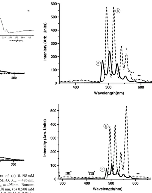
In order to get further insight into the solution proper-ties of the complexes, luminescence spectra of uranyl com-plexes in acetonitrile at both 291 and 77 K were recorded. The excitation spectra of the two complexes (Fig.2) display two main bands, one at 275 nm and the other at 225 nm, as well as a faint and broad feature centred at 325–330 nm, re-vealing both direct excitation into the uranyl ion (O→ U LMCT) [16] and indirect excitation from the ligand (absorp-tion spectrum,λmax= 275 nm). The phosphorescence spec-trum of UO2(B4bL4) is displayed on Fig.3(top) and reveals
the usual emission from lowest excited state of uranyl with five vibronic components at 495 (1), 516 (2), 540 (3), 564 (4, interfering with 2nd order Rayleigh scattering), and 593
(5) nm. Component 1 corresponds to the E(0−0) transition, formally a magnetic dipole transition, which acquires ap-preciable electric dipole character for non-centrosymmetric environments [16,44] it is red shifted by 13 nm with respect to uranyl nitrate, pointing to a sizeable uranyl-calixarene in-teraction.
In addition, a change of symmetry of the solvated uranyl cation (free uranyl in organic solvent) upon coordination to the calixarene (complexes) occurs since the intensity ratios of the vibronic bands are different with respect to [UO2(B6bL6)]2+in which the inner coordination sphere only
contains donor atoms from the calixarene. Vibronic bands are also broader (full width at half height, fwhh 7.7 vs. 6.5 cm−1) [6] pointing to a more fluxional edifice. Since the main vibronic spacing for 1 at 291 K (857 cm−1, see Table1) is the same as the one found for the 1: 1 complex with B6bL6, which has CN= 8, we postulate that 1 has also an
8-coordinate uranium centre in solution. The average vibronic spacing is related to the length of the O=U=O bond, rUO, by
the following equation [45]:
rUO= 10 650(νUO)−2/3+ 57.5 [pm]
And for both 1: 1 complexes with B4bL4and B6bL6, rUO=
177.8 and 178.6 pm, respectively are marginally longer than
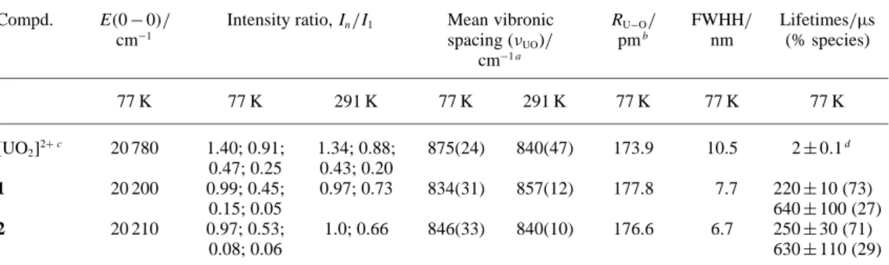
for the solvated uranyl ion, once more, in line with com-plexation by the calixarene. The emission spectrum of the 1: 2 complex 2 is very similar to the phosphorescence spec-trum of 1, apart slight differences in the intensity ratios of the vibronic components (Fig.3, Bottom), indicating a simi-lar chemical environment around the uranium ion, as far as the inner coordination sphere is concerned (CN= 8 for both
Fig. 2. Top: Excitation phosphorescence spectra of (a) 0.198 mM
B4bL4,λemi= 438 nm, (b) 5.1 mM UO2(NO3)2·6H2O, λem= 485 nm, (c) 0.347 mM UO2(NO3)2(B4bL4) 2H2O, 1, λemi= 495 nm. Bottom: similar spectra for (a) 0.198 mM B4bL4,λemi= 438 nm, (b) 0.508 mM UO2(NO3)2·6H2O,λem= 485 nm, (c) 0.198 mM UO2(B4bL4)2(NO3)2· 6H2O, 2,λemi= 495 nm. All spectra in frozen solutions of CH3CN at 77 K.
complexes).1ΠΠ∗ and3ΠΠ∗ remaining emissions
corres-pond to the coordinated calixarene ligands since no com-plete energy transfer from the ligand to uranyl occurs. The averaged uranyl-oxygen bond length calculated from the mean vibrational spacing is almost equal to the distance found for 1, within experimental error, while the fwhh is somewhat narrower, 6.7 nm (Table1), possibly indicating a more rigid coordination environment imposed by the two bound calixarenes and the absence of coordinated nitrate or water molecules.
Uranyl ion luminescent lifetimes are strongly depen-dent on the coordination environment. The luminescence decays for 1 and 2 in frozen acetonitrile are bi-exponential (Table1). The corresponding lifetimes are very similar: a short lifetime (0.22–0.25 ms) accounting for ≈ 70% of the emitted luminescence and a longer one (0.63–0.64 ms) representing ≈ 30%. An analogous situation with compa-rable lifetimes and populations has been reported for the complexes with B6bL6. Since the lifetime of un-complexed
uranyl is much shorter (< 2 µs, in organic solvents) [10] compared to complexed ones, and since the spectra of U(V) [46] and U(IV) [47] species are quite different,
dis-Fig. 3. Top: phosphorescence spectra of (a) 5.1 mM UO2(NO3)2·6H2O,
(λexc= 270 nm) and (b) 0.347 mM UO2(B4bL4)(NO3)2·2H2O, (λexc=
276 nm) in frozen solutions of CH3CN at 77 K; emission and
excita-tion slits: 5 nm. Bottom: similar spectra for (a) 0.508 mM UO2(NO3)2·
6H2O, (λexc= 270 nm) and (b) 0.198 mM UO2(B4bL4)2(NO3)2·6H2O, λexc= 278 nm; filter 290 nm, emission and excitation slits: 7 nm. Stars
denote second order Rayleigh scattering and double stars vibronic bands.1ΠΠ∗(singlet excited state) and3ΠΠ∗(triplet excited state.).
sociation of the complexes and reduction of uranyl can be ruled out; moreover, hydroxo species display much larger fwhh and can also be excluded [6]. Therefore, the bi-exponential decays reflect equilibria between two com-plexed species, possibly featuring different conformations of the calixarenes, one conformation being more “protective” than the other, leading to a longer lifetime.
3.3 X-photoelectron spectroscopy of uranyl complexes
The extent of the covalent and/or ionic character of the bonding, the coordination geometry and coordination num-bers, among others, determine the binding energies (BE) of the uranyl orbitals [48,49]. Therefore, XPS spectra of com-plexes 1 and 2 were recorded and are displayed on Figs.4
Table 1. Main luminescence parameters of uranyl and uranyl calixarene complexes in acetonitrile extracted from
phospho-rescence spectra.
Compd. E(0 − 0)/ Intensity ratio, In/I1 Mean vibronic RU−O/ FWHH/ Lifetimes/µs
cm−1 spacing (νUO)/ pmb nm (% species) cm−1 a 77 K 77 K 291 K 77 K 291 K 77 K 77 K 77 K [UO2]2+ c 20 780 1.40; 0.91; 1.34; 0.88; 875(24) 840(47) 173.9 10.5 2± 0.1d 0.47; 0.25 0.43; 0.20 1 20 200 0.99; 0.45; 0.97; 0.73 834(31) 857(12) 177.8 7.7 220± 10 (73) 0.15; 0.05 640± 100 (27) 2 20 210 0.97; 0.53; 1.0; 0.66 846(33) 840(10) 176.6 6.7 250± 30 (71) 0.08; 0.06 630± 110 (29)
a: Standard deviation (2σ) in parentheses; b: see text; c: in dry acetonitrile, this work; d: at room temperature, from Ref. [10].
Fig. 4. XPS spectra of UO2(B4bL4)(NO3)2·
2H2O.
Before interpreting these data, we stress the fact that all of the analytical tools used so far have demonstrated the pu-rity and stability (e.g. heating the KBr pellets did not alter the samples) of the complexes under investigation. High-vacuum treatment at room temperature led to removing the lattice water molecules but not the bonded ones. Irradiation times shorter than 15 min were used for XPS measurements in order to prevent the reduction of uranyl to lower oxi-dation state [48,50,51]. In addition, B4bL4 is neutral and
the P=O donors are linked to methyl groups which are less electron-donating substituents than phenyl groups, so that uranyl reduction as observed with OP(Phe)3 is unlikely to occur [51].
U(4f ) spectra
For both compounds, the uranium spectra reveal two broad peaks, U(4 f7/2) and U(4 f5/2), separated by 10.8 eV and
fea-turing shoulders on both sides. The spectra have been de-convoluted into Gaussian-Lorentzian functions and the best
fit yielded two main and well defined bands each flanked by a broad and weak shake-up satellite on the high binding en-ergy side and a well defined small band on the low-enen-ergy side. The two U(4 f5/2;7/2) doublets are labelled UI and UII.
The major one represents 81 and 74% of the bulk U con-centration for 1 and 2, respectively. In the case of U(4 f7/2)
the U proportions on the surface of the samples have also been measured and found to be the same as for the bulk sample (4: 1 and 3 : 1 ratios for 1 and 2, respectively). Bind-ing energies (BE) are quasi identical for both complexes, except UII(4 f ) which is 0.3 eV larger in 2, while the full
width at half height of all four peaks are the same. Some dif-ferences are seen between 1 (Fig.4a) and 2 (Fig.5a) with respect to the energies of the satellites,∆EI(sat) being equal
to 2.75 ( J= 7/2) and 3.05 eV (J = 5/2) for 1 compared to 3.52 and 3.31 eV for 2; similar data for ∆EII(sat) are 4.75
and 4.78 eV (1) and 5.04 and 4.83 eV (2). The position of the satellite peak with respect to the photoelectron peak de-pends on the energy difference between the ground state and the higher orbital to which the valence electrons of
ura-Table 2. Selected binding energies (BE) of uranium, oxygen, and phosphorus together with the
percentages of uranium atoms on the surface (IS) and in the sample (I).a
Levels UO2(NO3)2(B4bL4)·2H2O UO2(NO3)2(B4bL4)2·6H2O
BE/eV; IS, I (%), or FWHH/ BE/; IS, I (%), or FWHH/ ∆sat/eV eV ∆sat/eV eV UI4 f7/2 382.1; 5.2, 81.1 2.0 382.2; 2.8, 73.6 2.0 UII4 f7/2 380.4; 1.2, 18.9 2.0 380.7; 1.0, 26.4 2.0 ∆UI4 f7/2(sat) 2.8 3.5 ∆UII4 f7/2(sat) 4.8 5.0 UI4 f5/2 392.9 2.0 393.0 2.0 UII4 f5/2 391.2 2.0 391.5 2.0 ∆UI4 f5/2(sat) 3.05 3.3 ∆UII4 f5/2(sat) 4.8 4.8 O1s 531.2/532.8 1.8 530.9/532.6 1.8 P2s 132.8/134.1 1.7 132.5/133.8 1.7
a: Experimental conditions: spots diameter= 500 µm; 15 kV, 150 W.
Fig. 5. XPS spectra of UO2(B4bL4)2(NO3)2· 6H2O.
nium are excited, the valence of the elements and the type and number of its nearest-neighbours [48,49]. In particular, ∆E(sat) tends to decrease with increasing covalent charac-ter of the U-ligand bonds [52]. Reported average ∆E(sat) values are 4.0 (2.4–4.5), 6.6 (6–7), and 8 (7.8–8.5) eV for uranyl, U(IV), and U(V) compounds respectively [48–50,
52,53]. Although binding energy differences are larger than 4 eV for UII, they are well below 6 eV, so that we infer that
no reduction occurred in the samples, particularly in view of the other evidences reported above. The satellite features depend on the bonding environment and could therefore also be useful in estimating the covalent/ionic character of the U-ligand bonds; however a precise correlation is not presently at hand [50].
Moreover, binding energies for various oxidation states are often very close to each other. For instance, in the
uranium mineral brannerite, two U(VI) species in differ-ent structural environmdiffer-ents were iddiffer-entified, which give rise to two bands at 381.4, and 382.1 eV, while another band at 380.6 eV was assigned to U(V). Furthermore, in Na-substituted metaschoepite a band at 380.5 eV was assigned to U(IV). This demonstrates that there is a narrow border-line between the U(4 f ) bonding energies of different ox-idation numbers. On the other hand, two different U(VI) species were identified in this material characterized by U(4 f7/2) bands at 381.3 and 382.1 eV [48]. Regarding the
effect of X-ray irradiation, an exposure of several hours of metaschoepite only induced 5% reduction of U(VI) [48] while a 15-minute exposure of U(VI) deposited on mica did not produce enough reduced species to be detected [50]. The second set of signals is consequently assigned to a differ-ent coordination environmdiffer-ent for the uranium ions. Ligand
B4bL4has a cone conformation in acetonitrile which is
re-tained in its lanthanide complexes. However, luminescence studies of the uranyl complexes in frozen acetonitrile so-lutions revealed the presence of two structurally different species possibly differing by the calixarene conformation. It is noteworthy that the proportion of the emitting species with the longer lifetime, ≈ 30% (Table1) is in reasonably good agreement with that associated with UII(19–26%),
es-pecially given the fact that the medium is different (frozen solution vs. solid state).
O1 sand P2 pspectra
Spectra of the O1s and P2 p levels are broad, the former
re-vealing two resolved maxima while the latter are asymmetric on their high-energy side. The best fit in the deconvolu-tion of the O1s spectrum (Figs. 4b and 5b) yielded two
peaks with different intensities and FWHH= 1.8 eV. In add-ition to uranyl oxygen atoms, both complexes 1 (Fig.4b) and 2 (Fig.5b) have several types of oxygen atoms, phos-phoryl and ether groups from the calixarene molecules, nitrate and water molecules. It has recently been demon-strated that the nature of ligands influence the BE of uranyl oxygen atoms. In most compounds reported to date BE(O1s) lies in the range 530.8–532.4 eV (FWHH ≈ 2 eV);
exceptions are nitrate salts MUO2(NO3)3 (M= Cs, Rb)
and UO2(NO3)2·2H2O (533.6–533.9 eV, one peak reported)
while the oxide CaUO2O2has BE(O1S)= 530.2 eV [49,52].
We note that in uranium minerals, interstitial water and hy-droxyl groups have BE(O1s) in the range 532.1–533.8 eV
[49]. Therefore, we assign the bands centred at 531.2 and
530.9 eV to uranyl oxygen atoms and phosphoryl oxygen
atoms [54–56], while those centred at 532.8 and 532.6 eV correspond to the other oxygen atoms (phenoxy-, nitrate, water) for compounds 1 and 2, respectively. The higher en-ergy band in the O1s spectrum of 1 (Fig.4b) is less intense
compared to 2 (Fig.5b), which would be consistent with the smaller content of lattice water.
Similarly to the O1sspectra, the P2 p bands of 1 (Fig.4c)
and 2 (Fig.5c) can be de-convoluted into two components with different intensities. The BE of phosphorus in O=P groups coordinated to uranyl is affected by the U−OP bond strength. A stronger coordination of U−OP means signifi-cant PO→ U electron transfer, then the photoelectron en-ergy of P2 p increases. The more intense band at 132.8 eV
(1) and 132.5 eV (2) is therefore associated with a less strong U−OP bond in a complex with the less stable con-formation while the less intense band at 134.1 (1) and
133.8 eV(2) is associated with a stronger and shorter U−OP
bond.
3.4 Molecular modelling
In order to substantiate the experimental results described above, model calculations have been performed both in vacuo and in a polar solvent on the free calixarene and on the actinide complexes. Surprisingly, the predominant low-energy isomer calculated for B4bL4was a 1,3 alternate
conformer, even in highly polar water, while in less polar solvents such as CHCl3and CH3CN, NMR data are
consis-tent with a cone conformation. Such conformation was also
found for the acetonitrile adduct of this calixarene in the solid state by X-ray crystallography [36].
The structural versatility of actinide complexes in solu-tion and solid arises from the lack of strong crystal field effects for the 5 f electronic configurations as well as from their large ionic radii. The predominant ionic character of the bonding leads to a wide variety of coordination num-bers (CN) and symmetries. The uranyl ion is usually re-stricted to CN= 4–6 [1,13,16,57,58]. For CN= 6, due to the linearity of UO2
2+
, the six donor atoms are usu-ally located in the equatorial plane, but it is also com-mon that a distorted hexagonal bipyramidal polyhedron be found with the six donor atoms arranged in a puckered fashion.
Modelled uranyl calixarene molecules with AugMM3/ CONFLEX at vacuum or AugMM3/dynamics calculations at 300 K revealed U–OP, U–OH2and NO2–U lengths in the
ranges reported for similar complexes containing these types of donors (2.3–2.7 Å) but the uranyl bond angles (e.g. 169◦ for 1, 126◦for 2) were out of the accepted range for a hexa-coordinate uranyl complex (176–180◦) [13,16,57,58]. The modelling was based on the experimental data, therefore the structures of 1 and 2 were built for uranyl in a hexa-coordinate geometry.
The structure of 1, a monocationic complex [UO2B4bL4(NO3)(H2O)]1+ has four OP arms, one
mon-odentate nitrate and one water molecule coordinated to uranyl. Its molecular modelling yielded structural parame-ters as U−OP bond lengths, U−O=P bond angles varying from one coordinated OP arm to the other, pointing to four OP arms in an unsymmetrical arrangement around the uranyl in agreement with the spectroscopic results. For the structure of 2, a bicationic complex [UO2(B4bL4)2]2+ was
modelled in two arrangements: (i) with 4 OP arms of one calixarene and two of the other bound to the uranyl, and (ii) with three OP arms from each calixarene. In both cases, the minimum energy was high. Therefore, considering the affinity of water towards OP groups and the presence of six water molecules in 2, water molecules were linked to each one of the free OP arms. The most stable structure was that of three OP arms per calixarene coordinated to uranyl where the free OP of each calixarene was linked to one water molecule by weak bonding simulating hydro-gen bonding. This reduced three times the mobility of the complex. Both calixarenes are located in the first coordi-nation sphere of the uranyl and hydrogen bonded to water molecules, thus the structure of the complex can be written as [H2O···B4bL4→ UO2← B4bL4···H2O]2+.
In spite of the flaw concerning the O=U=O bond angle, the modelling reflects an 8-coordinate U(VI) ion in both complexes in agreement with the experimental results.
DFT calculations were attempted on complex 1 in order to find a more realistic O=U=O angle bond. The standard uranyl valence configuration 7s5 f 6d was found to be the most adequate for the ab initio calculations including rel-ativistic effects, which yielded an optimized molecule in which the linearity of uranyl is practically maintained but not the U−O bond length of uranyl. In fact, the O−U−O angle found (178.8◦) is comparable to the one for uranyl complexes with phosphoryl [59] and nitrate [60] donors (177–180◦) but which are highly asymmetric. Several
fac-tors exert symbiotic effects leading to this asymmetry: the size of the calixarene which does not permit complete in-clusion of the uranyl cation, steric and electronic restrictions due to the linear geometry of uranyl, the mono-coordinate mode of nitrate, and coordinated water molecules. The found geometrical parameters associated with the calcu-lated coordination polyhedron of 1 like U−OP bond lengths, PO−U−OP bond angles and dihedral angles do not allow us to propose the modeled molecule by DFT as the representa-tive of compound 1. Further interpretation was therefore not conducted.
3.5 Liquid–liquid extraction
Generally speaking, actinide cations are much better ex-tracted than rare earths by phosphinoylated calixarenes, which has been encouraging in the perspective of An/Ln separation in liquid–liquid or liquid–solid systems [6,22–31]. Therefore, in this work, the extraction ability of B4bL4
to-wards UO2 2+
, Y(III), La(III), and Eu(III) ions was also tested. The study was performed using three different aque-ous nitric phases (aqueaque-ous phases 1, 2 and 3, see experimen-tal) and two different calixarene concentrations in chloro-form corresponding to metal : ligand ratios of≈ 1 (organic phase 4) and ≈ 2.5 (phase 5). It was found that increas-ing the concentration of the calixarene by a factor 2.5 in going from organic phase 4 to phase 5 leads to a concomi-tant increase in the extraction of the uranyl cations (average increase is > 2.3-fold), in line with the formation of 1 : 2 complexes discussed above. Increasing the concentration of nitric acid in the aqueous phase from 1 to 3 M is quite detri-mental to uranyl extraction (see experidetri-mental) and blocks lanthanum and yttrium ion extraction. On the other hand, boosting sodium nitrate concentration from 0.5 to 3.5 M, the uranyl extraction is not substantially increased but its distribution ratio goes from 0.53 to 1.90. However, the in-crease in nitrate concentration gives rise to a remarkable improvement in La3+extraction, by a factor≈ 10 while Eu3+ extraction drops to 0. It has been recently demonstrated that the salting-out effect plays an important role in La3+
ex-traction with calix[6]arene B6bL6[6]. In fact, the behaviour
of B4bL4 as extractant for rare earths is similar to that
found for the p-tert-butylcalix[4]arene derivative contain-ing diphenyl-phosphinoyl groups [25] and others [26,30]. B4bL4 has also proved to be useful as a synergistic agent
in the solvent extraction of lanthanides with a pyrazolone derivative [39].
It has been found [6] that the calixarene concentration required for determining the stoichiometry of the extracted species has to be much larger than 3× 10⁻⁴M (up to ap-prox. 1× 10⁻³M, in CHCl3). In the present work, extraction
of uranyl with more concentrated calixarene solutions did not give good results due to an emulsion formed between the organic and aqueous phases so that a suitable graphic to evaluate the number of calixarene molecules bound to uranyl could not be built. However, extracted species with two pre-dominant stoichiometries: 1 M : 1 L and 1 M : 2 L have been reported for similar phosphinoylated calixarenes in chloro-form [6] and in m-nitrobenzotrifluoride [30]. According to the extraction behaviour of uranyl in presence of B4bL4, it is
anticipated that a 1 : 1 species was extracted.
4. Conclusions
The tetra-phosphinoylated p-tert-butylcalix[4]arene forms stable uranyl(VI) complexes with 1: 1 and 1 : 2 stoichiome-tries in organic media. A combination of several experimen-tal analytical techniques and theoretical modelling leads to the conclusion that coordination numbers for U(VI) ion are most probably 8 in these edifices. The uranyl complexes dis-play enhanced luminescence and longer lifetimes due to the complexation, which allows one to get information on the solution composition: for both stoichiometries, lifetime data point to the existence of two different species in solution fea-turing different conformations of the calixarene, as pointed out by molecular modelling. These species also exist in the solid state as proved by XPS data. It is noteworthy that the proportion of the emitting species with the longer lifetime (27–29%) is in reasonably good agreement with that asso-ciated with UII (19–26%), especially given the fact that the
medium is different (frozen solution vs. solid sate). There-fore, it is proposed that for 1 and 2, in solid and in solution, the U(VI) central ion is in an 8-coordinate geometry.
The B4bL4calixarene is a reasonably good extractant for
uranyl(VI) ions but a poor one for rare earths. Furthermore, the extraction behaviour and separation ability is much de-pendent on the initial conditions, pH and nitrate concentra-tion, so that modulation of the extraction process is easy. Further study on these systems using more lipophilic macro-cyclic receptors in which the wider rim of the calixarene is decorated with octyl substituents is in progress.
Acknowledgment. This work was supported through grants from
CONACYT (M´exico), project Nr. 36689-E and the Swiss National Science Foundation project SCOPES No 7BUPJ062293. We thank Q. E. Ricardo Soria from the Analytical department for his help in the uranium measurement by ICP and the technicians from the Chemistry Department of ININ.
References
1. Cotton, S.: Lanthanide and Actinide Chemistry. 1st
Edn., John Wi-ley & Sons Ltd., Chichester (2006).
2. Mishra, S.: Coord. Chem. Rev. 252, 1996 (2008).
3. Sutton, A. D., John, G. H., Sarsfield, M. J., Renshaw, J. C., May, L., Martin, L. R., Selvage, A. J., Collison, D., Helliwell, M.: Inorg. Chem. 43, 5480 (2004).
4. Billard, I.: Lanthanide and actinide solution chemistry as studied by time-resolved emission spectroscopy. In: Handbook on the
Physics and Chemistry and Rare Earth. (Gschneidner Jr., K. A.,
Bünzli, J.-C. G., Pecharsky, V. K., eds.) Elsevier Science B. V., Amsterdam (2003).
5. Thuery, P., Nierlich, M., Masci, B., Asfari, Z., Vicens, J.: J. Chem. Soc., Dalton Trans. 3151 (1999).
6. Ramirez, F. d. M., Varbanov, S., Padilla, J. P., Bünzli, J.-C. G.: J. Phys. Chem. B 112, 10976 (2008).
7. Choppin, G. R., Rizkalla, E. N.: Solution chemistry of actinides and lanthanides. In: Handbook on the Physics and Chemistry
of Rare Earths. (Gunzler, H., Eyring, L., Choppin, G. R.,
Lan-der, G. H., eds.) Elsevier Science Publ. B.V., Amsterdam (1994). 8. Edelstein, N. M.: J. Alloys Compd. 223, 197 (1995).
9. Moulin, C., Laszak, I., Moulin, V., Tondre, C. : Appl. Spectrosc.
52, 528 (1998).
10. Darmanyan, A. P., Khudyakov, I. V.: Photochem. Photobiol. 52, 293 (1990).
11. Servaes, K., De Houwer, S., Görller-Walrand, C., Binnemans, K.: Phys. Chem. Chem. Phys. 6, 2946 (2004).
12. De Houwer, S., Servaes, K., Görller-Walrand, C.: Phys. Chem. Chem. Phys. 5, 1164 (2003).
13. Umeda, K., Zukerman-Schpector, J., Isolani, P. C.: Polyhedron 25, 2447 (2006)
14. Zucchi, G., Maury, O., Thuery, P., Gumy, F., Bünzli, J.-C. G., Ephritikhine, M.: Chem. Eur. J. 15, 9686 (2009).
15. Denning, R. G.: Properties of the UO2n+ (n= 1, 2) ions. In: Gmelin’s Handbook of Inorganic Chemistry “U”. (Fluck, E., ed.)
Springer Verlag, Berlin (1983), A6, Sect. 2, p. 31. 16. Denning, R. G.: J. Phys. Chem. A 111, 4125 (2007).
17. Wiebke, J., Moritz, A., Cao, X., Dolg, M.: Phys. Chem. Chem. Phys. 9, 459 (2007).
18. Schlosser, F., Kruger, S., Rosch, N.: Inorg. Chem. 45, 1480 (2006).
19. Wiebke, J., Moritz, A., Glorius, M., Moll, H., Bernhard, G., Dolg, M.: Inorg. Chem. 47, 3150 (2008).
20. Boulet, B., Joubert, L., Cote, G., Bouvier-Capely, C., Cosson-net, C., Adamo, C.: Inorg. Chem. 47, 7983 (2008).
21. Baaden, M., Berny, F., Muzet, N., Troxler, L., Wipff, G.: In:
Cal-ixarenes for Separations. (Lumetta, G. J., Rogers, R. D.,
Ga-lopan, A. S., eds.) ACS Symposium Series No. 757, Am. Chem. Soc., Washington, D.C. (2000), Chapt. 6, p. 71ff.
22. Sliwa, W., Girek, T.: J. Inclus. Phenom. Macrocycl. Chem. 66, 15 (2010).
23. Mokhtari, B., Pourabdollah, K., Dallali, N.: J. Radioanal. Nucl. Chem. 287, 921 (2011).
24. Mokhtari, B., Pourabdollah, K., Dallali, N.: J. Inclus. Phenom. Macrocycl. Chem. 69, 1 (2011).
25. Yaftian, M. R., Burgard, M., Matt, D., Dieleman, C. B., Raste-gar, F.: Solv. Extr. Ion Exch. 15, 975 (1997).
26. Arnaud-Neu, F., Browne, J. K., Byrne, D., Marrs, D. J., McKer-vey, M. A., O’Hagan, P., Schwing-Weill, M.-J., Walker, A.: Chem. Eur. J. 5, 175 (1999).
27. Arnaud-Neu, F., Barboso, S., Byrne, D.: In: Calixarenes for
Sepa-rations. (Lumetta, G. J., Rogers, R. D., Galopan, A. S., eds.) ACS
Symposium Series No. 757, Am. Chem. Soc., Washington, D.C. (2000), Chapt. 12, p. 150ff.
28. Alexandratos, S. D., Natesan, S.: Ind. Eng. Chem. Res. 39, 3998 (2000).
29. Asfari, Z., Böhmer, V., Harrowfield, J. M., Vicens, J., Saadioui, M. (eds.): Calixarenes 2001. Kluwer Academic Publishers, Dord-recht, Boston, London (2001).
30. Karavan, M., Arnaud-Neu, F., Hubscher-Bruder, V., Smirnov, I., Kalchenko, V.: J. Inclus. Phenom. Macrocyc. Chem. 66, 113 (2010).
31. Talanova, G. G.: Ind. Eng. Chem. Res. 39, 3550 (2000). 32. Galletta, M., Baldini, L., Sansone, F., Ugozzoli, F., Ungaro, R.,
Casnati, A., Mariani, M.: Dalton Trans. 39, 2546 (2010). 33. Hall, I., Nicholson, G. P., Piper, T. J., Tay, D. M., Williams, D. R.:
Radiochim. Acta 69, 225 (1995).
34. Ramirez, F. d. M., Charbonnière, L. J., Muller, G., Scopelliti, R., Bünzli, J.-C. G.: J. Chem. Soc., Dalton Trans. 3205 (2001).
35. Ramirez, F. d. M., Charbonnière, L. J., Muller, G., Bünzli, J.-C. G.: Eur. J. Inorg. Chem. 2348 (2004).
36. Le Saulnier, L., Varbanov, S., Scopelliti, R., Elhabiri, M., Bünz-li, J.-C. G.: J. Chem. Soc., Dalton Trans. 3919 (1999).
37. Ramirez, F. d. M., Varbanov, S., C´ecile, C., Muller, G., Fatin-Rouge, N., Scopelliti, R., Bünzli, J.-C. G.: J. Chem. Soc., Dalton Trans. 4505 (2002).
38. Puntus, L. N., Chauvin, A.-S., Varbanov, S., Bünzli, J.-C. G.: Eur. J. Inorg. Chem. 2315 (2007).
39. Atanassova, M., Lachkova, V., Vassilev, N., Varbanov, S., Du-kov, I.: J. Inclus. Phenom. Macrocycl. Chem. 58, 173 (2007). 40. CERIUS2, Accelrys Software Inc., San Diego, CA, USA (2010).
41. Te Velde, G., Bickelhaupt, F. M., Baerends, E. J., Fonseca Guer-ra, C., Van Gisbergen, S. J. A., Snijders, J. G., Ziegler, T.: J. Com-put. Chem. 22, 931 (2001).
42. van Lenthe, E., Ehlers, A., Baerends, E. J.: J. Chem. Phys. 110, 8943 (1999).
43. Nakamoto, K.: Infrared and Raman Spectra of Inorganic and
Co-ordination Compounds. Part A. Theory and Applications in Inor-ganic Chemistry. John Wiley Interscience Publ., New York (1997).
44. Burrows, H. D., Miguel, M. D.: Adv. Colloid Interface Sci. 89, 485 (2001).
45. Bartlett, J. R., Cooney, R. P.: J. Mol. Struct. 193, 295 (1989). 46. Grossmann, K., Arnold, T., Ikeda-Ohno, A., Steudtner, R.,
Gei-pel, G., Bernhard, G.: Spectrochim. Acta A 72, 449 (2009). 47. Kirishima, A., Kimura, T., Nagaishi, R., Tochiyama, O.:
Radio-chim. Acta 92, 705 (2004).
48. Schindler, M., Hawthorne, F. C., Freund, M. S., Burns, P. C.: Geochim. Cosmochim. Acta 73, 2471 (2009).
49. Schindler, M., Hawthorne, F. C., Freund, M. S., Burns, P. C.: Geochim. Cosmochim. Acta 73, 2488 (2009).
50. Ilton, E. S., Boily, J. F., Bagus, P. S.: Surf. Sci. 601, 908 (2007). 51. Duval, P. B., Kannan, S.: J. Alloys Compd. 444, 673 (2007). 52. Teterin, A. Y., Teterin, Y. A.: Russ. Chem. Rev. 73, 541 (2004)
[Engl. translation].
53. Ilton, E. S., Haiduc, A., Cahill, C. L., Felmy, A. R.: Inorg. Chem.
44, 2986 (2005).
54. Bourbigot, S., Le Bras, M., Delobel, R., Gengembre, L.: Appl. Surf. Sci. 120, 15 (1997).
55. Jalil, P. A., Faiz, M., Tabet, N., Hamdan, N. M., Hussain, Z.: J. Catal. 217, 292 (2003).
56. Ignatova, M., Manolova, N., Lachkova, V.,Varbanov, S., Rash-kov, I.: Macromol. Rapid Commun. 29, 1871 (2008).
57. Deshayes, L., Keller, N., Lance, M., Navaza, A., Nierlich, M., Vig-ner, J.: Polyhedron 13 1725 (1994).
58. Thuery, P., Keller, N., Lance, M., Vigner, J. D., Nierlich, M.: New J. Chem. 19, 619 (1995).
59. de Aquino, A. R., Bombieri, G., Isolani, P. C., Vicentini, G., Zukerman-Schpector, J.: Inorg. Chim. Acta 306, 101 (2000). 60. Caville, C., Poulet, H.: J. Inorg. Nucl. Chem. 36, 1581 (1974).
![Fig. 1. (a) Calixarenes with phosphinoyl pen- pen-dant arms in the lower rims, (b) tetra-phosphino-ylated p-tert-butylcalix[4]arene, B 4 bL 4](https://thumb-eu.123doks.com/thumbv2/123doknet/14929049.664531/2.892.64.544.101.395/calixarenes-phosphinoyl-lower-tetra-phosphino-ylated-butylcalix-arene.webp)
![[PDF] Cours pdf de Le langage Haskell enjeux et pratique | Formation informatique](data:image/gif;base64,R0lGODlhAQABAIAAAP///wAAACH5BAEAAAAALAAAAAABAAEAAAICRAEAOw==)