Publisher’s version / Version de l'éditeur:
Journal of Sol-Gel Science and Technology, 32, December, pp. 323-326, 2004
READ THESE TERMS AND CONDITIONS CAREFULLY BEFORE USING THIS WEBSITE. https://nrc-publications.canada.ca/eng/copyright
Vous avez des questions? Nous pouvons vous aider. Pour communiquer directement avec un auteur, consultez la première page de la revue dans laquelle son article a été publié afin de trouver ses coordonnées. Si vous n’arrivez pas à les repérer, communiquez avec nous à PublicationsArchive-ArchivesPublications@nrc-cnrc.gc.ca.
Questions? Contact the NRC Publications Archive team at
PublicationsArchive-ArchivesPublications@nrc-cnrc.gc.ca. If you wish to email the authors directly, please see the first page of the publication for their contact information.
NRC Publications Archive
Archives des publications du CNRC
This publication could be one of several versions: author’s original, accepted manuscript or the publisher’s version. / La version de cette publication peut être l’une des suivantes : la version prépublication de l’auteur, la version acceptée du manuscrit ou la version de l’éditeur.
For the publisher’s version, please access the DOI link below./ Pour consulter la version de l’éditeur, utilisez le lien DOI ci-dessous.
https://doi.org/10.1007/s10971-004-5810-8
Access and use of this website and the material on it are subject to the Terms and Conditions set forth at
The effect of preparation method and calcination temperature on the
crystallite size and surface area of perovskite-type SrFeOx
Majid, Abdul; Tunney, Jim; Argue, Steven; Post, Mike
https://publications-cnrc.canada.ca/fra/droits
L’accès à ce site Web et l’utilisation de son contenu sont assujettis aux conditions présentées dans le site LISEZ CES CONDITIONS ATTENTIVEMENT AVANT D’UTILISER CE SITE WEB.
NRC Publications Record / Notice d'Archives des publications de CNRC:
https://nrc-publications.canada.ca/eng/view/object/?id=21adeeca-3fb3-41e0-86a6-2bd7136bef1a https://publications-cnrc.canada.ca/fra/voir/objet/?id=21adeeca-3fb3-41e0-86a6-2bd7136bef1aThe Effect of Preparation Method and Calcination Temperature
on the Crystallite Size and Surface Area of Perovskite-Type SrFeO
xABDUL MAJID, JIM TUNNEY, STEVE ARGUE AND MIKE POST
Institute for Chemical Process and Environmental Technology, National Research Council of Canada, Ottawa, Ontario K1A 0R9, Canada
abdul.majid@nrc-cnrc.gc.ca
Abstract. We have investigated the synthesis of perovskite-type SrFeOx(2.5 ≤x ≤ 3.0) using three preparative
methods: sol-gel, mechanochemical processing and solid state reactions at high temperature of the corresponding oxides. The sample obtained after calcination of the gel from sol-gel method, contained the least amount of strontium carbonate impurity. The amount of strontium carbonate impurity decreased with the increase in calcination temper-ature. Perovskites obtained have been characterized by X-ray diffraction (XRD) and nitrogen adsorption isotherms. Samples obtained from three methods have been compared with respect to calcination temperature, crystallite size and specific surface area.
Keywords: mechanochemical processing, perovskites, sol-gel, SrFeOx
Introduction
Since the discovery at Mobil [1, 2] of MCM-41, a meso-porous silica material, there has been an increasing in-terest to develop new ways to prepare high surface area mesoporous oxide materials including transition met-als oxides using wet chemical methods [3–5]. How-ever, relatively little of this work has targeted per-ovskites. This is surprising since many perovskites are of particular interest as they have many potential com-mercial applications such as materials for solid oxide fuel cells (SOFC’s) [6] and gas sensor devices [7–11]. In order to fully optimize the functionality of these materials, the achievement of controlled porous struc-tures is desirable, since these applications depend to a large extent on the surface area to volume ratio. More-over, it has been demonstrated that by decreasing the dimensionality of electroceramic materials, including many perovskites, to the 5–100 nm ranges, both the electronic and ionic transport properties may be dra-matically affected, leading to improved performance [12–14].
Issued as NRCC No. 46479.
The conventional method for the preparation of hetero-metallic oxides, such as perovskites, is the solid-state reaction at high temperatures of the correspond-ing binary metal oxides or carbonates. Given that the synthesis occurs at high temperatures, the powders pro-duced with this method are non-porous, coarse, with a non-uniformity of particle size and shape, and have a low specific surface area. The development of innova-tive processing methods through chemistry permits one to lower the preparation temperature and to improve ho-mogeneity and reproducibility of the ceramic products, with the synthesis of porous, ultrafine and chemically pure powders of mixed metal oxides at low temper-atures. Wet chemical methods and mechanochemical processing are two low temperature methods that fall into this category.
In this investigation, we have prepared the non-stoichiometric perovskite, SrFeOx (2.5 ≤ x ≤ 3.0)
[15, 16] using wet chemical methods, mechanochemi-cal processing and solid state reaction in order to com-pare the crystallite size and specific surface area of the samples. SrFeOxis a mixed ionic electronic conductor
in which the reversible oxygen nonstoichiometry may be exploited for gas sensor applications [11, 17–19].
324 Majid et al.
Experimental Methods
Materials
All reagents were obtained from Aldrich and used as re-ceived. The purity of the iron (III) oxide and strontium carbonate starting materials were greater than 99.9% (metals basis), Iron and strontium salts were kept and handled in an inert dry box under argon.
Hardened steel and zirconia vials and balls for mechanochemical syntheses were purchased from ATS Scientific Inc., Burlington, Ontario.
Preparation of SrFeOx
Solid State Reaction. Strontium and iron oxides were mixed in stoichiometric amounts (Sr:Fe = 1:1), ground together with an agate mortar and pestle and calcined at 1100◦
C in a muffle furnace. The resulting black prod-uct was repeatedly re-mixed, ground and calcined at 1100◦
C until the XRD pattern indicated the complete conversion to the SrFeOxperovskite phase.
Mechano-Chemical Method. Strontium oxide and iron (III) oxide (both 99.9%) were mixed in a stoichio-metric ratio of Sr:Fe of 1:1 in an agate mortar and pestle under argon in a dry box. The mixture was transferred to a hardened steel vial (size: 2.25′′×
3.0′′
) with steel balls in the dry box. The ball to-powder-weight ratio was kept between 12:1 to 6:1. The contents were agitated on a SPEX 8000 M mixer/mill for two hours at a time. Each time a small amount of sample was removed from the vial in a dry box for XRD investigation. The pro-cess was continued until the XRD pattern indicated the least amount of impurity (∼35 hrs). The reaction was repeated in zirconia vial with zirconia balls, for which a longer reaction time (>40 hrs) was required. This could be a consequence of the more effective sealing of the hardened steel vial versus that of the zirconia vial.
Mechano-Chemical Processing Followed by Calcination. The product obtained from mechano-chemical processing contained considerable quantities of SrCO3and Sr3Fe2O∼6.7(Ruddlesden-Popper phase)
as impurities. This was calcined at 600◦
C for 50 hrs.
Sol-Gel Method. For sol-gel, a modified Pechini’s method was used [20]. The nitrate salts of strontium and iron in a stoichiometric ratio of iron to strontium of 1:1 were transferred to a round bottom flask under
argon in a dry box. An appropriate amount of water was added to dissolve the two salts. Citric acid in a metal ions to citric acid mole ratio of 1:1 was then added and dissolved by stirring. The pH of the resultant metal citrate solution was adjusted to 6–7 by adding dilute ammonia solution drop wise. The solution was heated in a water bath while stirring with a magnetic stirrer to obtain a viscous solution. At this stage, ethylene glycol was added in a mole ratio of citric acid to ethylene glycol of 1:1.2. Heating and stirring was continued until the solution started solidifying forming a gel-like porous mass. At this stage the temperature was raised to 180–200◦
C to obtain a foamy dry mass. This was ground using an agate mortar and pestle using reagent grade acetone. This ground mass was dried and ashed at 400◦
C in a muffle furnace to completely remove the citrates and ethylene glycol that was left. A reddish brown coloured substance obtained was used as a precursor for SrFeOx. This precursor was calcined at
different temperatures in a muffle furnace to obtain SrFeOxas a blackish grey powder.
Measurements
The N2 adsorption-desorption isotherms were
mea-sured at 77 K on a Micromeritics ASAP2000 appara-tus. Before measurement, the samples were evacuated overnight at 120◦
C until the pressure was 10−6torr.
The density of the materials was determined by a py-cnometry measurement with helium using Micromerit-ics Accupyc 1330 apparatus.
X-ray powder diffraction data were collected be-tween 20◦≤
2θ ≤ 80◦
with a scan rate of 2◦
/min at room temperature on a Scintag XDS 2000 with a theta-theta geometry and a copper X-ray tube. The diffractome-ter had a pyrolytic graphite monochromator in front of the detector. The samples were mounted on a zero background sample holder made of an oriented sili-con wafer. The average crystallite size was determined from a convolution based full pattern fitting using the Topas software package [21]. Peaks were fitted using a simplified integral breadth method to account for both strain and crystallite size effects [22].
Results and Discussion
X-Ray Diffraction
The perovskite SrFeOx was prepared using four
Figure 1. XRD patterns for SrFeOx, prepared using various
methods.
oxides at 1100◦
C, (2) agitation of strontium and iron oxides on a Spex mixer (mechano-chemical process-ing), (3) mechanochemical processing followed by cal-cination at 600◦
C and (4) sol-gel processing followed by calcination at 600◦
C. Figure 1 shows XRD patterns for samples prepared from all four methods. The sol-gel processing was the only method yielding a rea-sonably pure phase of the SrFeO∼2.9perovskite phase,
as indicated from the XRD spectra shown in Fig. 1. The sample obtained from the solid state preparation method at 1100◦
C contained considerable amounts of Sr3Fe2O∼6.7 impurity. The removal of impurity
re-quired repeated re-mixing and calcination cycles. This is apparent from the XRD patterns (Fig. 2) which show that the amount of impurity is reduced considerably after calcination for 100 hrs at 1100◦
C. However, com-plete reduction of Sr3Fe2O∼6.7impurity required five
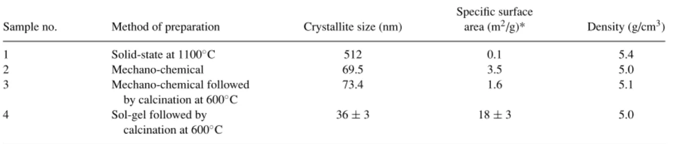
Table 1. Textural properties obtained for the SrFeOxproducts.
Specific surface
Sample no. Method of preparation Crystallite size (nm) area (m2/g)* Density (g/cm3)
1 Solid-state at 1100◦C 512 0.1 5.4 2 Mechano-chemical 69.5 3.5 5.0 3 Mechano-chemical followed 73.4 1.6 5.1 by calcination at 600◦C 4 Sol-gel followed by 36 ± 3 18 ± 3 5.0 calcination at 600◦C
*By N2adsorption/desorption isotherms at 77 K.
Figure 2. The effect of calcination time at 1100◦C on impurity
reduction from SrFeOx.
re-mixing and calcination cycles for a total of 200 hrs at 1100◦
C. The mechanochemical processing method produced even greater amounts of impurities than the solid-state method. The mechanochemical processing followed by calcination at 600◦
C reduced these im-purities significantly but resulted in additional impuri-ties of SrCO3 and Sr3Fe2O∼6.7 as suggested from the
XRD shown in Fig. 1. The remaining impurity could be removed by raising the calcination temperature to 900◦
C.
Crystallite Size
The average crystallite size of the samples prepared by the four methods was determined from the XRD data and is shown in Table 1. The samples obtained using
326 Majid et al.
solid state reaction of the oxides at high temperature had the greatest crystallite size of all samples. The crystallite size of the samples prepared using sol-gel methodology was the lowest. The samples pro-duced from mechano-chemical processing had ap-proximately double the crystallite size as compared to the samples obtained by sol-gel method. For the solid state method, long periods of calcination at higher temperatures results in a significant growth of crystallites.
Textural Properties
The textural properties of all samples prepared using the four methods were investigated by the N2 adsorp-tion/desorption isotherms at 77 K and are summarized in Table 1. A type III isotherm was obtained for all sam-ples suggesting a very weak adsorption interaction of which the fundamentals are not very well understood [23]. The data indicate the presence of both micropores and mesopores in these samples.
The samples prepared by sol-gel method exhibit the largest specific surface areas, which decreased with the increase in calcination temperature. This can be explained on the basis of the partial collapse of the micropore structure at higher temperatures [24]. The specific surface areas of the samples prepared by the solid-state method was the lowest.
Density
The density of the samples prepared by various methods did not vary significantly. The sample pre-pared by the solid-state method had the highest den-sity; the lowest density value was obtained for the samples prepared by sol-gel and mechanochemical processing.
Conclusion
The perovskite SrFeOx was successfully prepared by
sol-gel processing at much lower temperatures than with conventional solids-state methods. Samples pre-pared by the sol-gel method had greater surface area and lower crystallite size compared to the samples pre-pared by solid state methods.
Acknowledgments
The authors are grateful to Pascal L’Ecuyer, Gerry Pleizier, and Dr. Pamela Whitfield for some technical assistance.
References
1. J.S. Beck, J.C. Vartuli, W.J. Roth, M.E. Leonowicz, C.J. Kresge, K.D. Schmitt, C.T.W. Chu, D.H. Olson, E.W. Sheppard, S.B. McCullen, J.B. Higgins, and J.L. Schlenker, J. Amer. Chem. Soc.114, 10834 (1992).
2. J.S. Beck, J.C. Vartuli, G.J. Kennedy, C.J. Kresge, W.J. Roth, and S.E. Schramm, Chem. Mater6, 816 (1994).
3. Q.S. Huo, D.J. Margolese, U. Ciesla, P. Feng, T.E. Gier, P. Sieger, R. Leon, P.M. Petroff, F. Schuff, and G.D. Stucky, Nature368,
317 (1994).
4. Q.S. Huo, D.J. Margolese, U. Ciesla, D.G. Dermuth, and G.D. Stucky, Chem. Mater6, 1176 (1994).
5. F. Schuth, Chem. Mater.13, 3184 (2001).
6. R.M. Ormerod, Chem. Soc. Reviews32, 17 (2003).
7. H. Meixner and U. Lampe, Sensors and Actuators B,33, 198
(1996).
8. P.T. Moseley and D.E. Williams, Polyhedron9, 1615 (1989).
9. E. Traversa, P. Nunziante, M. Sakamoto, Y. Sadoaka, M.C. Carotta, and G.J. Martinelli, Mater. Res.3, 1335 (1998).
10. J.J. Tunney, M.L. Post, X. Du, and D. Yang, J. Electrochemical Soc.149, H113 (2002).
11. O. Grudin, R. Marinescu, L.M. Landsburger, G. Frolov, M. Kahrizi, J.D.N. Cheeke, S.M. Chehab, M.L. Post, J. Tunney, X. Du, and D.J. Segall, Vac. Sci., Technol.A20, 1100 (2002).
12. J. Maier, Solid State Ionics148, 367 (2002).
13. J. Schoonman, Solid State Ionics135, 5 (2000).
14. H.L. Tuller, J. Electroceramics1, 211 (1997).
15. Y. Takeda, K. Kanno, T. Takada, O. Yamamoto, M. Takano, N. Nakayama, and Y. Bando, J. Solid State Chem.63, 237 (1986).
16. J. Mizusaki, M. Okayasu, S. Yamauchi, and K. Fueki, J. Solid State Chem.99, 166 (1992).
17. J.J. Tunney and M.L. Post, J. Electroceramics5, 63 (2000).
18. M.L. Post, B.W. Sanders, and P. Kennepohl, Sensors and Actu-ators B13/14, 272 (1993).
19. T. Yu Chen, Y.F. Liu, Z.G. Sun, L. Xiong, S.B. Ming, N.B. Ji, and J. Zhou, Appl. Phys., A: Mater. Sci., Process64, 69 (1997).
20. K.K. Rao, T. Banu, M. Vithal, G.Y.S.K. Swamy, and K.R. Kumar, Materials Letters54, 205 (2002).
21. Bruker AXS, Topas V2.0: General Profile and Structure Analy-sis Software for Powder Diffraction Data, User Manula, Bruker AXS, Karlstruhe, Germany, 2000.
22. D. Balzar, inDefect and Microstructure Analysis from Diffrac-tion, edited by R.L. Snyder, H.J. Bunge, and J. Fiala, Interna-tional Union of Crystallography Monographs on Crystallogra-phy No. 10 (Oxford University Press, New York, 1999), p. 94. 23. J.D. Wright and N.A.J.M. Sommerdijk, Sol-Gel Materials:
Chemistry and Applications (Gordon and Breach Science Pub-lishers, Amsterdam, 2001).
24. G. Ennas, M.F. Casula, A. Falqui, D. Gatteschi, G. Marongiu, S. Marras, G. Piccaluga, and C. Sangregorio, J. Sol-Gel Sci. Tech.