HAL Id: tel-03092310
https://tel.archives-ouvertes.fr/tel-03092310
Submitted on 2 Jan 2021HAL is a multi-disciplinary open access
archive for the deposit and dissemination of sci-entific research documents, whether they are pub-lished or not. The documents may come from teaching and research institutions in France or abroad, or from public or private research centers.
L’archive ouverte pluridisciplinaire HAL, est destinée au dépôt et à la diffusion de documents scientifiques de niveau recherche, publiés ou non, émanant des établissements d’enseignement et de recherche français ou étrangers, des laboratoires publics ou privés.
Applications of Genomic and Epigenomic Signatures to
Identify Markers of Exogenous Exposures and Elucidate
their Potential Role in Cancer Aetiology
Hanane Omichessan
To cite this version:
Hanane Omichessan. Applications of Genomic and Epigenomic Signatures to Identify Markers of Exogenous Exposures and Elucidate their Potential Role in Cancer Aetiology. Quantitative Methods [q-bio.QM]. Université Paris Saclay (COmUE), 2019. English. �NNT : 2019SACLS558�. �tel-03092310�
Applications of genomic and epigenomic signatures
to identify markers of exogeneous exposures and
elucidate their potential role in cancer aetiology
University Paris-Saclay Doctoral Thesis
Prepared at the University of Paris-Sud
Doctoral school n°570 EDSP | Public Health
Doctoral Specialisation: BiostatisticsThesis presented and defended in Villejuif, the 17th of December 2019, by
Hanane Omichessan
Jury composition: Paolo Provero
Professor, University of Torino (Torino, Italy) President and Reporter
Marie-Aline Charles
Director of research, INSERM (Paris, France) Reporter
Valérie Chaudru
Associate professor, University of Evry (Evry, France) Examiner
Johanna Lepeule
Researcher, University of Grenoble (Grenoble, France) Examiner
Gianluca Severi
Director of recherche, INSERM (Villejuif, France) Thesis director
Vittorio Perduca
Associate professor,University of Paris-Descartes (Paris, France) Thesis co-director
NNT
:
20
19
SAC
LS5
5
8
Thesis prepared in the framework of the Public Health Doctoral Network coordinated by École des Hautes Études en Santé Publique (EHESP) and prepared within « Health across generations » team of the Center for Research in Epidemiology and Population Health (CESP), INSERM U1018:
Gustave Roussy – Espace Maurice Tubiana
114 rue Edouard Vaillant
F OREWORD
Following studies in biology carried out in Benin, then in molecular biology at the University of Evry Val d’Essonne, I started my training in bioinformatics and associated fields in 2014 by integrating the Master 1 mention bioinformatics, GENomics, Informatics and Mathematics for Health and Environment (GENIOMHE) of Paris-Saclay University.
During this course, I realized two internships including one in Germany at the department of bioinformatics within the Institute for Microbiology and Genetics, a component of Georges-August University of Göttingen. The second internship was with the INSERM U1018, “Health across generations” team of Gustave Roussy Institute, directed by Dr. Gianluca Severi. Under his supervision, I investigated the association between circulating levels of B vitamins and DNA methylation.
The “Health across generations” team conducts research projects related to the identification and analysis of the role of environment and lifestyle in the occurrence of women's cancers and other non-communicable diseases through E3N, a prospective cohort of almost 100.000 women. The team has recently started the recruitment of their husbands (E4N-G1), children (E4N-G2) and grandchildren (E4N-G3).
My pre-doctoral internships allowed me to gain experience in the analysis of genomics, epigenomics and epidemiological data and in the design of related studies. Following the obtention in july 2016 of a grant from the French National Institute of Cancer (INCa), I wanted to continue my research in the “Health across generations” team.
I did my thesis under the joint supervision of Drs. Gianluca Severi and Vittorio Perduca.
My doctoral work has been focused on the applications of genomic and epigenomic signatures to identify markers of exogenous exposures and elucidate their potential role in cancer aetiology. Data used included simulations, public repositories such as The Cancer Genome Atlas and those from to the French E3N prospective cohort.
This thesis is divided into 5 chapters. After a review of the concepts related to my work, recent advances in the study of mutational and epigenetic signatures in tumours will be described, followed by a chapter covering one most the most recent developments with regards to cancer genomics. The fourth chapter will report the investigations performed for the identification of novel markers of exposition to endocrine disruptors. And finally, a summary of the findings and the research perspectives will be presented.
A B S T R A C T
Background: Several risks factors have been identified for cancer, and it has been estimated that more than 40% of cases in developed countries are preventable through the modulation of known modifiable risk factors.
Objectives: The overall objective of this thesis was to demonstrate that the analysis of genomic and epigenomic data integrated with well-characterised exposure and lifestyle data may be used to identify markers of environmental exposures and lifestyle and may contribute to increase our understanding of cancer aetiology.
Results: We first describe how genomic and epigenomic signatures can be used to identify markers of exposure and decipher the aetiology of cancer. Then, we adopt the mutational signatures framework to contribute to the debate about the “bad luck” hypothesis for cancer and demonstrate that tobacco-related mutations are more strongly correlated with cancer risk than random mutations. We introduce a probabilistic model for the simulation of mutational signature data and compare the performance of the available methods for the identification of mutational signatures using both simulated and real data. Additionally, we introduce a new method for the identification of such signatures. Finally, we use methylation array data in an epidemiological study within the E3N cohort to investigate the association between exposure to Brominated Flame Retardants and Per- and polyfluoroalkyl substances, two organic pollutants that are known endocrine disrupting chemicals, and methylation in DNA from blood. Overall, our study does not provide evidence of methylation alterations at the level of the whole genome, in regions or in single CpGs. Suggestive evidence of alterations in the methylation of genes within plausible biological pathways (e.g. androgen response) warrants further investigations.
Conclusions: Our work on the methodological aspects of mutational signature research introduces an original framework for measuring the performance of tools for the identification of mutational signatures that may serve as reference for future methodological or applied research. Our applications of both mutational signature and methylome research demonstrate the usefulness of such tools to assess exposures and elucidate their role in cancer aetiology.
R E S U ME
Contexte : Plusieurs facteurs de risque de cancer ont été identifiés et il a été estimé que plus de 40% des cas dans les pays développés pourraient être évités en modifiant les facteurs de risque connus
Objectifs : L'objectif général de cette thèse était de démontrer que l’intégration de données génomiques et épigénomiques aux données détaillées sur les expositions environnementales et le mode de vie peut être utile pour identifier des biomarqueurs de ces facteurs et contribuer à augmenter notre connaissance de l'étiologie du cancer.
Résultats : Dans un premier temps, nous décrivons comment les signatures génomiques et épigénomiques peuvent être utilisées pour identifier des marqueurs d’exposition et déchiffrer l’étiologie du cancer. Ensuite, nous contribuons au débat relatif à l’hypothèse de la chance dans le développement du cancer et démontrons que les mutations induites par le tabagisme sont plus prédictives du risque de cancer que les mutations aléatoires. Nous introduisons un modèle probabiliste pour la simulation de données mutationnelles et comparons la performance des outils d’identification de ces signatures avec des données réelles et simulées. De plus, nous introduisons une nouvelle méthode pour l’identification des signatures mutationnelles. Enfin, nous utilisons les données de méthylation de la cohorte E3N pour étudier le lien entre l'exposition aux retardateurs de flamme bromés et aux composés perfluorés, deux substances classées parmi les perturbateurs endocriniens, et la méthylation de l’ADN sanguin. Globalement, notre étude ne fournit aucune preuve d'altérations globales du méthylome ou d'altérations à l’échelle des CpGs. Cependant, certains résultats suggèrent l’existence d'altérations de la méthylation de gènes impliqués dans des voies biologiques (ex., la réponse aux androgènes) et nécessitent des recherches supplémentaires.
Conclusions : Ce travail contribue à la recherche méthodologique portant sur les signatures mutationnelles en introduisant un protocole de mesure de performance et d’identification des signatures mutationnelles pouvant servir de référence à de futures études méthodologiques ou appliquées. Nos recherches sur les signatures mutationnelles et le méthylome démontrent l'utilité de tels outils pour évaluer les expositions et élucider leur rôle dans l'étiologie du cancer.
Mots clés : signatures mutationnelles, méthylation de l’ADN, perturbateurs endocriniens,
A K N O WL E D G E ME N T S
Foremost, I would like to extend my deepest thanks to my two supervisors, Dr. Gianluca Severi and Dr. Vittorio Perduca, for their involvement, enthusiasm, and constant encouragement and support at each phase of this PhD. I sincerely thank both of you for giving me the opportunity to work with you, for your precious research ideas and for all that I learnt during these years.
I sincerely thank Prof. Marine-Aline Charles for accepting to be part of my jury panel and to review my PhD thesis and Drs. Valéry Chaudru and Johanna Lepeule for accepting the role of examiners. I am also very grateful to Prof. Paolo Provero which in addition to being rapporteur, accepted the role of the president of my thesis committee. I am very honoured to have my PhD reviewed by such experienced researchers.
Thanks also to the Reviewers of the papers that have been published out of this PhD for their high-quality contribution that helped to improve greatly these articles, and thereby this thesis.
I wish to acknowledge the Institut National du Cancer (INCa) and the E3N team for their financial support during my PhD, as well as well as École des Hautes Études en Santé Publique (EHESP) for the funding of my travels within France, and between Paris, UK and Greece. My sincere thanks go to École Doctorale de Santé Publique (EDSP) of Paris-Saclay University, its former director Prof. Jean Bouyer, the current Prof. Florence Ménégaux and Fabienne Renoirt for their availability and support in the administrative aspects.
I would like to thank Dr. Marie-Christine Boutron-Ruault, the former director of the Health across Generations team, for welcoming me, first for a master internship, and finally during my PhD. I also sincerely thank Drs. Laura Baglietto and Francesca Manicini, for their valuable involvement, suggestions and recommendations. Thank you, Dr. Fanny Artaud, for your precious advices and Dr. Marina Kvaskoff for your kindness and friendship, and for welcoming me in your mentoring program. I would like to express my gratitude to Drs. Tania Di Gioia and Jessica Pericaud for welcoming me within their team and giving me the opportunities to have an overview of entrepreneurship, innovation and valorisation.
Thanks to my mentors Dr. Grégory Peignon and Françoise Touboul for their encouragement and support during this project and for the future prospects.
I thank all members of E3N team, for their help, support, and encouragement. I couldn’t have asked for better colleagues! A particular thank to Iris, my PhD twin who started this adventure at the same time with me three years ago. Thank you for all those crazy moments, these ups and downs shared together. Thank you, Roselyn, Sofiane, Doua, Mahamat, Emmanuelle, Amandine, Marie and Nasser for all the moments we shared together. I am truly grateful for your availability, kindness and friendship.
A special thanks to Solène, Emeline, Armelle, Monia, Fatou and Charlotte. It was amazing to work and have fun with you and others PhD candidate of EDSP, but also for Confédération des Jeunes
Chercheurs (CJC). I really enjoyed these dinner, bowling, party and among others discussions about
PhD’careers.
Thank you, Imane and Amira, for these laughs, smile, jokes. Thanks to my childhood friends and to all the others who have, from near and far, never stopped supporting me.
And of course, thank you, TH, for your constant support and patience.
My ultimate thanks go to my parents, brother and sisters for their love and patience, particularly during the last phase of this PhD.
Thank you, mum, for your unconditional support, which have been essential all the way through.
I am grateful to all those who will be interested in this doctoral work and who will read part or all of this manuscript.
S CI ENTI F I C P RODUCTI ON
PUBLISHED WORK
Omichessan H, Severi G, Perduca V. Computational tools to detect signatures of mutational processes
in DNA from tumors: a review and empirical comparison of performance. PLoS One. 2019 Sep 12;14(9): e0221235
Perduca V, Omichessan H, Baglietto L, Severi G. Mutational and epigenetic signatures in cancer tissue linked to environmental exposures and lifestyle. Curr Opin Oncol. 2018 Jan;30(1):61-67
Perduca V, Alexandrov LB, Kelly-Irving M, Delpierre C, Omichessan H, Little MP, Vineis P, Severi G. Stem cell replication, somatic mutations and role of randomness in the development of cancer. Eur
J Epidemiol. 2019 Jan 8
ARTICLES SUBMITTED
ManciniFR, Cano-SanchoG, Mohamed O, Cervenka I, OmichessanH, Marchand P, Boutron-Ruault
MC, Arveux P, Severi G, Antignac JP, Kvaskoff M. Plasma concentration of brominated flame retardants and breast cancer risk: A nested case-control study in the French E3N cohort.
ARTICLES WITH SUBMISSION IN PROGRESS
Omichessan H, Perduca V, Mancini FR, Baglietto L, Severi G. Association between Brominated Flame
Retardants and DNA methylation
Omichessan H, Perduca V, Mancini FR, Baglietto L, Severi G. Association between Per- and
polyfluorinated Alkylated Substances and DNA methylation OTHER PUBLICATIONS
Fedirko V, Jenab M, Méplan C, Jones JS, Zhu W, Schomburg L, Siddiq A, Hybsier S, Overvad K, Tjønneland A, Omichessan H et al.. Association of selenoprotein and selenium pathway genotypes with risk of colorectal cancer and interaction with Selenium status. Nutrients. 2019 Apr 25;11(4). pii: E935
Schmit SL, Edlund CK, Schumacher FR, Gong J, …, Omichessan H et al. Novel Common Genetic Susceptibility Loci for Colorectal Cancer. J Natl Cancer Inst. 2019 Feb 1;111(2):146-157. doi:
10.1093/jnci/djy099. PubMed PMID: 29917119
Campa D, Barrdahl M, Santoro A, Severi G, Baglietto L, Omichessan H et al. Mitochondrial DNA copy number variation, leukocyte telomere length, and breast cancer risk in the European Prospective Investigation into Cancer and Nutrition (EPIC) study. Breast Cancer Res. 2018 Apr 17;20(1):29 Perrier F, Novoloaca A, Ambatipudi S, Baglietto L, Ghantous A, Perduca V, Barrdahl M, Harlid S, Ong KK, Cardona A, Polidoro S, Nøst TH, Overvad K,Omichessan H et al. Identifying and correcting epigenetics measurements for systematic sources of variation. Clin Epigenetics. 2018 Mar 21;10:38. COMMUNICATIONS
Omichessan H, Severi G, Perduca V. Deciphering the signatures of mutational process in human
cancer: a review of algorithms and methods Rencontres scientifiques de l’EHESP – ISPED (13-14/03/18)
Omichessan H, Severi G, Perduca V. Computational tools to detect signatures of mutational processes
in DNA from tumours: a review and empirical comparison of performance – EACR 4th Conference on Cancer Genomics. Churchill College - Cambridge (23-26/06/19)
Omichessan H, Severi G, Perduca V. Computational tools to detect signatures of mutational processes
in DNA from tumours: a review and empirical comparison of performance – Mutographs International meeting. IARC - Lyon (11-12/07/19)
CO-SUPERVISION
Gabriele Alaimo. Master internship in molecular biotechnology – University of Torino Mutational signatures in cancer genomes and application to mesothelioma
A C T I V I T I E S O U T S I D E R E S E A R C H
DOCTORAL MISSION
Prospection officer
Direction de l’Orientation Professionnelle et des Relations Entreprises Paris-Sud (32 days)
• Marketing of training offer, cartography of training and associated skills
• Organization of events to promote professional insertion of young graduates
Valorisation officer
Société d’Accélération du Transfert de Technologies Paris-Saclay (32 days) • Presentation of the innovation offer within Paris-Saclay laboratories
• Promotion of the web portal with corporates and valorization at innovation show such as TechInnov or Vivatechnogy
VULGARISATION AND PROMOTION
Co-organizer of events related among others to health prevention and PhD careers.
ASSSOCIATION
Association des doctorants et docteurs de l’École Doctorale de Santé Publique : in charge of extra-scientific activities, then vice-president since April 2018
T A B L E O F C O N T E N T S
Foreword ... 1
Abstract ... 3
Résumé ... 5
Aknowledgements ... 7
Scientific production ... 9
Activities outside research ... 11
Table of Contents ... 13
List of Figures ... 19
List of Tables ... 21
List of Appendices ... 23
List of Abbreviations ... 25
Chapter I: ... 27
General introduction ... 27
1. Genomic signatures ... 311.1 Behind the concept of “mutational signatures” ... 31
1.1.1 Hallmarks of cancer ... 31
1.1.2 Somatic mutations and related therories ... 32
1.1.3 Base substitutions and genomic alterations ... 33
1.2 Mathematical modeling of a mutational process ... 35
1.2.1 Definition of mutational catalogues, spectra and signatures ... 35
1.2.2 Deciphering the signatures of mutational processes: de novo vs. refitting ... 37
1.3 COSMIC: Catalogue Of Somatic Mutations In Cancer ... 40
1.4 Experimental validation of mutational signatures ... 43
2. Epigenomic signatures ... 44
2.1 Introduction to epigenetics ... 44
2.1.1 Overview ... 44
2.1.2 DNA methylation and epigenetic mechanisms ... 45
2.2 Profiling DNA methylation ... 48
2.2.1 Methodological aspects ... 48
2.2.2 Beta-values and M-values in microarray analysis ... 49
2.3 How does lifestyle influence DNA methylation ... 51
3. Endocrine disruptors ... 52
3.1 Introduction to Persistent Organic Pollutants ... 52
3.1.1 Brominated Flame Retardants (BFRs) ... 53
3.2 Persistent Organic Pollutants and DNA methylation ... 62
4. Summary and Objectives ... 64
Chapter II: ... 67
Environment and lifestyle influence on molecular features ... 67
1. Environmental exposures associated mutational and epignetics signatures ... 71
1.1The exogeneous causes of mutational signatures ... 71
1.1.1 Tobacco ... 71
1.1.2 Aflatoxin B1 ... 72
1.1.3 Ionizing radiation ... 72
1.1.4 UV light ... 72
1.1.5 Aristolochic acid ... 73
1.2 Exposures related epigenetics signatures in tumour tissue ... 74
2. Exposure to smoking, lung adenocarcinoma development and the “bad” luck cancer theory ... 76
2.1 The “bad luck” debate: stem cell divisions, driver mutations and cancer risk ... 77
2.2 Predicting lung cancer risk via extrinsinc mutations ... 80
3. Conclusion ... 82
Chapter III: ... 83
Computational tools to detect signatures of mutational process ... 83
1. Context ... 87
2. Overview of avalaible tools for mutational signature analysis ... 88
2.1 De novo approaches ... 92
2.2 Refitting with known mutational signatures ... 93
2.3 Combining de novo and refitting procedure ... 94
3. Materials and experimental settings ... 95
3.1 The Cancer Genome Atlas ... 95
3.2 Our original refitting tool: MutationalCone ... 95
3.3 Simulation of a mutational catalogue ... 96
4. Comparison of algorithms performance ... 100
4.1. Specificity and sensitivity for de novo extraction and assignment ... 100
4.2 Bias of refitting procedures ... 101
5. Findings ... 102
5.1 Performance of de novo tools ... 102
5.1.1 Frobenius norm ... 102
5.1.2 Confusion matrices ... 104
5.2 Performance of refitting tools ... 110
Chapter IV: ... 115
Association between Persistent Organic Pollutants and DNA methylation ... 115
1. Materials: the E3N prospective cohort ... 119
1.1 Presentation of the cohort ... 119
1.2 Epidemiological data collected in E3N ... 119
1.2.1 Data collection ... 119
1.2.2 Dietary questionnaire ... 122
1.2.3 The E3N-TDS2 database on individual exposure to contaminants ... 122
1.3 Measurement of circulating levels of BFRs and PFASs ... 124
1.3.1 Design of the case-control study ... 124
1.3.2 Circulating levels of BFRs ... 124
1.3.3 Circulating levels of PFASs ... 125
1.4 Assessing DNA methylation in E3N ... 125
2. Statistical analyses ... 127
2.1. Descriptive statistics ... 127
2.1.1 Median, frequency and other basics statistics ... 127
2.1.2 Quantile-quantile plot ... 127
2.2 Association measures ... 127
2.2.1 Fixed vs. random effects ... 128
2.2.2 Mathematical definition of a linear mixed effects models ... 129
2.2.3 Statistical modeling ... 130
2.2.4 False Discovery Rate ... 130
2.2.5 Missing data ... 131
2.3 Gene Set Enrichment Analysis ... 131
2.3.1 Overview ... 131
2.3.2 The Molecular Signature Database ... 132
3. Methylation signatures of Brominated Flame Retardants ... 133
3.1 Approaches ... 133
3.1.1 Association between dietary exposure to BFRs and DNA methylation ... 133
3.1.2 Association between circulating levels of BFRs and DNA methylation ... 133
3.1.3 Enrichment analysis ... 134
3.2 Findings ... 134
3.2.1 Baseline characteristics of the study population ... 134
3.2.2 Epigenome-wide association study: BFRs and methylation of blood DNA ... 137
3.2.3 BFRs and global or regional methylation ... 140
3.2.4 BFRs and methylation alteration in specific pathways: Gene Set Enrichment Analyses 144 4. Methylation signatures of Per- and polyfluorinated Alkylated Substances ... 146
4.1.1 Association between dietary exposure to PFASs and DNA methylation ... 146
4.1.2 Association between circulating levels of PFASs and DNA methylation ... 146
4.1.3 Enrichment analysis ... 147
4.2 Findings ... 147
4.2.1 Baseline characteristics of the study population ... 147
4.2.2 Epigenome-wide association study: PFASs and methylation of blood DNA ... 149
4.2.3 PFASs and global or regional methylation ... 150
4.2.4 PFASs and methylation alterations in specific pathways: Gene Set Enrichment Analysis ... 154
5. Conclusion ... 155
5.1 Methylation signatures of Brominated Flame Retardants ... 155
5.2 Methylation signatures of Per- and polyfuorinated alkylated substances ... 156
Chapter V: ... 159
General discussion and future prospects ... 159
1. Synthesis ... 161 1.1 Genomic signatures ... 161 1.2 Epigenomic signatures ... 162 2. Research perspectives ... 163 2.1 Genomic signatures ... 163 2.2 Epigenomic signatures ... 163
3. Implication in public health ... 165
Appendices ... 167
1. Introduction ... 171
1.1 Les signatures mutationnelles ... 171
1.2 La méthylation de l’ADN ... 172
1.3 Les polluants organiques persistants ... 172
1.4 Objectifs ... 172
2. Matériels et méthodes ... 174
2.1 Identification des signatures mutationnelles ... 174
2.1.1 Aperçu des méthodes existantes ... 174
2.1.2 Simulation d’un catalogue mutationnel ... 174
2.1.3 La base de données TCGA ... 175
2.1.4 Évaluation de la performance des méthodes ... 175
2.2 Association entre pertubateurs endocriniens et méthylation de l’ADN ... 175
2.2.1 La cohorte E3N ... 175
2.2.2 Collection des données ... 175
2.2.3 Mesure du niveau circulants des BFRs et des PFASs ... 176
2.2.5 Gene Set Enrichment Analysis ... 176
2.2.6 Analyses statistiques ... 176
3. Résultats ... 177
3.1 Expositions environmentales associées aux signatures moléculaires ... 177
3.1.1 Expositions environmentales associées aux signatures mutationnelles et épigénétiques 177 3.1.2 Tabagisme, cancer du poumon et la role de la chance dans le dévelopement du cancer 177 3.2 Performance des algorithmes d’identification des signatures mutationnelles ... 178
3.3 Association entre pertubateurs endocriniens et méthylation de l’ADN ... 179
3.3.1 Association entre BFRs et méthylation de l’ADN ... 179
3.3.2 Association entre PFASs et méthylation de l’ADN ... 179
4. Discussion et conclusion ... 180
4.1 Expositions environmentales associées aux signatures moléculaires ... 180
4.2 Performance des algorithmes d’identification des signatures mutationnelles ... 180
4.3 Association entre pertubateurs endocriniens et méthylation de l’ADN ... 181
L I S T O F F I G U R E S
Figure I.1. The transformation process of normal cells to malignant cells. ... 31 Figure I.2. Somatic mutations leading to carcinogenesis ... 32 Figure I.3. 100 years of somatic mutations theory ... 33 Figure I.4. The 96 mutations types in a trinucleotide context ... 34 Considerations of the 6 types of base substitutions_ a DNA base is replaced by another (C>A, C>G, C>T, T>A, T>C and T>G) and the associated sequence context. ... 34 Figure I.5. Mutational catalogue and the individual signatures contribution to it ... 36 Figure I.6. Comparison of newly identified signatures with COSMIC signatures ... 38 Figure I.7. Cosine similarity plot of COSMIC signatures ... 39 Figure I.8. Overview of COSMIC tools ... 40 Figure I.9. Patterns of mutational signatures (v2 – March 2015): 30 SBS ... 41 Figure I.10. Patterns of mutational signatures (v3 – May 2019) : 49 SBS ... 42 Figure I.11. DNA methylation ... 45 Figure I.12. Micronutrient donors involved in one-carbon metabolism and subsequently in DNA methylation (one-carbon metabolism) ... 46 Figure I.13. Effect of DNA methylation on gene expression ... 47 Figure I.14. Evolution of next-generation sequencing-based techniques applied to DNA methylation profiling. ... 48 Figure I.15. Main DNA methylation techniques according to the type of DNA methylation measured (global or sequence-specific) and the principle of DNA methylation discrimination ... 48 Figure I.16. Chemical structures of major BFRs compounds ... 53 Figure I.17. Worldwide distribution of median PBDEs congeners indoor house dust concentrations 55 Figure I.18. Chemical structures of major PFASs compounds ... 58 Figure I.19. The occurrence of perfluoroalkyl acids in the global environment (including air, water, sediment and fish) ... 59 Figure I.20. Susceptibility windows of DNA-methylation due to environmental pollutants ... 62 Figure II.1. Number of new cancer cases attributable to lifestyle and environmental factors among adults aged 30 and over in France, 2015 ... 76 Figure II.2. Mutation aetiology in lung adenocarcinoma ... 78 Figure II.3. Somatic mutation and stem cell division theories of cancer ... 79 Figure III.1. Barplot with the number of mutations in each sample in four TCGA cohorts. Each bar represents a sample, with the number of mutations shown in the y-axis. ... 95 Figure III.2. Simulations of 563 lung adenocarcinoma catalogues according to different models ... 97 Figure III.4. Reconstruction errors and their variability due to stochastic steps in the algorithms with and without pre-treatment to moderate the effect of hypermutated samples. ... 103 Figure III.5. Simulation study: specificity of extraction methods and mapping on COSMIC signatures as the number of analyzed catalogues and the cosine cut-off h vary. ... 105 Figure III.6. Simulation study: sensitivity of extraction methods and mapping on COSMIC signatures as the number of analyzed catalogues and the cosine cut-off h vary. ... 106 Figure III.7. Simulation study: specificity of extraction methods and mapping on COSMIC signatures as the average number of mutations and the cosine cut-off h vary. ... 108 Figure III.8. Simulation study: sensitivity of extraction methods and mapping on COSMIC signatures as the average number of mutations and the cosine cut-off h vary. ... 109
Figure III.9. Running times of de novo tools. Methods were applied to subsets of the TCGA Lung cohort of different sizes. ... 110 Figure III.10. Simulation study: bias of the estimates of each signature contribution for several refitting methods. ... 111 Figure III.11. Running times of refitting tools. Methods were applied to subsets of the TCGA Lung cohort of different sizes. ... 112 Figure IV.1. Calendar of self-administrated questionnaires in E3N ... 121 Figure IV.2. Organization of chips within plate ... 128 Figure IV.3. Correlation between the different BFRs congeners for blood concentrations ... 137 Figure IV.4. Quantile-quantile plot for the association between circulating levels of BFRs and DNA methylation at 805 837 CpGs sites (N=168) ... 138 Figure IV.5. Quantile-quantile plot for association between estimated dietary exposure to BFRs and DNA methylation at 805.837 CpGs sites (N=162) ... 139 Figure IV.6. Quantile-quantile plot for association between circulating levels of PFASs and dietary exposure to PFASand DNA methylation at 805.837 CpGs sites ... 150
L I S T O F T A B L E S
Table I.1. Total contents in version 86 of the COSMIC database (August 2018). ... 40 Table I.2. Physicochemical properties of PBBs, PBDEs, and HBCDs ... 54 Table I.3. Physicochemical properties of PFOA and PFOS ... 58 Table II. Comparison between mutation rates, cumulative stem cell lifetime divisions, hazard ratios (HR) for cancer in smokers and mortality rates in smokers and never smokers, for the cancer sites for which information was available in all sources ... 81 Table III. Available tools for the detection of mutational signatures. ... 89 Table IV.1. Examples of random effects mixed-effects model formulas used in the lme4 R package. ... 129 Table IV.2. Baseline characteristics of the study population ... 135 Table IV.3. Distribution of BFRs concentrations in plasma (ng/g of lipids) and estimated dietary exposure to BFRs (ng/kg BW/day) in our study population (N=168 and N=162 respectively) ... 136 Table IV.4. Correlations between dietary exposure estimates and circulating levels of PBDEs
congeners (N=162) ... 137 Table IV.5. Linear mixed effect models for circulating levels or dietary exposure to BFRs and
genome-wide methylation M-value of 805 837 CpGs ... 141 Table IV.6. Linear mixed effect models for circulating levels of BFRs and median M-values across regions defined on the basis of their position relative to CpG islands and across functional genomic regions ... 142 Table IV.7. Linear mixed effect models for dietary exposure to BFRs and median M-values across regions defined on the basis of their position relative to CpG islands and across functional genomic regions ... 142 Table IV.8. Linear mixed effect models for circulating levels of BFRs and median M-values across regions defined on the basis of their position relative to CpG islands and across functional genomic regions. ... 143 Table IV.9. Linear mixed effect models for dietary exposure to BFRs and median M-values across regions defined on the basis of their position relative to CpG islands and across functional genomic regions. ... 143 Table IV.10. Gene set enrichment analysis results for genes that are positively or negatively
correlated to BFRs exposure ... 145 Table IV.11. Baseline characteristics of the study population ... 148 Table IV.12. Distribution of PFASs concentrations in serum (ng/mL) and estimated dietary exposure to PFASs (ng/kg BW/day) in our study population (N=168 and N=162 respectively) ... 148 Table IV.13. Correlation between the different PFASs congeners for blood concentrations and estimated dietary exposure separately ... 149 Table IV.14. Correlation between dietary exposure estimates and circulating levels of PFASs
congeners (N = 162) ... 149 Table IV.15. Linear model for circulating levels or dietary exposure to PFASs and genome-wide methylation of 805.837 CpGs ... 151
Table IV.16. Linear mixed effect models for circulating levels of PFASs and median M-values across regions defined on the basis of their position relative to CpG islands and across functional genomic regions. ... 152 Table IV.17. Linear mixed effect models for dietary exposure to PFASs and median M-values across regions defined on the basis of their position relative to CpG islands and across functional genomic regions. ... 152 Table IV.18. Linear mixed effect models for circulating levels of PFASs and median M-values across regions defined on the basis of their position relative to CpG islands and across functional genomic regions. ... 152 Table IV.19. Linear mixed effect models for dietary exposure to PFASs and median M-values across regions defined on the basis of their position relative to CpG islands and across functional genomic regions. ... 153 Table IV.20. Gene set enrichment analysis results for genes that are positively or negatively
L I S T O F AP P ENDI CES
Appendix 1. Résumé en français ... 169 Appendix 2. MutationalCone implementation ... 183 Appendix 3. Overview of gene sets in MSigDB ... 184 Appendix 4. Description of hallmarks associated with BFRs or PFASs exposure ... 186 Appendix 5. Top 20 CpGs associated with dietary exposure to HBCDs congeners ... 187 Appendix 6. Top 20 CpGs associated with dietary exposure to PBDEs congeners ... 189 Appendix 7. Top 20 CpGs associated with circulating levels of PBDEs congeners ... 194 Appendix 8. Top 20 CpGs associated with circulating levels of PBB-153 ... 197 Appendix 9. Top 20 CpGs associated with dietary exposure to PFASs congeners ... 198 Appendix 10. Top 20 CpGs associated with circulating levels of PFASs congeners ... 199
L I S T O F A B B R E V I A T I O N S
AA Aristolochic Acid
AIMS analysis of DNA methylation by amplification of intermethylated sites
AFB1 Aflatoxin B1
AHR aryl hydrocarbon receptor
AHRR aryl hydrocarbon receptor repressor
ANSES agence nationale de sécurité sanitaire de l’alimentation, de l’environnement et du travail
AuNPs Au nanoparticles
BFRs Brominated Flame Retardants
BMI Body Mass Index
BMIQ beta-mixture quantile
Bps base pairs
BS bisulfite
BS-Seq bisulfite sequencing
BSAS bisulfite amplicon sequencing
CGIs CpGs Islands
COBRA combined bisulfite restriction analysis
COSMIC Catalogue Of Somatic Mutations In Cancer
CNV Copy Number Variation
CYP1B1 Cytochrome P450 Family 1 Subfamily B Member 1
CSC cigarette smoke condensate
DNA deoxyribose nucleic acid
E3N Étude épidémiologique auprès de femmes de la MGEN
EDCs Endocrine-Disrupting Chemicals
EFSA European Food Safety Authority
ELISA enzyme-linked immunosorbent assay
ENTPD2 Ectonuclease triphosphate diphosphohydrolase 2
EPA Environnemental Protection Agency
FDR false discovery rate
FRs Flame Retardants
GD gestational day
GSEA Gene Set Enrichment Analysis
HBCD Hexabromocyclododecane
HBM Human Biomonitoring
HCC hepatocellular carcinoma
HPF hour post-fertilization
HPCE high-performance capillary electrophoresis
IHC immunohistochemistry
IL2 Interleukin 2
INCa Institut National du Cancer
IPCS International Programme on Chemical Safety
iPSC induced pluripotent stem cell
kb base pair
LC-MS liquid chromatography coupled with mass spectrometry
LME linear mixed effect
Log KOW octanol-water partition coefficient
LSCD Lifetime stem-cell divisions
MAF Mutation annotation format
MBD-Seq methyl-CpG binding domain sequencing
MCA methylated CpG island amplification
MD Mediterranean Diet
MeDIP methylated DNA immunoprecipitation
MeDIP-Seq methylated DNA immunoprecipitation sequencing
MFVF Mutation Feature Vector Format
MM Molecular Mass
MPF Mutation Position Format
MOE Margin Of Exposure
MS-AFLP methylation-sensitive amplification length polymorphism
MSP methyl-sensitive PCR
MYC proto-oncogene
NF-κB nuclear factor-kappa B
NGS next-generation sequencing
NSUMA next-generation sequencing of unmethylated Alu
NTL non-tumour lung tissue
oxBS.Seq oxidative bisulfite sequencing
PARK7 Parkinsonism associated deglycase
PBBs polybrominated biphenyls
PBDEs Polybrominated diphenyl ethers
PFOA Perfluorooctanoic acid
PFOS Perfluorooctanesulfonic acid
PFASs Per-Fluorinated Alkylated Substances
PND postnatal day
POPs persistent organic pollutants
PCAWG PanCancer Analysis of Whole Genomes
QUAlu quantification of unmethylated Alu
RE restriction enzyme
RRBS-Seq reduced representation bisulfite sequencing
RNA ribonucleic acid
RP-HPLC reversed-phase high-performance liquid chromatography
RRBS reduced representation bisulfite sequencing
SAM S-Adenosyl-l-Methionine
SBS Single Base Substitution
SCDTC stem cell division theory of cancer
SMT Somatic Mutation Theory
SNVs Single Nucleotide Variants
STAT5 Signal transducer and activator of transcription 5
TAB-Seq TET-associated bisulfite sequencing
TCGA The Cancer Genome Atlas
TDS2 Second French Total Diet Study
TFs Transcription Factors
TNF Tumour Necrosis Factor
TSS1500 within 1500 bps of a transcription start site
TSS200 within 200 bps of a transcription start site
TSG Tumour Suppressor Gene
TSL total serum lipids
TSS transcription start site
VCF Variant Call Format
WGBS whole genome bisulfite sequencing
C H A P T E R I :
This chapter serves as an introduction to most of the concepts discussed in my dissertation and will be divided into four sections, with the first three presenting background knowledge and recent advances about genomic and epigenomic signatures, and the last outlining the specific objectives and results of my thesis. Firstly, this introductive chapter will focus on genomics signatures, and in particular cancer mutational signatures, with a brief summary of concepts behind their definitions, mathematical modeling and identification. Next, we will discuss the best-studied epigenetic signatures, DNA methylation, focusing on methodological aspects and the influence lifestyle has on it. Finally, the third section will summarize current knowledge about brominated flame retardants and Per- and polyfluorinated alkylated substances, two classes of endocrine disrupting chemicals, and provide information about their impact on human health, as well as current developments in their molecular epidemiology.
This chapter does not review any of the articles that have been published or submitted as part
of this thesis as these will be presented in the following chapters.
1. Genomic signatures ... 31
1.1 Behind the concept of “mutational signatures” ... 31 1.2 Mathematical modeling of a mutational process ... 35 1.3 COSMIC: Catalogue Of Somatic Mutations In Cancer ... 40 1.4 Experimental validation of mutational signatures ... 43
2. Epigenomic signatures ... 44
2.1 Introduction to epigenetics ... 44 2.2 Profiling DNA methylation ... 48 2.3 How does lifestyle influence DNA methylation ... 51
3. Endocrine disruptors ... 52
3.1 Introduction to Persistent Organic Pollutants ... 52 3.2 Persistent Organic Pollutants and DNA methylation ... 62
1. GENOMIC SIGNATURES
1.1 BEHIND THE CONCEPT OF “MUTATIONAL SIGNATURES”
1.1.1 HALLMARKS OF CANCER
Living organisms are continuously exposed to a myriad of DNA damaging agents that can impact health and modulate disease-states1 such as cancer which induce modifications in human genome resulting in an abnormal cell growth. In France, 382,000 new cases and 157,400 deaths have been observed in 20182. Cancer encompasses more than 100 distinct diseases with diverse risk factors and epidemiology which originate from most of the cell types and organs of the human body and which are characterized by relatively unrestrained proliferation of cells that can invade beyond normal tissue boundaries and metastasize to distant organs3. This complexity points to a set of questions and investigations mainly related to regulatory mechanisms carcinogenesis that further lead to the identification of ten alterations in cell physiology that collectively dictate malignant growth and are shared by most and perhaps all types of human tumours4.
Also known as “hallmarks of cancer”, each of these physiologic changes represents novel capabilities acquired during tumour development and in particular the successful breaching of anticancer defense mechanisms hardwired into cells and tissues. These subsequent changes may explain why cancer is relatively rare during an average human lifetime. Six years later after the introduction of the original hallmarks, a revisited version consisting in seven categories was further proposed by Fouad and Aanei5. These hallmarks were defined as acquired evolutionary, advantageous characteristics that complementarily promote transformation of phenotypically normal cells into malignant ones and that promote progression of malignant cells while sacrificing/exploiting host tissue (Figure I.1).
Figure I.1. The transformation process of normal cells to malignant cells.
Adopted from Fouad and Anei5
1.1.2 SOMATIC MUTATIONS AND RELATED THERORIES
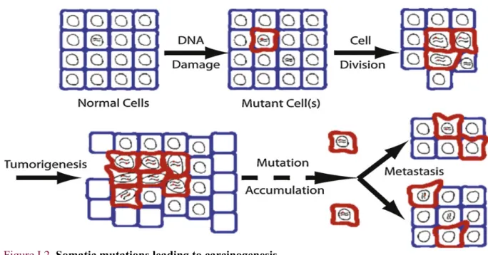
Somatic mutations are defined as changes in the DNA sequence that are not passed on to the offspring through the germline3. Most current approaches in cancer research are based on Somatic Mutation Theory (SMT) that views somatic mutations as an epiphenomenon or a post-carcinogenesis event5,6. Briefly, cellular defects (mainly through to DNA damage) induce uncontrolled cell divisions that lead to the development of carcinogenesis suggesting that cancer is due to the accumulation of somatic mutations7 (Figure I.2).
Figure I.2. Somatic mutations leading to carcinogenesis
Adopted from Kennedy and colleagues7
Historically, the SMT was first postulated in 1914 suggesting that a combination of chromosomal defects should result in cancer, followed by a proposal that mutations could cause cancer.
Two decades later, the understanding of the molecular structure of DNA lead to the 1-hit (mutation), 2-hit and hyper-mutation theories First, it was postulated that a person who inherits a mutant allele (1-2-hit) must experience a second somatic mutation (2-hit) to initiate carcinogenesis before further studies shown that for most cancer, more mutations are required (1953-2014). In 2007, they were categorized in two groups termed as “drivers”, those that confer a large selective advantage for tumour development and progression, and “passengers”, those that confer weaker selective advantage or are truly neutral in that they do not affect cancer cells’ survival.
Together, they both constitute a record of all cumulative DNA damage and repair activities occurred during the cellular lineage of the cancer cell8. A recent elaboration on the SMT was proposed in 2015 by Vogelstein and Tomasetti9 who suggested that cancer development is an event that can be attributed to “bad luck” through accumulation of “enough” mutations that cause cancer.
This controversial claim will be discussed in chapter II and a summary of 100 years of research on the SMT can be found below10 (Figure I.3).
Figure I.3. 100 years of somatic mutations theory
Modified from Brücher and Jamall10
1.1.3 BASE SUBSTITUTIONS AND GENOMIC ALTERATIONS
Cancer is a complex disease that involves mutant cells originating from a DNA modification in a single normal cell. Such modification is then propagated through cell divisions and accumulates with further DNA modifications finally leading to abnormal, cancerous cells3. Such somatic mutations include Single Nucleotide Variants (SNVs), insertions or deletions, Copy Number Variation (CNV) and chromosomal aberrations and are not to be confounded with those inherited and transmitted from parents (germline mutations). It is important to note that SNVs are different from SNPs (Single Nucleotide Polymorphisms). SNPs are single nucleotides substitutions expected to be present in a certain fraction of a given population and at the same position in both normal or cancer cells, while SNVs are only present in tumour cells and are likely shared in individuals with the same cancer.
As previously mentioned, somatic mutations can be endogenous, thus resulting from genome instability or deficiency in a DNA repair mechanism, or exogenous, that is due to environmental exposure such as tobacco smoking or UV light. For instance, UV light is known to induce DNA damage through C>T substitutions and could lead to a genotoxic stress that induces genome instability, while tobacco smoking induces T>A mutations.
With the development and the improvement of sequencing technologies collectively referred to as
High-Throughput Sequencing (HTS) and the availability of cancer exome and genome data from most human
cancers, much has been learnt about somatic mutations.
Among all of them, a particular focus has been placed on Single Base Substitutions (SBS) that have been classified in six types according to the mutated pyrimidine base (C or T) in a strand-symmetric model of mutation. Such 6 substitutions (C>A, C>G, C>T, T>A, T>C and T>G) may be further
Chromosome defects 1914 Mutation 1928 DNA Doppelhelix 1952/1953 1-hit theory 1953 2-hit theory 1971 Driver and passenger mutations 2007 Hper mutation theory 2014 Bad luck 2015
classified in different types when considering the sequence pattern in which they are located (sequence context). For practical reasons, the sequence context is typically defined using the 5’ and 3’ bases proximal to the mutated base, that results in substitutions being classified in 96 types (6 ∗ 4 ∗ 4) (Figure I.4).
Figure I.4. The 96 mutations types in a trinucleotide context
Considerations of the 6 types of base substitutions_ a DNA base is replaced by another (C>A, C>G, C>T, T>A, T>C and T>G) and the associated sequence context.
It has been hypothesized that mutational processes leave specific patterns of somatic mutations, so-called mutational signatures. To identify such patterns from the substitutions measured from cancer samples, computational models, such as matrix decomposition algorithms or probabilistic models, have been developed. The first of such methods was published in 2013 by Alexandrov and colleagues11, and, as for most of all the other models that followed, is based on the idea that a mutational signature can be seen as a probability distribution of the 96 types of mutations or more according to the length of the sequence context. Mutational signatures contribute to the total mutational burden of a cancer genome, commonly referred to as mutational “catalogue” or “spectrum” in the recent computational biology literature. A C>A A C C>A A G C>A A T C>A A A C>A C C C>A C G C>A C T C>A C A C>A G C C>A G G C>A G T C>A G A C>A T C C>A T G C>A T T C>A T C>A A C G T A C G T A C>G A C C>G A G C>G A T C>G A T>A A C G T A C G T A C>G G C C>G G G C>G G T C>G G A C>G C C C>G C G C>G C T C>G C A C>G T C C>G T G C>G T T C>G T A C G T A C G T C>G A C G T A C G T A C>T A C C>T A G C>T A T C>T A C>T A C>T C C C>T C G C>T C T C>T C A C>T G C C>T G G C>T G T C>T G A C>T T C C>T T G C>T T T C>T T A T>A T C T>A T G T>A T T T>A T A T>A G C T>A G G T>A G T T>A G A T>A C C T>A C G T>A C T T>A C A T>A A C T>A A G T>A A T T>A A A C G T A C G T A T>C A C T>C A G T>C A T T>C A T>C A T>C G C T>C G G T>C G T T>C G A T>C C C T>C C G T>C C T T>C C A T>C T C T>C T G T>C T T T>C T A C G T A C G T A T>G A C T>G A G T>G A T T>G A T>G A T>G C C T>G C G T>G C T T>G C A T>G G C T>G G G T>G G T T>G G A T>G T C T>G T G T>G T T T>G T 5’ 3’ Su bs tit ut io n typ e 16 types of mutation for each substitution type
1.2 MATHEMATICAL MODELING OF A MUTATIONAL PROCESS
1.2.1 DEFINITION OF MUTATIONAL CATALOGUES, SPECTRA AND SIGNATURES
The mutational catalogue representing the total mutational burden of a genome (or exome) $ is defined as a vector (&'(, … , &'+)-, where each &'. is the number of mutations of type / found in the genome
and K, the number of possible mutation types, is equal to 96. The superscript T denotes the transpose of a matrix so that vectors are thought as column vectors. In this setting, information about mutation locations in the sequence is lost and the catalogue is built by comparing the sequence to a reference sequence in order to detect mutations and then by simply counting the occurrences of each type. The reference sequence can either be a standard reference (e.g. the assembly GRCh38 of 2013 also known as hg38 or the previous one GRCh37 with reference to hg19) or a sequence from a “normal” tissue from the same individual (e.g. DNA from blood or from normal tissue surrounding tumours when available). For the purposes of the present thesis, the generic term “samples” will be used for both genomes and exomes as the concepts and models used may be applied to both.
The basic idea underlying all computational models proposed is that the mutational catalogue of a sample results from the combination of all the mutational processes operative during lifetime, and therefore it can be seen as the weighted superposition of simpler mutational signatures, each uniquely corresponding to a specific process. The weight is larger if the process has a larger role in the final catalogue of mutations: for example, mutagens that last longer, are more intense, generate poorly repaired DNA lesions, mutate more genes, or also act as selection pressures favoring mutant cells. Formally, the signature of a mutational process 0 is a vector 12= (12(, … , 1
2+)-, where each 12.
represents the probability that the mutational process will induce a mutation of type /. In other words, 12. is the expected relative frequency of type / mutations in genomes exposed to 0.
Note that ∑+ 12.
.5( = 1 and 0 ≤ 12. ≤ 1 for all /.
The intensity of the exposure to a mutational process 0 in a sample $ is measured by the number of mutations 9'2 in $ that are due to 0. For this reason, 9
'2 is referred to as the “exposure” of $ to 0. It is
important to notice that the term “exposure” does not refer here to the exposure to a mutagen per se, because it also includes the likelihood that an unrepaired DNA lesion will cause a mutation. The expected number of mutations of type / due to the process 0 in sample $ is therefore 12.9
'2. If sample
$ has been exposed to : mutational processes, then the total number of mutations of type / is : &'. = ∑ 1
2. ;
where ='. is an error term reflecting sampling variability and non-systematic errors in sequencing or
subsequent analyses.
Matrix notation is effectively used when dealing with several samples and signatures. In this situation, the collection of G samples is represented by the > × @ matrix, with catalogues in columns:
A = B
&(( &C( … &D(
⋮ ⋮ ⋮
&(+ &
C+ … &D+
F, Figure I5.A)
the : signatures are represented by the > × : matrix
G = B 1(( 1 C( … 1;( ⋮ ⋮ ⋮ 1(+ 1 C+ … 1;+ F, Figure I.5.B)
and the exposures by the : × @ matrix
H = B 9(( 9 C( … 9D( ⋮ ⋮ ⋮ 9(; 9 C; … 9D; F. Figure I.5.C)
Equation (1) then becomes : A ≈ G × H where we omitted the error term.
Figure I.5. Mutational catalogue and the individual signatures contribution to it
A) Mutational catalogue of a breast cancer genome PD4107a12. B) The catalogue is the result of the linear combination of COSMIC signatures 2, 3 and 8 with some additional noise. C) Relative burden of each signature.
1.2.2 DECIPHERING THE SIGNATURES OF MUTATIONAL PROCESSES: DE NOVO VS. REFITTING
De novo signature extraction methods aim at estimating G and H given A . Non-negative matrix factorization (NMF) is an appealing solution to this unsupervised learning problem, because, by definition, all involved matrices are non-negative. NMF was popularized in 1999 by Lee and Seung and has become a widely used tool for the analysis of high dimensional data, mainly image processing or recognition and text mining.
In the context of mutational signatures, NMF identifies two matrices G and H that minimize the distance between A and G × H . In particular, NMF finds an approximated solution to the non-convex optimization problem:
KL$&M0NOP, ROP||A − G × H||UC, (2)
where the Frobenius matrix norm of the error term is considered.
We recall that the Frobenius norm of a matrix is simply the square root of the sum of the squares of all the matrix elements.
NMF requires the number of signatures :, an unknown parameter, to be predefined or estimated. An approach for selecting this parameter consists in obtaining a factorization of A for several of its values and then choosing the best : with respect to some performance measure such as the reconstruction error or the overall reproducibility. NMF is at the core of the Wellcome Trust Sanger Institute (WTSI) Mutational Signature Framework, the first published method for signature extraction11. An alternative to numerical approaches based on NMF is given by statistical modelling and algorithms. With these latter approaches, the number of mutations of a given type can be modelled by a Poisson distribution
&'. ∼ W XY 1 2. ; 25(
9'2Z
where mutational processes are assumed to be mutually independent.
This latter independence hypothesis simplifies the mathematics but does not necessarily hold in practice, where mutation processes are likely to interfere with each other (e.g. distinct defective DNA repair processes). In order to estimate H and G, it has been proposed to consider H as latent data and G as a matrix of unknown parameters and to apply an expectation-maximization algorithm13 or use Bayesian approaches14. One important advantage of statistical approaches is the availability of model selection techniques for the choice of :.
The refitting approaches consider that the signatures G are known and the goal is to estimate H given A and G. Refitting can be done for individual mutational catalogues (i.e. individual samples) and, from a linear algebra perspective, can be seen as the problem of projecting a catalogue living in the K-dimensional vector space (the space spanned by all mutation types) onto its subset of all linear
combinations of the given mutational signatures having non-negative coefficients (the cone spanned by the given signatures).
A current practice consists in first performing a de novo extraction of signatures followed by a comparison of the newly identified signatures with the reference signatures (e.g. the COSMIC signatures introduced in the next section) by means of a similarity score, typically cosine similarity ranging from 0 (completely different) to 1 (identical)10,11. A “novel” signature is considered to reflect a specific reference signature if the similarity is larger than a fixed cut-off. If similarity is observed with more than one reference signature, the one with the largest value of similarity is chosen (Figure I.6).
Figure I.6. Comparison of newly identified signatures with COSMIC signatures
Signatures a-g were identified in a de novo extraction using the maftools16 R package from the The Cancer Genome Atlas lung adenocarcinoma cohort which include 563 cancer genomes at the date of selection. The novel signatures were then compared to the 30 signatures validated in the COSMIC database in terms of cosine similarity. Each signature is then assigned to the most similar COSMIC signature provided that their cosine similarity is above a fixed threshold. For instance, signature f is matched to signature 5 at a cut-off of 0.75 but is considered as a completely new signature if the cut-off is at 0.80. Also note that a unique assignment can be controversial: for instance, signature g is similar both to signatures 12 and 26 (Figure I.7).
This assignment step crucially depends on the choice of the cut-off ℎ that has been so far inconsistent in the literature with some studies using a value of 0.7517 whereas others 0.8018,19. Another difficulty is that different signatures might have very close cosine similarity, as it happens also between COSMIC signatures, so that a unique assignment is not always possible. This shows that mutational signatures are a useful mathematical construct that, however, might have biological ambiguous meaning.
1.3 COSMIC: CATALOGUE OF SOMATIC MUTATIONS IN CANCER
The Catalogue Of Somatic Mutations In Cancer (COSMIC) available at http://
cancer.sanger.ac.uk/cosmic/signatures, is the world’s largest and most comprehensive resource for
exploring the impact of somatic mutations in human cancer. Built in 2004, the database and website have been developed to store somatic mutation data in a single location and display the data and other information related to human cancer.
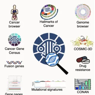
In addition to coding mutations, COSMIC covers all the genetic mechanisms by which somatic mutations promote cancer (Figure I.8). In parallel, the Cancer Gene Census (CGC) describes a curated catalogue of genes driving every form of human cancer using the ten hallmarks as proposed by Hanahan and Weinberg4.
Figure I.8. Overview of COSMIC tools
Adopted from COSMIC
Data within COSMIC are updated constantly and released on a regular, three-monthly cycle, guaranteeing four releases per year20. As example, one of the last updates (Table I.1, August 2018) includes almost 6 million coding mutations across 1.4 million tumour samples.
Table I.1. Total contents in version 86 of the COSMIC database (August 2018).
Adopted from Tate and colleagues20
1 391 372 Tumour samples
5 977 977 Coding Mutations
26 251 Manually Curated Publications
19 368 Gene Fusions
35 480 Whole Genomes/Exomes across 457 studies/papers
1 179 545 Copy Number Variants
9 147 833 Gene Expression Variants
7 879 142 Differentially Methylated CpGs
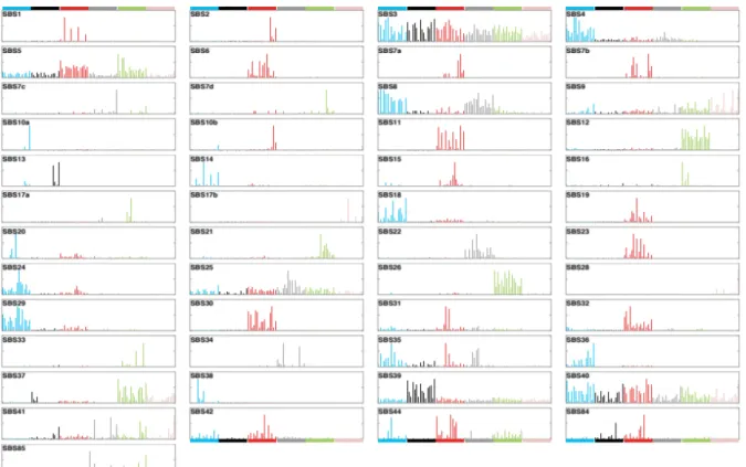
The application of the mutational signature’s framework to tens of thousands of genomes and exomes from 40 different cancers types from large data repositories such as TCGA (The Cancer Genome Atlas), has led to the identification of 30 mutational signatures (Figure I.9) characterized by a unique probability profile across the 96 mutation types. These validated mutational signatures are listed in a repertory on the COSMIC website and have been widely used as references (Mutational signatures v2).
Figure I.9. Patterns of mutational signatures (v2 – March 2015): 30 SBS
More recently, Alexandrov et al. have introduced an updated set of signatures identified from an even larger collection of both exome and whole-genome sequences (including the sequences from the PanCancer Analysis of Whole Genomes also known as PCAWG project) using two different methods (a new version of the original framework and a Bayesian alternative21). The new repertory includes 49 mutational signatures (Mutational signatures v3, Figure I.10) based on SBS as in the previous version, and also mutational signatures built in the context of other types of mutations such as Double Base Substitutions or DBS (11 signatures), clustered based substitutions (4 signatures) and small insertions and deletions (17 signatures).
Figure I.10. Patterns of mutational signatures (v3 – May 2019) : 49 SBS
1.4 EXPERIMENTAL VALIDATION OF MUTATIONAL SIGNATURES
Since the publication of the first work about mutational signatures in 201311, multiple algorithms have been developed, leading to similar but not identical results, a source of concern for researchers interested in this type of analysis. Conceptually, this is not surprising: mutational signatures are naturally defined in terms of non-negative matrix factorization, a well-known ill-posed problem (a unique solution does not exist). Although this limitation has cast doubts on the biological validity of mutational signatures, this has been somehow validated using experimental and computational approaches by Zou and colleagues22. Sufficiently detailed tumour catalogues and mutagen spectra might yield patterns that are unique to a tumour type or mutagen, and therefore become “true” signatures that allow backward inference from the tumour to the mutagen. Mutational signatures data in combination with epidemiological information may provide useful insights to identify the causes of cancer23,24. The utility of the current models of substitution mutational signatures is also shown in a recent experimental work based on a human induced pluripotent stem cell (iPSC) line that provides evidence for the possibility to identify the agents responsible for some specific mutational signatures25. In such work, Kucab and colleagues compared iPSCs treated and untreated with 79 known or suspected environmental carcinogens and identified specific substitution mutational signatures for around half of such carcinogens. Some of such signatures were similar to those identified in human tumour DNA.
2. EPIGENOMIC SIGNATURES
2.1 INTRODUCTION TO EPIGENETICS
2.1.1 OVERVIEW
The word “epigenetics” literally means “in addition to changes in the genetic sequence” 26. Epigenetics thus encompasses a wide range of mechanisms at the molecular level that can influence gene expression without involving changes to the underlying DNA sequence. As a matter of fact, even if every cell in a given individual contains the same DNA sequence, the molecular pattern leading to gene expression and protein synthesis is different. For instance, brain and lung cells are characterized by different physiological mechanisms and thus require different patterns of gene expression.
Reflecting how cells translate the information contained in the genetic sequence, are common to many organisms and is essential to their physiological functions. Aberrant modifications of epigenetic processes may have major adverse health and behavioral effects. Indeed, one of the most interesting fact of epigenetics is that its marks or states in cells change in response to outside influences. Studying epigenetic processes may therefore be helpful in addressing key questions such as: why are some foods good for our health while others are unhealthy particularly for groups of individuals? How does physical activity exert beneficial effects on several health outcomes? How do particular environmental exposures or psycho-social stress exert their detrimental effects on health?
Epigenetics is essentially additional information layered on top of the genetic sequence of the four nucleotides that makes up our DNA. Important modifications are the addition of molecules (methyl groups) or proteins (called histones) to the DNA sequence. Sometimes, epigenetic modifications are stable and passed on to future generations. Though DNA sequence is fairly permanent, and as previously mentioned, epigenetic modifications in other instances are dynamic and change in response to environmental stimuli. Thus, epigenetic is the study of mitotically heritable yet potentially reversible, molecular modifications to DNA and chromatin without alteration to the underlying DNA sequence27. There are multiple epigenetics mechanisms that may play a role in gene regulation machinery but the most studied and well-known remain histone modifications and DNA methylation. These are two process crucial to normal development and differentiation of distinct cell lineages in the adult organism, that if modified by exogeneous influences, and, as such, can contribute to or be the result of environmental alterations of phenotype or pathophenotype28. Other modifications include RNA