HAL Id: hal-02352143
https://hal.sorbonne-universite.fr/hal-02352143
Submitted on 6 Nov 2019
HAL is a multi-disciplinary open access
archive for the deposit and dissemination of
sci-entific research documents, whether they are
pub-lished or not. The documents may come from
teaching and research institutions in France or
abroad, or from public or private research centers.
L’archive ouverte pluridisciplinaire HAL, est
destinée au dépôt et à la diffusion de documents
scientifiques de niveau recherche, publiés ou non,
émanant des établissements d’enseignement et de
recherche français ou étrangers, des laboratoires
publics ou privés.
ulipristal acetate (UPA) on the endometrium
Kamila Kolanska, Justine Varinot, Geoffroy Canlorbe, Christine Bergeron,
Arsène Mekinian, Perrine Capmas, Martin Koskas, Emile Daraï, Selim
Aractingi, Sofiane Bendifallah, et al.
To cite this version:
Kamila Kolanska, Justine Varinot, Geoffroy Canlorbe, Christine Bergeron, Arsène Mekinian, et al..
Absence of predictable long-term molecular effect of ulipristal acetate (UPA) on the endometrium.
Reproductive BioMedicine Online, Elsevier, 2019, 38 (5), pp.825-834. �10.1016/j.rbmo.2018.12.013�.
�hal-02352143�
Short title: UPA and the endometrium
1
Absence of predictable long-term molecular impact of ulipristal acetate (UPA) on the
2
endometrium
3
4
Kamila Kolanskaa,b, Justine Varinotc, Geoffroy Canlorbeb,d, Christine Bergerone, Arsène Mekinianf,
5
Perrine Capmasg, Martin Koskash,Emile Daraïa,b, Selim Aractingib,i, Sofiane Bendifallaha,b, Nathalie
6
Chabbert-Buffeta,b
7
a
Service de gynécologie obstétrique et médecine de la reproduction, Hôpital Tenon, AP-HP,
8
Sorbonne Université, 4 rue de la Chine, 75020 Paris, France
9
b
INSERM UMRS 938, Faculté de médecine Pierre et Marie Curie, Site Saint-Antoine, 27 rue Chaligny,
10
75571 PARIS cedex 12, France
11
c Service d’anatomopathologie, Hôpital Tenon, AP-HP, Sorbonne Université, 4 rue de la Chine, 75020
12
Paris, France
13
d
Service de chirurgie et cancérologie gynécologique et mammaire, Hôpitaux Universitaires
Pitié-14
Salpêtrière, Charles-Foix, Sorbonne Université, 47/83, boulevard de l'Hôpital, 75013 Paris, France
15
e
Cerba Laboratory, 95066 Cergy Pontoise, France
16
f
Service de médecine interne, Hôpital Saint Antoine, AP-HP, 184 rue du Faubourg Saint Antoine,
17
75012 Paris, Sorbonne Université, France
18
g
Service de gynécologie obstétrique, CHU de Bicêtre, AP-HP, 78, rue du Général-Leclerc, 94270 Le
19
Kremlin-Bicêtre, France
20
h
Service de gynécologie obstétrique, CHU de Bichat, AP-HP, 75018 Paris, France
21
i Faculté de médecine Paris 5 Descartes, 12 rue de l’Ecole de Médecine, 75006 Paris, France
22
23
Address for correspondence:
24
Nathalie Chabbert-Buffet, MD PhD
25
Service de gynécologie obstétrique et médecine de la reproduction
26
Hôpital Tenon, AP-HP
27
4 rue de la Chine
28
75020 Paris, France
29
Tel: +33 1 56 01 77 48 Email: nathalie.chabbert-buffet@aphp.fr
ABSTRACT
31
Research Question: To describe the effects of ulipristal acetate (UPA) on the expression of
32
endometrial proliferation and maturation markers.
33
Design: A total of 45 endometrium-containing blocks of hysterectomy samples from non-menopausal
34
women with a diagnosis of moderate to severe symptoms of uterine fibroids: 14 women operated on at
35
the end of a three-month course of UPA; 4 women who had discontinued UPA treatment 1-12 months
36
before surgery; 27 control unexposed samples (14 in the proliferative and 13 in the secretory phase).
37
Immunohistochemical staining of Ki67, vascular endothelial growth factor-receptor 2 (VEGFR2),
38
estradiol receptor (ER), PR, interleukin-15 (IL-15), indoleamin-2,3-dioxygenase (IDO) and C-C motif
39
chemokine ligand-2 (CCL2) markers were analysed in both endometrial compartments and layers.
40
Results: Under UPA, ER and PR expression is similar to the proliferative phase in both layers,
41
although with a decrease in cell proliferation. IL-15, IDO and CCL2 expressions are similar to the
42
proliferative phase suggesting a progesterone-antagonist effect of UPA. VEGFR2 staining suggests a
43
trend to a mixed agonist-antagonist effect. No significant difference is observed in the post-UPA
44
proliferative phase group as compared to the control group in both layers of the endometrium.
45
Conclusion: The effect of three-month UPA treatment is mostly progesterone receptor (PR)
46
antagonist-like. After treatment discontinuation there are no signs of any long-term effects of this
47
molecule on both endometrial proliferation and maturation. Therefore UPA may be administered to
48
women willing to conceive in the short term without consequences for further implantation.
49
50
KEYWORDS: Endometrium, ulipristal acetate, basalis, functionalis, maturation
51
52
ABBREVIATIONS:
53
CCL2 – C-C motif chemokine ligand-2
54
ER – estradiol receptor55
IDO – indoleamin-2,3-dioxygenase56
IL-15 – interleukin-1557
PAEC – progesterone receptor modulator associated endometrial changes
58
PR – progesterone receptor
SPRM – selective progesterone receptor modulator
60
UPA – ulipristal acetate
61
VEGFR2 – vascular endothelial growth factor-receptor 2
62
63
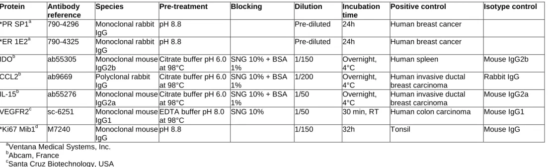
ACKNOWLEDGEMENTS
64
We thank Pr Nathalie Lédée for many productive discussions, and Anita Rodenas (Department of
65
Pathology, Tenon University Hospital) and Michèle Oster (UMRS 938) for their technical contributions.
66
We thank Felicity Kay for professional editing of the manuscript.
67
Study funding/competing interest(s): This work was in part funded by an educational grant from
68
Gedeon Richter, France. Gedeon Richter employees were not involved in the design of the project or
69
the interpretation of the results, or in any of the steps of the publication process, including writing of
70
the manuscript.
71
N.C.-B., C.B., M.K. and E.D. are board members of Gedeon Richter France SAB. All other authors
72
have no conflicts of interest to declare.
73
74
INTRODUCTION
75
Recently, the use of ulipristal acetate (UPA), a selective progesterone receptor modulator (SPRM) has
76
been approved in women of reproductive age as a treatment for symptomatic myomas before surgical
77
intervention or as alternative to surgery, using a long-term sequential regimen (Donnez et al., 2012).
78
Currently, the use of SPRMs is being evaluated in women who wish to conceive subsequently
79
(Donnez and Dolmans, 2016). SPRMs act as progesterone receptor ligands able to induce agonistic
80
or antagonistic activities according to the target cell (Bouchard et al., 2011).
81
During UPA treatment the hormonal profile is modified, with blunted progesterone levels, while
82
estradiol levels remain in the early follicular phase range (Chabbert-Buffet et al., 2007). In addition
83
95% of patients experience amenorrhea (Donnez et al., 2012). Therefore, the menstrual cycle phase
84
cannot be defined stricto sensu under UPA.
85
Endometrial morphology during UPA treatment has been described as non-physiological secretory
86
(Mutter et al., 2008; Williams et al., 2012) due to the combined progesterone receptor agonist
87
antagonist activity of the molecule. The effects of UPA on morphology and gene expression in the
88
superficial layer of the endometrium have been described in one study, consistent with UPA acting as
89
a PR antagonist, although without any proliferative effects (Whitaker et al., 2017). However, the
90
superficial layer undergoes a monthly shedding, regenerates from the basal layer, which acts as “the
91
memory” of the endometrium, and finally, during the secretory phase, undergoes decidualization, a
92
crucial step to allow embryo implantation (Gellersen et al., 2007; Perrier D’hauterive et al., 2002).
93
Immune cells play a critical role in the regulation of cellular proliferation, tissue remodelling and
94
angiogenesis in a direct and an indirect response to steroid hormones (Croxatto et al., 2014;
95
Szekeres-Bartho et al., 2009). Their proliferation and activation are regulated under the action of
96
progesterone by locally produced molecules, such as interleukin 15 (IL-15) (Okada et al., 2000;
97
Wilkens et al., 2013), C-C motif ligand 2 (CCL2) (Szekeres-Bartho et al., 2009), and indoleamine
2,3-98
dioxygenase (IDO) (Kudo et al., 2001; Munn et al., 1999). Progesterone stimulates the expression of
99
IL-15 (Kitaya et al., 2000) and VEGFR2 (Lai et al., 2015) and the secretion of CCL2 (Caballero-Campo
100
et al., 2002) in the human endometrium. In an in vitro model of endometrial culture progesterone
101
increases IL-15 secretion (Okada et al., 2000) and decreases IDO expression (Kudo et al., 2004). The
102
angiogenesis of endometrial tissue is also regulated by immune cells-produced angiogenic factors,
such as vascular endothelial growth factor A (VEGFA) which interacts with its receptor VEGFR-2
104
(Lash et al., 2016).
105
The impact of UPA in the basal layer of the endometrium and its potential post-treatment impact is
106
currently poorly understood, and may impair the whole endometrial maturation process in post
107
treatment cycles.
108
Since previous data do not provide information on the impact the molecule has, either on the basal
109
layer or on the post-treatment endometrium, the aim of this study was to describe the possible effects
110
of UPA on proliferation and maturation markers in both layers of the endometrium, under the action of
111
UPA as well as after the end of treatment.
MATERIAL AND METHODS
113
Patients and samples
114
Adequate endometrium-containing blocks of hysterectomy samples from non-menopausal women
115
aged 40–52 years, with a diagnosis of moderate to severe symptoms of multiple uterine myoma with a
116
combination of subserous and/or intramural and/or submucous fibroids requiring hysterectomy, were
117
retrospectively collected in three university hospital surgical gynaecology units (Department of
118
Gynaecology Obstetrics and Reproductive Medicine, Tenon Hospital; Department of Gynaecology
119
Obstetrics, Bicêtre University Hospital and Department of Gynaecology Obstetrics, Bichat University
120
Hospital). Four groups of patients were included in the study. The control groups, not exposed to
121
hormonal treatments or UPA, included samples of superficial and basal layers of endometrium
122
obtained in the proliferative UPA proliferative phase group, n = 14) and secretory phases
(No-123
UPA secretory phase group, n = 13), classified on the basis of histological examination (Bouchard et
124
al., 1991; Noyes et al., 1975).The UPA group (n = 14) included women operated on at the end of a
125
three-month course of UPA (Esmya, Gedeon Richter, Paris, France, 5mg daily). The post-UPA
126
proliferative phase group (n = 4) was composed of women who had been treated with UPA for at least
127
three months, and who had discontinued treatment 1–12 months before surgery with a proliferative
128
phase histological examination classification.
129
The mean ages for the No-UPA proliferative phase, No-UPA secretory phase, UPA and post-UPA
130
groups were, respectively, 47.2 ± 2.8 years, 46.9 ± 3.0 years, 46.9 ± 2.6 years and 45.8 ± 1.5 years, p
131
= 0.796. BMI was 27.2 kg/m2 ± 5.5, 26.7 kg/m2 ± 5.3, 27.7 kg/m2 ± 6.8 and 26.3 kg/m2 ± 2.0, p = 0.995
132
for the No-UPA proliferative phase, No-UPA secretory phase, UPA and post-UPA groups, respectively,
133
and parity was 1.8 ± 1.0 versus 1.5 ± 1.3 versus 1.7 ± 1.1 versus 0.0 ± 0.0, p = 0.200, respectively.
134
Among evaluated samples from the UPA group only two included focal progesterone receptor
135
modulator associated endometrial changes (PAEC) lesions.
136
The Institutional Review Board allowed retrospective sample analysis and approved the study. All
137
women gave written informed consent.
138
139
Histological analysis and endometrial dating
Paraffin-embedded tissue sample sections of 10μm were processed and stained with haematoxylin
141
and eosin by standard methods. All evaluations were performed under blinded conditions by one
142
expert pathologist (CB). Endometrial differentiation was assessed by histological dating (Noyes et al.,
143
1975) and evaluation of ER and PR expressions (Bouchard et al., 1991). Presence of progesterone
144
receptor modulator associated endometrial changes (PAEC) was noted.
145
146
Immunohistochemistry
147
We selected proliferation (Ki67), angiogenesis (vascular endothelial growth factor-receptor 2
148
[VEGFR2]), immune pathways (interleukin-15 [IL-15], indoleamin-2,3-dioxygenase [IDO], C-C motif
149
chemokine ligand-2 [CCL-2]) and endocrine maturation (ER, PR) markers on the basis of their major
150
functions in human endometrial homeostasis (Ban et al., 2013; Dimitriadis et al., 2005; Jones et al.,
151
1998; Kayisli et al., 2002; Kitaya et al., 2000; Lai et al., 2015; Munn et al., 1999; Sedlmayr et al., 2002).
152
Sample sections of 5μm were subjected to deparaffinisation.and antigen retrieval (Supplemental Table
153
I). All sections were subjected to quenching of endogenous peroxidase activity by incubation with 3%
154
hydrogen peroxide (GPR Rectapur VWR Chemicals, Radnor, PA, USA), and non-specific activity was
155
blocked with appropriate serum before incubation with antibodies specific to IDO, CCL2, IL-15 and
156
VEGFR2 (Table A.1). Appropriate matched IgG was applied as a negative control.
157
The primary antibodies were detected using an avidin-biotin peroxidase technique with anti-rabbit or
158
anti-mouse secondary antibodies as appropriate and Vectastain Elite ABC kit (Vector Laboratories,
159
Inc., Burlingame, CA, USA). The reaction was developed with 3,3’-diaminobenzidine (DAB; Vector
160
Laboratories, Inc.). Sections were counterstained with Mayer’s haematoxylin and mounted with
Immu-161
Mount (Thermo Scientific Shandon, Kalamazoo, MI, USA). Positive and negative controls
162
(replacement of the primary antibody by appropriate non-immune serum) were included in each
163
staining run.
164
The immunostaining of PR, ER and Ki67 was performed using the automated method Automate
165
BenchMark XT, Roche Ventana®, (Ventana Medical Systems, Inc., Rotkreuz, Switzerland) (Table A.1).
166
The demasking was done by heating at pH 8.8. The slides were incubated over 24h with pre-diluted
167
anti-PR and anti-ER antibodies and over 32h with anti-Ki67 antibody (dilution 1/150). Then the slides
168
were incubated with anti-rabbit and anti-mouse antibodies and revealed by UltraView Universal DAB
(ref. 760-500, Ventana Medical Systems, Inc.). Representative images were captured using a Nikon
170
Eclipse 90i microscope equipped with a Nikon DSFi1 camera (Nikon Ltd, UK).
171
172
Semi-quantitative analysis of immunostaining
173
Blinded interpretation of immunostained sections was performed by an expert pathologist (JV). The
174
percentage of stained cells was evaluated at 200x magnification. The stromal cells and glandular
175
epithelium were evaluated separately in each layer of the endometrium. The percentage of
176
immunostained cells was noted. For Ki67 a score from 1–3 was used, where 1 corresponded to <5%
177
of stained cells, 2 to 5–50% of stained cells and 3 to >50% of stained cells.
178
To determine the protein expression profile under UPA treatment, immunostaining was compared
179
between the UPA group and the No-UPA proliferative and secretory phase groups. In order to analyse
180
the possible persistent endometrial effect of UPA, the post-UPA proliferative phase group was
181
compared to the control No-UPA proliferative phase group. In addition, the effects of UPA were
182
evaluated in both endometrial layers and in both cellular compartments, i.e. stromal cells and the
183
glandular epithelium.184
185
Statistical analysis186
The Kruskal-Wallis test was performed to compare the continuous variables concerning study group
187
characteristics. The qualitative variables were analysed using the chi-square test. The
semi-188
quantitative evaluation of the immunostaining for each cellular type in each layer of the endometrium
189
was expressed as a percentage and was compared using the Mann-Whitney test.
190
All reported p values are two-sided and p < 0.05 was considered statistically significant. All analyses
191
were performed using the GraphPad Prism 7.
192
193
RESULTS
194
UPA treatment induces epithelial ER and PR expression in both layers of the endometrium
195
ER and PR are well known markers of progesterone-induced endometrial epithelium maturation. Their
196
expression was decreased in the No-UPA secretory phase as compared to the No-UPA proliferative
197
phase in the glandular epithelium in both layers (65.8% ± 29.4 versus 100.0% ± 0.0, p < 0.0001 and
198
37.1% ± 38.8 versus 100.0% ± 0.0, p < 0.0001 for ER and PR, respectively in the superficial layer and
199
88.3% ± 21.2 versus 100.0% ± 0.0, p = 0.03 and 73.3% ± 22.3 versus 100.0% ± 0.0, p < 0.0001 for
200
ER and PR, respectively in the basal layer) as previously described in women without any uterine
201
disorder (Bouchard et al., 1991) (Figure 1). However, ER and PR staining in the No-UPA secretory
202
phase was significantly lower in the superficial layer when it was compared with the basal layer
203
(65.8% ± 29.4 versus 88.3% ± 21.3, p = 0.04 and 37.1% ± 38.8 versus 73.3% ± 22.3, p = 0.02,
204
respectively) (Figure A.1).
205
Under UPA, the ER immunostaining level was very intense in stromal and epithelial cells, in both
206
endometrial layers (Figure 1) and the percentage of stained cells was generally increased as
207
compared to the No-UPA secretory phase, except for the stromal compartment of the basal layer
208
(90.0% ± 17.5 versus 67.5% ± 31.7, p = 0.04 and 98.6% ± 5.3 versus 65.8% ± 29.4, p = 0.0004 for the
209
stromal and epithelial compartments, respectively in the superficial layer and 92.9% ± 15.4 versus
210
77.5% ± 24.2, p = 0.06 and 100.0% ± 0.0 versus 88.3% ± 21.3, p = 0.03 for the stromal and epithelial
211
compartments, respectively, in the basal layer) (Figure 1).
212
The percentage of PR labelled cells in the UPA group was significantly increased in the epithelial cells
213
of both layers as compared to the No-UPA secretory phase (100.0% ± 0.0 versus 37.1% ± 38.8, p <
214
0.0001 and 100.0% ± 0.0 versus 73.3% ± 22.3, p < 0.0001 for the superficial and basal layers,
215
respectively) (Figure 1). This progesterone-antagonist pattern was not observed in the stromal
216
compartment of either layer.
217
ER and PR expressions in the UPA and post-UPA groups were comparable to the control No-UPA
218
proliferative phase group in both cellular populations of both layers (Figures 1 and A.1).
219
There was no layer-specific regulation of ER and PR under UPA (Figure A.1).
220
221
UPA treatment has no impact on cell proliferation in the basal layer
In the superficial layer in the UPA group, the percentage of Ki67 marked epithelial cells was
223
significantly increased in comparison to the No-UPA secretory phase group and significantly
224
decreased as compared to the No-UPA proliferative phase group (1.5 ± 0.5 versus 1.2 ± 0.6, p = 0.03
225
and 1.5 ± 0.5 versus 2.1 ± 0.9, p = 0.047, respectively). In the stroma, Ki67 staining was decreased in
226
comparison to the No-UPA proliferative phase (1.1 ± 0.4 versus 1.8 ± 0.8, p = 0.02) (Figure 2).
227
In contrast, in the basal layer, Ki67 staining in the study groups remained unchanged in both cellular
228
compartments.
229
Ki67 staining score in the epithelial cells in the No-UPA proliferative phase and UPA groups was lower
230
in the basal layer as compared to the superficial layer (2.1 ± 0.9 versus 1.4 ± 0.6, p = 0.0204 and 1.5 ±
231
0.5 versus 1.1 ± 0.3, p = 0.0329 for the No-UPA proliferative phase and UPA groups, respectively)
232
(Figure 3).
233
234
UPA has a mixed agonist and antagonist effect on angiogenesis marker in both endometrial
235
layers
236
We observed a layer-specific pattern of progesterone-regulated VEGFR2 expression. In the glandular
237
compartment VEGFR2 was down regulated in the No-UPA secretory phase group as compared to the
238
No-UPA proliferative phase group in the superficial layer (61.5% ± 50.6 versus 95.0% ± 18.7, p = 0.03),
239
but this pattern was not observed in the basal layer. In contrast, VEGFR2 was regulated in the stromal
240
compartment of the basal layer but not in the superficial layer (27.9% ± 36.0 versus 4.6% ± 11.3, p =
241
0.047 for the No-UPA proliferative and secretory phases, respectively, in the stromal compartment of
242
the basal layer) (Figure 2).
243
In the UPA group the regulation of VEGFR2 expression was compartment- and layer-specific. In the
244
superficial layer there was a trend to a proliferative phase-like profile in the glandular epithelium, while
245
in the stromal compartment the trend was towards a secretory phase-like profile, although these
246
results did not reach significance. In the post-UPA proliferative phase group VEGFR2 immunostaining
247
was similar to that observed in the No-UPA proliferative control group in both layers of the
248
endometrium with the exception of the basal layer stromal compartment (Figure 2).
249
250
UPA treatment has an anti-progesterone effect on immunological markers
IL-15 expression was not statistically differentially expressed in the No-UPA proliferative and secretory
252
phases in either compartment or layer, although a trend to a decreased expression in the No-UPA
253
secretory phase was observed (Figure 4, Figure A.2). IDO expression was down regulated by
254
progesterone in the glandular epithelium of both layers (Figure 4, Figure A.2). UPA administration
255
resulted in a proliferative phase-like expression profile of IL-15 and IDO in both layers (Figure 4,
256
Figure A.2). In the post-UPA proliferative phase group their expression was similar to the No-UPA
257
control group in all analysed compartments.
258
CCL2 expression profile was similar in both endometrial layers, showing an increased staining in the
259
stromal compartment during the No-UPA secretory phase. In the UPA and post-UPA proliferative
260
phase groups, a proliferative phase-like CCL2 expression pattern was observed in both endometrial
261
layers (Figure 4, Figure A.2).
DISCUSSION
263
Our results show that under UPA, ER and PR expression mirrors the proliferative phase in both layers,
264
although with a decrease in cell proliferation. In fact, in the UPA group the ER immunostaining was
265
generally increased as compared to the No-UPA secretory phase, except for the stromal compartment
266
of the basal layer. The percentage of PR labelled cells in the UPA group was significantly increased in
267
the epithelial cells of both layers as compared to the No-UPA secretory phase.
268
IL-15, IDO and CCL2 expressions mirror the proliferative phase, as a proliferative phase-like
269
expression profile of IL-15, IDO and CCL2 was observed in both layers suggesting a
progesterone-270
antagonist effect of UPA. In contrast VEGFR2 staining suggests a trend to a mixed agonist-antagonist
271
effect, since an intermediary aspect was observed.
272
No significant difference is observed in the post-UPA proliferative phase group as compared to the
273
proliferative phase control group in both layers of the endometrium.
274
This is the first report evaluating the effect of UPA on both layers of the human endometrium. In
275
addition, samples obtained after treatment discontinuation were also evaluated.
276
The long-term impact of SPRM administration on the endometrium is debatable, as morphologically
277
reversible benign alterations – PAEC (Mutter et al., 2008; Williams et al., 2012) – are observed in 11–
278
26% of patients treated by UPA, based on endometrial biopsy evaluation (Donnez et al., 2012). In our
279
study we observed PAECs in 14% of patients treated with UPA. The significance and impact of
280
PAECs on endometrial functions is still incompletely understood since they are also observed in
281
approximately 10% of women with myomas before any treatment with UPA (Williams et al., 2012).
282
Long-term daily administration of UPA results in blunted progesterone levels, while oestradiol levels
283
are constantly in the early follicular phase range (Chabbert-Buffet et al., 2007), which has raised the
284
question of a potential proliferative effect.
285
Endometrial proliferation (Ki67) and maturation markers (ER, PR, AR, HAND2, FOXO1, HOXA 10,
286
PTEN) have recently been described in endometrial biopsies of women under UPA treatment
287
(Whitaker et al., 2017). Of note, although the short-term effects of mifepristone have been widely
288
studied (for example, in the review by Danielsson et al (Danielsson et al., 2003)), its long-term and
289
post-treatment effects are likewise imperfectly understood, while continuous administration of
asoprisnil, another SPRM, results in an original pattern of expression of endometrial maturation
291
markers (Wilkens et al., 2013).
292
In the previous report of UPA effects in the superficial layer (Whitaker et al., 2017) the authors
293
reported that ER expression mirrored the proliferative phase in stromal and epithelial cells. In the
294
present study, a similar impact of UPA on ER expression profile was observed suggesting an
anti-295
progestin effect of UPA. No effect on ER expression profile was found in the post-UPA proliferative
296
group as compared to the control proliferative group in either endometrial layer, which suggests that
297
UPA impact on ER expression does not persist after treatment discontinuation.
298
Dose- and molecule-specific SPRM effects on PR expression have been described previously
299
(Danielsson et al., 2003; Wilkens et al., 2013). Under UPA, Whitaker et al. (Whitaker et al., 2017)
300
reported that PR expression does not phenocopy either the secretory or proliferative endometrium
301
with a compartment-specific profile in the superficial layer, however in the glandular epithelium PR
302
expression was modified towards a proliferative phase profile. In the present study, under UPA PR
303
expression is proliferative phase-like in both layers. No specific effect was seen after treatment
304
discontinuation. This suggests that UPA has a reversible anti-progestin effect on PR expression.
305
Our results on the impact of UPA on cell proliferation are different from those of Whitaker (Whitaker et
306
al., 2017). In the superficial layer these authors reported a decrease in Ki67 expression under UPA as
307
compared to the proliferative phase in stromal cells, while it was unchanged in the glandular
308
epithelium. In the present study, Ki67 was down regulated compared to the proliferative phase in
309
stromal cells as well, while in the glandular epithelium it was distinct from both the proliferative and
310
secretory phases, suggesting a mixed progesterone agonist-antagonist effect. Altogether, these
311
results suggest an anti-proliferative effect of UPA in the stromal compartment and a specific
312
intermediate effect in the glandular epithelium of the superficial layer of the human endometrium.
313
Importantly, non-specific regulation of Ki67 expression was observed in the basal layer during UPA
314
treatment or after discontinuation, suggesting the absence of any long-term effect of UPA on cell
315
proliferation.
316
The discrepancies between our results and those of Whitaker and colleagues may be due to clinical
317
differences such as a distinct timing in the secretory phase of control samples. In addition, our
318
assessment of Ki67 staining was not automated. However, its evaluation was reproducible and
performed blinded. VEGFR2 is a progesterone-regulated marker. While in the superficial layer
320
VEGFR2 expression was down regulated in the glandular epithelium during the secretory phase,
321
which is consistent with a previous report in the literature (Lai et al., 2015), in the basal layer a distinct
322
and specific pattern of VEGFR2 was observed. VEGFR2 expression was unchanged in the glandular
323
epithelium and down regulated in the stromal cells in the secretory phase.
324
Under UPA there was a trend to a proliferative phase-like profile in the glandular epithelium in the
325
superficial layer. In the stromal compartment the course was towards a secretory phase-like profile in
326
both layers. In the post-UPA proliferative phase group VEGFR2 expression was broadly similar to that
327
observed in the proliferative control group. This is the first report on VEGFR2 staining under UPA.
328
Clinically, UPA treatment results in the rapid control of abnormal uterine bleeding and amenorrhea is
329
observed in 91% of patients (Donnez et al., 2012). The mechanism behind this effect is still
330
incompletely understood. UPA impact on angiogenesis has been assessed by Ravet and team (Ravet
331
et al., 2008). These authors reported a UPA effect similar to the progesterone action on vascular
332
maturation and proliferation, that is, integrity of the collagen-rich fibrillar network and no change in the
333
relative vascular area or localisation and level of VEGF-A expression.
334
IDO plays a major role in tryptophan degradation (Kudo et al., 2001; Munn et al., 1999), an important
335
pathway in regulatory T lymphocyte generation and immunosuppression. IDO expression was down
336
regulated in the secretory phase in both endometrial layers as reported previously in the superficial
337
layer (Sedlmayr et al., 2002).
338
IL-15, which regulates proliferation and activation of immune cells, showed a trend to a decreased
339
expression in the secretory phase, while CCL2, which stimulates immune cell migration, displayed a
340
trend to an increased expression in the stromal compartment in the secretory phase. There was no
341
layer-specific pattern of expression. In the literature these markers have been evaluated in
342
miscellaneous situations. In women with either uterine leiomyomas or cervix cancer, IL-15 was
343
detected in the proliferative phase, primarily in the glandular epithelium (Kitaya et al., 2000). Its
344
production increases in stromal cells in the vicinity of spiral arteries during the secretory phase (Kitaya
345
et al., 2000). In healthy women IL-15 mRNA expression was increased in total endometrial samples
346
during the secretory phase (Wilkens et al., 2013). CCL2 expression was increased from the early
347
proliferative phase in endometrial biopsies from unselected women (Jones et al., 1997). These two
348
markers are specifically implicated in endometrial maturation, while ER and PR participate in
endometrial regeneration as well. The differences between our results and the literature regarding the
350
superficial layer might be explained by the fact that in our study endometrial samples were obtained
351
from hysterectomies of selected patients with symptomatic myomas, while different physiological and
352
pathological conditions were included in other studies. It is known that uterine myomas can have a
353
specific effect on expression of endometrial molecular markers (Ikhena and Bulun, 2018; Rackow and
354
Taylor, 2010).
355
Asoprisnil and UPA administration have been reported to result in the reduction of IL15 mRNA
356
(Whitaker et al., 2017; Wilkens et al., 2013). No data is available on the impact of SPRMs on IDO or
357
CCL2 expression. In the present study, a proliferative phase-like effect of UPA on IL-15, IDO and
358
CCL2 expression was found. In the post-UPA proliferative phase group a proliferative phase-like
359
expression profile of these markers suggests a limited impact of UPA on the immunomodulatory
360
processes of endometrial maturation. This is in accordance with data available on emergency
361
contraception with UPA, showing that this molecule does not interfere with embryo implantation
362
(Berger et al., 2015; Gemzell-Danielsson et al., 2014). A series of post-UPA treatment pregnancies
363
has been described in women treated for myomas (Luyckx et al., 2014) and a case report of
364
pregnancy during UPA treatment has recently been published (Hrgovic et al., 2018). UPA may
365
therefore be administered to women willing to conceive in the short term without any consequences
366
for further implantation. .
367
The major strength of our study is the large number of patients from a homogeneous population of
368
women with symptomatic myomas included in the analysis. The analysis of hysterectomy samples
369
permitted the evaluation of both endometrial layers. The immunostaining was assessed by a blinded
370
expert.
371
The limitations of this study include its retrospective design and the absence of hormonal assessment
372
of the menstrual cycle phase. Control sample numbers were limited, as symptomatic myomas require
373
hormonal treatment. The post-UPA group was composed of women in proliferative phase, because
374
the majority of patients without hormonal treatment are operated on in the first phase of the menstrual
375
cycle. Since all included patients were experiencing severe myoma-related symptoms, our data
376
cannot be extrapolated to other clinical situations. Quantification of markers’ expression was
semi-377
quantitative since paraffin-embedded samples did not allow Western Blot analysis.
379
CONCLUSION
380
A three-month UPA course exerts a specific, compartment- and layer-dependent effect on the human
381
endometrium. Although in the superficial layer the effect of UPA is mostly PR antagonist-like, our
382
observations regarding the basal layer during treatment and after its discontinuation suggest the
383
absence of a long-term effect of this molecule on endometrial proliferation and maturation.
384
385
APPENDICES
386
Table A.1. Primary antibodies used in the study
387
Protein Antibody reference
Species Pre-treatment Blocking Dilution Incubation
time
Positive control Isotype control
*PR SP1a 790-4296 Monoclonal rabbit
IgG
pH 8.8 Pre-diluted 24h Human breast cancer
*ER 1E2a 790-4325 Monoclonal rabbit
IgG
pH 8.8 Pre-diluted 24h Human breast cancer
IDOb ab55305 Monoclonal mouse
IgG2b Citrate buffer pH 6.0 at 98°C SNG 10% + BSA 1% 1/150 Overnight, 4°C
Human spleen Mouse IgG2b
CCL2b ab9669 Polyclonal rabbit
IgG Citrate buffer pH 6.0 at 98°C SNG 10% + BSA 1% 1/200 Overnight, 4°C
Human invasive ductal breast carcinoma
Rabbit IgG
IL-15b ab55276 Monoclonal mouse
IgG2a Citrate buffer pH 6.0 at 98°C SNG 10% + BSA 1% 1/50 Overnight, 4°C
Human invasive ductal breast carcinoma
Mouse IgG2a
VEGFR2c sc-6251 Monoclonal mouse
IgG1
EDTA buffer pH 8.0 at 98°C
SNG 10% 1/50 30 min, RT Human colon carcinoma Mouse IgG1
*Ki67 Mib1d M7240 Monoclonal mouse
IgG
pH 8.8 1/150 32h Tonsil Mouse IgG
a
Ventana Medical Systems, Inc.
388
b
Abcam, France
389
c
Santa Cruz Biotechnology, USA
390
d
DAKO
391
*Automate BenchMark XT, Roche Ventana®, (Ventana Medical Systems, Inc.)
392
393
IL-15, interleukin-15; IDO, indoleamin-2,3-dioxygenase; CCL2, C-C motif chemokine ligand-2; VEGFR2, vascular endothelial growth factor-2; SNG, serum
394
normal goat (Clinisciences); BSA, bovine serum albumin (Sigma-Aldrich); EDTA, ethylenediaminetetraacetic acid (Ivitrogen); RT, room temperature
Figure A.1. Oestrogen (ER) and progesterone (PR) receptor expression is layer dependent in
396
the epithelial compartment.
397
The quantification of ER and PR immunostaining expressed as a percentage of marked cells in each
398
endometrial layer. * p< 0.05. Error bars: mean ± SD.
399
Under UPA, the percentage of ER stained cells in the stroma was 90.0% ± 17.5 in the superficial layer
400
versus 92.9% ± 15.4 in the basal layer (p = 0.7186) and in the epithelium 98.6% ± 5.3 in the superficial
401
versus 100.0% ± 0.0 in the basal layers (p = 1). The percentage of PR stained cells in the stroma was
402
96.4% ± 9.3 in the superficial layer versus 97.9% ± 8.0 in the basal layer (p = 1) and in the epithelium
403
100.0% ± 0.0 in the superficial versus 100.0% ± 0.0 in the basal layers (p = 1).
404
UPA, ulipristal acetate
405
406
Figure A.2. VEGFR2, IL-15, IDO and CCL2 expression in both endometrial layers.
407
The quantification of IL-15, IDO and CCL2 immunostaining expressed as a percentage of marked cells
408
in each endometrial layer. * p< 0.05, ** p < 0.005. Error bars: mean ± SD.
409
VEGFR2, vascular endothelial growth factor-receptor 2; IL-15, interleukin-15; IDO,
indoleamin-2,3-410
dioxygenase; CCL2, C-C motif chemokine ligand-2
411
412
VITAE
413
Nathalie Chabbert-Buffet, MDPhD, is a reproductive medicine and endocrinology specialist, working at
414
a university hospital - Obstetrics Gynaecology and Reproductive Medicine department, Assistance
415
Publique Hôpitaux de Paris, Sorbonne University. Research topics are focused more specifically on
416
endometrial physiology and diseases, and endometrial impact of hormone treatments.
417
418
KEY MESSAGE
419
Ulipristal acetate (UPA) effects on proliferation and maturation markers in both endometrial layers
420
during treatment and after discontinuation suggest the absence of long-term effect.
REFERENCES
422
423
Ban, Y., Chang, Y., Dong, B., Kong, B., and Qu, X., 2013. Indoleamine 2,3-dioxygenase levels at the
424
normal and recurrent spontaneous abortion fetal-maternal interface. J. Int. Med. Res. 41, 1135–1149.
425
Berger, C., Boggavarapu, N.R., Menezes, J., Lalitkumar, P.G.L., and Gemzell-Danielsson, K., 2015.
426
Effects of ulipristal acetate on human embryo attachment and endometrial cell gene expression in an
427
in vitro co-culture system. Hum. Reprod. 30, 800–811.
428
Bouchard, P., Marraoui, J., Massai, M.R., Medalie, D.A., De Ziegler, D., Perrot-Applanat, M., Frydman,
429
R., and Bergeron, C., 1991. Immunocytochemical localization of oestradiol and progesterone
430
receptors in human endometrium: a tool to assess endometrial maturation. Baillieres Clin Obstet
431
Gynaecol. 5, 107–115.
432
Bouchard, P., Chabbert-Buffet, N., and Fauser, B.C.J.M., 2011. Selective progesterone receptor
433
modulators in reproductive medicine: pharmacology, clinical efficacy and safety. Fertil. Steril. 96,
434
1175–1189.
435
Caballero-Campo, P., Domínguez, F., Coloma, J., Meseguer, M., Remohí, J., Pellicer, A., and Simón,
436
C. 2002. Hormonal and embryonic regulation of chemokines IL-8, MCP-1 and RANTES in the human
437
endometrium during the window of implantation. Mol. Hum. Reprod. 8, 375–384.
438
Chabbert-Buffet, N., Pintiaux-Kairis, A., Bouchard, P., and VA2914 Study Group, 2007. Effects of the
439
progesterone receptor modulator VA2914 in a continuous low dose on the
hypothalamic-pituitary-440
ovarian axis and endometrium in normal women: a prospective, randomized, placebo-controlled trial. J.
441
Clin. Endocrinol. Metab. 92, 3582–3589.
442
Croxatto, D., Vacca, P., Canegallo, F., Conte, R., Venturini, P.L., Moretta, L., and Mingari, M.C., 2014.
443
Stromal cells from human decidua exert a strong inhibitory effect on NK cell function and dendritic cell
444
differentiation. PLoS ONE. 9, e89006.
445
Danielsson, K.G., Marions, L., and Bygdeman, M., 2003. Effects of mifepristone on endometrial
446
receptivity. Steroids. 68, 1069–1075.
447
Dimitriadis, E., White, C.A., Jones, R.L., and Salamonsen, L.A., 2005. Cytokines, chemokines and
448
growth factors in endometrium related to implantation. Hum. Reprod. Update. 11, 613–630.
449
Donnez, J., and Dolmans, M.-M., 2016. Uterine fibroid management: from the present to the future.
450
Hum. Reprod. Update. 22, 665–686.
451
Donnez, J., Tatarchuk, T.F., Bouchard, P., Puscasiu, L., Zakharenko, N.F., Ivanova, T., Ugocsai, G.,
452
Mara, M., Jilla, M.P., Bestel, E., et al., 2012. Ulipristal acetate versus placebo for fibroid treatment
453
before surgery. N. Engl. J. Med. 366, 409–420.
454
Gellersen, B., Brosens, I.A., and Brosens, J.J., 2007. Decidualization of the human endometrium:
455
mechanisms, functions, and clinical perspectives. Semin. Reprod. Med. 25, 445–453.
456
Gemzell-Danielsson, K., Berger, C., and Lalitkumar, P.G., 2014. Mechanisms of action of oral
457
emergency contraception. Gynecol. Endocrinol. 30, 685–687.
458
Hrgovic, Z., Habek, D., Cerkez Habek, J., Hrgovic, I., Jerkovic Gulin, S., and Gulin, D., 2018.
459
Spontaneous pregnancy during ulipristal acetate treatment of giant uterine leiomyoma. J Clin Pharm
460
Ther. 43, 121–123.
461
Ikhena, D.E., and Bulun, S.E., 2018. Literature Review on the Role of Uterine Fibroids in Endometrial
462
Function. Reprod. Sci. 25, 635–643.
463
Jones, R.K., Searle, R.F., Stewart, J.A., Turner, S., and Bulmer, J.N., 1998. Apoptosis, bcl-2
464
expression, and proliferative activity in human endometrial stroma and endometrial granulated
465
lymphocytes. Biol. Reprod. 58, 995–1002.
Jones, R.L., Kelly, R.W., and Critchley, H.O., 1997. Chemokine and cyclooxygenase-2 expression in
467
human endometrium coincides with leukocyte accumulation. Hum. Reprod. 12, 1300–1306.
468
Kayisli, U.A., Mahutte, N.G., and Arici, A., 2002. Uterine chemokines in reproductive physiology and
469
pathology. Am. J. Reprod. Immunol. 47, 213–221.
470
Kitaya, K., Yasuda, J., Yagi, I., Tada, Y., Fushiki, S., and Honjo, H., 2000. IL-15 expression at human
471
endometrium and decidua. Biol. Reprod. 63, 683–687.
472
Kudo, Y., Boyd, C.A., Sargent, I.L., and Redman, C.W., 2001. Tryptophan degradation by human
473
placental indoleamine 2,3-dioxygenase regulates lymphocyte proliferation. J. Physiol. (Lond.). 535,
474
207–215.
475
Kudo, Y., Hara, T., Katsuki, T., Toyofuku, A., Katsura, Y., Takikawa, O., Fujii, T., and Ohama, K., 2004.
476
Mechanisms regulating the expression of indoleamine 2,3-dioxygenase during decidualization of
477
human endometrium. Hum. Reprod. Oxf. Engl. 19, 1222–1230.
478
Lai, T.-H., Vlahos, N., Shih, I.-M., and Zhao, Y., 2015. Expression Patterns of VEGF and Flk-1 in
479
Human Endometrium during the Menstrual Cycle. J. Reprod. Infertil. 16, 3–9.
480
Lash, G.E., Bulmer, J.N., Li, T.C., Innes, B.A., Mariee, N., Patel, G., Sanderson, J., Quenby, S., and
481
Laird, S.M., 2016. Standardisation of uterine natural killer (uNK) cell measurements in the
482
endometrium of women with recurrent reproductive failure. J. Reprod. Immunol. 116, 50–59.
483
Luyckx, M., Squifflet, J.-L., Jadoul, P., Votino, R., Dolmans, M.-M., and Donnez, J., 2014. First series
484
of 18 pregnancies after ulipristal acetate treatment for uterine fibroids. Fertil. Steril. 102, 1404–1409.
485
Munn, D.H., Shafizadeh, E., Attwood, J.T., Bondarev, I., Pashine, A., and Mellor, A.L., 1999. Inhibition
486
of T cell proliferation by macrophage tryptophan catabolism. J. Exp. Med. 189, 1363–1372.
487
Mutter, G.L., Bergeron, C., Deligdisch, L., Ferenczy, A., Glant, M., Merino, M., Williams, A.R.W., and
488
Blithe, D.L., 2008. The spectrum of endometrial pathology induced by progesterone receptor
489
modulators. Mod. Pathol. 21, 591–598.
490
Noyes, R.W., Hertig, A.T., and Rock, J., 1975. Dating the endometrial biopsy. Am. J. Obstet. Gynecol.
491
122, 262–263.
492
Okada, H., Nakajima, T., Sanezumi, M., Ikuta, A., Yasuda, K., and Kanzaki, H., 2000. Progesterone
493
enhances interleukin-15 production in human endometrial stromal cells in vitro. J. Clin. Endocrinol.
494
Metab. 85, 4765–4770.
495
Perrier D’hauterive, S., Charlet-Renard, C., Goffin, F., Foidart, M., and Geenen, V., 2002. [The
496
implantation window]. J Gynecol Obstet Biol Reprod (Paris). 31, 440–455.
497
Rackow, B.W., and Taylor, H.S., 2010. Submucosal uterine leiomyomas have a global effect on
498
molecular determinants of endometrial receptivity. Fertil. Steril. 93, 2027–2034.
499
Ravet, S., Munaut, C., Blacher, S., Brichant, G., Labied, S., Beliard, A., Chabbert-Buffet, N., Bouchard,
500
P., Foidart, J.-M., and Pintiaux, A., 2008. Persistence of an intact endometrial matrix and vessels
501
structure in women exposed to VA-2914, a selective progesterone receptor modulator. J. Clin.
502
Endocrinol. Metab. 93, 4525–4531.
503
Sedlmayr, P., Blaschitz, A., Wintersteiger, R., Semlitsch, M., Hammer, A., MacKenzie, C.R., Walcher,
504
W., Reich, O., Takikawa, O., and Dohr, G., 2002. Localization of indoleamine 2,3-dioxygenase in
505
human female reproductive organs and the placenta. Mol. Hum. Reprod. 8, 385–391.
506
Szekeres-Bartho, J., Halasz, M., and Palkovics, T., 2009. Progesterone in pregnancy; receptor-ligand
507
interaction and signaling pathways. J. Reprod. Immunol. 83, 60–64.
Whitaker, L.H.R., Murray, A.A., Matthews, R., Shaw, G., Williams, A.R.W., Saunders, P.T.K., and
509
Critchley, H.O.D., 2017. Selective progesterone receptor modulator (SPRM) ulipristal acetate (UPA)
510
and its effects on the human endometrium. Hum. Reprod. 32, 531–543.
511
Wilkens, J., Male, V., Ghazal, P., Forster, T., Gibson, D.A., Williams, A.R.W., Brito-Mutunayagam, S.L.,
512
Craigon, M., Lourenco, P., Cameron, I.T., et al., 2013. Uterine NK cells regulate endometrial bleeding
513
in women and are suppressed by the progesterone receptor modulator asoprisnil. J. Immunol. 191,
514
2226–2235.
515
Williams, A.R.W., Bergeron, C., Barlow, D.H., and Ferenczy, A., 2012. Endometrial morphology after
516
treatment of uterine fibroids with the selective progesterone receptor modulator, ulipristal acetate. Int.
517
J. Gynecol. Pathol. 31, 556–569.
518
519
FIGURE CAPTIONS
520
521
Figure 1. Ulipristal acetate (UPA) treatment modifies the expression of the oestrogen receptor
522
(ER) and progesterone receptor (PR) in both layers of the endometrium.
523
Representative images showing immuno-localisation of ER and PR in the superficial (A-D, I-L) and
524
basal (E-H, M-P) layers of the endometrium of women with symptomatic fibroids during No-UPA
525
proliferative and secretory phases, at the end of the cycle of 3 months of UPA treatment and after
526
discontinuation. (-) Negative control: human breast cancer without expression of PR and ER,
527
respectively. Magnification x200 (scale bar = 100 μm).
528
The quantification of ER (Q and S) and PR (R and T) immunostaining expressed as a percentage of
529
marked cells in each endometrial layer. * p< 0.05, ** p < 0.005, *** p < 0.001. Error bars: mean ± SD.
530
SC, stromal cells; GE, glandular epithelium.
531
532
Figure 2. Ulipristal acetate (UPA) treatment has no impact on Ki67 expression in the basal layer
533
and has a mixed agonist-antagonist effect on vascular endothelial growth factor-receptor 2
534
(VEGFR2).
535
Representative images showing immuno-localisation of Ki67 and VEGFR2 in the superficial (A-D, I-L)
536
and basal (E-H, M-P) layers of the endometrium of women with symptomatic fibroids during No-UPA
537
proliferative and secretory phases, at the end of the cycle of 3 months of UPA treatment and after
538
discontinuation. (-) Negative control: Ki67 – non-specific antibody isotype control, VEGFR2 – human
539
endometrium + non-specific antibody isotype control. Magnification x200 (scale bar = 100 μm). The
540
quantification of Ki67 (Q and S) and VEGFR2 (R and T) immunostaining expressed as a percentage
541
of marked cells in each endometrial layer. * p< 0.05, ** p < 0.005. Error bars: mean ± SD.
542
SC, stromal cells; GE, glandular epithelium.
543
544
Figure 3. Ki67 expression is layer dependent.
545
The quantification of Ki67 immunostaining expressed as a percentage of marked cells in each
546
endometrial layer. * p< 0.05, ** p < 0.005. Error bars: mean ± SD.
547
UPA, ulipristal acetate
548
549
Figure 4. Ulipristal acetate (UPA) treatment has an anti-progesterone effect on immunological
550
markers.
551
Representative images showing immuno-localisation of IL-15, IDO and CCL2 in the superficial (A-D,
I-552
L and Q-T) and basal (E-H, M-P and U-X) layers of the endometrium of women with symptomatic
553
fibroids in the No-UPA proliferative phase, the secretory phase, the UPA and the post-UPA groups. (-)
554
Negative control: human endometrium + non-specific antibody isotype control. Magnification x200
555
(scale bar = 100 μm).
556
The quantification of IL-15 (Y and AB), IDO (Z and AC) and CCL2 (AA and AD) immunostaining
557
expressed as a percentage of marked cells in each endometrial layer. * p< 0.05, ** p < 0.005. Error
558
bars: mean ± SD.
559
SC, stromal cells; GE, glandular epithelium; IL-15, interleukin-15; IDO, indoleamin-2,3-dioxygenase;
560
CCL2, C-C motif chemokine ligand-2.
561
562
563
Figure 1.
564
565
566
567
Figure 2.
568
569
570
571
572
573
Figure 3.
574
575
576
577
578
Figure 4.
579
580
581
582
Figure A.1.
583
584
585
586
587
Figure A.2.
588
589
590
Author photo