HAL Id: hal-02445473
https://hal.archives-ouvertes.fr/hal-02445473
Submitted on 7 Dec 2020HAL is a multi-disciplinary open access
archive for the deposit and dissemination of sci-entific research documents, whether they are pub-lished or not. The documents may come from teaching and research institutions in France or abroad, or from public or private research centers.
L’archive ouverte pluridisciplinaire HAL, est destinée au dépôt et à la diffusion de documents scientifiques de niveau recherche, publiés ou non, émanant des établissements d’enseignement et de recherche français ou étrangers, des laboratoires publics ou privés.
1H, 13C and 15N backbone resonance assignment of the
human BRCA2 N-terminal region
Manon Julien, Simona Miron, Aura Carreira, François-Xavier Theillet, Sophie
Zinn-Justin
To cite this version:
Manon Julien, Simona Miron, Aura Carreira, François-Xavier Theillet, Sophie Zinn-Justin. 1H, 13C and 15N backbone resonance assignment of the human BRCA2 N-terminal region. BIOMOLECULAR NMR ASSIGNMENTS, 2020, 14 (1), pp.79-85. �10.1007/s12104-019-09924-8�. �hal-02445473�
1
H, 13C and 15N backbone resonance assignment of the human BRCA2 N-terminal region Manon Julien1,2, Simona Miron1, Aura Carreira2,3,4, François-Xavier Theillet1, Sophie Zinn-Justin1
1
Institute for Integrative Biology of the Cell (I2BC), CEA, CNRS, Univ Paris-Sud, Université Paris-Saclay, Gif-sur-Yvette Cedex, France
2
Paris Sud University, Paris-Saclay University CNRS, UMR3348, F-91405 Orsay, France
3 Institut Curie, PSL Research University, UMR3348, F-91405, Orsay, France
4
CNRS, UMR3348, F-91405, Orsay, France
Abstract
The Breast Cancer susceptibility protein 2 (BRCA2) is involved in mechanisms that maintain genome stability, including DNA repair, replication and cell division. These functions are ensured by the folded C-terminal DNA binding domain of BRCA2 but also by its large re-gions predicted to be disordered. Several studies have shown that disordered rere-gions of BRCA2 are subjected to phosphorylation, thus regulating BRCA2 interactions through the cell cycle. The N-terminal region of BRCA2 contains two highly conserved clusters of phos-phorylation sites between amino acids 75 and 210. Upon phosphos-phorylation by CDK, the cluster 1 is known to become a docking site for the kinase PLK1. The cluster 2 is phosphorylated by PLK1 at least at 2 positions. Both of these phosphorylation clusters are important for mitosis progression, in particular for chromosome segregation and cytokinesis. In order to identify the phosphorylated residues and to characterize the phosphorylation sites preferences and their functional consequences within BRCA2 N-terminus, we have produced and analyzed the BRCA2 fragment from amino acid 48 to amino acid 284 (BRCA248-284). Here, we report the
assignment of 1H, 15N, 13CO, 13C and 13C NMR chemical shifts of this region. Analysis of these chemical shifts confirmed that BRCA248-284 shows no stable fold : it is intrinsically
dis-ordered, with only short, transient -helices.
Keywords. BRCA2, mitosis, breast cancer, intrinsically disordered protein, phosphorylation,
Biological context
BReast CAncer susceptibility 2 (BRCA2) is a tumor suppressor gene identified in 1994, com-monly mutated in hereditary breast and ovarian cancers (Wooster et al. 1994). BRCA2 was initially characterized as a DNA repair protein involved in the recruitment and loading of the recombinase RAD51, which, in turn, drives the homologous recombination process at DNA double-strand breaks (DSB) (Thorslund et al. 2007, Moynahan et al. 2010, Jensen et al. 2010). Two decades later, BRCA2 is mainly described as a key platform protein for genome stability contributing to DNA repair, telomere maintenance, stressed replication fork stabilization (Fradet-Turcotte et al. 2016) and mitosis (Daniels et al. 2004, Mondal et al. 2012, Choi et al. 2012, Venkitaraman et al. 2014, ). These functions are ensured by the folded C-terminal DNA binding domain of BRCA2 (Yang et al. 2002) and the large regions predicted to be disordered that contain several protein binding motifs. Among them, two short fragments were already structurally characterized in complex with their partner: one of the repeated BRC motifs BRC4 bound to RAD51 (Pellegrini et al. 2002) and BRCA2fragment between amino acid 21 and amino acid 39 interacting with PALB2 (Oliver et al. 2009). Both of these BRCA2 seg-ments form α-helices upon binding to their partner. Here, we focus on the N-terminal region of BRCA2, which contains 1000 amino acids with a structure that remain elusive. It is pre-dicted to be disordered (Figure 1.B) and contains two clusters of phosphorylation (Figure
1.A) identified by mass spectrometry (https://www.phosphosite.org/) between amino acid 75
and amino acid 80 for cluster 1, and amino acid 190 and aa 210 for cluster 2. These two clus-ters are very well conserved from mammals to fishes and their biological relevance is still unclear. The cluster 1 was reported to be phosphorylated by Cyclin-Dependant Kinase (CDK) at position T77 during late S-phase to mitosis (Yata, et al. 2014). This phosphorylation gener-ates a genuine Polo-Like Kinase 1 (PLK1) docking site (Takaoka et al. 2014, Yata et al. 2014) suggested to promote further phosphorylation by PLK1 both on BRCA2 itself (Lin et al. 2003, Takaoka et al. 2014) and on BRCA2 partners such as RAD51 (Yata et al. 2014). How-ever, biochemical data are missing to describe to which extent this BRCA2 phosphorylation by CDK influences later phosphorylation kinetics by PLK1. The cluster 2 is phosphorylated by PLK1 during mitosis (Lin et al. 2003). This regulates BRCA2 interaction with P300/CBP-associated factor (P/CAF) (Lin et al. 2003). It also creates a supplementary PLK1 docking site that ensures chromosome segregation (Ehlen et al. BioRxiv). Furthermore, phosphorylation of S193 by PLK1 leads to the recruitment of BRCA2 at the Flemming body, an important step that warrants a complete cytokinesis (Daniels et al. 2004, Takaoka et al. 2014). While these phosphorylation events have been observed to dynamically regulate these BRCA2 functions throughout the cell cycle, little is known about the structure of the BRCA2 N-terminal region and the phosphorylated residues are only partially identified. Here, we report the assignment of the 1H, 15N, 13Cα 13Cβ and 13CO NMR chemical shifts of the WT BRCA248-284 fragment.
Methods and experiments
Protein expression and purification
Five fragments spanning the human WT BRCA2 N-terminal phosphorylation clusters were designed: a fragment from amino acid 53 to amino acid 131 (BRCA253-131), a fragment from
amino acid 53 to amino acid 228 (BRCA253-228), a fragment from amino acid 48 to amino acid
218 in which all cysteines are mutated into alanines (BRCA248-218(C4A)) a fragment from
ami-no acid 190 to amiami-no acid 284 (BRCA2190-284) and a fragment from amino acid 48 to amino
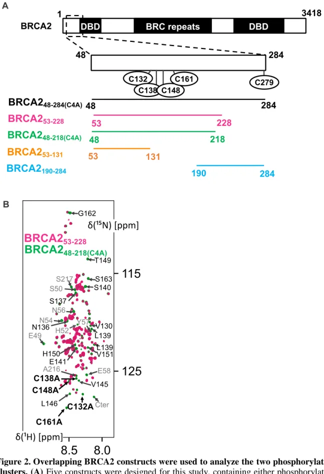
acid 284 with the first four cysteines mutated into alanines (BRCA248-284(C4A)) (Figure 2.A).
All fragments were expressed in Escherichia coli BL21 (DE3) Star using a pETM13 vector (BRCA253-228, BRCA248-218(C4A) and BRCA248-284(C4A)), a pGEX-6P-1 vector (GE Healthcare,
BRCA2190-284) or a pET-41b vector (BRCA253-131). cDNA of BRCA253-131, BRCA253-228,
Bacteria were grown in M9 medium supplemented with 15NH4Cl (0.5 g/L) and 13C-glucose (2
g/L) as sole sources of nitrogen and carbon. Recombinant expression was induced at an opti-cal density of 0.6-0.8 using 1 mM isopropyl β-D-1-thiogalactopyranoside during 4 hours at 37°C. Cells were harvested by centrifugation, resuspendend in lysis buffer (20 mM Tris, 150 mM NaCl, 1 mM EDTA, 5 mM DTT, 1 mM PMSF, pH 8) and lysed by sonication. The solu-ble fraction was obtained upon centrifugation of the whole cell lysate at 20,000 g during 15 min at 4°C. BRCA253-131, BRCA253-228 and BRCA2190-284 were produced with a N-terminal
GST tag followed by either a TEV cleavage site (ENLYFQG) or a PreScission cleavage site (LEVLFQGP) and purified by Gluthatione Sepharose affinity chromatography. The tag was cleaved by the TEV protease (BRCA253-131 and BRCA253-228) or the PreScission protease
(BRCA2190-284). The BRCA253-131 and BRCA2190-284 samples were then boiled at 95°C during
10 min to remove the GST tag, centrifuged 10 minutes at 16,000g, and the supernatant was later injected on a gel filtration column (Highload 16/60 Superdex 75pg; GE Healthcare) equilibrated with buffer A (50 mM HEPES, 1 mM EDTA, 2 mM dithiothreitol, pH 7.0). The influence of boiling on structure was verified by 1H-15N HSQC NMR (data not shown). BRCA253-228 precipitates after cleavage, and therefore was pelleted by centrifugation (10
minutes, 16,000g), then resuspended into buffer A supplemented with 8 M urea and finally diluted 10 times with buffer A containing 10 mM -mercaptoethanol before injection on a gel filtration column (Highload 16/60 Superdex 75pg; GE Healthcare) equilibrated with 50 mM HEPES, 1 mM EDTA, 5 mM tris(2-carboxyethyl)phosphine (TCEP), pH 7.0. BRCA2
48-218(C4A) and BRCA248-284(C4A) were expressed with a N-terminal octa-histidine tag and purified
by Ni-NTA affinity chromatography. The tag was cleaved using the TEV protease and the sample was injected on a gel filtration column (Highload 16/60 Superdex 75 pg; GE Healthcare) equilibrated with buffer A at pH 7.0 or buffer A at pH 6.4 for assignment of BRCA248-218(C4A). Samples were concentrated to 200-400 M for assignment experiments
(BRCA253-131, BRCA248-218(C4A) and BRCA2190-284) and to 50 M for BRCA253-228 and
BRCA248-284(C4A) characterization.
NMR Spectroscopy
NMR experiments were performed on uniformly 15N and 13C labelled fragments in buffer A at pH 7.0 for BRCA253-131, BRCA248-218(C4A), BRCA2190-284 and BRCA248-284(C4A) (H2O:D2O
ratio 90:10), in buffer A at pH 6.4 (H2O:D2O ratio 95:5) for BRCA248-218(C4A) and in 50 mM
HEPES, 1 mM EDTA, 5 mM TCEP, pH 7.0 (H2O:D2O ratio 95:5) for BRCA253-228. All
sam-ples were supplemented with 50 M Sodium trimethylsilylpropanesulfonate (DSS). NMR experiments were recorded at 283 K on a 600 MHz Bruker Advance II spectrometer and a 700 MHz Bruker Advance Neo spectrometer, both equipped with a triple resonance cryogeni-cally cooled probe. Spectra were referenced using DSS 1H chemical shifts (Wishart el al. 1995) and 1H, 13C and 15N resonance frequencies were assigned using 2D 1H-15N SOFAST-HMQC, 3D BEST-HNCACB, CBCA(CO)NH, BEST-HNCO, BEST-HN(CA)CO and HN(CO)(CA)NH experiments. The data were processed using Topspin 3.6 (Bruker) and ana-lyzed with CCPNMR Analysis (Vranken et al. 2005). The assignments were deposited in the BioMagResBank (http://www.bmrb.wisc.edu/) under the following codes: 50077 for BRCA248-218(C4A), 50078 for BRCA253-131 and 50079 for BRCA2190-284.
Assignments and data deposition
To facilitate the assignment of the human BRCA2 region from amino acid 48 to amino acid 284, we designed and produced three overlapping fragments: BRCA253-131 centered on the
phosphorylation cluster 1, BRCA253-228 containing the two phosphorylation clusters and
BRCA2190-284 that contains only the cluster 2 (Figure 2.A). BRCA253-131 and BRCA2190-284
BRCA253-228 was prone to aggregation due to the oxidation of its four solvent-exposed
cysteines. These cysteines are not conserved within BRCA2 from fishes to human. Therefore, we designed the construct BRCA248-218(C4A), which corresponds to the fragment from amino
acid 48 to amino acid 218 with all cysteines mutated into alanines (C132A, C138A, C148A and C161A; see Figure 2A). The 1H-15N HSQC spectra of BRCA253-228 and BRCA248-218(C4A)
overlap to a large extent: we observed differences only in the vicinity of the mutated residues and N-ter or C-ter ends (Figure 2.B). This shows that the alanine mutations did not modify the average structural ensemble of the peptide. Then, we assigned the 1H, 15N, 13Cα, 13Cβ and
13
CO chemical shifts of the fragments BRCA253-131, BRCA248-218(C4A) and BRCA2190-284 using
a series of 3D heteronuclear NMR experiments. We obtained high assignment coverages along the sequences of fragments BRCA253-131, BRCA248-218(C4A) and BRCA2190-284: 94 %,
97% and 96% of 1H-15N pairs, 87 %, 98 % and 95% of 13Cα, 99%, 96 % and 97 % of 13Cβ and 96 %, 98 %, 95 % of 13CO resonances were assigned, respectively. Figure 3 shows a very good crosspeak superimposition between the 1H-15N spectra of every fragment and that of BRCA248-284(C4A). The narrow range of backbone amide 1H chemical shifts (between 7.5 and
8.5 ppm) for all BRCA2 fragments reveals their disordered behavior. Furthermore, only the crosspeaks corresponding to the N-terminal or C-terminal residues of each fragment differ from those of the largest construct of BRCA2, i.e. BRCA248-284(C4A). The secondary structure
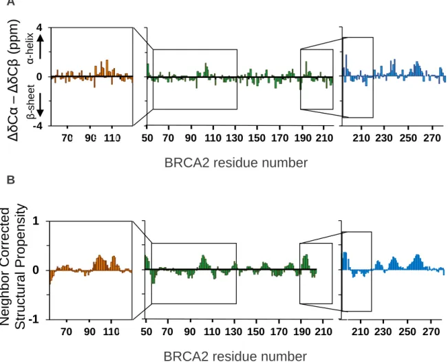
analysis, based on 13Cα and 13Cβ chemical shifts and the neighbor corrected structural pro-pensity method (Tamiola et al. 2010, Tamiola et al. 2012), confirms the absence of a stable fold for BRCA253-131, BRCA248-218(C4A) and BRCA2190-284 (Figure 4). We observed α-helical
propensities of about 25 % around residues 100-110 and 255-260. We concluded that the fragment of BRCA2 from amino acid 48 to amino acid 284 is disordered and that its shorter segments BRCA253-131, BRCA248-218(C4A) and BRCA2190-284 have the same structural
proper-ties when isolated or within BRCA248-284(C4A). From this set of data, it is now possible to
mon-itor phosphorylation reactions within the two conserved BRCA2 clusters of phosphorylation using 1H-15N and 1H-13C NMR spectroscopy.
Acknowledgments
This work was supported by CEA, CNRS, University Paris South and ENS Paris Saclay, by the French Infrastructure for Integrated Structural Biology (https://www.structuralbiology.eu/
networks/frisbi, grant number ANR-10-INSB-05-01, Acronym FRISBI) and by the French National Research Agency (ANR; research grant FUNBRCA2).
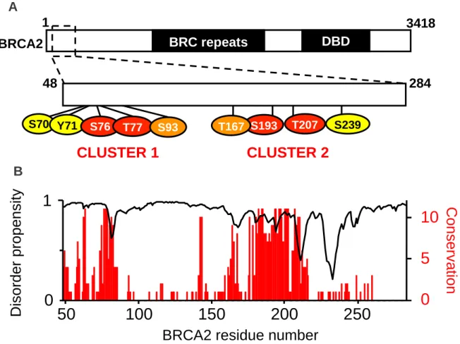
Figure 1. The N-terminal BRCA2 fragment includes 2 phosphoclusters well-conserved from mammals to fishes. (A) BRCA2 is composed of i) a 1000 residues long N-terminal
region containing the PALB2 binding site (from amino acid 21 to amino acid 39) and a DNA binding domain (DBD) (from amino acid 250 to amino acid 500) (Von Nicolai et al. 2016), ii) an intermediate region made of 8 successive BRC repeats (from amino acid 1002 to amino acid 2085 and iii) a folded C-terminal DBD (from amino acid 2500 to amino acid 3200. The N-terminal region of the protein contains several phosphorylation sites (https://www.phosphosite.org/) organized in two phosphorylation clusters. Alignment of 30 BRCA2 sequences from fishes to mammals revealed that 4 of these positions are 100% con-served: Ser76, Thr77, Ser193 and Thr207. We focused on the BRCA2 region from amino acid 48 to amino acid 284, which includes these 4 phosphosites. In this region, residues identified as phosphorylated in more than one mass spectrometry study are indicated in red if conserved in all 30 sequences, in orange if conserved in more than 80% and yellow if conserved in less than 80% of the sequences. (B) The disorder propensity and conservation of the BRCA2 re-gion from amino acid 48 to amino acid 284 are displayed as a function of the sequence. The disorder propensity was calculated using SPOT-Disorder (Hanson et al. 2016). A score of 1 corresponds to a predicted disorder propensity of 100%. The conservation score was calculat-ed using Jalview 1.0 (Clamp et al. 2004). A score of 11 corresponds to a position identical in 100% of the sequences, while a score of 1 indicates that only one chemical criteria (size, hy-drophobicity, global charge) is common to all the variants.
0
5
10
0
1
4 8 52 56 60 64 68 27 76 80 84 88 92 96 1 0 0 1 0 4 1 0 8 1 1 2 1 1 6 1 2 0 1 2 4 1 2 8 1 3 2 1 3 6 1 4 0 1 4 4 1 4 8 1 5 2 1 5 6 1 6 0 1 6 4 1 6 8 1 7 2 1 7 6 1 8 0 1 8 4 1 8 8 1 9 2 1 9 6 2 0 0 2 0 4 2 0 8 2 1 2 2 1 6 2 2 0 2 2 4 2 2 8 2 3 2 2 3 6 2 4 0 2 4 4 2 4 8 2 5 2 2 5 6 2 6 0 2 6 4 2 6 8 2 7 2 2 7 6 2 8 0BRCA2 residue number
C
o
n
s
e
rv
a
tio
n
D
is
o
rd
e
r
p
ro
p
e
n
s
it
y
3418 BRC repeats DBD 1 A B 48 284 S193 T207 T167 S239 S70 Y71 S76 T77 S93 BRCA21
0
CLUSTER 1
CLUSTER 2
50 100 150 200 250
Figure 2. Overlapping BRCA2 constructs were used to analyze the two phosphorylation clusters. (A) Five constructs were designed for this study, containing either phosphorylation
cluster 1 (BRCA253-131) or cluster 2 (BRCA2190-283) or both (BRCA253-228, BRCA248-218(C4A)
and BRCA248-284(C4A)). BRCA253-228 comprises 4 cysteines (C132, C138, C148, C161). In
(B) The 1H-15N SOFAST-HMQC spectra of BRCA253-228 (pink) at 50 M in buffer A
contain-ing TCEP and BRCA248-218 C4A (green) at 50 M in buffer A containing DTT are
superimpos-able, peaks overlap for residues spanning the whole sequence except around the mutated posi-tions (labelled in black) and close to the N-terminal or C-terminal ends (labelled in grey). These spectra were recorded at 283 K and pH 7.0 on a Bruker 700 MHz spectrometer.
Figure 3. Superimposition of the constructs spanning the BRCA2 region from amino acid 48 to amino acid 284 confirms the intrinsically disordered behavior of this region.
The 1H-15N SOFAST-HMQC spectra of (A) BRCA253-131 (orange), (B) BRCA2 48-218(C4A)
(green) and (C) BRCA2190-284 (blue) are superimposed over BRCA248-284(C4A) in the different
panels. All peptides are at 50 M in buffer A and spectra were recorded at 283 K and pH 7.0 on a Bruker 700 MHz spectrometer. Only chemical shifts of residues close to the extremities vary between shorter constructs and BRCA248-284(C4A). In order to simplify the figure, peaks
corresponding to these residues are not labelled, however, they can easily be highlighted by comparaison of chemical shift values available on the BMRB.
Figure 4. Analysis of the secondary propensity BRCA253-131 (orange), BRCA2 48-218(C4A)
(green) and BRCA2190-284 (blue) based on the Cα and Cβ chemical shifts.
(A) Analysis of experimental Cα and Cβ chemical shifts compared to predicted values for a disordered region (http://nmr.chem.rug.nl/ncIDP) (Tamiola et al. 2010). (B) Analysis based on the calculator available on the website http://linuxnmr02.chem.rug.nl/ncSPC/ (Tamiola, 2012) and the library of Tamiola, Acar and Mulder (2010), with an average window of 5 resi-dues. A B -4 0 4 70 90 110 210 230 250 270
BRCA2 residue number
α -h e lix β -s h e e t Δ δC α – Δ δ C β (p p m ) -4 0 4 49 7069 90 110 130 150 170 190 21089 109 133 153 173 193 214 50 -4 0 4 55 75 97 119 -1 -0,5 0 0,5 1 70 90 110 210 230 250 270
BRCA2 residue number
N e ig h b o r C o rr e c te d S tr u c tu ra l P ro p e n s it y -1 -0,5 0 0,5 1 4950 7069 90 110 130 150 170 190 21089 109 131 151 171 191 211 -1 0 1
References
Choi, E., Park, P., Lee, H., Lee, Y., Kang, G., Lee, J., Han, W., Lee, H., Noh, D., Lekomtsev, S., Lee, H. (2012). BRCA2 Fine-Tunes the Spindle Assembly Checkpoint through Re-inforcement of BubR1 Acetylation Developmental Cell 22(2), 295-308. https://dx.doi.org/10.1016/j.devcel.2012.01.009
Clamp, M., Cuff, J., Searle, S., Barton, G. (2004). The Jalview Java alignment editor Bioin-formatics 20(3), 426-427. https://dx.doi.org/10.1093/bioinBioin-formatics/btg430
Daniels, M., Wang, Y., Lee, M., Venkitaraman, A. (2004). Abnormal Cytokinesis in Cells Deficient in the Breast Cancer Susceptibility Protein BRCA2 Science 306(5697), 876-879. https://dx.doi.org/10.1126/science.1102574
Ehlen, A. Martin, C. Miron, S. Julien, M. Theillet, FX. Ropars, V. Sessa, G. Beaurepere, R. Boucherit, V. Duchambon, P. El Marjou, A. Zinn-Justin, S. Carreira, A. 2019. Proper chromosome alignment depends on BRCA2 phosphorylation by PLK1. BioRxiv. https://doi.org/10.1101/265934
Fradet-Turcotte, A., Sitz, J., Grapton, D., Orthwein, A. (2016). BRCA2 functions: from DNA repair to replication fork stabilization Endocrine-Related Cancer 23(10), T1-T17. https://dx.doi.org/10.1530/erc-16-0297
Hanson, J., Yang, Y., Paliwal, K., Zhou, Y. (2016). Improving protein disorder prediction by deep bidirectional long short-term memory recurrent neural networks Bioinformatics https://dx.doi.org/10.1093/bioinformatics/btw678
Jensen, R., Carreira, A., Kowalczykowski, S. (2010). Purified human BRCA2 stimulates RAD51-mediated recombination Nature 467(7316), 678-683. https://dx.doi.org/10.1038/nature09399
Lin, H., Ting, N., Qin, J., Lee, W. (2003). M Phase-specific Phosphorylation of BRCA2 by Polo-like Kinase 1 Correlates with the Dissociation of the BRCA2-P/CAF Complex Journal of Biological Chemistry 278(38), 35979-35987. https://dx.doi.org/10.1074/jbc.m210659200
Mondal, G., Rowley, M., Guidugli, L., Wu, J., Pankratz, V., Couch, F. (2012). BRCA2 Local-ization to the Midbody by Filamin A Regulates CEP55 Signaling and Completion of
Cytokinesis Developmental Cell 23(1), 137-152.
https://dx.doi.org/10.1016/j.devcel.2012.05.008
Moynahan, M., Jasin, M. (2010). Mitotic homologous recombination maintains genomic sta-bility and suppresses tumorigenesis Nature Reviews Molecular Cell Biology 11(3), 196-207. https://dx.doi.org/10.1038/nrm2851
Oliver, A., Swift, S., Lord, C., Ashworth, A., Pearl, L. (2009). Structural basis for recruitment of BRCA2 by PALB2 EMBO reports 10(9), 990-996. https://dx.doi.org/10.1038/embor.2009.126
Pellegrini, L., Yu, D., Lo, T., Anand, S., Lee, M., Blundell, T., Venkitaraman, A. (2002). In-sights into DNA recombination from the structure of a RAD51–BRCA2 complex Na-ture 420(6913), 287-293. https://dx.doi.org/10.1038/naNa-ture01230
Takaoka, M., Saito, H., Takenaka, K., Miki, Y., Nakanishi, A. (2014). BRCA2 Phosphory-lated by PLK1 Moves to the Midbody to Regulate Cytokinesis Mediated by Nonmuscle Myosin IIC Cancer Research 74(5), 1518-1528. https://dx.doi.org/10.1158/0008-5472.can-13-0504
Tamiola, K., Mulder, F. (2012). Using NMR chemical shifts to calculate the propensity for structural order and disorder in proteins Biochemical Society Transactions 40(5), 1014-1020. https://dx.doi.org/10.1042/bst20120171
Tamiola, K., Acar, B., Mulder, F. (2010). Sequence-Specific Random Coil Chemical Shifts of Intrinsically Disordered Proteins Journal of the American Chemical Society 132(51), 18000-18003. https://dx.doi.org/10.1021/ja105656t
Thorslund, T., West, S. (2007). BRCA2: a universal recombinase regulator Oncogene 26(56), 1210870. https://dx.doi.org/10.1038/sj.onc.1210870
Venkitaraman, A. (2014). Cancer Suppression by the Chromosome Custodians, BRCA1 and BRCA2 Science 343(6178), 1470-1475. https://dx.doi.org/10.1126/science.1252230 Vranken, W., Boucher, W., Stevens, T., Fogh, R., Pajon, A., Llinas, M., Ulrich, E., Markley,
J., Ionides, J., Laue, E. (2005). The CCPN data model for NMR spectroscopy: Devel-opment of a software pipeline Proteins: Structure, Function, and Bioinformatics 59(4), 687-696. https://dx.doi.org/10.1002/prot.20449
Wishart, D., Bigam, C., Yao, J., Abildgaard, F., Dyson, H., Oldfield, E., Markley, J., Sykes, B. (1995). 1H, 13C and 15N chemical shift referencing in biomolecular NMR Journal of Biomolecular NMR 6(2), 135-140. https://dx.doi.org/10.1007/bf00211777
Wooster, R., Neuhausen, S., Mangion, J., Quirk, Y., Ford, D., Collins, N., Nguyen, K., Seal, S., Tran, T., Averill, D., et, a. (1994). Localization of a breast cancer susceptibility gene, BRCA2, to chromosome 13q12-13 Science 265(5181), 2088-2090. https://dx.doi.org/10.1126/science.8091231
Yang, H., Jeffrey, P., Miller, J., Kinnucan, E., Sun, Y., Thomä, N., Zheng, N., Chen, P., Lee, W., Pavletich, N. (2002). BRCA2 Function in DNA Binding and Recombination from a BRCA2-DSS1-ssDNA Structure Science 297(5588), 1837-1848. https://dx.doi.org/10.1126/science.297.5588.1837
Yata, K., Bleuyard, J., Nakato, R., Ralf, C., Katou, Y., Schwab, R., Niedzwiedz, W., Shira-hige, K., Esashi, F. (2014). BRCA2 Coordinates the Activities of Cell-Cycle Kinases to Promote Genome Stability Cell Reports 7(5), 1547-1559. https://dx.doi.org/10.1016/j.celrep.2014.04.023