ARTICLE
Altered control of T follicular helper (Tfh) cells can lead to generation of autoantibodies and autoimmune manifestations.
Signaling pathways that selectively limit pathogenic responses without affecting the protective function of Tfh cells are
unknown. Here we show that the ATP-gated ionotropic P2X7 receptor restricts the expansion of aberrant Tfh cells and
the generation of self-reactive antibodies in experimental murine lupus, but its activity is dispensable for the expansion
of antigen-specific Tfh cells during vaccination. P2X7 stimulation promotes caspase-mediated pyroptosis of Tfh cells and
controls the development of pathogenic ICOS
+IFN-γ–secreting cells. Circulating Tfh cells from patients with systemic
lupus erythematosus (SLE) but not primary antiphospholipid syndrome (PAPS), a nonlupus systemic autoimmune disease,
were hyporesponsive to P2X7 stimulation and resistant to P2X7-mediated inhibition of cytokine-driven expansion. These
data point to the P2X7 receptor as a checkpoint regulator of Tfh cells; thus, restoring P2X7 activity in SLE patients could
selectively limit the progressive amplification of pathogenic autoantibodies, which deteriorate patients’ conditions.
P2X7 receptor restrains pathogenic Tfh cell
generation in systemic lupus erythematosus
Caterina E. Faliti1,2, Roberta Gualtierotti3,4, Elsa Rottoli1,5, Maria Gerosa3,4, Lisa Perruzza1,2, Andrea Romagnani1,2, Giovanni Pellegrini6, Benedetta De Ponte Conti1,5, Riccardo L. Rossi7, Marco Idzko8, Emilia M.C. Mazza9, Silvio Bicciato9, Elisabetta Traggiai10, Pier Luigi Meroni3,4,11, and Fabio Grassi1,5,7
Introduction
T follicular helper (Tfh) cells are a specialized subset of effector CD4 T cells that play a crucial role in the generation of protective antibody responses against pathogens. However, dysfunctional Tfh cells can activate autoantibody-producing B cells that cause autoimmunity (Yu and Vinuesa, 2010; Craft, 2012; Crotty, 2014). Understanding the regulatory mechanisms that ensure the ho-meostatic control of Tfh cell activation can provide insight for manipulating T cell–dependent antibody responses in autoim-mune conditions. The Tfh cell differentiation program is imple-mented by up-regulation of inducible T cell costimulator (ICOS) that induces the transcription factor Bcl6 (Nurieva et al., 2008; Choi et al., 2011). Bcl6 in turn promotes CXCR5 expression and migration of the developing Tfh cell to the B cell follicle (Choi et al., 2011; Pepper et al., 2011). The concomitant down-regulation of CCR7 and P-selectin glycoprotein ligand 1 (PSGL-1) allows the T cell to exit the T cell zone and colocalize with B cells. The ICOS– ICO SL interaction is important in mediating Tfh cell migration to the B cell follicle (Xu et al., 2013). Antigen presentation and ICO SL
expression by B cells are instrumental to the expansion of Tfh cells, resulting in germinal center (GC) formation.
ATP is a ubiquitous extracellular messenger that can act also as a danger-associated molecular pattern; it activates puriner-gic receptors in the plasma membrane termed P2 receptors. The P2X7 receptor subtype is an ATP-gated nonselective cationic channel characterized by dual gating: whereas P2X7 stimulation with ATP in the hundred-micromolar range leads to opening of a cytolytic pore and cell death, receptor exposure to low concentra-tions of ATP (e.g., micromolar range) results in small-amplitude currents (Khadra et al., 2013). The P2rx7 gene, encoding for P2X7, is widely expressed, with the highest levels in cells from nervous and immune systems. Tfh cells express high levels of P2X7 in the plasma membrane; in the Peyer’s patches (PPs) of the small in-testine, they are exposed to extracellular concentrations of ATP that promote cell death via P2X7. Consequently, Tfh cells with deletion of P2rx7 show resistance to extracellular ATP (eATP)–in-duced pore opening and cell death. The improved helper activity
Correspondence to Fabio Grassi: fabio.grassi@ irb .usi .ch; Pier Luigi Meroni: pierluigi.meroni@ unimi .it.
© 2019 Faliti et al. This article is distributed under the terms of an Attribution–Noncommercial–Share Alike–No Mirror Sites license for the first six months after the publication date (see http:// www .rupress .org/ terms/ ). After six months it is available under a Creative Commons License (Attribution–Noncommercial–Share Alike 4.0 International license, as described at https:// creativecommons .org/ licenses/ by -nc -sa/ 4 .0/ ).
1Institute for Research in Biomedicine, Università della Svizzera Italiana, Bellinzona, Switzerland; 2Graduate School for Cellular and Biomedical Sciences, University
of Bern, Bern, Switzerland; 3Department of Clinical Science and Community Health, University of Milan, Milan, Italy; 4Lupus Clinic, IAS ST-Istituto Gaetano Pini,
Milan, Italy; 5Department of Medical Biotechnology and Translational Medicine, University of Milan, Milan, Italy; 6Laboratory for Animal Model Pathology, Institute
of Veterinary Pathology, Vetsuisse Faculty, University of Zurich, Zurich, Switzerland; 7Istituto Nazionale Genetica Molecolare “Romeo ed Enrica Invernizzi,” Milan,
Italy; 8Division of Pulmonology, Department of Medicine II, Medical University of Vienna, Vienna, Austria; 9Department of Life Sciences, University of Modena and
Reggio Emilia, Modena, Italy; 10Novartis Institute for Biomedical Research, Basel, Switzerland; 11Istituto Auxologico Italiano, Milan, Italy.
on January 17, 2019
jem.rupress.org
Downloaded from
http://doi.org/10.1084/jem.20171976
of P2rx7−/− Tfh cells results in enhanced GC reaction, IgA secre-tion, and binding to commensals (Proietti et al., 2014).
It is not clear whether eATP might influence Tfh cells at in-flammatory sites, where it is present at high concentrations (Wilhelm et al., 2010). We addressed this issue in chronic in-flammation elicited by pristane injection that causes a lupus-like syndrome in mice (Satoh and Reeves, 1994; Reeves et al., 2009). We show that lack of P2X7 in Tfh cells significantly worsened the disease by enhancing the generation of autoantibodies. Notably, circulating Tfh cells from patients with SLE were almost insensi-tive to P2X7-mediated control. In contrast, Tfh cells from patients with primary antiphospholipid syndrome (PAPS) were inhibited by P2X7 stimulation, suggesting that impaired P2X7 activity se-lectively contributes to the immunopathogenesis of SLE.
Results
P2rx7 deletion exacerbates immunopathology in experimental murine lupus
Several key features of SLE can be induced in mice by a single i.p. injection of the hydrocarbon oil 2,6,10,14-tetramethylpen-tadecane (commonly known as “pristane”; Satoh and Reeves, 1994; Reeves et al., 2009), which provokes peritoneal inflamma-tion, production of antinuclear antibodies (ANAs) and glomer-ulonephritis. P2rx7−/− mice treated with pristane showed more severe splenomegaly (Fig. 1 A). Pristane-induced lupus (PIL) is characterized by peritoneal lipogranulomas, ectopic lymphoid structures that sustain autoantibody production (Nacionales et al., 2009; Weinstein et al., 2013). We observed more widespread lipogranulomas and enhanced glomerular damage in P2rx7−/− compared with WT mice (Fig. 1 C). Consistent with these findings, proteinuria levels (Fig. 1 B) and IgG immunocomplexes as well as complement C3 deposits in the glomeruli (Fig. 1 D) were markedly increased in mice lacking P2X7. Overall, these results indicate that P2rx7 deletion exacerbates pristane-induced immunopathology. P2rx7 deletion leads to enhanced secretion of self-reactive antibodies and abnormal GC reaction
Administration of pristane results in hypergammaglobulinemia and production of ANAs, a hallmark of SLE (Satoh and Reeves, 1994; Reeves et al., 2009). The serum concentrations of IgG, par-ticularly IgG1 and IgG2B subtypes, were significantly augmented in P2rx7−/− with respect to WT mice, whereas IgG2C, IgM, and IgG3 were comparable in the two groups of animals (Fig. 2 A). Indirect immunofluorescence assay with HEp-2 cells revealed predomi-nant staining patterns classified as “domipredomi-nant homogenous” and “mixed homogeneous/nucleolar” with sera from P2rx7−/− mice, suggesting robust generation of autoantibodies directed against nuclear components, whereas sera from WT mice preferentially showed cytoplasmic and mixed cytoplasmic/nucleolar patterns with minimal reactivity (8% of tested sera) to homogeneous/ nuclear antigens (Fig. 2 B). Semiquantitative detection of IgG re-active to SLE-specific self-antigens showed significant increases of self-reactive IgG in sera from pristane-treated P2rx7−/− mice, which showed values more similar to those of autoimmune-prone Murphy Roths Large/Lymphoproliferative (MRL/lpr) than to WT mice (Fig. 2 C).
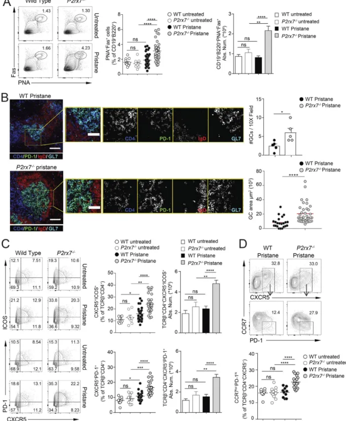
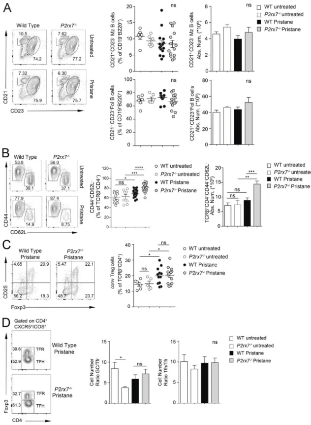
The generation of self-reactive antibodies might originate from dysregulated immune checkpoint function during B cell differentiation. Analysis of the splenic B cell compartment at 8 mo after pristane administration did not reveal differences in the distribution of follicular and marginal zone B cells between WT and P2rx7−/− mice (Fig. S1 A). Conversely, splenic plasma cells (Fig. 2 E) as well as IgG-secreting cells (Fig. 2 D) were more abundant in P2rx7−/− mice. Serum autoantibodies can be gen-erated through both extrafollicular and GC-derived pathways, the latter likely promoting somatic hypermutation and IgG af-finity maturation. The contribution of GC B cells to PIL immu-nopathogenesis has not been directly explored so far. Splenic PNA+Fas+ GC B cells were unaltered in pristane-treated WT mice with respect to untreated animals at 8 mo after pristane admin-istration. However, these cells were significantly increased in P2rx7−/− mice (Fig. 3 A), as were the number and size of GCs de-tected by immunofluorescence staining of spleen cryosections (Fig. 3 B). These experiments suggest that protracted GC reaction could be responsible for the enrichment of autoreactive IgG in P2rx7−/− mice.
P2rx7 deletion favors the generation of pathogenic Tfh and kidney-infiltrating T cells in PIL
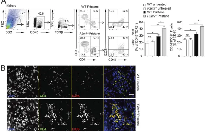
Pristane administration induced a significant expansion of ef-fector/memory CD4 cells in P2rx7−/− mice compared with WT mice (Fig. S1 B). Among effector T cells, Tfh cells are specialized to provide help to B cells and crucially contribute to GC regu-lation. Aberrant expansion of Tfh cells is a common feature of SLE as well as of several spontaneous and induced models of murine lupus. Indeed, flow cytometry and immunofluorescence confirmed the follicular localization (Fig. 3 B) and expansion of Tfh cells expressing ICOS and PD-1 (Fig. 3 C) in pristane-treated P2rx7−/− compared with WT mice. Down-regulation of CCR7 and high expression of PD-1 define the immunopathogenic Tfh cell phenotype in lupus-prone sanroque mice (He et al., 2013). Anal-ogously, CCR7lo/−PD-1hi Tfh cells were significantly increased in the spleen of pristane-treated P2rx7−/− with respect to WT mice (Fig. 3 D). ICOS amplification results in spontaneous autoim-mune manifestations in sanroque mice (Vinuesa et al., 2005) and increased IgG production as well as multiorgan inflamma-tion in autoimmune-prone MRL/lpr mice (Odegard et al., 2008; Teichmann et al., 2015). ICOS-expressing CD4 cells promote ne-phritis in lupus-prone mice (Odegard et al., 2009) and were de-tected in renal biopsy of patients with lupus nephritis (Hutloff et al., 2004; Cohen et al., 2008). In P2rx7−/− mice, kidney-infil-trating ICOS+ CD4 cells were significantly increased following pristane administration (Fig. S2, A and B), likely contributing to the severe glomerular damage observed in these mutant mice.
Immunosuppressive T regulatory (T reg) cells limit the ex-pansion of autoreactive cells and are considered as potential therapeutic tools in autoimmune conditions. Their defect might contribute to lupus pathogenesis, although a specific role in human SLE is debated (Ohl and Tenbrock, 2015). We did not de-tect differences in T reg cell representation in the spleen between pristane-injected WT and P2rx7−/− mice (Fig. S1 C). Moreover, we addressed whether T follicular regulatory (Tfr) cells, which represent specialized T reg cells controlling GC reaction (Chung
et al., 2011; Linterman et al., 2011; Wollenberg et al., 2011), were affected in P2rx7−/− mice. However, we did not detect differences in Tfh/Tfr or GC B/Tfr cell ratios in the spleen of treated animals (Fig. S1 D), suggesting that P2rx7−/− Tfr cells efficiently expanded concomitantly to enhanced Tfh and GC B cells expansion. There-fore, it seems unlikely they might contribute to deregulated GC reaction in P2rx7−/− mice.
P2X7 activity limits the expansion of Tfh cells in PIL but not during conventional immunization
A peculiarity of P2X7 among the P2 family of ATP-gated ionic channels is the low affinity for ATP; therefore relatively high concentrations of ATP (μM range) are required for its activa-tion. Under physiological conditions, eATP is virtually absent
in tissue interstitia. However, stress, injuries or inflammation considerably enhance the concentration of eATP that acts as a danger-associated molecular pattern. To address whether P2X7-mediated control of Tfh cell expansion was peculiar for the inflammatory and immunopathogenic environment elicited by pristane administration but not immunization with a con-ventional antigen, we immunized WT and P2rx7−/− mice with NP16-OVA in MF59, a potent ATP-releasing adjuvant (Vono et al.,
2013; Fig. 4 A). In contrast to the enhanced response to pristane, we detected an impaired expansion of Tfh cells and OVA-pep-tide329–337–specific CD4 cells (Fig. 4 B) as well as NP-specific GC B cells (Fig. 4 C) in the draining lymph nodes of P2rx7−/− mice 7 d after immunization. Boost immunization at 14 d and analysis after 7 d showed the persistent reduction of antigen-specific B Figure 1. Increased lupus nephritis severity in P2rx7−/− mice. (A) Spleen weight of untreated WT (n = 7), P2rx7−/− (n = 6), pristane-treated WT (n = 19), and
P2rx7−/− (n = 18) mice. (B) Proteinuria score of pristane-treated WT and P2rx7−/− mice. Each dot represents an individual mouse, and horizontal lines represent
median values. WT untreated (n = 8), P2rx7−/− untreated (n = 8), WT pristane (n = 47), P2rx7−/− pristane (n = 53). (C) Representative pristane-induced glomerular
injury in periodic-acid-Schiff–stained kidney sections (bar, 50 µm) with proportion of affected glomeruli (left graph) and glomerular injury score (right graph). WT pristane (n = 10), P2rx7−/− pristane (n = 15). (D) Confocal microscopy of kidneys stained for IgG (green) and complement C3 (red) to detect glomerular immune
deposits. Rag1−/− mice were used as negative control for IgG staining (bars, 20 µm). Histograms show means ± SEM for glomerular IgG (top) and C3 depositions
(bottom) of three independent experiments. Rag1−/− Ctrl (n = 2), WT untreated (n = 3), P2rx7−/− untreated (n = 3), WT pristane (n = 8), P2rx7−/− pristane (n = 10).
cells in the spleen of mutant animals (Fig. 4 D) concomitant to significantly reduced abundance of both high- and low-affinity IgG-secreting B cells specific for NP4 and NP41 antigen, respec-tively (Fig. 4 D). These cells as well as OVA-peptide329–337 –spe-cific CD4 cells were also significantly reduced in P2rx7−/− mice 7 d after subcutaneous injection of NP16-OVA in PBS at day 95 after primary immunization (Fig. 4 E). These results suggest that en-hanced GC reaction and hyper-IgG observed in pristane-treated P2rx7−/− mice are not general features of the adaptive immune response triggered in the absence of P2X7 but characterize the immunopathogenic response triggered by pristane. Since P2X7 is important in promoting the proinflammatory activation of APCs during antigen priming of adaptive immunity (Wilhelm et
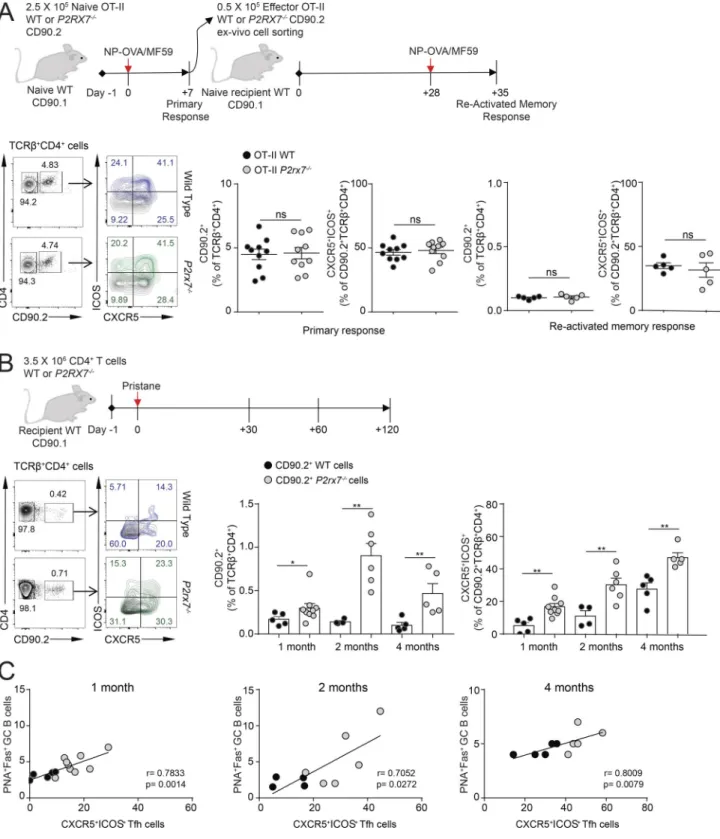
al., 2010), the defective antigen responsiveness of P2rx7−/− mice could be due to impaired APC activation. To rule out an intrinsic defect of P2rx7−/− Tfh cells during antigen priming, we trans-ferred either WT or P2rx7−/− TCR transgenic CD90.2 OT-II CD4 cells (specific for OVA peptide 323–339) into congenic CD90.1 WT mice and repeated the immunization with NP16-OVA in MF59. The analysis of primary as well as reactivated memory response revealed undistinguishable frequencies of antigen-specific CD4 cells in mice adoptively transferred with WT or P2rx7−/− OT-II cells (Fig. 5 A). Conversely, the transfer of CD90.2 WT or P2rx7−/− naive T CD4 cells into congenic CD90.1 WT animals and subsequent injection of pristane (to reproduce the features of ex-perimental lupus within this adoptive transfer system) recapit-Figure 2. Increased serum autoantibodies and splenic plasma cells in P2rx7−/− mice. (A) Serum Ig isotype concentrations in untreated (n = 6 for IgM and IgA, n = 16 for total IgG, n = 12 for IgG subclasses) and pristane-treated (n = 15 for IgM and IgA, n = 40 for total IgG, n = 15 for IgG subclasses) mice by ELI SA. Mean ± SEM are shown. Two-tailed nonparametric Mann–Whitney U test. (B) Representative immunofluorescence of HEp-2 cells and relative distribution of ANA IgG staining patterns with sera from pristane-treated WT and P2rx7−/− mice. Bar, 50 µm. The number in the circle indicates the number of analyzed mice.
Fisher's exact test with χ2. (C) ELI SA (QUA NTA-Lite ANA) for the semiquantitative detection of self-reactive IgG. Each dot represents an individual mouse, and
horizontal lines represent median values. MRL/lpr sera were used as positive controls. MRL/lpr (n = 5), WT untreated (n = 11), P2rx7−/− untreated (n = 11), WT
pristane (n = 29), P2rx7−/− pristane (n = 33). Two-tailed Mann–Whitney U test. Distribution of sera from the indicated mice as nonreactive or reactive in the assay
is shown. (D) Absolute number of IgG-secreting cells by ELI SPOT assay in spleens from the indicated mice. Mean ± SEM of three independent experiments. WT untreated (n = 4), P2rx7−/− untreated (n = 4), WT pristane (n = 7), P2rx7−/− pristane (n = 9). (E) Representative contour plots, relative frequency and absolute
number of splenic plasma cells gated as TCRβ−CD138+CD44+ cells from untreated WT (n = 6) and P2rx7−/− (n = 5) mice and treated WT (n = 17) and P2rx7−/− (n
Figure 3. Increased GC reaction and Tfh cells in P2rx7−/− mice. (A) Representative contour plots for PNA and Fas staining on gated CD19+B220+ splenocytes,
and frequency and absolute number in untreated (n = 12) and treated (n = 37) WT as well as P2rx7−/− mice. (B) Representative images of spleen sections from
pristane-treated WT and P2rx7−/− mice stained with anti-IgD, -CD4, –GL-7, and –PD-1 antibodies (bar, 100 µm) and inset zooming on GC cells (bar, 50 µm). The
number of GCs per field at 10-fold magnification in individual mice (n = 5) and GC areas in WT (n = 22) and P2rx7−/− (n = 37) mice are shown in right panels.
Two-tailed Mann–Whitney U test. Mean ± SEM are shown in bar graphs. (C) Representative contour plots and frequency and absolute number of CXCR5+ICOS+
(untreated WT, n = 10; P2rx7−/−, n = 9; treated WT, n = 30; and P2rx7−/−, n = 34 mice) and CXCR5+PD-1+ (untreated WT, n = 9; P2rx7−/−, n = 8; treated WT, n
= 17; and P2rx7−/−, n = 22 mice) cells within gated TCRβ+CD4+ splenocytes. Bar graphs: mean ± SEM. Two-tailed Mann–Whitney U test. (D) Representative
contour plots for CCR7 and PD-1 among CXCR5+ CD4 T cells, and frequency of CCR7lo/–PD-1hi CD4 Tfh cells in the spleen of indicated mice. WT untreated (n =
8), P2rx7−/− untreated (n = 13), WT pristane (n = 10), P2rx7−/− pristane (n = 17). Two-tailed Mann–Whitney U test. Each dot in graphs represents an individual
ulated the significant increase of donor P2rx7−/− compared with WT Tfh cells (Fig. 5 B). Notably, Tfh cell expansion significantly correlated with GC B cell abundance (Fig. 5 C) in the recipient treated hosts. The contrasting data obtained with the two in vivo adoptive transfer models, OVA immunization versus pristane ad-ministration, support the hypothesis that P2X7 plays an exquisite role in limiting the expansion of potentially pathogenic Tfh cells upon inflammation and chronic exposure to self-antigens, but its activity does not influence Tfh cell response within vaccination. P2X7 intrinsically regulates the aberrant generation of Tfh cells and GC-derived autoantibodies
We investigated whether self-reactive IgG induced by pristane originated from a GC-dependent or -independent pathway. To address this issue, first we analyzed the impact of the treatment in Icos−/−P2rx7−/− mice, which are devoid of Tfh cells and cannot generate GCs (Proietti et al., 2014). As expected, in the absence of ICOS-dependent generation of Tfh cells we did not detect varia-tions in GC B cell abundance with respect to untreated animals (Fig. 6 A). Consistent with a pathogenic role of ICOS+ Tfh cells in PIL, Icos−/−P2rx7−/− mice showed significantly reduced self-reac-tive IgG in serum (Fig. 6 B) and proteinuria (Fig. 6 C) compared with single mutant P2rx7−/− mice. Hence, in the absence of Tfh cells and GCs, P2rx7 deletion in B cells does not apparently in-fluence the generation of pathogenic self-reactive IgG levels as observed in P2rx7−/− mice. These data suggest that the proportion of extrafollicular self-reactive IgG accounts only for a minority of the repertoire of autoreactive antibodies generated by pristane injection and that the vast majority of self-reactive IgG detected in the sera from pristane-treated P2rx7−/− mice originate from GCs and depend on the presence of P2X7-deficient Tfh cells.
To definitely restrict the function of P2X7 in T cells in limit-ing the generation of immunopathogenic IgG in a GC-competent host, we repeated PIL in mice with conditional deletion of P2rx7 in T cells (Cd4-Cre P2rx7fl/fl). Analogously to P2rx7−/−, Cd4-Cre P2rx7fl/fl mice showed increased Tfh and GC B cells in PPs con-sistent with impaired regulation of Tfh cells by intestinal ATP (Proietti et al., 2014). When compared with Cd4-Cre P2rx7WT/WT, these mice recapitulated increased splenomegaly and protein-uria following pristane administration, as previously observed in P2rx7−/− versus WT mice (Fig. 1, A and B; and Fig. 7, A and B), suggesting these two phenotypic traits of PIL are influenced by P2X7 in T cells. Aging (33-wk-old) Cd4-Cre P2rx7fl/fl mice showed reduced IgG-secreting cells (Fig. 7 E) and GC B cells (Fig. 7 F) in the spleen compared with Cd4-Cre P2rx7WT/WT mice. Moreover, serum IgG concentration did not increase upon pristane admin-istration, as observed in P2rx7−/− mice (Fig. 7 C). It is possible that this feature of PIL is not T cell intrinsic but depends on the lack of P2X7 in other cells. Moreover, we cannot definitely ex-clude that the deregulation of other T cell subsets by lack of P2X7 activity might contribute to the phenotype of pristane-treated Cd4-Cre P2rx7fl/fl mice. Nonetheless, the significant worsen-ing of selected immunopathological features of SLE, includworsen-ing ANA serum concentration (Fig. 7 D) and IFN-γ–secreting ICOS+ PSGL-1lo/− CD4 cells (Fig. 7 H; see below) in Cd4-Cre P2rx7fl/fl mice, together with the observed phenotype of Icos−/−P2rx7−/− animals following pristane administration, suggests that P2X7 activity is
crucial in restraining the T cell helper function to B cells bearing self-reactive IgG.
Augmented IFN-γ secretion by PSGL-1lo/− committed Tfh cells
in pristane-treated P2rx7−/− mice
The lineage-defining transcription factor Bcl6 orchestrates Tfh cell differentiation and function via complex and only partially understood mechanisms (Hatzi et al., 2015). Bcl6-dependent down-regulation of PSGL-1 is part of the Tfh cell differentia-tion program (Poholek et al., 2010). In MRL/lpr mice, ICOS-de-pendent interaction with B cells was shown to promote PSGL-1 down-regulation and autoantibody production (Odegard et al., 2008). Analogously to these mice, we found a significant increase of PSGL-1lo/− cells in pristane-treated P2rx7−/− mice (Fig. 6 D). Concomitant Icos deletion abolished PSGL-1 down-regulation (Fig. 6 D), suggesting that ICOS-dependent and protracted func-tional activity in P2rx7−/− cells is responsible for the generation of PSGL-1lo/− committed Tfh cells. Expression of IL-21 is considered a key feature of normal (Nurieva et al., 2008) as well as patho-genic Tfh cells (McGuire et al., 2011; Yang et al., 2013; Kim et al., 2015). However, other proinflammatory cytokines such as IFN-γ (Lee et al., 2012; Domeier et al., 2016; Jackson et al., 2016) or IL-17 (Ding et al., 2013) were implicated in aberrant Tfh cell function. Intracellular staining for IL-21, IL-17, and IFN-γ showed a pref-erential increase in the percentage of IFN-γ–secreting CD4 cells in pristane-treated P2rx7−/− mice (Fig. 6 E) that was abolished by Icos deletion (Fig. 7 J). Accordingly, we observed a selective enrichment of IFN-γ–secreting cells in ICOS+ and PSGL-1lo/− CD4 cells (Fig. 7 I). As mentioned above, an analogous enrichment was also observed in mice with conditional deletion of P2rx7 in CD4 cells (Fig. 7 H). According to previous data showing the relative resistance of P2rx7−/− CD4+ T cells to differentiate along the Th17 pathway (Schenk et al., 2011), we detected decreased percentages of IL-17–secreting cells in pristane-treated P2rx7−/− mice as well as decreased percentages of IL-21–secreting cells with respect to WT mice (Fig. 7 J). These observations are consistent with the re-ported role of IFN-γ–secreting Tfh cells in contributing to lupus pathology (Lee et al., 2012).
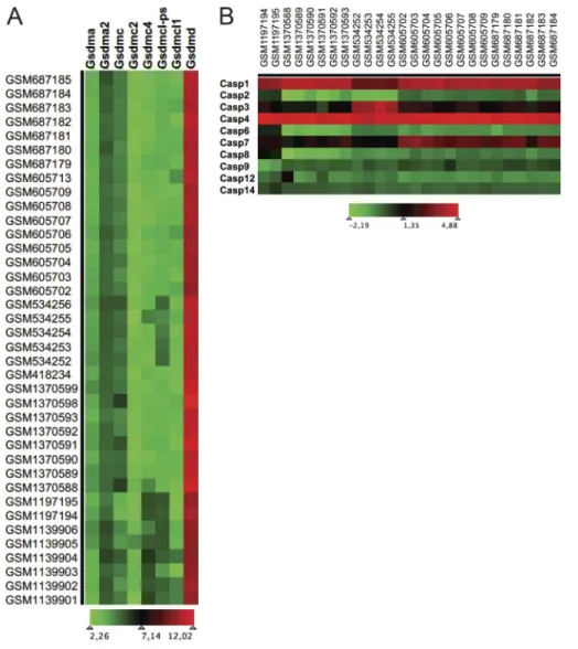
P2X7-mediated pyroptosis of Tfh cells via caspase-mediated activation of gasdermin D
Online monitoring of cell permeability to YO-PRO-1 by flow cy-tometry upon stimulation with the P2X7 agonist 3′-O-(4-benzoyl) benzoyl ATP (BzATP) is used to assess cellular sensitivity to death mediated by cytolytic pore opening. Tfh cells from spleen of pris-tane-treated WT but not P2rx7−/− mice were sensitive to P2X7 stimulation in this assay (Fig. 8 A). In T cells, P2X7-mediated pore opening and cell death was associated with features characteris-tic of pyroptosis (Taylor et al., 2008), a form of death executed by gasdermin D (Gsdmd) upon caspase-mediated cleavage and relief of autoinhibition of the pore forming N terminus (Kayagaki et al., 2015; Shi et al., 2015). A search of gene expression profiles in public datasets revealed Gsdmd as the most widely represented member among the gasdermin family in CD4 naive and Tfh cells (Fig. S3 A). In addition, the analysis of caspase expression in CD4 naive versus Tfh cells revealed the selective up-regulation of caspase-1 and -4 in Tfh cells (Fig. S3 B). Since Gsdmd is a
sub-Figure 4. Impaired primary, secondary, and memory responses to NP-OVA in P2rx7−/− mice. (A) Scheme of the immunization protocol and time points
of analysis. (B) Frequency and absolute number of CXCR5+ICOS+ Tfh cells in the draining lymph nodes from untreated or immunized WT and P2rx7−/− mice at
1 wk after s.c. injection of NP-OVA in MF59 (WT unimmunized, n = 5; P2rx7−/− unimmunized, n = 5; WT NP-OVA, n = 8; P2rx7−/− NP-OVA, n = 8), representative
contour plots of staining with human CLIP and OVA tetramers, and statistics of OVA tetramer positive cells (n = 4). (C) Frequency and absolute number of GC B cells (same samples as above) gated as PNAhiB220+ among CD19+ B cells (upper bar graphs) and of NP-positive GC B cells (lower bar graphs) with
strate of caspase-1 and -4, this observation suggests that Tfh cells could be sensitive to Gsdmd pore-forming activity and execute pyroptosis in Tfh cells upon exposure to BzATP. Accordingly, BzATP stimulation of Tfh cells isolated from pristane-treated WT but not P2rx7−/− mice resulted in caspase activation (Fig. 8 B) and generation of cleaved Gsdmd (Fig. 8 C), suggesting that P2X7 limits Tfh cell survival in eATP-rich microenvironment via Gsd-md-mediated pyroptotic cell death.
Phenotypic and functional distinction of circulating Tfh cells in SLE and PAPS donors
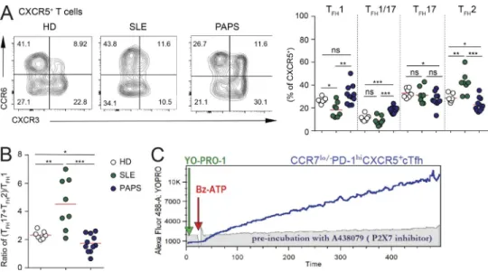
As previously shown (He et al., 2013), we observed significantly increased frequencies of the CCR7lo/−PD-1hi subset of circulat-ing Tfh (cTfh) cells in our cohort of SLE patients (Table S1 and Fig. 9 A). Interestingly, the analysis of these cells in patients suffering from PAPS did not reveal differences with respect to healthy donors (HDs; Fig. 9 A). Moreover, quantification of CXCL13 in the blood as a Tfh cells derived biomarker of GC ac-tivity in humans (Havenar-Daughton et al., 2016) showed the significant increase in SLE but not PAPS with respect to HDs (Fig. 9 B). These results support a role for deregulated Tfh cells in autoantibody generation and pathogenesis in SLE, but not PAPS (where SLE features are not observed even in the presence of antiphospholipid antibodies, including lupus anticoagulant, anti-cardiolipin, or anti-β2-glycoprotein-I antibodies; Ruiz-Irastorza et al., 2010). To investigate possible differences in cTfh cell function in SLE and PAPS, we used a chemokine receptor signature, which classifies these cells into three majors subsets: efficient B cell helper cells, defined as cTfh2 (CXCR3−CCR6−) and cTfh17 (CXCR3−CCR6+) cells, which are increased in some auto-immune disorders, including SLE, and nonefficient B cell helper cTfh1 (CXCR3+CCR6−) cells (Ueno et al., 2015; Blanco et al., 2016). We confirmed a significant increase in cTfh2 and a concom-itant decrease in cTfh1 cells in SLE patients. In contrast, PAPS patients were characterized by the increase in a cTfh1/Tfh17 “hybrid” subset that was negatively associated with disease ac-tivity in SLE (Choi et al., 2015; Fig. S4 A). SLE and PAPS patients showed an opposite trend of the ratio between the sum of cTfh2 and cTfh17 cells with cTfh1 cells compared with HDs. Whereas SLE patients showed a significant increase of this value, as pre-viously described (Le Coz et al., 2013), PAPS patients displayed a significant reduction, thereby defining a functional distinction of cTfh cells in these two autoantibody-mediated pathological conditions (Fig. S4 B).
Impaired P2X7 receptor activity unleashes Tfh cell expansion in SLE
To address possible differences in sensitivity to eATP via P2X7 in cTfh cells from SLE versus PAPS, we tested YO-PRO-1 permeabil-ity in flow cytometry upon stimulation with BzATP. This assay
is sensitive in detecting P2X7 activity in human cTfh cells, and preincubation with the P2X7 inhibitor A438079 completely abol-ished the dye uptake (Fig. S4 C). These experiments revealed the significant reduction of BzATP-sensitive cTfh cells and impair-ment of YO-PRO-1 permeability in SLE patients with respect to both healthy and PAPS subjects (Fig. 9 C). YO-PRO-1 permeability by P2X7 stimulation inversely correlated with the frequency of CCR7lo/−PD-1hi cTfh cells (Fig. 9 D). Principal component analysis (PCA) for the frequencies of CCR7lo/−PD-1hi and YO-PRO-1+ cells as well as YO-PRO-1 uptake at 450 s identified two main clusters: while HDs and PAPS segregated close to each other, SLE samples were clearly differentiated along the first component (Fig. 9 E). These results indicate that impaired P2X7 activity might be re-sponsible for the increase in cTfh cells in SLE. In line with the reduced sensitivity to pore formation, P2RX7 mRNA was signifi-cantly reduced in cTfh cells from SLE with respect to both healthy and PAPS donors, suggesting that P2RX7 is selectively down-reg-ulated in SLE (Fig. 10 B). Stimulation of sorted CD4 naive T cells and cTfh cells from HDs with anti-CD3/CD28 antibodies induced progressive up-regulation of P2RX7 transcripts, which were significantly higher in ex vivo sorted and in vitro activated cTfh compared with naive T cells (Fig. 10 A). This up-regulation is maintained also by shifting cells in medium with IL-2 after 48-h TCR stimulation (Fig. 10 A). The propensity of effector/memory CD4 cells to expand in an antigen-independent fashion can be scored in vitro by the analysis of cell proliferation in response to cytokines (Geginat et al., 2001). Upon stimulation with IL-7 and IL-15, CXCR5+ circulating T cells from SLE patients proliferated more robustly than cells isolated from healthy or PAPS subjects and were significantly more resistant to inhibition of prolifer-ation by BzATP (Fig. 10 C). Analogous defective inhibition was observed also in purified CCR7lo/−PD-1hi cells (Fig. 10 D).
It was recently shown that CD4 naive T cells can differentiate into Tfh cells and expand in vitro upon exposure to activin A and IL-12 (Locci et al., 2016). The generation and expansion of Tfh cells in this assay was significantly inhibited by the addition of BzATP (Fig. 10 E). However, CD4 cells from SLE donors generated Tfh cells with the same efficiency irrespective of the presence of BzATP, suggesting that Tfh cells in SLE are poised to be defective in sensing eATP as an inflammatory cue that should limit their function (Fig. 10 E). Altogether, these results point to a role for P2X7 as a checkpoint regulator for Tfh cells that appears to be compromised in SLE.
Discussion
The identification of the sanroque mutation (that disrupts a repressor of ICOS) in mice unraveled the causal role of deregu-lated Tfh cells and GCs in developing high titers of autoantibodies and lupus pathology (Vinuesa et al., 2005). We hypothesize that WT unimmunized (n = 2), P2rx7−/− unimmunized (n = 2), WT NP-OVA (n = 4), and P2rx7−/− NP-OVA (n = 4). Absolute number of NP-specific IgG secreting cells
in the spleen (WT NP-OVA, n = 8; and P2rx7−/− NP-OVA, n = 9) and BM (WT NP-OVA, n = 7; and P2rx7−/− NP-OVA, n = 8) of immunized mice. (E) Frequency
of OVA tetramer positive cells among CD4+ T cells (n = 5) and absolute number of NP-specific IgG secreting cells in the spleen of the indicated mice. WT and
P2rx7−/− no recall (n = 4), WT and P2rx7−/− NP-OVA (n = 5). Unpaired Student’s t test. Each dot in graphs represents an individual mouse, and horizontal lines
Figure 5. Selective expansion of P2rx7−/− Tfh cells upon pristane injection but not conventional immunization. (A) Scheme of the adoptive transfer and immunization experiment; representative plots of donor CD4+ and CXCR5+ICOS+ T cells within transgenic cells recovered from the draining lymph nodes of
recipient mice 1 wk after primary immunization and frequency of recovered donor cells within individual mice after primary immunization (10 mice per group) and secondary response (five mice per group; mean ± SEM). (B) Scheme of the adoptive transfer and pristane administration experiment; representative plots of CD90.2+ donor CD4+ T cells from spleen at 2 mo after transfer, frequency of recovered donor CXCR5+ICOS+ CD4+ T cells within individual mice at 1, 2, and
4 mo after injection of pristane (mean ± SEM). Each dot in graphs represents an individual mouse. Two-tailed Mann–Whitney U test. *, P < 0.05; **, P < 0.01. (C) Correlation between GC B cells and donor Tfh cell frequencies at the indicated time points. The correlation coefficient r and the respective P value were calculated with nonparametric Spearman test. Each dot in graphs represents an individual mouse.
P2X7-mediated regulation of Tfh cells can limit potentially patho-genic GC activity. The abundance of CCR7lo/−PD-1hi cTfh cells in the blood correlates with active Tfh cell differentiation (He et al., 2013). These cells were selectively enriched and resistant to
P2X7-mediated cell death in SLE but not PAPS patients. Excessive IFN-γ promotes Tfh cell accumulation and lupus-associated pa-thology (Lee et al., 2012). Our results indicate that P2X7 activity might contribute to inhibition of IFN-γ secretion and offset the Figure 6. P2X7-mediated control of PSGL-1 down-regulation and IFN-γ secretion in pristane-treated mice. (A) Frequency of PNA+Fas+ GC splenic B
cells (untreated WT, n = 3; P2rx7−/−, n = 3; and Icos−/−P2rx7−/−, n = 3; pristane-treated WT, n = 9; P2rx7−/−, n = 9; and Icos−/−P2rx7−/−, n = 5). (B) Serum ANA IgG
detection by ELI SA (MRL/lpr, n = 2; untreated WT, n = 4; P2rx7−/−, n = 4; and Icos−/−P2rx7−/−, n = 5; pristane-treated WT, n = 9; P2rx7−/−, n = 10; and Icos−/−
P2rx7−/−, n = 10). (C) Proteinuria score (untreated WT, n = 3; P2rx7−/−, n = 3; and Icos−/−P2rx7−/−, n = 4; pristane-treated WT, n = 8; P2rx7−/−, n = 8; and Icos−/−
P2rx7−/−, n = 8) in the indicated mice at 33 wk after pristane injection. Each dot represents an individual mouse, and horizontal lines represent median values.
(D) Representative contour plots for PSGL-1 and CD62L on splenic CD4+ T cells, frequency and absolute number (mean ± SEM) of PSGL1lo/−CD62L− cells from
untreated WT (n = 12), P2rx7−/− (n = 10), Icos−/−P2rx7−/− (n = 5), treated WT (n = 38), P2rx7−/− (n = 37), and Icos−/−P2rx7−/− (n = 8) mice. (E) Contour plots show
representative intracellular staining for IL-21, IL-17, and IFN-γ on gated CD4+ICOS+PSGL1lo/− cells from spleens of treated WT and P2rx7−/− mice. Statistics from
three independent experiments are shown (mean ± SEM, untreated mice, n ≥ 4; treated mice, n ≥ 8). Two-tailed Mann–Whitney U test. *, P < 0.05; **, P < 0.01; ***, P < 0.001; ****, P < 0.0001. ns, not significant.
Figure 7. Increased PIL severity and IFN-γ secretion by ICOS+PSGL-1lo/− CD4 T cells in mice with conditional deletion of P2rx7 in T cells. (A) Spleen weight of untreated CD4-Cre P2rx7WT/WT (n = 5), CD4-Cre P2rx7fl/fl (n = 12), pristane-treated CD4-Cre P2rx7WT/WT (n = 21), and CD4-Cre P2rx7fl/fl (n = 15) mice.
(B) Proteinuria score. (C) Serum IgG concentration. (D) Semiquantitative detection of self-reactive IgG by ELI SA (QUA NTA-Lite ANA) in the same mice. (E–G) Absolute numbers of IgG-secreting cells (E) and frequencies and absolute numbers of CD19+B220+PNA+ Fas+ (F) and TCRβ+CD4+CXCR5+ICOS+ (G) cells in the
spleen of the same mice. (H) Representative contour plots for ICOS and PSGL-1 staining among CD4+ T cells from spleen and frequency of IFN-γ secreting
cells within the ICOS+PSGL-1lo/− CD4+ T cells of the same mice. Each dot represents an individual mouse; median or mean ± SEM is shown. Two-tailed Mann–
risk of GC-driven autoimmunity by chronic Tfh cell stimulation. Notably, IFN-γ deficiency did not impair Tfh cell response against immunization with foreign antigen (Lee et al., 2012). According to a dichotomy of P2X7 in regulating Tfh cell function in acute stimulation by cognate antigen versus chronic exposure to in-flammatory environments, immunization with OVA did not re-sult in any difference in the expansion of OT-II Tfh cells both in primary and secondary response. Whereas P2X7 activity does not affect Tfh cell response to “physiological” stimulation by exoge-nous antigens, it appears to limit immunopathological responses. Therefore, restoring P2X7 competence in Tfh cells might improve immunopathology in systemic autoimmunity.
The progressive nature of SLE is characterized by the accu-mulation of new types of autoantibodies up to the clinical onset of the disease. Moreover, the appearance of specific autoan-tibodies, namely anti-Sm and anti–nuclear ribonucleoprotein antibodies, tends to coincide with the clinical manifestations of SLE. These observations led to the modeling of disease evolution into three distinct phases of benign autoimmunity, pathogenic autoimmunity, and clinical illness (Arbuckle et al., 2003). The genetic as well as environmental factors influencing the develop-ment of pathogenic autoimmunity in SLE are not defined. SLE is a polygenic disease; strong candidates that may cause the disease seem to be located across chromosomal bands 12q24.1 to 12q24.3 (Nath et al., 2004). Together with other potential candidate genes in SLE pathogenesis, P2RX7 is located within this chromosomal region. Human P2RX7 is highly polymorphic and contains a large set of single nucleotide polymorphisms that affect P2X7 activity. Haplotypes containing the Ala348>Thr polymorphism (rs1718119) are characterized by a gain-of-function effect (Stokes et al., 2010). Interestingly, this polymorphism was associated with lower risk of SLE in a Chinese population (Chen et al., 2013). CD4 cells from patients with SLE, but not rheumatoid arthritis, exhibited diminished P2X7-dependent ATP-mediated cell death compared with healthy controls (Portales-Cervantes et al., 2010). We have shown a diminished sensitivity of Tfh cells from SLE but not PAPS patients to cytolytic pore opening by P2X7 pharmaco-logical agonist, suggesting that down-regulation of P2X7 might contribute to a selective Tfh cell dysfunction associated with SLE. Notably, aging Fas-deficient MRL/lpr mice, which develop a lupus-like syndrome, show a drastically reduced sensitivity to ATP-mediated stimulation of P2X7 in T cells associated with progressive lymphoproliferation (Le Gall et al., 2012). Although we cannot exclude that other mechanisms contribute to the im-paired response of Tfh cells to P2X7 stimulation in SLE patients, the observed reduction of P2RX7 mRNA constitutes a possible cause. This reduction might be correlated to the pronounced changes in microRNAs that characterize SLE and affect lym-phocyte function and Ig responses (Zan et al., 2014). In innate immune system, P2X7 has a well-established role as a trigger for inflammatory cytokines release, a feature that fostered the
de-velopment of P2X7 inhibitors to be used as therapeutic drugs in autoimmune diseases (Ferrari et al., 2006). Our results suggest that P2X7 inhibition in chronic inflammatory conditions where GC activity is enhanced might promote the inappropriate expan-sion of potentially pathogenic Tfh cells.
Materials and methods
Mice
C57/BL6J, P2rx7−/− (B6.129P2-P2rx7tm1Gab/J), Icos−/− (B6.129P2- Icostm1Mak/J), OT-II (B6.Cg-Tg(TcraTcrb)425Cbn/J), CD45.1 (B6.SJL-Ptprca Pepcb/BoyJ), CD90.1 (B6.PL-Thy1a/CyJ), and Cd4-cre (B6.Cg-Tg(Cd4-Cd4-cre)1Cwi/BfluJ) mice were from The Jackson Laboratory. The P2rx7fl/fl mice were from the European Mouse Mutant Archive (EMMA ID 05116; Skarnes et al., 2011). Cd4-Cre P2rx7fl/fl mice were generated by crossing the two strains. All mice were bred in the specific pathogen–free facility at the Insti-tute for Research in Biomedicine (IRB), Bellinzona, Switzerland. All animal experiments were performed in accordance with the Swiss Federal Veterinary Office guidelines and authorized by the Cantonal Veterinary.
PIL
SLE was induced by a single i.p. injection of 0.5 ml pristane to 8-wk-old WT or P2rx7−/− female mice. Mice were monitored for clinical signs of SLE until the end of the study (33 wk after dosing) and examined for lesions consistent with SLE by kidney histology and immunofluorescence, presence of serum autoanti-bodies by ELI SA, and cell subsets composition by flow cytometry. Proteinuria was measured using Bayer Albustix reagent strips on collected fresh spots of urine from each mouse. Score from 0 to 4 was applied as follows: 0, none; 1, trace; 2, 30 mg/ml; 3, 100 mg/ ml; and 4, ≥500 mg/ml.
Immunization and adoptive transfer of CD4 cells
8-wk-old female mice were s.c. injected with 50 µg of NP16-OVA (BIO SEA RCH Technologies) in MF59 (AddaVax; InvivoGen). Draining lymph nodes and spleen were collected 1 wk after injec-tion, and cells were analyzed by flow cytometry. For the analysis of secondary response, the same immunogen was administered at day 14 after primary immunization and cells were analyzed after 1 wk. For analysis at 102 d after primary immunization, 50 µg of NP16-OVA in PBS was administered s.c.; OVA-peptide329–337 –spe-cific CD4 and NP-spe–spe-cific IgG-secreting spleen cells were quan-tified 1 wk later. For adoptive transfer of OT-II cells, CD4+ cells were enriched with anti-CD4 coated magnetic beads (Miltenyi Biotec) and sorted as CD4+CD8−CD25−CD44−CD62L+ naive cells on a FAC SAria. 8-wk-old CD90.1 mice were injected intravenously with 2.5 × 10⁵ sorted cells containing at least 90% OT-II+ cells. Recipient mice were immunized 24 h later with a subcutaneous injection of 50 µg of NP16-OVA (BIO SEA RCH Technologies) 1:1 in
and frequency of IFN-γ–secreting cells within the indicated subsets of untreated and treated mice. Statistics from three independent experiments are shown. Mean ± SEM (untreated mice, n ≥ 4; treated mice, n ≥ 8). (J) Representative contour plots for intracellular staining of IL-21, IL-17, and IFN-γ in CD4+ T cells from
spleen of pristane-treated WT, P2rx7−/−, and Icos−/−P2rx7−/− mice and statistics of frequencies. Mean ± SEM (n = 2 independent experiments with at least five
MF59 (AddaVax; InvivoGen). For retransfer of activated OT II ef-fector cells, CD4-enriched cells were sorted for CD90.2 cells, and 5 × 10⁴ cells were injected intravenously into naive CD90.1 hosts. To reactivate memory OT-II cells, recipient mice received a sub-cutaneous injection of 50 µg of NP16-OVA in MF59 28 d after sec-ondary transfer, and draining lymph nodes were analyzed 1 wk later. For adoptive transfer of polyclonal CD4+ T cells from either WT or P2rx7−/− mice, spleens were collected, and CD4+ cells were enriched with anti-CD4 coated magnetic beads (Miltenyi Biotec)
and then sorted as CD4+CD8−CD19−B220−CD11c−CD11b− cells on a FAC SAria. 3.5 × 10⁶ cells were injected into CD90.1 WT mice, and 24 h later, mice were injected i.p. with 0.5 ml pristane. Spleens were collected and analyzed 1, 2, or 4 mo after injection.
Histochemistry and immunohistochemistry
All animals were euthanized with CO2, followed by exsanguina-tion. For histological evaluation, the kidneys were removed, fixed in 10% neutral buffered formalin for 48 h, trimmed, dehydrated, Figure 8. P2X7-mediated caspase activation and Gsdmd cleavage in Tfh cells. (A) Time monitoring of YO-PRO-1 uptake after stimulation with BzATP in ICOS−PD-1− and ICOS+PD-1+ cells within CXCR5+ CD4+ T cells from spleen of WT and P2rx7−/− mice at 33 wk after pristane administration and frequency of
responding cells in the two subsets from WT mice (n = 14). (B) Analysis by flow cytometry and statistics of caspase activation in purified WT and P2rx7−/− Tfh
cells upon stimulation with BzATP as indicated (n = 4). (C) Representative Western blot of full-length and caspase-cleaved Gsdmd (FL and Nterm, respectively), and actin on the same cells as in B, either untreated or stimulated with BzATP. A shorter exposure for GsdmFL is shown. Histograms show the statistics of
GsdmdNterm relative expression normalized on GsdmFL in the indicated conditions (n = 3). Bar graphs: mean ± SEM. Two-tailed Mann–Whitney U test. *, P <
and embedded in paraffin wax. Sections of 3–5-µm thickness were prepared, mounted on glass slides, deparaffinized in xy-lene, and rehydrated through graded alcohols, before staining with H&E or periodic-acid-Schiff reagent. Slides were evaluated in a blinded fashion by a board-certified veterinary pathologist (G. Pellegrini). Glomerular injury was assessed by light micros-copy on 50 consecutive glomeruli/mouse using a histopatholog-ical scoring system modified from Wang et al. (1999). Briefly, a grade of 0 indicated normal glomerular histology; scores 1 to 3 in-dicated progressive severe glomerular injury, i.e., score 1: mildly thickened and hypereosinophilic mesangium, multiple clusters of nuclei, mild reduction in the number of glomerular capillar-ies; score 2: moderately thickened and hypereosinophilic
mesan-gium, moderate diffuse hypercellularity, marked reduction in the number of glomerular capillaries, occasional inflammatory cells, mostly neutrophils; and score 3: noticeable enlargement of the glomerular tuft, abnormal glomerular shape (irregular rather than round), severely thickened and hypereosinophilic mesangium, occasional adhesions of the glomerular tuft to the Bowman’s capsule (synechiae), severe diffuse hypercellularity, loss of capillaries, occasional inflammatory cells, mostly neu-trophils. To detect glomerular immune complexes by immuno-fluorescence, kidneys were removed and immediately frozen in OCT (Tissue-Tek). Cryostat sections (4 µm) were stained with the following antibodies: AF488 goat anti–mouse IgG (highly cross-adsorbed; Southern Biotech), rat anti-mouse C3 (RmC11H9) Figure 9. Frequency of cTfh cells and P2X7 activity in healthy subjects and SLE and PAPS patients. (A) Representative dot plots for CXCR5+ cells and
CCR7lo/−PD-1hi cells within cTfh cells of the indicated subjects and frequency of CCR7lo/−PD-1hi cells within cTfh cells in healthy (HD), SLE, and PAPS subjects.
Two-tailed Mann–Whitney U test. (B) Serum CXCL13 concentration in HDs, SLE, and PAPS subjects. Two-tailed Mann–Whitney U test. (C) Representative time monitoring of YO-PRO-1 uptake in CD14−CD4+CD45RA−CXCR5+CCR7lo/−PD-1hi cTfh cells from PBMCs of the indicated subjects after stimulation with BzATP.
Statistics of YO-PRO-1–permeable cells (two-tailed Mann–Whitney U test) and YO-PRO-1 uptake expressed as fold increase of mean fluorescence intensity at 250 and 450 s (two-way ANO VA multiple comparisons, with Bonferroni’s correction) are shown. (D) Correlation of fold increase in YO-PRO-1 uptake at 450 s and percentage of cTfh cells. The correlation coefficient r and the respective P value were calculated with nonparametric Spearman test. (E) PCA of HDs and SLE and PAPS patients according to % cTfh cells, % YO-PRO-1+ cTfh cells, and YO-PRO-1 uptake at 450 s. Areas of spreading are highlighted to better visualize
segregation of groups of subjects. Each dot in graphs represents a subject, and horizontal lines represent median values. *, P < 0.05; ***, P < 0.001; ****, P < 0.0001. ns, not significant.
followed by AF647 labeled goat anti–rat IgG (cross-adsorbed against mouse IgG; Southern Biotech). To detect renal infiltrat-ing CD4 T lymphocytes, the followinfiltrat-ing antibodies were applied to the sections: rat anti-mouse CD4 (clone: GK1.5; BD PharMingen) followed by AF488 goat anti–rat IgG (Invitrogen), hamster an-ti-mouse ICOS (clone: C398.4A; eBioscience) followed by AF594 goat anti–hamster IgG (Invitrogen). Slides were mounted with Gelvatol Mounting Media and stained with DAPI for the detection of nuclei. Images were acquired using a Leica TCS SP5 confocal microscope, with a HCX PL APO 40×/1.25 N.A. oil-immersion
ob-jective. Quantification was performed with ImageJ open-source software (National Institutes of Health; Schindelin et al., 2012). Regions of interest containing the glomeruli were manually de-fined, and the background was excluded by applying a threshold on the intensity; the area above the threshold in these regions of interest was measured in three different fields of view for each sample and is expressed as percentage of the total glomerulus area. For histological analysis of GCs, spleens were frozen in OCT. Cryostat sections (4 µm) were stained with the following antibodies: AF594 anti–mouse IgD (clone: 11-26c.2a; BioLegend), Figure 10. P2RX7 expression and function in HDs and SLE and PAPS patients. (A) P2RX7 transcript quantification in CD4 naive and cTfh cells at different times after in vitro stimulation with anti-CD3/28 antibodies and upon shifting cells in IL-2 after 48-h stimulation. Data are from cells sorted from three different HDs. Unpaired Student’s t test. (B) P2RX7 transcripts in sorted CXCR5+ CD4 T cells from healthy (HD), SLE, and PAPS donors (n = 10). Two-tailed Mann–Whitney
U test. A.U., arbitrary units. (C) Representative histograms of CellTrace Violet (CTV) dilution in purified CXCR5+CD4+ cells from the indicated subjects after 7-d
culture in the presence of IL-7 and IL-15 either without or with BzATP. Percentages of proliferating cells within the marker are indicated. Statistics for prolifer-ation and inhibition of proliferprolifer-ation by BzATP. Each dot represents a subject, and horizontal lines represent median values. Two-tailed Mann–Whitney U test. (D) Percentage inhibition of proliferation by BzATP in sorted CCR7lo/−PD-1hi Tfh cells from HDs and SLE individual subjects at day 7 of stimulation with IL-7 and
IL-15. Number of tested donors is indicated in the graphs. Each dot represents a subject, and horizontal lines represent median values. Two-tailed Mann–Whit-ney U test. (E) Representative contour plots for in vitro differentiation of naive CD4 cells from HDs and SLE donors (n = 4) into Tfh cells in the presence of the indicated stimuli and statistics (Student’s t test). Bar graphs: mean ± SEM. *, P < 0.05; **, P < 0.01; ***, P < 0.001. ns, not significant.
AF647 anti-mouse GL-7 (clone: GL-7; BioLegend), BV405 ti-mouse CD4 (clone: GK1.5; BioLegend), unconjugated goat an-ti-mouse PD-1 Ig (Novusbio) followed by AF488 rabbit anti–goat Ig (Invitrogen). Images were collected on a laser-scanning confo-cal microscope with a HCX PL APO 40×/1.25 N.A. oil-immersion objective. GC area was measured using ImageJ (Schindelin et al., 2012). To score GC frequency, total GC numbers were quantitated in three separate 10× fields of view of 2.4 mm2 (1,550 µm × 1,550 µm) per spleen section.
Preparation of single-cell suspension from kidney
Kidneys were removed, minced, and digested with Liberase (0.14 mg/ml; Roche) and DNase I (0.03 mg/ml; Sigma-Aldrich) in HBSS medium, for 30–40 min at 37°C. Tissue pieces were then pressed through a 70-µm strainer to obtain single-cell suspensions. Cells were enriched in leukocytes using an 80/20 Percoll gradient and centrifuged at 800 g for 20 min at room temperature. Lymphoid fractions were collected at the interphase of the Percoll gradient and used for flow cytometry.
Mouse cell isolation and flow cytometry
Single-cell suspensions were prepared by passing spleens through a 70-µm nylon mesh. After centrifugation at 1,500 rpm for 5 min, red blood cells were lysed in ACK-lysis buffer for 7 min. Lysis buffer was neutralized by washing once in staining buffer. Cells were stained with the following monoclonal antibodies: biotin-conjugated anti-CXCR5 (clone: 2G8; BD), PE-labeled anti ICOS (clone: 7E.17G9; BD) or PerCPCy5-labeled anti-ICOS (clone: C398.4A; BioLegend), APC-labeled anti PD-1 (clone: RMPI-30; BioLegend), Percp-eFluor710–labeled anti-CD3 (Clone: 17A2; eBioscience), AF488-labeled TCRβ (clone: H57-597; BioLegend), APC-Cy7–labeled anti-CD4 (clone: RM4-5; BioLegend), BV421 anti-CD162 (PSGL1; clone 2PH1; BD), PeCy7-labeled anti-CD25 (clone: PC61; BioLegend), PE-labeled anti-CD44 (clone: IM7; BioLegend), APC-labeled CD62L (clone: MEL-14; eBioscience), and BV421-labeled anti-CD197 (CCR7; clone: 4B12; BioLegend), APC-Cy7–labeled anti-CD19 (clone: 6D5; BioLegend), BV405-la-beled B220 (clone: RA3-6B2; BioLegend), PE-laBV405-la-beled anti-Fas (clone: Jo2; BD), fluorescein-labeled peanut agglutinin (PNA; FL-10-71; Vectorlabs), biotinylated anti-CD275 (ICO SL; clone: HK5.3; BioLegend), PE-labeled anti-CD138 (clone: 281-2; BD), APC-la-beled anti-CD11c (clone: N418; eBioscience), APC-eFluor780– labeled CD45.2 (clone: 104; eBioscience), and APC-Cy7–labeled anti-CD90.2 (clone: 30-H12; BioLegend). APC-labeled streptavi-din was purchased from BioLegend and efluo405-labeled strepta-vidin from eBioscience. Intracellular staining was performed using the BD Cytofix/Cytoperm and Perm/Wash buffers or, for intracellular FoxP3 (FITC-labeled, clone: FJK-16s; eBioscience) staining, the eBioscience FoxP3 staining buffer set. For intra-cellular staining of IL-21 (R&D Systems), IFN-γ (PeCy7-labeled, clone: XMG1.2; eBioscience), and IL-17A (PerCP-Cy5–labeled, clone: eBio17B7; eBioscience), 5 × 10⁶ splenocytes were cultured for 5 h at 37°C in 24-well plates in 2 ml culture medium contain-ing ionomycin (750 ng/ml) and PMA (20 ng/ml). For the last 4 h, Monensin (1,000× solution; eBioscience) was added to the cultures. IL-21 was detected with a recombinant mouse IL-21R subunit/human IgG1 Fc chimera (R&D Systems) with goat
an-ti-human Fcγ conjugated to AF488 (Jackson ImmunoResearch). For detection of activated caspases, Tfh cells were incubated with FITC-VAD-FMK (CaspGlow Fluorescein Active Caspase Staining Kit; BioVision) for 30–45 min at 37°C in RPMI-1640 according to the manufacturerʼs protocol. Cells were then washed and resus-pended in PBS for analysis by flow cytometry. Samples were ac-quired on an LSRFortessa (BD Biosciences) flow cytometer. Data were analyzed using FlowJo software (TreeStar).
Tetramer staining
Tetramers were provided by the National Institutes of Health Te-tramer Core Facility. Cells were stained for 2 h at 37°C with the following APC-labeled tetramers: I-Ab chicken OVA329–337 AAH AEI NEA, I-Ab chicken OVA328–337 HAA HAE INEA, I-Ab chicken OVA259–277 IIN FEK LTE WTS SNV MEER, and I-Ab human CLIP87–101 PVS KMR MAT PLL MQA. All OVA tetramers gave comparable re-sults in flow cytometry.
ELI SA and ELI SPOT assays
For total Ig isotype determination, ELI SA plates (Corning 96-Well Half Area Flat Bottom Polystyrene High Bind) were coated for 3 h at room temperature with purified goat anti–mouse IgG, IgM, and IgA antibodies (Southern Biotech) used at a con-centration of 10 µg/ml. After four washes with PBS 0.025% Tween-20 and blocking with PBS 1% BSA for 1 h at room tem-perature, samples and standards (relative unlabeled mouse Ig; Southern Biotech) were diluted and incubated at room temperature for 4 h. Specific secondary goat anti–mouse Ig conjugated with alkaline-phosphatase were added after four washes with PBS and 0.025% Tween-20 and incubated for 2 h at room temperature. Plates were washed again, and the assay was developed with Sigma 104 phosphatase substrate. Plates were read at 405 nm. IgG-secreting cells were detected using ELI SPOT assay: 96-well plates (Millipore, MSI PS4510 Sterile, hy-drophobic high protein binding Immobilon-P membrane) were coated with 10 µg/ml purified goat anti–mouse IgG (Southern Biotech) for 2 h at room temperature. After three washes with PBS solution, plates were blocked with PBS and 1% BSA and incubated for 30 min at 37°C. Serial dilutions of splenocytes were added in a final volume of 200 µl B cell medium (RPMI, 10% Hyclone serum) and left at 37°C for 16 h. Subsequently, plates were washed three times with PBS and 0.25% Tween-20 and four times with PBS and incubated for 2 h at room tem-perature with biotinylated goat anti–mouse IgG (Southern Bio-tech). After washing, avidin-peroxidase (HRP; Sigma-Aldrich) was added and left for 1 h at room temperature. The assay was developed with AEC (Sigma-Aldrich). For quantification of IgG-secreting cells, plates were acquired, counted, and quality controlled using an ELI SPOT reader and ImmunoSpot 5.1 soft-ware (CTL Europe). For ANA detection, both indirect immuno-fluorescence assay ANA-Hep-2 fixed cells (ORG 870; Orgentec) and microwell ELI SA plates coated with highly purified indi-vidual antigens plus extracts from Hep-2 nuclei and nucleoli (QUA NTA Lite; Inova Diagnostics) were used. Briefly, fixed Hep-2 cells were incubated with mouse sera (diluted 1:100), and ANAs were detected with AF488-labeled goat anti–mouse IgG (South-ern Biotech). Slides were stained with DAPI and captured with a
Nikon Eclipse E800 upright microscope, with a 20×/0.75 N.A. ob-jective. Images were analyzed with ImageJ open-source software for pattern recognition (Schindelin et al., 2012). The kit ELI SA (QUA NTA Lite) for the semiquantitative detection of ANAs in human serum was used and adapted for the detection of mouse ANAs. The antigens include chromatin (double-stranded DNA [dsDNA] and histones), Sm/RNP, SS-A, SS-B, Scl-70, centromere, PCNA, Jo-1, mitochondria (M-2), and ribosomal-P protein, as well as the extracts. Sera were diluted at 1:200 in ANA sample diluent and detected with HRP-conjugated goat anti–mouse IgG (Southern Biotech). Plates were read for the absorbance at 450 nm within 1 h after addition of the stop solution.
Western blot
Tfh cells were isolated from mesenteric lymph nodes and PPs, washed with ice-cold PBS, and lysed with RIPA buffer 1× (Sigma) supplemented with protease inhibitor cocktail (Roche). Samples were centrifuged at 14,000 rpm for 10 min at 4°C and snap fro-zen. Cleared protein lysate was denatured with loading buffer supplemented with 0.1 µM DTT for 10 min at 65°C. Samples were run on precast 4–12% bis-Tris protein gels (Bio-Rad) and then transferred onto polyvinylidene difluoride membranes using Trans-Blot Turbo Transfer System (Bio-Rad). Membranes were blocked with 10% (wt/vol) nonfat dry milk (Bio-Rad) and 0.1% Tween-20 in TBS and incubated with appropriate antibod-ies in TBS with 0.1% Tween-20 for 16 h at 4°C. The following antibodies were used: anti-GSD MDC1 (clone: A-7, sc-393656; Santa Cruz Biotechnology) and anti-actin (A2066; Sigma). Bound antibodies were revealed by incubation with secondary HRP-conjugated anti-mouse (7076, Cell Signaling Technology) or anti-rabbit (7074; Cell Signaling Technology) IgG antibodies in TBS with 0.1% Tween-20. Membranes were developed using the Pierce ECL Western blotting substrate (32209; Thermo Sci-entific), signals were detected with the ImageQuant LAS 4000 system in the standard acquisition mode (GE Healthcare Life Sciences), and bands were quantified using the Multi Gauge Analysis tool (Fujifilm).
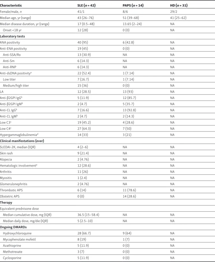
Human samples and patients
Peripheral blood samples from patients with SLE and PAPS were obtained after informed consent was provided (local ethics com-mittee approval no. 192_2016bis) during regular follow-up visits at the Lupus Clinic, IAS ST-Istituto Gaetano Pini, University of Milan, Italy. Inclusion criteria were age >18 yr, fulfillment of the American College of Rheumatology 1997 classification criteria for SLE (Hochberg, 1997) and 2006 classification criteria for an-tiphospholipid syndrome (APS; Miyakis et al., 2006; Table S1). Exclusion criteria were treatment with B cell–depleting agents or experimental drugs. Disease activity was determined with SLE disease activity index (SLE DAI-2K; Gladman et al., 2002); clinical and laboratory data were obtained during the routine follow-up. Patients were characterized for antiphospholipid an-tibodies (Lupus Anticoagulant, anticardiolipin IgG and IgM, and anti-β2GPI IgG and IgM; Andreoli et al., 2015) as well as ANA, anti-ENA, and anti-dsDNA antibodies (Ingegnoli et al., 2014). All experiments performed on human blood samples from HDs were approved by the ethics committee of Fondazione IRC CS Ca’
Granda Ospedale Maggiore Policlinico, and informed consent was obtained from all subjects.
Human cell isolation, stimulation, and flow cytometry
Peripheral blood mononuclear cells (PBMCs) were isolated from blood by density-gradient centrifugation using Ficoll-Paque Plus gradient (GE Healthcare). CD4+ T cells were enriched by posi-tive selection using human CD4 MicroBeads (Miltenyi Biotec), and then CD4+ T cell subsets were sorted to 99% purity with a FAC SAria (BD). For in vitro TCR stimulation, CD4+ naive and CXCR5+ cells were stimulated with anti-CD3 (5 µg/ml) and an-ti-CD28 (1 µg/ml) mAbs. PBMCs were stained with the following antibodies: PE-Texas Red–labeled anti-CD4 (clone: MHCD0417; Life Technologies), QD655-labeled anti-CD45RA (clone: MEM-56; Life Technologies), PE-labeled anti-CCR7 (clone: G043H7; BioLegend), purified mouse monoclonal IgG2b anti-CXCR5 (BRL-1, clone: 51505; R&D Systems) revealed by FITC-labeled anti–mouse IgG2b (Southern Biotech), PE-cyanine 5 (PeCy5)–labeled an-ti-CD183 (CXCR3; clone: 1C6/CXCR3; BD), PE-labeled anti-CD196 (CCR6; clone: 11A9; BD), BV785-labeled anti-CD279 (PD-1; clone: EH12.2H7; BioLegend). PeCy5-labeled anti-CD56 (clone: A07789; Beckman Coulter), anti-CD14 (clone: A07765; Beckman Coulter), anti-CD19 (clone: HIB19; BioLegend), anti-CD25 (clone: BC96; BioLegend), and anti-CD8 (clone: HIT8a; BioLegend) were in-cluded as a dump channel to exclude contaminant cells. Time monitoring of YO-PRO-1 uptake
106 human PBMCs were stained with the appropriate antibod-ies, washed, resuspended in RPMI 1640 complete medium, and loaded with YO-PRO-1 iodide (Life Technologies) at a final concentration of 5 µM. The YO-PRO-1 uptake following cell stimulation with 1 mM BzATP (Sigma) was monitored on an LSR-Fortessa for 480 s, and the kinetics was analyzed using FlowJo software (TreeStar). Murine splenocytes (106) were stimulated with 0.1 mM BzATP.
In vitro cytokine-driven proliferation assay
For cytokine-driven proliferation assay, sorted CXCR5+ or CCR7lo/−PD-1hi CD4 T cells were labeled with 5 µM Cell Trace Violet (Life Technologies) in PBS for 20 min at 37°C; staining was blocked by adding an equal volume of filtered prewarmed FBS. Cells were centrifuged at 1,500 rpm for 5 min and plated at a minimal density of 25,000 cells/well in RMPI 1640 complete medium. Plated cells were stimulated with recombinant IL-7 and IL-15 (R&D Systems) at 25 ng/ml, with BzATP at a final concen-tration of 100 µM when indicated. Cell proliferation and viability were assessed after 7 d using LSRFortessa.
In vitro CD4 cell differentiation to Tfh cell
CD4+ cells were enriched from PBMCs by positive selection with anti-CD4 MicroBeads (Miltenyi Biotec). Naive T cells were sorted on a FAC SAria (BD) to 99% purity as CD8−CD25−CD14− CD19−CD4+CD45RA+CCR7+ cells, activated by Dynabeads Human T-Activator CD3/CD28 (Life Technologies), and cultured with recombinant activin A (100 ng/ml), human IL-12 (5 ng/ml), IL-7 (4 ng/ml; all from R&D Systems), and BzATP where indicated, in AIM-V medium (Life Technologies), as described (Locci et al.,