HAL Id: hal-02565216
https://hal.archives-ouvertes.fr/hal-02565216
Submitted on 10 Nov 2020
HAL is a multi-disciplinary open access
archive for the deposit and dissemination of sci-entific research documents, whether they are pub-lished or not. The documents may come from teaching and research institutions in France or abroad, or from public or private research centers.
L’archive ouverte pluridisciplinaire HAL, est destinée au dépôt et à la diffusion de documents scientifiques de niveau recherche, publiés ou non, émanant des établissements d’enseignement et de recherche français ou étrangers, des laboratoires publics ou privés.
Pentagonal Bipyramidal Ln(III) Complexes Containing
an Axial Phosphine Oxide Ligand: Field-induced
Single-ion Magnetism Behavior of the Dy(III) Analogues
Pankaj Kalita, Naushad Ahmed, Arun Kumar Bar, Sourav Dey, Anukul Jana,
Gopalan Rajaraman, Jean-Pascal Sutter, Vadapalli Chandrasekhar
To cite this version:
Pankaj Kalita, Naushad Ahmed, Arun Kumar Bar, Sourav Dey, Anukul Jana, et al.. Pentagonal Bipyramidal Ln(III) Complexes Containing an Axial Phosphine Oxide Ligand: Field-induced Single-ion Magnetism Behavior of the Dy(III) Analogues. Inorganic Chemistry, American Chemical Society, 2020, 59 (9), pp.6603-6612. �10.1021/acs.inorgchem.0c00751�. �hal-02565216�
1
Pentagonal Bipyramidal Ln(III) Complexes Containing an Axial Phosphine Oxide Ligand: Field-induced Single-ion Magnetism Behavior of the Dy(III) Analogues
Pankaj Kalita,a,b Naushad Ahmed,‡b Arun Kumar Bar,a Sourav Dey,‡c Anukul Jana,b Gopalan Rajaraman*c, Jean-Pascal Sutter*d and Vadapalli Chandrasekhar*b,e
aSchool of Chemical Sciences, National Institute of Science Education and Research, HBNI, Bhubaneswar -752050, India.
bTata Institute of Fundamental Research Hyderabad, Gopanpally, Hyderabad-500107, India. cDepartrment of Chemistry, Indian Institute of Technology Bombay, Mumbai-400 076, India d Laboratoire de Chimie de Coordination du CNRS (LCC-CNRS),, Université de Toulouse, CNRS, Toulouse, France.
eDepartment of Chemistry, IIT Kanpur, Kanpur 208016, India.
*vc@tifrh.res.in; vc@iitk.ac.in; sutter@lcc-toulouse.fr; rajaraman@chem.iitb.ac.in
‡Authors contributed equally in this manuscript
Abstract
A series of neutral homologous complexes [(L)Ln(Cy3PO)Cl] {where Ln = Gd (1), Tb (2), Dy (3) and Er (5)} and [(L)Dy(Ph3PO)Cl] (4) [H2L = 2,6-diacetylpyridine bis-benzoylhydrazone] have been isolated. In these complexes, the central lanthanide ion possesses a pentagonal bipyramidal (PBP) geometry with an overall pseudo D5h symmetry. The coordination environment around the lanthanide ion comprises of three nitrogen and two oxygen donors in an equatorial plane. The axial positions are taken up by a phosphine oxide (O donor) and a chloride ion. Among these compounds, the Dy(III) (3 and 4) analogues were found to be field-induced single-ion magnets.
2
Introduction
There has been a significant renaissance in the chemistry of the rare earth elements because of their applications in catalysis1, photophysical properties2 and in magnetic materials3. In recent years lanthanide-4and some actinide complexes5 are finding increasing utility as molecular magnets (single-molecule- and single-ion magnets; SMMs and SIMs). These molecular systems, once magnetized, retain their magnetization even after the removal of the external magnetic field and are characterized by a slow reversal of magnetization below certain temperatures.6 This is because in SMMs, on application of a magnetic field, a double-well potential comprising of the various mj or ms states with an energy barrier (Ueff) is generated which prevents the reversal of magnetization below certain temperatures (the blocking temperature, TB).7 Various relaxation mechanisms including quantum tunnelling are prevalent to allow the magnetization to be lost.8
The evidence of SMM behaviour in a molecular Tb(III) complex, where the Tb(III) is sandwiched by two phthalocyanine ligands, was first reported by Ishikawa and co-workers.9 One of the intrinsic features of the lanthanide elements is that the 4f electrons are deeply buried inside the [Xe] core and are considerably shielded by the 5s and 5p electrons. This results in an almost unquenched orbital angular momentum (L) which couples with the spin angular momentum (S) giving rise to the total angular momentum, J.4d, 10 Unlike transition metal ions, the magnitude of spin-orbit coupling in the case of 4f metal ions is comparatively much larger than the crystal field and which splits the ground 2S+1LJ term into different J multiplets. Although the crystal field effects are small, it has a significant impact on removing the degeneracy of the (2J + 1) mJ microstates corresponding to each of the J multiplets. Since the dynamics of magnetization relies on the relative energies of the ground J manifold, therefore, a suitable crystal field renders the requirement of large splitting between the energy levels giving rise to high energy barrier for magnetization reversal.11
3
Soon after this discovery, many mononuclear Ln(III) complexes were reported to be SMMs with high energy barriers (Ueff) and high blocking temperatures (TB).12 Among various types of lanthanide complexes, the mononuclear complexes are of considerable interest as they provide a very good understanding of the influence of the ligand field on the observed magnetic properties. The recent reports on mononuclear Dy(III) complexes [Dy(Cpttt)2]+ (Cpttt=C5H2tBu3-1,2,4) and [Dy(CpiPr5)(Cp*)]+ (CpiPr5 = penta-iso-propylcyclopentadienyl, Cp* = pentamethylcyclopentadienyl) revealing the highest magnetization blocking temperatures of 60 K and 80 K respectively has further spurred activity in this area of mononuclear Ln(III) complexes.13
Although ligand fields are much smaller compared to spin-orbit coupling among lanthanide complexes, ironically the former happens to be the most decisive in controlling the performance of SMMs/SIMs. The spatial distribution of the electrons in the different 4f orbitals leads to inherent anisotropic shapes in the Ln(III) ions (except Gd, Eu, La, and Lu). Based on an electrostatic argument, Rinehart and Long have proposed a qualitative model that assists in the designing of SMMs/SIMs.14 According to this model, an axial ligand field stabilizes the oblate-shaped Ln(III) ions while a prolate-shaped Ln(III) ion requires an equatorial ligand field because such a ligand field minimizes the electrostatic repulsion between the ligands and the metal center and maximizes the molecular magnetic anisotropy. Using this clue a large number of monometallic Ln(III) complexes were prepared with interesting magnetic properties.15 Among them, the pseudo-linear pentagonal bipyramidal complexes in the DyO7, DyClO6, DyXN4O2 (X = Cl and Br), and DyN5O2 coordination environment with strong axial ligand field and weak equatorial ligand field stand out as the most effective systems for the observation of high energy barriers of magnetization reversal.16
4
We have been utilizing various types of multidentate ligands for the synthesis of mononuclear Ln(III) complexes. In our previous work, we have synthesized mononuclear pentagonal bipyramidal Ln(III) complexes by employing a pentadentate chelating ligand that provides a rigid equatorial plane.17 The axial sites in these complexes were occupied by the chloride ions which are considerably weak field ligands compared to N and O donors present in the ligand backbone. We have thoroughly studied the magnetic properties of the Dy(III), Tb(III) and the diluted Dy(III) (in an isostructural Y(III) host) complexes which reveal the molecular origin of slow magnetic relaxation in the Dy(III)derivative with an energy barrier of magnetization reversal of 70 K.17 To understand the role of the axial ligands in this system we have now prepared a series of neutral mononuclear PBP complexes, [(L)LnIII(R3PO)Cl] [(Ln = Gd (1), Tb (2), Dy (3), Er (5); R = cyclohexyl) and (Ln = Dy (4); R = phenyl)] where one phosphine oxide ligand replaces one of the two chloride ligands in the axial sites. Herein, we report the synthesis, structural characterization and magnetic properties of 1-5. The difference in the magnetic properties has been analyzed by ab initio CASSCF/RASSI-SO/SINGLE_ANISO calculation which provides the insight to design the potential SIM.
Experimental Section
Materials and methods. All the reagents and solvents used for the syntheses were used as received from commercial sources. The organic ligand 2,6-diacetylpyridine bis-benzoylhydrazone (H2L) was synthesized following a reported procedure.18 Fourier transform infrared (FT-IR) spectroscopy was performed with a Bruker FT-IR spectrometer. Elemental analyses were performed with a Perkin–Elmer 2400 series II instrument. Powder X-ray diffraction study was performed on finely ground polycrystalline material with Bruker D8 Advance Powder X-ray diffractometer.
5
Magnetic Measurements. Magnetic measurements for all the samples were carried out with a Quantum Design MPMS 5S SQUID magnetometer in the temperature range 2−300 K. The measurements were performed on polycrystalline samples. The crystalline powders of the complexes were mixed with grease (except for Gd derivative) and put in gelatin capsules. The temperature dependences of the magnetization were measured in an applied field of 1kOe, and the isothermal field dependence of the magnetizations were collected up to 5 T. The molar susceptibility (
χ
M) was corrected for sample holder and for the diamagnetic contribution of all the atoms by using Pascal’s tables. AC susceptibility data have been collected in zero field and with applied fields in the frequency range 1–1500 Hz.X-ray crystallographic studies. The single-crystal X-ray diffraction data of 1-5 were collected on a Rigaku Xtal LAB X-ray Diffractometer system equipped with a CCD area detector and operated at 30 W power (50 kV, 0.6 mA) to generate MoKα radiation (λ = 0.71073 Å) at 120(2) K. Data were integrated using CrysAlisPro software with a narrow frame algorithm. Data were subsequently corrected for absorption by the program SCALE3 ABSPACK scaling algorithm.13a All the structures were solved by the direct methods in SHELXTL19 and refined by the full-matrix least-squares method on F2 (SHELXL-2014)20 using the Olex-2 software.21 All the non-hydrogen atoms were refined with anisotropic thermal parameters. All the hydrogen atoms were included in idealized positions, and a riding model was used. All the mean plane analyses and crystallographic figures have been generated using the DIAMOND software (version 3.2k).22 The crystal data and refinement parameters for 1–5 are summarized in Table 1. More details on the crystallographic data are given in the X-ray crystallographic files in the CIF format.
Synthesis
General procedure. The following general protocol was employed for the synthesis of complexes 1-5.
6
The organic ligand, H2L (1 eq.) was suspended in 30 mL of EtOH and cyclohexyl/phenyl phosphine oxide (1 eq.) was added to it. To this white cloudy solution, the respective LnCl3·6H2O (1 eq.) salts were added which results in a yellow solution. The reaction mixture was then heated under reflux conditions for 1 h and allowed to cool to room temperature. To this solution 2 eq. of NEt3 was added and the solution further stirred at room temperature for 10 minutes. The solvent was evaporated to dryness and the resulting yellow precipitate was washed with diethyl ether. The dried yellow precipitate was then dissolved in 10 mL of EtOH and filtered. The filtrate was kept under vapor diffusion with diethyl ether to afford needle-shaped crystals suitable for X-ray crystallography after one week. The stoichiometry of the reactants involved in each reaction, yield of the products, and their characterization data are provided below:
[(L)Gd(Cy3PO)Cl] (1). H2L (0.040 g, 0.100 mmol), GdCl3·6H2O (0.037 g, 0.100 mmol), Cy3PO (0.030 g, 0.100 mmol), and Et3N (28 μL, 0.200 mmol) were used. Yield: 0.053 g, 60% (based on Gd). M.P.: >250 ℃. IR (KBr ν/cm-1): 3439(br), 3062(w), 2929(s), 2852(m), 1632(w), 1587(m), 1552(m), 1503(s), 1446(m), 1411(m), 1371(s), 1324(m), 1297(m), 1258(w), 1197(w), 1169(m), 1148(m), 1103(s), 1069(w), 1040(s), 987(w), 895(m), 854(w), 809(m), 744(m), 716(s), 679(s), 650(w), 534(m). Anal. Calcd for C41H52Cl1N5O3P1Gd1 (886.57): C, 55.55; H, 5.91; N, 7.90. Found: C, 55.21; H, 6.36; N, 7.61.
[(L)Tb(Cy3PO)Cl] (2). H2L (0.040 g, 0.100 mmol), TbCl3·6H2O (0.037 g, 0.100 mmol), Cy3PO (0.030 g, 0.100 mmol), and Et3N (28 μL, 0.200 mmol) were used. Yield: 0.059 g, 67% (based on Tb). M.P.: >250 ℃. IR (KBr ν/cm-1): 3441(br), 3064(w), 2927(s), 2854(m), 1634(w), 1587(m), 1552(m), 1505(s), 1446(m), 1409(m), 1368(s), 1326(m), 1299(m), 1256(w), 1197(w), 1169(m), 1148(m), 1105(s), 1067(w), 1040(s), 987(w), 897(m), 856(w), 809(m), 744(m), 714(s), 679(s), 652(w), 532(m). Anal. Calcd for C41H52Cl1N5O3P1Tb1 (888.25): C, 55.44; H, 5.90; N, 7.88. Found: C, 55.02; H, 6.56; N, 7.75.
7
[(L)Dy(Cy3PO)Cl] (3). H2L (0.040 g, 0.100 mmol), DyCl3·6H2O (0.038 g, 0.100 mmol), Cy3PO (0.030 g, 0.100 mmol), and Et3N (28 μL, 0.200 mmol) were used. Yield: 0.061 g, 69% (based on Dy). M.P.: >250 ℃. IR (KBr ν/cm-1): 3443(br), 3064(w), 2929(s), 2852(m), 1630(w), 1587(m), 1554(m), 1505(s), 1446(m), 1411(m), 1368(s), 1326(m), 1299(m), 1258(w), 1197(w), 1171(m), 1150(m), 1105(s), 1067(w), 1042(s), 989(w), 897(m), 854(w), 809(m), 744(m), 714(s), 679(s), 650(w), 532(m). Anal. Calcd for C41H52Cl1N5O3P1Dy1 (891.82): C, 55.22; H, 5.88; N, 7.85. Found: C, 54.85; H, 6.39; N, 7.71.
[(L)Y0.90Dy0.10(Cy3PO)Cl] (3'): Anal. Calcd for C41H52Cl1N5O3P1Y0.90Dy0.10: C, 60.19; H, 6.41; N, 8.56. Found: C, 59.91; H, 6.52; N, 8.39.
[(L)Dy(Ph3PO)Cl] (4). H2L (0.040 g, 0.100 mmol), DyCl3·6H2O (0.038 g, 0.100 mmol), Ph3PO (0.028 g, 0.100 mmol), and Et3N (28 μL, 0.200 mmol) were used. Yield: 0.064 g, 72% (based on Dy). M.P.: >250 ℃. IR (KBr ν/cm-1): 3429(br), 3054(w), 2919(s), 1632(w), 1587(m), 1552(m), 1499(m), 1438(m), 1409(), 1366(s), 1325(m), 1297(m), 1258(w), 1160(w), 1122(m), 1093(m), 1067(s), 1044(w), 989(w), 897(m), 809(m), 744(m), 714(s), 691(), 650(2), 540(m). Anal. Calcd for C41H34Cl1N5O3P1Dy1 (873.68): C, 56.37; H, 3.92; N, 8.02. Found: C, 55.98; H, 3.61; N, 7.89
[(L)Er(Cy3PO)Cl] (5). H2L (0.040 g, 0.100 mmol), ErCl3·6H2O (0.038 g, 0.100 mmol), Cy3PO (0.030 g, 0.100 mmol), and Et3N (28 μL, 0.200 mmol) were used. Yield: 0.065 g, 73% (based on Er). M.P.: >250 IR (KBr ν/cm-1): 3447(br), 3068(w), 2929(s), 2852(m), 1636(w), 1587(m), 1554(m), 1505(s), 1446(m), 1413(m), 1366(s), 1326(m), 1299(m), 1260(w), 1199(w), 1169(m), 1152(m), 1107(s), 1067(w), 1044(s), 989(w), 897(m), 854(w), 809(m), 746(m), 714(s), 679(s), 650(w), 534(m). Anal. Calcd for C41H52Cl1N5O3P1Er1 (896.58): C, 54.93; H, 5.85; N, 7.78. Found: C, 54.65; H, 6.33; N, 7.53.
8
Computational Details
Post-Hartree-Fock ab initio calculations were carried out on the X-ray crystal structures of all complexes using the CASSCF+RASSI-SO+SINGLE_ANISO approach as implemented in MOLCAS 8.2 programme package.23 The relativistic effects of the lanthanide ions have been incorporated using the DKH Hamiltonian.24 The basis set of all the atoms (Table S8 in ESI) has been taken from the ANO-RCC library implemented in MOLCAS 8.2 suite. First, we have performed CASSCF calculations by considering 8, 9 and 11 electrons in the seven 4f orbitals of Tb(III), Dy(III) and Er(III) ions in their respective complexes. Using this active space, we have computed 7 septet, 140 quintet and 195 triplet states for Tb(III), 21 sextet states for Dy(III), 35 quartet and 112 doublet states for Er(III) ion. These spin free states of each complex were mixed by RASSI-SO to calculate the spin-orbit energy levels. Finally, the g tensors and mechanism of magnetization relaxations were estimated using the SINGLE_ANISO which interfaced with the RASSI-SO energies. In a nutshell, CASSCF/RASSI-SO/SINGLE_ANISO method was used to compute the magnetic anisotropy, energy of the spin free and spin-orbit states and the magnetic relaxation dynamics.
Results and Discussion
Synthetic aspects. The PBP geometry is regarded as one of the most promising coordination geometries around the Ln(III) center that can bring axiality in the ground state of Ln(III)ions provided the axial sites are occupied by comparatively strong donor ligands. We have previously reported the synthesis of mononuclear pentagonal bipyramidal Ln(III) complexes using a pentadentate chelating ligand which effectively provides a rigid equatorial plane. In these complexes, the two axial sites were occupied by chloride ions which can be regarded as weak field ligands compared to the N and O donor atoms of the pentadentate chelating
9
ligand. Theoretical studies accompanied by experimental evidence show that oblate shaped Ln(III) ions show high energy barriers of magnetization in the PBP geometry when the axial sites are occupied by relatively strong donor ligands compared to the equatorial sites. Keep this in mind we have chosen tri-alkyl/aryl phosphine oxides to replace the chloride ions in the axial sites. Accordingly, when we treated the ligand H2L with lanthanide chlorides in the presence of phosphine oxides followed by addition of base we obtained neutral mononuclear [(L)LnIII(R3PO)Cl] (Ln = Dy Tb, Gd, Er when R = cyclohexyl; Ln = Dy when R = phenyl ) complexes (Scheme 1).
Scheme 1. Reaction scheme for the synthesis of 1–5.
X-ray Crystallography
The complexes 1, 3 and 5 crystallize in the monoclinic crystal system with P21/c (for
3) and P21/n (for 1 and 5) space groups whereas the complexes 2 and 4 crystallize in the triclinic crystal system with P-1 space group. Crystallographic data and refinement parameters of all the complexes are given in Table S1. The overall molecular structures of the complexes 1-5 are essentially identical. The molecular structure of complex 3 is shown in Figure 1, while those of 1, 2, 4 and 5 are given in the Supporting Information (Figures S1-S4). In view of the structural similarities present in the complexes we discuss below the molecular structures of complexes 3 and 4.
10
Figure 1. Molecular structure of complex 3. (The H atoms are removed for clarity)
The complexes are formed by the coordination action of ligand pyridyl N atom, two imino N atoms, and two carboxy O atoms in the equatorial positions. One of the two axial sites is occupied by one chloride anion in both the two complexes. The remaining axial site is occupied by one Cy3PO ligand in the case of 3 and Ph3PO ligand in the case of 4. The ligand upon chelation with the Ln(III) ions generates four five-membered rings revealing its excellent ability to stabilize the Ln(III) ions in its pentagonal coordination environment. The equatorial Dy–O/N bond distances are in the range of 2.259(2)–2.462(2) Å for 3 and 2.282(2)–2.456(2) Å for 4. The Dy−Oaxial bond distances are 2.237(2) for 3 and 2.275(2) for 4. The Dy−Cl bond distances are 2.625(8) Å for 3 and 2.622 (7) Å for 4. Interestingly, the Dy−Oaxial bond distance in both 3 and 4 are shorter compared to the Dy−Oequatorial distances (Table S2 and Table S4) indicating the strong-field nature of the phosphine oxide ligand in comparison to the equatorial oxygen donors. The Ophos−Dy−Cl bond angles are 169.62(5)° for 3 and 174.07(5)° for 4. The immediate coordination environment the Ln(III) ions are analyzed with Continuous-Shape Measures using the SHAPE program.25 It reveals a distorted pentagonal bipyramid geometry around the Dy(III) ions with D5h (pseudo) CF symmetry (Table S3, see ESI). The pentagonal bipyramidal geometry of the Dy(III) ion in complex 3 is shown in Figure 2 (left). The shortest intermolecular
11
Ln‧‧‧‧‧Ln distance in 1, 3 and 4 is 8.47 Å, 8.56 Å, and 8.80 Å respectively as revealed in the solid-state packing diagram (see Figure 2 (right) for complex 3 and Figures S5 and S6 for 1 and 4). The solid state phase purity of the complex 3' was confirmed by powder X-ray diffraction studies (Figure S7 see ESI). The selected bond lengths and bond angles of complexes 1, 2, 4 and 5 are summarized in Table S4 (see ESI).
Figure 2. (left) Coordination polyhedra of Dy(III) and (right) solid-state packing diagram of complex 3.
Magnetic Properties
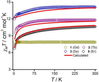
The temperature dependence of the molar magnetic susceptibility (χM) for 1-3 and 5 are plotted in Figure 3 and the field dependence of the magnetization for these compounds can be found in Figure S9. The corresponding behavior for 4 is summarized in Figure S8. The values of the product of the molar magnetic susceptibility with temperature, χMT (in cm3 mol-1 K) found at 300 K are 7.85 (Gd), 11.79 (Tb), 14.14 and 14.10 (Dy), and 11.18 (Er), in good accordance with the expected values expected for the isolated ions (i.e. 7.88, 11.81, 14.17, and 11.48 cm3 mol-1 K, respectively). For the Tb, Dy, and Er derivatives the χMT slowly decreases as T is lowered in agreement with the anticipated crystal field effect. The absence of any contribution from intermolecular exchange interactions is confirmed by the perfect Curie behavior down to 2 K for Gd. For the Tb and Dy derivatives, the field-dependent
12
magnetization at 2-5 K show a fast rise at lower field regions and remain almost unchanged above 15 kOe (at 2 K) to reach 4.8 µB (2), 5.14 (3), and 5.09 (4) at high field (5 T). To probe the dynamics of the magnetization relaxation, AC susceptibility behavior was investigated without and with applied static fields. No out-of-phase component (χ″M) were found for the Tb and Er derivatives down to 2 K (see Figure S10 in ESI). However, the Dy complexes 3 and 4 exhibited a χ″M signal but no maximum was observed above 2 K (see Figure S10 in ESI).
Figure 3. Temperature dependence of the product of the molar magnetic susceptibility with temperature, χMT, under 1 kOe applied magnetic field for complexes 1-3 and 5. Solid red lines represent the ab initio calculated data. The computed data for 5 was reduced by 2% to meet the experimentally observed.
Such a behaviour was suggesting relaxation driven by QTM which was reduced by applying a static field. For 3, the optimum applied field was estimated at HDC = 1 kOe (See Figure S11). AC susceptibility study carried out in this field within the frequency domain 1–1500 Hz yielded well-defined maxima for χ″M between 4 and 18 K (Figure 4). Relaxation times (τ)
13
have been assessed by fitting χ″M = f(Frq) for different temperatures with an extended Debye model, best fit parameters are gathered in Table S5. The very small a parameter is indicative for a narrow distribution width for the relaxation time over the whole temperature domain, suggesting that mainly one relaxation process is operative. The temperature dependence of the relaxation time, plotted in log scale in Figure 4, develops to a more linear variation above 10 K, which is the behavior anticipated for a thermally activated process (Orbach). Deviation from linearity for lower T indicates that other processes also come into play. Analysis of the behavior over the whole T range was obtained by summing the contributions of the Orbach, Raman, and direct processes (τ = τ0 exp(Ueff/kBT) + 1/(CTn) + 1/(AT). The latter were required to reproduce the lower T behavior. Best fit gave a thermal energy barrier for magnetization reversal, Ueff/kB = 204 ± 3 K with τ0 = (6 ± 1)×10-9 s, C = 0.015 K-1s-1, n = 4.5, and A = -1.30 s-1. (a) (b) 0 0.5 1 1.5 2 2.5 5 10 15 20 25 1.0 Hz 3.0 Hz 10 Hz 20 Hz 30 Hz 50 Hz 70 Hz 100 Hz 160 Hz 250 Hz 400 Hz 642 Hz 997 Hz 1488 Hz χ M ( cm 3 m ol -1 ) T (K) χM' χM'' H AC = 3 Oe; HDC= 1 kOe 0 0.2 0.4 0.6 0.8 1 1.2 1.4 1 10 100 1000 χ M '' (c m 3 m ol -1 ) Frq (Hz) 4 K 18 K
14
(c) (d)
Figure 4. (a,b) AC susceptibility behaviors for 3; (c) temperature dependence of the relaxation times for 3, and (d) for diluted compound 3'; the solid red lines are the best fits (see text).
The dilution of 3 in a matrix of isomorphous Y complex, 3' hereafter, gave very similar results (see Figure S12 in ESI). For this sample however, a maximum was observed in the χ″M = f(T) behavior in zero field but a QTM contribution was discernible at low temperature. This was suppressed upon applying a small DC field of 750 Oe. The temperature dependence of the relation times between 2 and 17.5 K (Figure 4 and Table S6) parallels that obtained for the pure Dy derivative 3, and contributions of the Orbach, Raman, and direct processes had to be taken into account to reach a good modeling. Best fit to the experimental data for 3' yielded Ueff/kB = 208 ± 5 K, τ0 = (6 ± 2)×10-9 s, C = 4.3×10⁻³ K-1s-1, n = 5.0, and A = -0.30 s-1. The comparison of the behavior for 3 and its diluted form 3' shows that the observed behavior is clearly of molecular origin.
The replacement of OPCy3 for OPPh3 in the apical position of Dy appeared to have no significant incidence on the magnetic behaviours; the AC susceptibility features for 4 (Figure
10-5 10-4 10-3 10-2 10-1 100 101 2 4 6 8 10 12 14 16 18 3 best fit τ (s ) T (K) 10-5 10-4 10-3 10-2 10-1 100 101 2 4 6 8 10 12 14 16 18 3(Y) best fit τ (s ) T (K)
15
S13 and Table S7 in ESI) are very similar to that obtained for 3. To reproduce the temperature dependence of the relaxation time for 4 required to consider contributions from Orbach, Raman and direct processes, best fit to the experimental behavior gave Ueff/kB = 241 ± 7 K, τ0 = (2.3 ± 0.9)×10-10 s, C = (5.97 ± 0.03)×10-3 K-1 s-1, n = 5.1, and A = 0.2 ± 0.7 s-1.
It is satisfying to see that the energy barrier for magnetization reversal, Ueff/kB, for the Dy(III) complexes reported herein is significantly increased with respect to the homologue complex with Cl– ligands in the apical positions. This could be attributed to the stronger axial field due to the phosphine oxide ligand. A detailed comparison of the bond and magnetic properties of the present complexes with the reported pentagonal bipyramidal Dy(III) complexes having other equatorial ligands (including monodentate ones) and axial groups of various crystal field strengths are given in Table 1.
Table 1. Selected bond and magnetic parameters of PBP Dy(III) complexes
Sl. No. Complex Lax–Ln–L(°) ax Average axial Ln– O/Ln–X distance (Å) Average equatorial Ln– O/Ln–N/Ln–X distance (Å) Ueff (K) (Hdc ) τo (s) Ref. 1 [Dy(OtBu) 2(py)5][BPh4] 155.80 2.112 2.557 1815 (0 Oe) 1.17 × 10 –12 [16e] 2 [Dy(bbpen)Br] 155.79 2.163 2.586/2.851 1025 (0 Oe) 4.21 × 10 –12 [16c] 3 [Dy(tBuO)Cl(THF) 5] [BPh4]·2THF 178.28 2.043/2.662 2.410 950 (0 Oe) 3 × 10 −12 [26] 4 [Dy(bbpen)Cl] 154.24 2.166 2.584/2.682 708 (0 Oe) 9.46 × 10 –11 [16c] 5 [L1 2Dy(H2O)5][I]3·(L1)2· H2O 177.9 2.205 2.363 651 (0 Oe) 5.63 × 10⁻¹² [16b] 6 [Dy(L2) 2(H2O)5]2∙Br6∙ 2L2∙2H2O 177.82 2.221 2.354 556 (0 Oe) 9.33 × 10⁻¹² [27] 7 [Dy(Cy3PO)2(H2O)5]Br3·
2(Cy3PO)·2H2O·2EtOH
179.04 2.200 2.352 543
(0 Oe) 2.0 × 10
16 8 [Dy(CyPh2PO)2(H2O)5] Br3·2(CyPh2PO)·EtOH· 3H2O 174.2 2.217 2.364 508 (0 Oe) 8.6 × 10⁻¹² [29] 9 [Dy(Cy3PO)2(H2O)5]Cl3·
(Cy3PO)·H2O·EtOH
175.79 2.219 2.359 472 (0 Oe) 8.7 × 10 −12 [28] 10 [(NCN)DyCl2(THF)2] 176.50 2.596 2.448/2.668 2.393 (Dy–C) (0 Oe) 335 6 × 10 –10 [30] 11 [DyCl2(THF)5][BPh4] 179.68 2.577 2.390 80.6 (0 Oe) 4.1 × 10 –10 [31] 12 [Dy(Bpen)Cl(OPhCl2NO 2)2] 165.60 2.174 2.523/2.616 86 (1 kOe) 4.65 × 10 −7 [32] 13 [(H2L3)Y0.94Dy0.06Cl2] 166.32 5.643 2.264/2.444 70 (0.5 kOe) 1.9 × 10 –6 [17] 14 [(L4)Dy(Cy 3PO)Cl] 169.62 2.237/2.625 2.265/2.458 204 (1 kOe) 6.0 × 10⁻⁹ This work 15 [(L4)Dy(Ph 3PO)Cl] 174.07 2.276/2.623 2.283/2.460 241 (1.5 kOe) 2.3 × 10⁻¹⁰ This work L1 = (tBuPO(NHiPr)
2; L2: hexamethylphosphoric triamide; H2bbpen =
N,N′-bis(2-hydroxybenzyl)-N,N′-bis(2-methylpyridyl)ethylenediamine); NCN: [2,6-(2,6-C6H3R2N5CH)2C6H3]–; Bpen: N,N′‐bis(2‐
methylenepyridinyl)ethylenediamine; H4L3: 2,6-diacetylpyridine bis-salicylhydrazone; H2L4:
2,6-diacetylpyridine bis-benzoylhydrazone
Computational study
In order to rationalize the experimentally observed magnetization relaxation, we have performed ab initio CASSCF/RASSI+SO/SINGLE_ANISO calculations23 on the X-ray structures of complexes 2-5 and the previously reported complex 617 in which both the axial positions were occupied by the Cl– ligands. The X-ray analysis revealed that complexes 2 and 5 consist of two molecules in the asymmetric unit and we have performed our calculations on the one molecule for each of the complexes.
17
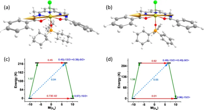
Figure 5. Orientation of the anisotropy axis of Dy(III) ion in complex 3 (a) and 4 (b). (c) and (d) represent the plausible relaxation mechanism for 3 and 4 respectively. The red arrows show the QTM and TA-QTM via ground and higher excited KD respectively. The sky dotted arrows show the Orbach process for the relaxation. The green arrows show the possible mechanism of magnetic relaxation. The blue characters imply the mJ composition of the KDs.
Table 2. The computed energy and the associated g-tensors of the low lying KDs generated from 6H15/2 state of complexes 3, 4 and 6.
Energy (K) gx gy gz The angle of gzz axis between
the ground and higher excited KDs (°) Complex 3 0.0 0.013 0.030 19.741 222.6 0.910 1.684 13.977 16.2 277.1 0.661 3.360 11.281 19.3 334.9 0.044 1.050 13.447 87.8 516.4 1.005 3.829 12.313 3.6 599.6 2.148 5.995 13.311 92.5 700.0 0.467 1.645 12.249 90.7 903.6 0.145 0.743 17.531 90.4 Complex 4 0.0 0.020 0.041 19.734 222.9 0.954 2.781 13.614 3.7 284.1 0.627 1.875 11.962 6.0
18 398.2 1.326 1.612 16.936 88.6 518.3 0.969 4.094 12.524 16.4 624.5 11.040 6.990 1.617 89.9 724.2 1.384 3.050 11.564 91.1 903.2 0.108 0.870 17.209 90.1 Complex 6 0.0 0.009 0.016 19.741 211.0 0.454 1.287 14.174 0.0 281.1 0.683 0.816 12.236 0.0 509.2 1.955 3.683 11.188 90.0 635.5 0.010 2.340 16.111 90.0 704.3 1.061 3.553 10.520 90.0 829.9 3.445 5.293 8.380 90.0 942.3 1.253 1.728 15.285 90.0
Figure 6. (a) The beta electron density of complex 3. (b) The beta electron density of complex 4. Colour code: Dy-yellow, Cl-green, P-violet, O-red, N-blue, C-grey, H-white.
The computed energy of the eight low lying KDs generated from 6H15/2 state spans from zero to 903 Kin complexes 3 and 4 and 942 K in complex 6 (Table 2). The g tensors; gx gy 0 and gz 20 for complexes 3, 4 and 6 demonstrate the Ising behavior of Dy(III) ion in these complexes. The gzz axis of ground KD is found to be lying on the equatorial plane due to the strong crystal field generated from the equatorial ligand (Figure 5 and S14). We have plotted the beta electron density of Dy in complexes 3, 4 and 6 using the procedure introduced by Ruiz and coworkers.33 Since the beta electron density is found to orient along the axial Cl– Dy–O bond (Cl–Dy–Cl bond in 6), the gzz anisotropy axis is oriented along the perpendicular direction of it to minimize the electrostatic repulsion (Figure 6). The large thermally assisted
19
quantum tunneling (TA-QTM) value of 0.41, 0.64 and 0.24 µB in the first excited KDs
suggest the relaxation will occur ideally through the first excited state KD (Figure 5 and S14). This suggests Ucal value of 222.6, 222.9 and 211.0 K for complexes 3, 4 and 6 respectively. These values are consistent with the estimate obtained from the experiments except for 6 where Ucal values are overestimated (for 3 208 K, 4 241 K and 6 70 K). We have also analyzed the composition of the computed wave function to get more insight about the extent of mixing of the mJ levels. The ground KD is found to be consist of mJ = ±15/2 ( 97%) with the negligible (≈ 0.02%) mixing with mJ = ±11/2 states. The strong mixing in the first excited KD leads to very large TA-QTM which forces the complex to relax via this state.
The ab initio computed energy barrier for the magnetization reversal does not affect significantly when one Cl– ion in the axial position (complex 6) is replaced by the neutral Cy3PO or Ph3PO ligand (complexes 3 and 4 respectively). The energy spectrum depends on the ligand field generated from the equatorial ligands which is found to be same in all the complexes. We have analyzed the LoProp charges to get more insight into the axiality of the three complexes. The LoProp charge of the axial oxygen atoms is found to be larger than the equatorial nitrogen and oxygen atoms in 3 and 4 (Table S9 and Figure S15). More importantly, the ratio of the av. axial/equatorial LoProp charges is found to be almost same in the complexes 3 and 4 which is consistent with similar Ucal values estimated in these complexes. The LoProp charge of the axial chlorine atoms and equatorial oxygen atoms is found to be very close in 6 which imply the less axial nature of this complex compared to 3 and 4. However the av. axial/equatorial LoProp charge ˃1.7 implies significant axiality in this complex.
The crystal field parameter of the three complexes has been estimated using the Steven formalism ( = ∑ ∑ , where and is computed crystal field parameter and
20
Stevens operator respectively) as implemented in SINGLE_ANISO suite to get more insight on the relaxation mechanism. The axial crystal field parameter is found to be larger in 4 compare to 3 implies the stronger axiality in 4 which agrees well with the Ueff value (Table S10). However the value in complex 6 is found to be similar implies lower axiality here compare to 3 and 4.
Furthermore, we have modeled the complex 3a, 4a and 6a (where the equatorial ligand has been removed) from complexes 3, 4 and 6 respectively to achieve the axial limit in each of these complexes. The anisotropy axis is found to be oriented along the Cl–Dy–O bond due to the absence of equatorial ligand field (Figure S16). The beta electron density is found to be oblate in nature and the gzz axis is oriented along the axial Cl–Dy–O bond (Cl–Dy–Cl bond in
6) to minimize the electrostatic repulsion (Figure 7). The ab initio calculations on these models reveal that the energy splitting of the eight KDs generated from 6H15/2 state raises to 2864, 2787 and 2616 K for 3a, 4a and 6a respectively (Table S11). Absence of equatorial ligand field leads to higher LoProp charge in the Dy center and metal coordinated oxygen atoms revealing the axial limit that can be achieved in these set of complexes (Table S12). It is also reflected in the computed crystal field parameter in which larger value is found compare to original complexes (Table S13). The magnetization relaxation is found to occur via fourth excited KD due to the strong transverse anisotropy which leads to Ucal value of 2540, 2471 and 2248 K in 3a, 4a and 6a respectively (Figure S17).
21
Figure 7. (a) The beta electron (spin-down) density of model 3a. (b) The beta electron density of model 4a. (c) The beta electron density of model 6a. Colour code: Dy-yellow, Cl-green, P-violet, O-red, N-blue, C-grey, H-white.
In the case of complex 2, a large tunnel splitting (∆tun) of 2.26 cm-1 is found between the two pseudo ground KDs which triggers the magnetization relaxation via the ground state (Table S14). The ground state anisotropy axis is found to be lying on the equatorial plane due to the strong equatorial ligand field (Figure S18). In case of complex 5 the computed large QTM value of 0.22 µB between the ground state KD also suggests the relaxation via ground state
which is supported by no out-of-phase signals in ac susceptibility (Figure S18 and Table S15).
Summary
Mononuclear 4f complexes (GdIII, DyIII, ErIII and TbIII) have been assembled using a multifunctional chelating ligand. The ligand enforces a pentagonal equatorial architecture around the metal ion. The two axial sites are taken up by a chloride and a phosphine oxide ligand. Thus, the overall coordination geometry around the lanthanide metal ion is distorted pentagonal bipyramidal. Magnteic studies on these complexes reveal that the DyIII analogues
22
show a slow relaxation of magnetization under the presence of small DC fields. A large tunneling gap between the ground as well as consequent excited non-Kramers doublets of TbIII and a very high quantum tunneling between the ground state Kramers doublets in the ErIII complex prevents these complexes from revealing a single-ion magnet behavior. The strategy adopted in the present instance of using a rigid ligand that enforces a pentagonal equatorial plane around the lanthanide ion is clearly effective in enabling the designed assembly of single-ion magnets. An improvement in this strategy would be to effect modifications in the ligand design to decrease the effective crystal field in the equatorial plane and increase it in the axial sites. We are currently examining these possibilities.
Acknowledgements
V. C. is thankful to the Department of Science and Technology, New Delhi, India, for a National J. C. Bose Fellowship. P. K. is thankful to National Institute of Science Education and Research, Bhubaneswar and Tata Institute of Fundamental Research, Hyderabad for doctoral and post-doctoral fellowships respectively. N.A is thankful to Tata Institute of Fundamental Research, Hyderabad for a post-doctoral fellowship. S. D. thanks UGC for SRF fellowship. GR would like to acknowledge SERB for funding (CRG/2018/000430).
Supporting Information
CCDC 1989266 (1), 1989267(2), 1989268(3), 1989270(4), 1989271(5) contain crystallographic supplementary data for this paper. These data can be obtained free of charge from The Cambridge Crystallographic Data Centre via www.ccdc.cam.ac.uk/data_request/cif.
X-ray crystallographic table of complexes 1–5; molecular structures of complexes 1, 2, 4 and 5: crystal packing diagrams of 1 and 4. table of bond distances and angles; table of continuous shape measures calculations; powder X-ray diffraction pattern of 3'. details of magnetic properties of 1−5; details of computaional calculations of complexes 1−5 (PDF).
23
References
1. (a) Robinson, J. R.; Fan, X.; Yadav, J.; Carroll, P. J.; Wooten, A. J.; Pericàs, M. A.; Schelter, E. J.; Walsh, P. J. Air- and Water-Tolerant Rare Earth Guanidinium BINOLate Complexes as Practical Precatalysts in Multifunctional Asymmetric Catalysis. J. Am. Chem. Soc. 2014, 136, 8034-8041; (b) Chen, S.; Yan, D.; Xue, M.; Hong, Y.; Yao, Y.; Shen, Q. Tris(cyclopentadienyl)lanthanide Complexes as Catalysts for Hydroboration Reaction toward Aldehydes and Ketones. Org. Lett. 2017, 19, 3382-3385; (c) Halter, D. P.; Palumbo, C. T.; Ziller, J. W.; Gembicky, M.; Rheingold, A. L.; Evans, W. J.; Meyer, K. Electrocatalytic H2O Reduction with f-Elements: Mechanistic Insight and Overpotential Tuning in a Series of Lanthanide Complexes. J. Am. Chem. Soc. 2018, 140, 2587-2594; (d) Nagae, H.; Aoki, R.; Akutagawa, S.-n.; Kleemann, J.; Tagawa, R.; Schindler, T.; Choi, G.; Spaniol, T. P.; Tsurugi, H.; Okuda, J.; Mashima, K. Lanthanide Complexes Supported by a Trizinc Crown Ether as Catalysts for Alternating Copolymerization of Epoxide and CO2: Telomerization Controlled by Carboxylate Anions. Angew. Chem. Int. Ed. 2018, 57, 2492-2496; (e) Schmidt, B. M.; Pindwal, A.; Venkatesh, A.; Ellern, A.; Rossini, A. J.; Sadow, A. D. Zwitterionic Trivalent (Alkyl)lanthanide Complexes in Ziegler-Type Butadiene Polymerization. ACS Catal. 2019, 9, 827-838.
2. (a) Bünzli, J.-C. G. On the design of highly luminescent lanthanide complexes. Coord. Chem. Rev. 2015, 293-294, 19-47; (b) Hirai, Y.; Nakanishi, T.; Kitagawa, Y.; Fushimi, K.; Seki, T.; Ito, H.; Hasegawa, Y. Luminescent Europium(III) Coordination Zippers Linked with Thiophene-Based Bridges. Angew. Chem. Int. Ed. 2016, 55, 12059-12062; (c) Wei, H.; Zhao, Z.; Wei, C.; Yu, G.; Liu, Z.; Zhang, B.; Bian, J.; Bian, Z.; Huang, C. Antiphotobleaching: A Type of Structurally Rigid Chromophore Ready for Constructing Highly Luminescent and Highly Photostable Europium Complexes. Adv. Funct. Mater. 2016,
24
26, 2085-2096; (d) Kovacs, D.; Lu, X.; Mészáros, L. S.; Ott, M.; Andres, J.; Borbas, K. E. Photophysics of Coumarin and Carbostyril-Sensitized Luminescent Lanthanide Complexes: Implications for Complex Design in Multiplex Detection. J. Am. Chem. Soc. 2017, 139, 5756-5767; (e) Martinić, I.; Eliseeva, S. V.; Nguyen, T. N.; Pecoraro, V. L.; Petoud, S. Near-Infrared Optical Imaging of Necrotic Cells by Photostable Lanthanide-Based Metallacrowns. J. Am. Chem. Soc. 2017, 139, 8388-8391; (f) Xiang, S.; Bao, D.-X.; Wang, J.; Li, Y.-C.; Zhao, X.-Q. Luminescent lanthanide coordination compounds with pyridine-2,6-dicarboxylic acid. J. Lumin. 2017, 186, 273-282; (g) Qiao, Y.; Schelter, E. J. Lanthanide Photocatalysis. Acc. Chem. Res. 2018, 51, 2926-2936; (h) Du, Y.; Jiang, Y.; Sun, T.; Zhao, J.; Huang, B.; Peng, D.; Wang, F. Mechanically Excited Multicolor Luminescence in Lanthanide Ions. Adv. Mater. 2019, 31, 1807062.
3. (a) Strnat, K.; Hoffer, G.; Olson, J.; Ostertag, W.; Becker, J. J. A Family of New Cobalt‐Base Permanent Magnet Materials. J. Appl. Phys. 1967, 38, 1001-1002; (b) Vincent, R.; Klyatskaya, S.; Ruben, M.; Wernsdorfer, W.; Balestro, F. Electronic read-out of a single nuclear spin using a molecular spin transistor. Nature 2012, 488, 357-360; (c) Woodruff, D. N.; Winpenny, R. E. P.; Layfield, R. A. Lanthanide Single-Molecule Magnets. Chem. Rev. 2013, 113, 5110-5148; (d) Kiefl, E.; Mannini, M.; Bernot, K.; Yi, X.; Amato, A.; Leviant, T.; Magnani, A.; Prokscha, T.; Suter, A.; Sessoli, R.; Salman, Z. Robust Magnetic Properties of a Sublimable Single-Molecule Magnet. ACS Nano 2016, 10, 5663-5669; (e) Zhu, Z.; Guo, M.; Li, X.-L.; Tang, J. Molecular magnetism of lanthanide: Advances and perspectives. Coord. Chem. Rev. 2019, 378, 350-364.
4. (a) Coutinho, J. T.; Monteiro, B.; Pereira, L. C. J. In Lanthanide-Based Multifunctional Materials; Martín-Ramos, P., Ramos Silva, M., Ed.; Elsevier: 2018; pp 195-231; (b) Dey, A.; Kalita, P.; Chandrasekhar, V. Lanthanide(III)-Based Single-Ion Magnets.
25
ACS Omega 2018, 3, 9462-9475; (c) Lu, J.; Guo, M.; Tang, J. Recent Developments in Lanthanide Single-Molecule Magnets. Chem. Asian J. 2017, 12, 2772-2779; (d) Kahn, O. Molecular Magnetism. Willey VCH: 1993.
5. (a) Meihaus, K. R.; Long, J. R. Actinide-based single-molecule magnets. Dalton Trans. 2015, 44, 2517-2528; (b) Future Directions for Transuranic Single Molecule Magnets. Inorganics 2018, 6, 26; (c) Coutinho, J. T.; Perfetti, M.; Baldovi, J. J.; Antunes, M. A.; Hallmen, P. P.; Bamberger, H.; Crassee, I.; Orlita, M.; Almeida, M.; van Slageren, J.; Pereira, L. C. J. Spectroscopic Determination of the Electronic Structure of a Uranium Single-Ion Magnet. Chem. Eur. J. 2019, 25, 1758-1766; (d) King, D. M.; Cleaves, P. A.; Wooles, A. J.; Gardner, B. M.; Chilton, N. F.; Tuna, F.; Lewis, W.; McInnes, E. J. L.; Liddle, S. T. Molecular and electronic structure of terminal and alkali metal-capped uranium(V) nitride complexes. Nat. Commun. 2016, 7, 13773; (e) Chatelain, L.; Tuna, F.; Pécaut, J.; Mazzanti, M. A zig-zag uranyl(v)–Mn(ii) single chain magnet with a high relaxation barrier. Chem. Commun. 2015, 51, 11309-11312.
6. (a) Layfield, R. A.; Murugesu, M. Lanthanides and Actinides in Molecular Magnetism. Wiley‐VCH Verlag GmbH & Co. KGaA 2015; (b) Gao, S. E. Molecular Nanomagnets and Related Phenomena. Springer-Verlag Berlin Heidelberg: 2015; (c) Gatteschi, D.; Sessoli, R.; Villain, J. Molecular Nanomagnets. 2006.
7. Benelli, C.; Gatteschi, D. Introduction to Molecular Magnetism: From Transition Metals to Lanthanides. Wiley-VCH.
8. Bartolomé, E.; Arauzo, A.; Luzón, J.; Bartolomé, J.; Bartolomé, F. In Handbook of Magnetic Materials; Brück, E., Ed.; Elsevier: 2017; Vol. 26, pp 1-289.
26
9. Ishikawa, N.; Sugita, M.; Ishikawa, T.; Koshihara, S.-y.; Kaizu, Y. Lanthanide Double-Decker Complexes Functioning as Magnets at the Single-Molecular Level. J. Am. Chem. Soc. 2003, 125, 8694-8695.
10. Cotton, S. A. Lanthanides and Actinides. Macmillan, London: 1991.
11. (a) Liddle, S. T.; van Slageren, J. Improving f-element single molecule magnets. Chem. Soc. Rev. 2015, 44, 6655-6669; (b) Chilton, N. F. Design Criteria for High-Temperature Single-Molecule Magnets. Inorg. Chem. 2015, 54, 2097-2099; (c) Ungur, L.; Chibotaru, L. F. Strategies toward High-Temperature Lanthanide-Based Single-Molecule Magnets. Inorg. Chem. 2016, 55, 10043-10056; (d) Liu, J.-L.; Chen, Y.-C.; Tong, M.-L. Symmetry strategies for high performance lanthanide-based single-molecule magnets. Chem. Soc. Rev. 2018, 47, 2431-2453.
12. (a) Day, B. M.; Guo, F.-S.; Layfield, R. A. Cyclopentadienyl Ligands in Lanthanide Single-Molecule Magnets: One Ring To Rule Them All? Acc. Chem. Res. 2018, 51, 1880-1889; (b) Harriman, K. L. M.; Murugesu, M. An Organolanthanide Building Block Approach to Single-Molecule Magnets. Acc. Chem. Res. 2016, 49, 1158-1167; (c) Bar, A. K.; Kalita, P.; Singh, M. K.; Rajaraman, G.; Chandrasekhar, V. Low-coordinate mononuclear lanthanide complexes as molecular nanomagnets. Coord. Chem. Rev. 2018, 367, 163-216.
13. (a) Guo, F.-S.; Day, B. M.; Chen, Y.-C.; Tong, M.-L.; Mansikkamäki, A.; Layfield, R. A. Magnetic hysteresis up to 80 kelvin in a dysprosium metallocene single-molecule magnet. Science 2018, 362, 1400-1403; (b) Goodwin, C. A. P.; Ortu, F.; Reta, D.; Chilton, N. F.; Mills, D. P. Molecular magnetic hysteresis at 60 kelvin in dysprosocenium. Nature 2017, 548, 439-442; (c) Guo, F.-S.; Day, B. M.; Chen, Y.-C.; Tong, M.-L.; Mansikkamäki, A.;
27
Layfield, R. A. A Dysprosium Metallocene Single-Molecule Magnet Functioning at the Axial Limit. Angew. Chem. Int. Ed. 2017, 56, 11445-11449.
14. Rinehart, J. D.; Long, J. R. Exploiting Single-Ion Anisotropy in the Design of f-Element Single-Molecule Magnets. Chem. Sci. 2011, 2, 2078-2085.
15. (a) Zhang, P.; Zhang, L.; Wang, C.; Xue, S.; Lin, S.-Y.; Tang, J. Equatorially Coordinated Lanthanide Single Ion Magnets. J. Am. Chem. Soc. 2014, 136, 4484-4487; (b) Sun, W.-B.; Yan, P.-F.; Jiang, S.-D.; Wang, B.-W.; Zhang, Y.-Q.; Li, H.-F.; Chen, P.; Wang, Z.-M.; Gao, S. High symmetry or low symmetry, that is the question – high performance Dy(iii) single-ion magnets by electrostatic potential design. Chem. Sci. 2016, 7, 684-691; (c) Chen, Y.-C.; Liu, J.-L.; Wernsdorfer, W.; Liu, D.; Chibotaru, L. F.; Chen, X.-M.; Tong, M.-L. Hyperfine-Interaction-Driven Suppression of Quantum Tunneling at Zero Field in a Holmium(III) Single-Ion Magnet. Angew. Chem. Int. Ed. 2017, 56, 4996-5000; (d) Gupta, S. K.; Murugavel, R. Enriching lanthanide single-ion magnetism through symmetry and axiality. Chem. Commun. 2018, 54, 3685-3696.
16. (a) Kalita, P.; Acharya, J.; Chandrasekhar, V. Mononuclear pentagonal bipyramidal Ln(III) complexes: Syntheses and magnetic properties. J. Magn. Magn. Mater. 2020, 498, 166098; (b) Gupta, S. K.; Rajeshkumar, T.; Rajaraman, G.; Murugavel, R. An air-stable Dy(iii) single-ion magnet with high anisotropy barrier and blocking temperature. Chem. Sci. 2016, 7, 5181-5191; (c) Liu, J.; Chen, Y.-C.; Liu, J.-L.; Vieru, V.; Ungur, L.; Jia, J.-H.; Chibotaru, L. F.; Lan, Y.; Wernsdorfer, W.; Gao, S.; Chen, X.-M.; Tong, M.-L. A Stable Pentagonal Bipyramidal Dy(III) Single-Ion Magnet with a Record Magnetization Reversal Barrier over 1000 K. J. Am. Chem. Soc. 2016, 138, 5441-5450; (d) Jiang, Z.; Sun, L.; Yang, Q.; Yin, B.; Ke, H.; Han, J.; Wei, Q.; Xie, G.; Chen, S. Excess axial electrostatic repulsion as
28
a criterion for pentagonal bipyramidal DyIII single-ion magnets with high Ueff and TB. J. Mater. Chem. C 2018, 6, 4273-4280; (e) Ding, Y.-S.; Chilton, N. F.; Winpenny, R. E. P.; Zheng, Y.-Z. On Approaching the Limit of Molecular Magnetic Anisotropy: A Near-Perfect Pentagonal Bipyramidal Dysprosium(III) Single-Molecule Magnet. Angew. Chem. Int. Ed. 2016, 55, 16071-16074.
17. Bar, A. K.; Kalita, P.; Sutter, J.-P.; Chandrasekhar, V. Pentagonal-Bipyramid Ln(III) Complexes Exhibiting Single-Ion-Magnet Behavior: A Rational Synthetic Approach for a Rigid Equatorial Plane. Inorg. Chem. 2018, 57, 2398-2401.
18. Pelizzi, C.; Pelizzi, G. Investigation into aroylhydrazones as chelating agents. Synthesis and structural characterization of a tin(IV) complex with 2,6-diacetylpyridine bis(salicyloylhydrazone). J. Chem. Soc., Dalton Trans. 1980, 1970-1973.
19. Sheldrick, G. M. SHELXT-Integrated Space-Group and Crystal-Structure Determination. Acta Crystallogr., Sect. A: Found. Adv. 2015, 71, 3-8.
20. Sheldrick, G. M. Crystal Structure Refinement with SHELXL. Acta Crystallogr., Sec. C: Struct. Chem. 2015, 71, 3-8.
21. Dolomanov, O. V.; Bourhis, L. J.; Gildea, R. J.; Howard, J. A. K.; Puschmann, H. OLEX2: a complete structure solution, refinement and analysis program. J. Appl. Crystallogr. 2009, 42, 339-341.
22. Brandenburg, K.; Putz, H. DIAMOND, version 3.2; Crystal Impact GbR: Bonn, Germany, 1997−2014.
29
23. Aquilante, F.; Autschbach, J.; Carlson, R. K.; Chibotaru, L. F.; Delcey, M. G.; De Vico, L.; Fdez. Galván, I.; Ferré, N.; Frutos, L. M.; Gagliardi, L.; Garavelli, M.; Giussani, A.; Hoyer, C. E.; Li Manni, G.; Lischka, H.; Ma, D.; Malmqvist, P. Å.; Müller, T.; Nenov, A.; Olivucci, M.; Pedersen, T. B.; Peng, D.; Plasser, F.; Pritchard, B.; Reiher, M.; Rivalta, I.; Schapiro, I.; Segarra-Martí, J.; Stenrup, M.; Truhlar, D. G.; Ungur, L.; Valentini, A.; Vancoillie, S.; Veryazov, V.; Vysotskiy, V. P.; Weingart, O.; Zapata, F.; Lindh, R. Molcas 8: New capabilities for multiconfigurational quantum chemical calculations across the periodic table. J. Comput. Chem. 2016, 37, 506-541.
24. Reiher, M. Relativistic Douglas–Kroll–Hess theory. Wiley Interdiscip. Rev. Comput. Mol. Sci. 2012, 2, 139-149.
25. (a) SHAPE: Continuous Shape Measures calculation, 2.1; Electronic Structure Group, Universitat de Barcelona, Spain, 2013; (b) Cirera, J.; Ruiz, E.; Alvarez, S. Continuous Shape Measures as a Stereochemical Tool in Organometallic Chemistry. Organometallics 2005, 24, 1556-1562.
26. Ding, Y.-S.; Yu, K.-X.; Reta, D.; Ortu, F.; Winpenny, R. E. P.; Zheng, Y.-Z.; Chilton, N. F. Field- and temperature-dependent quantum tunnelling of the magnetisation in a large barrier single-molecule magnet. Nat. Commun. 2018, 9, 3134.
27. Li, L.-L.; Su, H.-D.; Liu, S.; Xu, Y.-C.; Wang, W.-Z. A new air- and moisture-stable pentagonal-bipyramidal DyIII single-ion magnet based on the HMPA ligand. Dalton Trans. 2019, 48, 2213-2219.
30
28. Chen, Y.-C.; Liu, J.-L.; Ungur, L.; Liu, J.; Li, Q.-W.; Wang, L.-F.; Ni, Z.-P.; Chibotaru, L. F.; Chen, X.-M.; Tong, M.-L. Symmetry-Supported Magnetic Blocking at 20 K in Pentagonal Bipyramidal Dy(III) Single-Ion Magnets. J. Am. Chem. Soc. 2016, 138, 2829-2837.
29. Chen, Y.-C.; Liu, J.-L.; Lan, Y.; Zhong, Z.-Q.; Mansikkamäki, A.; Ungur, L.; Li, Q.-W.; Jia, J.-H.; Chibotaru, L. F.; Han, J.-B.; Wernsdorfer, Q.-W.; Chen, X.-M.; Tong, M.-L. Dynamic Magnetic and Optical Insight into a High Performance Pentagonal Bipyramidal DyIII Single-Ion Magnet. Chem. Eur. J. 2017, 23, 5708-5715.
30. Guo, Y.-N.; Ungur, L.; Granroth, G. E.; Powell, A. K.; Wu, C.; Nagler, S. E.; Tang, J.; Chibotaru, L. F.; Cui, D. An NCN-pincer ligand dysprosium single-ion magnet showing magnetic relaxation via the second excited state. Sci. Rep. 2014, 4, 5471.
31. Long, J.; Selikhov, A. N.; Mamontova, E.; Lyssenko, K. A.; Guari, Y.; Larionova, J.; Trifonov, A. A. Single-molecule magnet behaviour in a Dy(iii) pentagonal bipyramidal complex with a quasi-linear Cl–Dy–Cl sequence. Dalton Trans. 2019, 48, 35-39.
32. Li, M.; Wu, H.; Yang, Q.; Ke, H.; Yin, B.; Shi, Q.; Wang, W.; Wei, Q.; Xie, G.; Chen, S. Experimental and Theoretical Interpretation on the Magnetic Behavior in a Series of Pentagonal-Bipyramidal DyIII Single-Ion Magnets. Chem. Eur. J. 2017, 23, 17775-17787.
33. (a) Aravena, D.; Ruiz, E. Shedding Light on the Single-Molecule Magnet Behavior of Mononuclear DyIII Complexes. Inorg. Chem. 2013, 52, 13770-13778; (b) Norre, M. S.; Gao, C.; Dey, S.; Gupta, S. K.; Borah, A.; Murugavel, R.; Rajaraman, G.; Overgaard, J. High-Pressure Crystallographic and Magnetic Studies of Pseudo-D5h Symmetric Dy(III) and
31
Ho(III) Single-Molecule Magnets. Inorg. Chem. 2020, 59, 717-729; (c) Dey, S.; Rajaraman, G. In silico design of pseudo D5h actinide based molecular magnets: role of covalency in magnetic anisotropy. J. Chem. Sci. 2019, 131, 124.
32
Supporting Information
Table S1. Crystallographic data and refinement parameters of 1–5.
1 2 3 4 5 Empirical formula C41H52Cl1Gd1N5O3P 1 C84H116Cl2N12O8P2Tb 2 C90H124Cl2Dy2N10O8P 2 C41H34Cl1Dy1N5O3P 1 C86H116Cl2Er2N10O8P 2 Formula weight (gmol-1) 886.54 1872.56 1931.82 873.65 1885.24 Temperature (K) 120.00(10) 120.00(10) 120(2)K 120(2) 120.00(10) Crystal system Monoclinic Triclinic Monoclinic Triclinic Monoclinic
Space group P21/n P-1 P21/c P-1 P21/n
Unit cell lengths (Å) a = 20.1164(5) b = 9.6283(2) c = 22.3601(6) a = 10.0872(3) b = 18.3148(5) c = 23.4884(6) a = 18.6952(5) b = 9.9898(3) c = 24.5545(7) a = 8.8046(2) b = 11.5978(2) c = 18.9669(3) a = 23.5494(8) b = 10.0618(3) c = 36.0847(11) Unit cell angles (°)
β = 113.594(3) α = 84.704(2) β = 89.758(2) γ = 83.504(2) β = 91.430(2) α = 93.4930(10) β = 101.8670(10) γ = 103.418(2) β = 94.409(3) Volume (Å3) 3968.81(19) 4293.0(2) 4584.4(2) 1831.60(6) 8524.9(5) Z 4 2 2 2 8 Density (calculated) 1.484 1.449 1.399 mg/m3 1.584 1.469 Absorption coefficient 1.823 1.794 1.769 mm-1 2.203 2.116 F(000) 1812.0 1924.0 1988.0 874.0 3864.0 Crystal size (mm) 0.17 × 0.14 × 0.1 0.18 × 0.14 × 0.07 0.3 × 0.08 × 0.01 mm3 0.34 × 0.11 × 0.09 0.21 × 0.12 × 0.09 2θ range for data
collection (°) 5.5 to 58.818 5.244 to 49.998 5.412 to 57.664° 4.884 to 58.042 4.892 to 58.068 Reflections collected 62624 91078 53867 27626 96210 Index ranges -27 ≤ h ≤ 24, -11 ≤ k ≤ 12, -30 ≤ l ≤ 27 -11 ≤ h ≤ 11, -21 ≤ k ≤ 21, -27 ≤ l ≤ 27 -24 ≤ h ≤ 23, -13 ≤ k ≤ 8, -33 ≤ l ≤ 30 -11 ≤ h ≤ 9, -15 ≤ k ≤ 15, -24 ≤ l ≤ 24 -30 ≤ h ≤ 32, -13 ≤ k ≤ 13, -45 ≤ l ≤ 44 Independent reflections 9730 [Rint =
0.0382] 15077 [Rint = 0.0576] 10841 [Rint = 0.0587] 8459 [Rint = 0.0448] 19876 [Rint = 0.0840] Data/Restrain/Paramete r 9730/0/471 15077/0/999 10841/0/518 8459/0/471 19876/0/999 Goodness-of-fit on F2 1.031 1.159 1.040 1.055 1.026 Final R indices [I>2sigma(I)] R1 = 0.0228, wR2 = 0.0448 R1 = 0.0492, wR2 = 0.1197 R1 = 0.0346, wR2 = 0.0644 R1 = 0.0293, wR2 = 0.0648 R1 = 0.0470, wR2 = 0.0936
R indices (all data) R1 = 0.0289, wR2 = 0.0465 R1 = 0.0549, wR2 = 0.1223 R1 = 0.0571, wR2 = 0.0704 R1 = 0.0348, wR2 = 0.0668 R1 = 0.0898, wR2 = 0.1079 R1=∑|F − F |/ ∑ F ; wR2 = ∑[w(F − F )] /[w(F ) ]
33
Figure S1. Molecular structure of complex 1. (H-atoms are omitted for clarity)
Figure S2. Molecular structure of complex 2. (H-atoms are omitted for clarity)
34
Figure S4. Molecular structure of complex 5. (H-atoms are omitted for clarity)
Figure S5. Solid-state packing diagram of complex 1 viewed along the crystallographic c axis. (H-atoms are omitted for clarity)
Figure S6. Solid-state packing diagram of complex 4 viewed along the crystallographic c axis (H-atoms are omitted for clarity).
35
Table 2. Bond distance and bond angle parameters of complex 3.
Bond distances (Å) Bond angles (°)
Dy1−Cl1 2.625(8) Dy1−O1 2.272(2) Dy1−O2 2.259(2) Dy1−O3 2.237(2) Dy1−N3 2.462(2) Dy1-N2 2.464(2) Dy1−N4 2.448(2) O2−Dy1−Cl1 93.19(5) O2−Dy1−O1 100.23(7) O2−Dy1−N3 130.25(7) O2−Dy1−N2 165.17(7) O2−Dy1−N4 65.59(7) O3−Dy1−Cl1 169.62(5) O3−Dy1−O1 92.19(7) O3−Dy1−O2 90.35(7) O3−Dy1−N3 84.83(7) O3−Dy1−N2 91.41(7) O3−Dy1−N4 81.19(7) N3−Dy1−Cl1 85.44(6) N3−Dy1−N2 64.57(7) N2−Dy1−Cl1 87.67(6) O1−Dy1−Cl1 96.78(6) O1−Dy1−N3 129.37(7) O1−Dy1−N2 65.00(7) O1-Dy1-N4 164.10(7) N4−Dy1−Cl1 91.39(6) N4−Dy1−N3 64.74(7)
Table S3. Continuous shape analysis
Complex
Structure!
HP-7 HPY-7 PBPY-7 COC-7 CTPR-7 JPBPY-7 JETPY-7
1 31.572 19.350 2.233 7.665 6.207 6.298 18.718 2 32.740 20.143 1.732 7.872 6.409 5.652 20.810 3 32.944 22.380 1.446 8.229 6.678 5.549 21.175 4 32.742 21.540 1.505 7.661 6.338 5.725 21.267 5 32.781 20.809 1.353 7.774 6.403 5.232 21.450 !HP-7: Heptagon (D
7h); HPY-7: Hexagonal pyramid (C6v); PBPY-7: Pentagonal bipyramid (D5h); COC-7: Capped octahedron (C3v); CTPR-7: Capped trigonal prism (C2v); JPBPY-7: Johnson pentagonal bipyramid J13 (D5h); JETPY-7: Johnson elongated triangular pyramid J7 (C3v)
36
Table S4. Crystallographic details of 1, 2, 4 and 5.
Coordination geometry Bond lengths (Å) Bond angles (°)
Distorted PBP geometry of GdIII center in 1 Gd1–Cl1 2.645(5) Gd1–O1 2.330(12) Gd1–O2 2.313(12) Gd1–O3 2.271(13) Gd1–N3 2.510(15) Gd1–N2 2.522(15) Gd1–N4 2.480(15) O1–Gd1–Cl1 97.10(3) O1–Gd1–N3 126.15(4) O1–Gd1–N2 63.55(5) O1–Gd1–N4 167.95(5) O2–Gd1–Cl1 87.60(3) O2–Gd1–O1 105.63(4) O2–Gd1–N3 128.20(5) O2–Gd1–N2 167.59(5) O2–Gd1–N4 64.78(5) O3–Gd1–Cl1 174.67(3) O3–Gd1–O1 87.87(5) O3–Gd1–O2 89.22(5) O3–Gd1–N3 91.00(5) O3–Gd1–N2 96.12(5) O3–Gd1–N4 84.82(5) N3–Gd1–Cl1 87.61(3) N3–Gd1–N2 63.08(5) N2–Gd1–Cl1 87.84(4) N4–Gd1–Cl1 89.96(4) N4–Gd1–N3 63.68(5) Distorted PBP geometry of TbIII center in 2 Tb1–Cl1 2.622(12) Tb1–O1 2.291(3) Tb1–O2 2.306(3) Tb1–O3 2.228(4) Tb1–N4 2.472(4) Tb1–N3 2.500(4) Tb1–N2 2.495(4) O1–Tb1–Cl1 93.57(10) O1–Tb1–O2 102.50(13) O1–Tb1–N4 167.36(13) O1–Tb1–N3 128.57(13) O1–Tb1–N2 64.66(14) O2–Tb1–Cl1 90.53(10) O2–Tb1–N4 65.20(13) O2–Tb1–N3 128.85(13) O2–Tb1–N2 167.16(14) O3–Tb1–Cl1 175.22(9) O3–Tb1–O1 91.19(13) O3–Tb1–O2 87.95(13) O3–Tb1–N4 85.66(13) O3–Tb1–N3 88.25(13) O3–Tb1–N2 92.04(13) N4–Tb1–Cl1 89.59(10) N4–Tb1–N3 63.65(13) N4–Tb1–N2 127.62(14) N3–Tb1–Cl1 89.18(10) N2–Tb1–Cl1 90.44(10) Distorted PBP geometry of DyIII center in 4 Dy1–Cl1 2.622(7) Dy1–O2 2.282(2) Dy1–O3 2.276(2) Dy1–O1 2.285(2) Dy1–N4 2.456(2) Dy1–N2 2.457(2) Dy1–N3 2.467(2) O2–Dy1–Cl1 94.12(5) O2–Dy1–O1 100.00(6) O2–Dy1–N4 65.20(7) O2–Dy1–N2 164.01(7) O2–Dy1–N3 129.84(7) O3–Dy1–Cl1 174.07(5) O3–Dy1–O2 89.99(7) O3–Dy1–O1 88.02(6) O3–Dy1–N4 88.02(7) O3–Dy1–N2 83.31(7) O3–Dy1–N3 85.96(7) O1–Dy1–Cl1 95.46(5) O1–Dy1–N4 164.67(7) O1–Dy1–N2 65.38(7) O1–Dy1–N3 129.71(7) N4–Dy1–Cl1 89.83(5) N4–Dy1–N2 128.73(8) N4–Dy1–N3 64.71(8) N2–Dy1–Cl1 93.75(5) N2–Dy1–N3 64.32(7)
37 Distorted PBP geometry of ErIII center in 5 Er1–Cl1 2.591(11) Er1–O1 2.259(3) Er1–O3 2.195(3) Er1–O2 2.268(3) Er1–N3 2.444(4) Er1–N4 2.420(4) Er1–N2 2.432(4) O1–Er1–Cl1 94.97(9) O1–Er1–O2 97.78(11) O1–Er1–N3 130.69(11) O1–Er1–N4 63.78(11) O1–Er1–N2 65.83(12) O3–Er1–Cl1 174.16(8) O3–Er1–O1 90.83(11) O3–Er1–O2 89.86(11) O3–Er1–N3 86.83(12) O3–Er1–N4 85.42(12) O3–Er1–N2 91.81(12) O2–Er1–Cl1 89.99(8) O2–Er1–N3 131.42(11) O2–Er1–N4 66.48(11) O2–Er1–N2 163.55(12) N3–Er1–Cl1 88.89(9) N4–Er1–Cl1 89.16(9) N4–Er1–N3 64.95(12) N4–Er1–N2 129.97(12) N2–Er1–Cl1 89.99(9)
38
Figure S8. Magnetic behavior for 4. (a) Temperature dependence of
χ
MT and (b) field dependence of the magnetization (inset), solid lines represent the ab initio computed data using MOLCAS 8.2 program.Figure S9. Field dependence of the magnetization (A-C) for complexes 2-3 and 5 respectively, solid lines are the ab initio computed data using MOLCAS 8.2 program.
39
Figure S140. temperature dependence of the AC susceptibility in zero field and in an applied field of 1 kOe or 750 Oe for 2 (a), 3 (b), 3’ (c), 4 (c), and 5 (e).
(a) (b) (c) (d) (e) 0 0.5 1 1.5 2 2.5 3 3.5 0 5 10 15 20 Complex 2 (Tb) HDC = 0 H DC = 1 kOe χ M ( cm 3 m ol -1 ) T (K) χM' χM'' H AC = 3 Oe, Frq: 1 kHz 0 1 2 3 4 5 6 0 5 10 15 20 Complex 3 (Dy) χM' in zero field χM'' in zero field χM' with 1 kOe χM'' with 1 kOe χ M ( cm 3 m ol -1 ) Temperature (K) H AC = 3 Oe, Frq: 1 kHz 0 0.1 0.2 0.3 0.4 0.5 0.6 0.7 0.8 0 5 10 15 20 25 30 Complex 3' (Dy in Y) χM', H = 0 χM'', H = 0 χM', HDC = 750 Oe χM', HDC = 750 Oe χ M ( cm 3 m ol -1 ) T (K) H AC = 3 Oe, Frq: 1 kHz 0 1 2 3 4 5 0 5 10 15 20 Complex 4 (Dy) χM', H = 0 χM'', H = 0 χM', HDC = 1 kOe χM', HDC = 1 kOe χ M ( cm 3 m ol -1 ) T (K) HAC = 3 Oe, Frq: 1 kHz 0 0.5 1 1.5 2 2.5 3 0 5 10 15 20 Complex 5 (Er) no DC field H DC = 1 kOe χ M ( cm 3 m ol -1 ) T (K) HAC = 3 Oe, Frq: 1 kHz χM' χM''
40
Figure S10. Field dependence of the AC susceptibility (left), and of the relaxation time,
τ
(right), for 3 at 8 K. The largerτ
is reached for 1 kOe which was used as applied DC field in the AC studies for this complex.0 0.001 0.002 0.003 0.004 0.005 0.006 0 2000 4000 6000 8000 1 104 τ ( s) H (Oe)
41
Table S5. Best fit parameters of the Debye model to the
χ
″M = f(frq) behaviors for 3.χ
T stands for the isothermal susceptibility,χ
S for the adiabatic susceptibility,τ
for the relaxation time, and α accounts for the distribution width of the relaxation time.42 (a)
(b) (c)
Figure S12. Magnetic behavior for 3'. The composition in DyIII for the sample was estimated by adjusting the magnetization reached for 3' under 5 t at 2 K (blue plot in (a)) to that of 3 at same temperature (red plot).
0 1 2 3 4 5 6 0 10 20 30 40 50 2 K 3 K 5 K 3 at 2 K M ( µ B ) H (kOe) % Dy: 11.8 0 0.5 1 1.5 2 5 10 15 20 25 1.0 Hz 2.0 3.0 5 7 10 20 30 50 70 100 160 250 400 642 997 1488 χ M ( cm 3 m ol -1 ) T (K) χM' χM'' H AC = 3 Oe; HDC = 750 Oe 0 0.2 0.4 0.6 0.8 1 1.2 1.4 1.6 0.1 1 10 100 1000 χ M '' (c m 3 m ol -1 ) Frq (Hz) 3 K 19 K HAC = 3 Oe; HDC = 750 Oe
43
Table S4. Best fit parameters of the Debye model to the
χ
″M = f(frq) behaviors for 3'.χ
T stands for the isothermal susceptibility,χ
S for the adiabatic susceptibility,τ
for the relaxation time, and α accounts for the distribution width of the relaxation time.44
(a) (b)
(c) (d)
Figure S12. AC susceptibiliy behavior for 4.
0 0.5 1 1.5 2 2.5 5 10 15 20 25 1.0 Hz 3.0 10.0 20.0 30 50 70 100 160 250 400 642 997 1488 χ M ' ( cm 3 m ol -1 ) T (K) HAC = 3 Oe; HDC = 1.5 kOe 0 0.2 0.4 0.6 0.8 1 1.2 1.4 5 10 15 20 25 1.0 Hz 3.0 Hz 10 Hz 20 Hz 30 Hz 50 Hz 70 Hz 100 Hz 160 Hz 250 Hz 400 Hz 642 Hz 997 Hz 1488 Hz χ M '' (c m 3 m ol -1 ) T (K) HAC = 3 Oe; HDC = 1.5 kOe 0 0.2 0.4 0.6 0.8 1 1.2 1.4 1 10 100 1000 2.0 K 3 4 4.5 5 5.5 6 6.5 7 8 9 10 11 12 13 14 15 16 17 18 K χ M '' (c m 3 m ol -1 ) Frq (Hz) 100 101 102 103 104 105 5 10 15 1/ τ ( s -1 ) T (K)
best fit parameters:
∆eff/kB = 241 +/-7 K
τ0 = (2.3+/-0.9)x 10-10 s
C = (59.7+/-0.3 )10-4 s-1 n = 5.1
AHm = 0.2 +/- 0.7 s-1 relaxation processes considered: Orbach + Raman + direct
τ -1
45
Table S7. Best fit parameters of the Debye model to the
χ
″M = f(frq) behaviors for 4.χ
Tstands for the isothermal susceptibility,
χ
S for the adiabatic susceptibility,τ
for the relaxationtime, and α accounts for the distribution width of the relaxation time.
Table S8. Basis set used for all the elements in our calculation.
Elements Basis set
H H.ANO-RCC...2s. C C.ANO-RCC...3s2p. N N.ANO-RCC...4s3p2d1f. O O.ANO-RCC...4s3p2d1f. P P.ANO-RCC...4s3p. Cl Cl.ANO-RCC...5s4p2d1f. Tb Tb.ANO-RCC...8s7p5d3f2g1h. Dy Dy.ANO-RCC...7s6p4d2f1g.