Construction of Polynuclear Lanthanide (Ln = Dy III , Tb III , and Nd III ) Cage Complexes Using Pyridine − Pyrazole-Based Ligands: Versatile Molecular Topologies and SMM Behavior
Sukhen Bala,
†Mousumi Sen Bishwas,
‡Bhaskar Pramanik,
§Sumit Khanra,
§Katharina M. Fromm,
∥Pankaj Poddar,
‡and Raju Mondal*
,††
Department of Inorganic Chemistry, Indian Association for the Cultivation of Science, Kolkata 700032, India
‡
Physical & Materials Chemistry Division, CSIR-National Chemical Laboratory, Dr. Homi Bhabha Road, Pune 411008, India
§
Department of Chemical Sciences, Indian Institute of Science Education and Research, Kolkata, Mohanpur 741246, India
∥
Department of Chemistry, University of Fribourg, Chemin du Muse e 9, CH-1700 Fribourg, Switzerland ́
ABSTRACT: Employment of two di ff erent pyridyl − pyrazolyl-based ligands a ff orded three octanuclear lanthanide(III) (Ln = Dy, Tb) cage compounds and one hexanuclear neodymium(III) coordination cage, exhibiting versatile molecular architectures including a butter fl y core. Relatively less common semirigid pyridyl − pyrazolyl-based asymmetric ligand systems show an interesting trend of forming polynuclear lanthanide cage complexes with di ff erent coordination environments around the metal centers. It is noteworthy here that construction of lanthanide complex itself is a challenging task in a ligand system as soft N-donor rich as pyridyl − pyrazol. We report herein some lanthanide complexes using ligand containing only one or two O-donors compare to fi ve N-coordinating sites. The resultant multinuclear lanthanide complexes show interesting magnetic and spectroscopic features originating from di ff erent spatial arrangements of the metal ions. Alternating current (ac) susceptibility measurements of the two dysprosium complexes display
frequency- and temperature-dependent out-of-phase signals in zero and 0.5 T direct current fi eld, a typical characteristic feature of single-molecule magnet (SMM) behavior, indicating di ff erent energy reversal barriers due to di ff erent molecular topologies. Another aspect of this work is the occurrence of the not-so-common SMM behavior of the terbium complex, further con fi rmed by ac susceptibility measurement.
■ INTRODUCTION
Coordination cage compounds
1−5are of unabated interest in SMMs,
2,3magnetocaloric e ff ects,
4and in luminescence studies.
5Among other applications, SMMs are of considerable promise as molecular spintronics devices for high-density data storage.
6An escalating research interest has been developed to the design of lanthanide-based complexes, not only because of their intriguing structural architectures but also owing to their interesting magnetic
2−4,7−12and luminescence properties.
5,7,10Furthermore, highly luminescent complexes of Ln
IIIions employ multidentate organic ligands as chromophore, which act as antennae to sensitize the weakly luminescent metal centers.
5Hence, ligand design can play a crucial role not only to synthesize polynuclear cage complexes having versatile molecular topologies but also to signi fi cantly tune the magnetic and luminescent behaviors of the lanthanide complexes.
Because of their signi fi cant anisotropy arising from large unquenched orbital angular momentum, lanthanide ions have become attractive candidates for SMMs.
8Homometallic lanthanide compounds containing Dy
IIIhave received consid-
erable attention, and polynuclear complexes with variable nuclearity are known in literature.
3Dy compounds are known to exhibit a slow relaxation, among which the Dy
5square-based pyramid exhibits an energy barrier as high as 528 ± 11 K,
3aclearly indicating that lanthanide compounds are highly promising for the development of SMMs with high-energy reversal barrier.
The final structural outcome can be engineered at a molecular level through subtle modi fi cation in the ligand backbone as it is known that the ligand in fl uences the magnetic properties of paramagnetic complexes. There is a continuous need for the design of novel structures with versatile topologies that will improve the knowledge of structure − property relationships. In this regard, pyridyl − pyrazolyl-based frame- works have attracted signi fi cant attention in stabilizing polynuclear cage complexes,
1,13,14with an overwhelming majority of transition metal-based complexes. However, even
Published in ,QRUJDQLF&KHPLVWU\±which should be cited to refer to this work.
http://doc.rero.ch
a cursory inspection would reveal that corresponding lanthanide chemistry is unexplored to date, except for a very few random examples.
5i−mIt is well-known that lanthanide ions have high a ffi nity for hard donor atoms such as oxygen.
Needless to mention, construction of lanthanide complex itself is a challenging task in a ligand system as soft N-donor rich as pyridyl−pyrazol. Notwithstanding these difficulties, the pyr- idyl − pyrazole-based ligands have a natural tendency to form polynuclear cages with metal ions, which are particularly important for SMM materials. This is particularly true considering the fact that the magnetic relaxation rates, the essence of SMMs, are extremely sensitive to tiny distortions of the coordination geometry in lanthanide complexes.
11Our earlier report on pyridyl − pyrazolyl-based cages containing di ff erent coordination geometry of transition metals is highly relevant here.
14The obvious question then arose as to whether lanthanide cage complexes could be produced with similar pyridyl − pyrazolyl-based ligands. Although, one potential obstacle to this strategy would be the coordinating nature of the ligands themselves with only one or two hard O-donors compare to five sof t N-coordinating sites.
To study this question, we extend our work along these lines by employing two pyridyl−pyrazolyl-based ligands, namely, 3- (pyridin-2-yl)-N ′ -((pyridin-2-yl) methylene)-1H-pyrazole-5- carbohydrazide (H
2L
1) and N ′-(2-hydroxybenzylidene)-3-(pyr- idin-2-yl)-1H-pyrazole-5-carbohydrazide (H
3L
2). Both ligands have two interrelated characteristic features, a semirigid backbone and the possibility to ful fi ll variable coordination modes, so that, depending on the coordination requirements, the ligands adopt di ff erent conformations via bending, stretching, or twisting of the rings. We envisioned that such a unique blend of asymmetric nature, conformational freedom as well as coordination flexibility of the semirigid ligands may direct the synthesis of polynuclear lanthanide complexes.
Indeed, four new polynuclear lanthanide (III) (Ln = Dy
III, Tb
IIIand Nd
III) cage complexes of versatile molecular topologies were obtained using two pyridyl−pyrazolyl-based asymmetric, semirigid ligands. Their SMM nature and luminescence properties further validate the usefulness of this type of ligands for the study of lanthanide chemistry.
■ EXPERIMENTAL SECTION
All reagents used in the present work were obtained from commercial sources and were used without further puri
fication unless otherwise stated. Fourier transform IR spectra were obtained on a Nicolet MAGNA-IR 750 spectrometer with samples prepared as KBr pellets.
C, H, and N microanalyses were performed with a 2400 Series-II CHN Analyzer; Perkin−Elmer, USA, and UV−visible studies were performed in PerkinElmer Lambda 950 UV
−vis instrument. Magnetic data of polycrystalline samples were collected by MPMS (Evercool, 7 T) by Quantum Design, and
1H NMR was performed by Bruker Spectrometer operating at 400 MHz in deuterated dimethyl sulfoxide solvent. The photoluminescence measurements of solid samples were performed with the Horiba Jobin Yvon Fluoromax-3 spectrometer using steady-state 450 W Xe lamps as the excitation source, and excitation of the samples was done in UV region at 270 nm. Mass spectra were recorded on a Q-ToF Micro YA263 high resolution (Waters Corporation) mass spectrometer by positive ion mode electrospray ionization and the spectra were collected in methanol and water (1:1).
Synthesis of H2L1.
The ligand 3-(pyridin-2-yl)-N′-((pyridin-2- yl)methylene)-1H-pyrazole-5-carbohydrazide (H
2L
1) was synthesized by re
fluxing 3-(pyridin-2-yl)-1H-pyrazole-5-carbohydrazide (0.203 g, 1.0 mmol) and 2-pyridinecarboxaldehyde (0.107 g, 1.0 mmol) in ethanol at 80
°C for 6 h using acetic acid as catalyst. After the solutioncooled, a white precipitate formed, which was
filtered and dried.(Yield: 77% based on carbohydrazide). Anal. Calcd for C
15H
12N
6O (H
2L
1) (%): C, 61.64; H, 4.10; N, 28.76. Found: C, 61.67; H, 4.05; N, 28.67. IR (400−4000 cm
−1): 3436(b), 3299(s), 3074(s), 1674(s), 1533(s), 1226(s) .
1H NMR (500 MHz, [(CD
3)
2SO],
δ): 14.174 (s,1H, pyz−H), 12.096 (s, 1H, NH), 8.650 (s, 1H, imine-H), 7.385−
7.998 (8H, py-H). Electrospray ionization mass spectrometry (ESI- MS) m/z (M + H): 293.1258
Synthesis of H3L2.
The ligand N
′-(2-hydroxybenzylidene)-3- (pyridin-2-yl)-1H-pyrazole-5-carbohydrazide (H
3L
2) was synthesized by re
fluxing 3-(pyridin-2-yl)-1H-pyrazole-5-carbohydrazide(0.203 g, 1.0 mmol) and salicylaldehyde(0.122 g, 1.0 mmol) in ethanol at 80
°C for 6 h using acetic acid as catalyst. After the solution cooled, a light yellow precipitate formed, which was
filtered and dried in air. (Yield:82% based on carbohydrazide) Anal. Calcd for C
16H
13N
5O
2(H
3L
2) (%):C, 62.54; H, 4.24; N, 22.27. Found: C, 62.25; H, 4.32; N, 22.53.
IR (400−4000 cm
−1): 3429(b), 3275(s), 2995(s), 1654(s), 1614(s), 1541(s), 1467(s), 1400(s), 1274(s), 1205(s), 1163(s).
1H NMR (500 MHz, [(CD
3)
2SO],
δ) 14.169(s, 1Hpyz-H), 12.184(s, 1H, NH),11.427(s, 1H, phenolic-OH), 8.797(s, 1H, imine-H), 7.329−7.983 (4H, py-H), 6.895−7.312 (4H, Ar−H). ESI-MS m/z (M + H): 308.
Synthesis of Complex 1 ([DyIII8(μ3-OH)4(L1)4(DEA)4Cl4]).
H
2L
1(0.0292 g, 0.1 mmol), DyCl
3·6H2O (0.0377g, 0.1 mmol), and diethanolamine (DEA, 0.021 g, 0.2 mmol) were dissolved in a solvent mixture containing 3 mL of methanol and 3 mL of dimethylformamide (DMF). Trimethylamine (0.02 g, 0.2 mmol) was used as base, and the mixture was stirred for half an hour and then heated in an oil bath at 80
°C for 6 h. The yellow solution was
filtered after it cooled, and the solution was kept undisturbed for crystallization. After one week rod- shaped yellow crystals of complex 1 were obtained, which were suitable for X-ray data collection, washed with small amount of methanol, and dried. (Yield: 78%). Anal. Calcd for 1 (%): C, 29.63; H, 2.79; N, 12.45. Found: C, 29.32; H, 2.87; N, 12.14. IR (400−4000 cm
−1): 3394(b), 2933(mb), 2846(ms), 1656(s), 1596(s), 1512(s), 1467(s), 1434(s), 1386(s), 1309(s), 1251(s), 1157(s), 1049(s).
Synthesis of Complex 2 ([TbIII8(μ3-OH)4(L1)4(DEA)4Cl4]).
H
2L
1(0.0292 g, 0.1 mmol), TbCl
3·6H2O (0.0374g, 0.1 mmol), and DEA (0.021 g, 0.2 mmol) were dissolved in a solvent mixture containing 3 mL of methanol and 3 mL of DMF. Triethylamine (0.02 g, 0.2 mmol) was used as base, and the mixture was stirred for half an hour and then heated in an oil bath at 80
°C for 6 h. The yellow solution was
filtered after it cooled, and the solution was kept undisturbed for crystallization. Rod-shaped yellow crystals of complex 2 were obtained within 5 d that were suitable for X-ray data collection, washed with small amount of methanol, and dried. (Yield: 77%). Anal. Calcd for 2 (%): C, 29.85; H, 2.61; N, 12.83. Found: C, 30.17; H, 2.72; N, 12.47.
IR (400−4000 cm
−1): 3382(b), 3249(ms), 2844(s), 1656(s), 1596(s), 1566(s), 1506(s), 1465(s), 1433(s), 1386(s), 1348(s), 1309(s), 1249(s), 1157(s), 1047(s).
Synthesis of Complex 3 ([DyIII8(μ3-OH)4(L2)6(DMF)4(H2O)8]).
H
3L
2(0.0307 g, 0.1 mmol) and DyCl
3·6H2O (0.0377 g, 0.1 mmol) were dissolved in a solvent mixture containing 3 mL of methanol and 3 mL of DMF. Triethylamine (0.02 g, 0.2 mmol) was used as base, and the mixture was stirred for half an hour and then heated in an oil bath at 80
°C for 6 h. The light brown solution was filtered and keptundisturbed for crystallization. Needle-shaped brown crystals of complex 3 were obtained within two that were suitable for X-ray data collection, washed with small amount of methanol, and dried.
(Yield: 72%). Anal. Calcd for 3 (%): C, 35.64; H, 3.13; N, 13.09.
Found: C, 35.81; H, 3.16; N, 12.91. IR (400
−4000 cm
−1): 3377(vb), 2927(ms), 1658(s), 1602(s), 1442(s), 1371(s), 1261(s), 1201(s), 1153(s), 1103(s), 1043(s).
S y n t h e s i s o f C o m p l e x 4 ( [ N dI I I6(μ3- OH)2(L2)4(L2H)2(DMF)2(H2O)5]).
H
3L
2(0.0307 g, 0.1 mmol) and NdCl
3·6H
2O (0.025 g, 0.1 mmol) were dissolved in a solvent mixture containing 3 mL of methanol and 3 mL of DMF. Triethylamine (0.02 g, 0.2 mmol) was used as base, and the mixture was stirred for half an hour and then heated in an oil bath at 80
°C for 6 h. The light brownsolution was
filtered and kept undisturbed for crystallization. Needle-shaped brown crystals of complex 4 were obtained within two weeks
http://doc.rero.ch
that were suitable for X-ray data collection, washed with small amount of methanol, and dried. (Yield: 67%). Anal. Calcd for 4 (%): C, 41.78;
H, 3.65, 15.43. Found: C, 41.85; H, 3.81; N, 15.35. IR (400−4000 cm
−1): 3386(b), 2925(ms), 1654(s), 1604(s), 1544(s), 1471(s), 1438(s), 1386(s), 1296(s), 1249(s), 120(s)1, 1153(s), 1103(s), 1039(s).
X-ray Crystallography.
X-ray diffraction intensities for com- pounds 1−4 were collected at 120 K on Bruker APEX-2 CCD and diffractometer using Mo Kα radiation and processed using SAINT.
The structures were solved by direct methods in SHELXS and refined by full matrix least-squares on F
2in SHELXL.
15Crystallographic data are summarized in
Table S1, and additional crystallographicinformation is available in the
Supporting Information.■ RESULTS AND DISCUSSION
Synthesis. The ligands can be prepared in high yields by Schi ff base reactions of 3-(pyridin-2-yl)-1H-pyrazole-5-carbo- hydrazide with 2-pyridinecarboxaldehyde or salicylaldehyde in ethanol using acetic acid as catalyst. The lanthanide complexes were synthesized under similar conditions using LnCl
3· 6H
2O (Ln = Dy, Tb, Nd) and H
2L
1or H
3L
2at 80 °C. When DyCl
3· 6H
2O reacted with the ligand H
2L
1in the presence of DEA in a 1:1:2 molar ratio and with NEt
3as base, crystals of [Dy
III8(μ
3- OH)
4(L
1)
4(DEA)
4Cl
4] (1) were obtained. The homologue [Tb
III8(μ
3-OH)
4(L
1)
4(DEA)
4Cl
4]) (2) was obtained when TbCl
3· 6H
2O was used under similar reaction conditions. The reaction of DyCl
3·6H
2O with 1 equiv of ligand H
3L
2in DMF and NEt
3as base a ff orded the crystals of [Dy
III8( μ
3-OH)
4(L
2)
6- (DMF)
4(H
2O)
8] (3). Changing the lanthanide ion to NdCl
3· 6H
2O leads to [Nd
III6( μ
3-OH)
2(L
2)
4(L
2H)
2(DMF)
2(H
2O)
5] (4). All the complexes are characterized by elemental analysis, IR spectroscopy, and X-ray single-crystal di ff raction analyses.
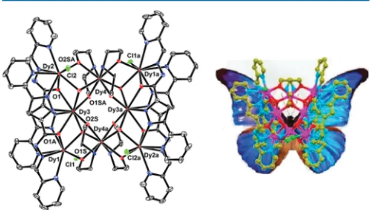
Description of Molecular Structures. Single-crystal X-ray analysis reveals that 1 crystallizes in the monoclinic C2/c space group and consists of a discrete neutral [Dy
III8( μ
3-OH)
4(L
1)
4- (DEA)
4Cl
4] entity and methanol molecules as cocrystallizing solvent. The molecular structure of 1 without the solvent molecules is depicted in Figure 1, and selected geometrical
parameters are provided in Table S2. Eight Dy
IIIions are held together by four μ
3-OH
−ligands (O1WS, O2WS, and symmetry equivalents) to form the [Dy
8(OH)
4] core as shown in Figure 2, which is composed of four triangular Dy
3units sharing vertices with Dy···Dy separations of 3.820 Å for Dy2 ··· Dy4, 3.764 Å for Dy3 ··· Dy4, and 3.848 Å for Dy1 ··· Dy4.
These μ
3-OH
−centered triangular units are similar to those of earlier reported Dy(III) compounds.
3In the {Dy
3( μ
3-OH)}
cores de fi ned by the Dy − O bonds, Dy − O − Dy angles, and Dy ··· Dy separations, the geometric parameters with Dy − O bonds varying from 2.352(6) to 2.435(6) Å, the Dy − O − Dy angles span from 101.2(2) ° to 110.8(2) ° , and Dy ··· Dy separations range from 3.7323(11) Å to 3.8792(11) Å. Each triangular unit is further connected by eight μ
2- η
2alkoxide oxygen atoms (O1S, O2S, and symmetry equivalents) from the four deprotonated diethanolamine coligands, where the Dy − O bond lengths are found to lie in the range from 2.253(6) to 2.368(6) Å and are in agreement with the literature.
3,9−12Four amide oxygen atoms (O1, O1A, and symmetry equivalents) of the dianionic ligand {L
1} are also bridging in a μ
2- η
2fashion, connecting the Dy
IIIcenters. Peripheral ligations are provided by the nitrogen atoms from the pyridine and pyrazole motifs.
Dy1, Dy2, and their symmetry equivalents are further coordinated by terminal Cl
−ions with Dy − Cl bond lengths of 2.674(2) and 2.703(2) Å.
The major impetus for this work was to validate the usefulness of the pyridyl − pyrazole-based ligands in designing polynuclear lanthanide complexes with di ff erent coordination environments. We are grati fi ed to note that our assumptions were correct. The dysprosium ions exhibit di ff erent coordina- tion environments; Dy1 and Dy2 are eight-coordinated with a DyO
3N
4Cl
1coordination motif, and the geometry around them can be best described as distorted square antiprism, whereas Dy3 and Dy4 are also eight-coordinated but in DyO
6N
2coordination motif with a bicapped trigonal prism geometry, as represented in Figure 3. As far as topology is concerned, the
Dy
8core closely resembles a butter fl y (Figure 1) wherein the orientation of ligands can be envisaged as wings and Dy4 coordinated to the DEA-part as body.
Complex 2 also crystallizes in the monoclinic C2/c space group, and the lattice consists of a discrete neutral [Tb
III8( μ
3- OH)
4(L
1)
4(DEA)
4Cl
4] molecule. The molecular structure of 2 is isostructural to that of 1 including the butter fl y core shown in Figure 1. Perspective view of 1 with thermal ellipsoids at the 50%
probability level (left) and butterfly topology (right). Hydrogen atoms are omitted for clarity.
Figure 2. Representation of the Dy
III8(left) and Tb
III8core (right) in 1 and 2, respectively.
Figure 3. Representations of bicapped trigonal prism and distorted square antiprismatic geometry around Dy in 1.
http://doc.rero.ch
Figure S4, and selected geometrical parameters are represented in Table S2. The {Tb
3( μ
3-OH)} units display Tb − O bonds in the range from 2.336(6) to 2.425(5) Å, the Tb−O−Tb angles vary from 101.6(2) ° to 110.1(2) ° , and the Tb ··· Tb separations range from 3.735 to 3.889 Å. The Tb−O bond distances in 2 are in the range of 2.251(5) − 2.501(5) Å, while Tb − N
pyand Tb−N
pzare in the range of 2.515(7)−2.777(7) Å and 2.411(7) − 2.465(7) Å, respectively. The bond lengths are comparable to those of previously reported Tb
IIIcomplex- es.
6,10aThe Tb − Cl bond lengths are 2.645(3) and 2.685(3) Å.
The different coordination environments around the Tb
IIIcenters are reminiscent of the Dy compound 1.
Both Tb2 and Tb4 are eight-coordinated with a TbO
3N
4Cl
1coordination motif, and the geometry around them can be best described as distorted square antiprismatic, whereas Tb1 and Tb3 are also eight-coordinated but with a TbO
6N
2coordina- tion motif with a bicapped trigonal prism geometry as shown in Figure S5. The {Tb
III8} core is presented in Figure 2.
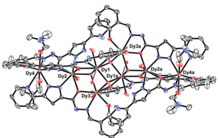
In contrast to 1 and 2, complex 3 crystallizes in the triclinic P1 ̅ space group and consists of a discrete [Dy
III8( μ
3- OH)
4(L
2)
6(DMF)
4(H
2O)
8] (3) molecule. Replacement of the terminal pyridyl group with a salicylic phenolic group in the ligand system did bring some drastic changes in the crystal structure of 3. The Dy
8core of compound 3 adopts a staircase- type arrangement. The molecular structure of 3 is presented in Figure 4, and selected geometrical parameters are given in
Table S2. Inspection of molecular structure indicates that six Dy
IIIions are held together by four μ
3-OH
−and six μ
2-OR- ligands {L
2}, where the [Dy
6(μ
3-OH)
4(μ
2-OR)
6]
8+core consists of two [Dy
3( μ
3-OH)( μ
2-OR)
2] triangular units in an
“edge-to-edge” arrangement linked by two μ
3-OH
−ions and two deprotonated alcohol oxygen atoms from the {L
2} ligands.
The dysprosium triangles Dy1Dy2Dy3 and symmetry equiv- alent are linked by μ
3-OH
−ions (O1S and counterpart) capping above and below the plane of the triangle, with Dy ··· Dy distances of 3.927, 3.9530, and 3.722 Å. Two sides of the triangles are further bridged by two deprotonated alkoxo oxygen atoms (O2 and symmetry equivalent), while the remaining side is coordinated by a μ
3-OH
−ion (O6S and counterpart). The equivalent oxygen atoms (O6S) together with two alkoxo oxygen atoms link the two dysprosium triangular units, which results in an edge-to-edge arrangement of the dysprosium triangles. Dy2 and symmetry equivalents are further bridged to Dy4 and symmetry equivalent, respectively,
via two pyrazolyl nitrogen atoms (N2, N3, and symmetry equivalent) in a bidentate fashion alongside an oxygen atom (O1A and counterpart) in μ
2-η
2leading to the Dy
8IIIcage complex; the core structure is presented in Figure 5. Note that the topology in 3 is quite different from the Dy
8cage in 1. The coordination is completed by oxygen atoms from the water and DMF solvent molecules.
Alternatively, the core structure of 3 can be described as four incomplete heterocubane entities “ Dy
3O
4” , which miss one Dy ion to complete the heterocubane entities and are fused to each other via Dy
2O
2-faces. The “ terminal ” Dy2 atom (and its symmetry equivalent) is further connected to Dy4 via two bridging N atoms of the pyrazolyl ligands and a bridging O atom.
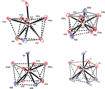
The values of the Dy − O and Dy − N bond lengths cover the ranges from 2.221(8)−2.491(6) and 2.386(8)−2.690(9) Å, respectively. The Dy − O − Dy angles are comprised between 99.9(2)° and 123.2(3)°. In an excellent agreement with our assumption, the dysprosium ions display again di ff erent coordination environments. Dy1 and Dy3 possess an eight- coordinated DyO
7N
1motif, and the geometry around them can be best described as bicapped trigonal prism, whereas Dy2 has a DyO
5N
3coordination motif displaying bicapped trigonal prism geometry. The geometry around Dy4 and its symmetry equivalent is di ff erent with a nine-coordinated DyO
4N
5, which can best be described as monocapped square antiprismatic. The coordination spheres of the dysprosium ions are illustrated in Figure 6.
Introduction of neodymium as lanthanide ion results in an interesting cage complex with a Nd
6core, instead of an octanuclear core as observed earlier for 1 − 3. It is noteworthy here that, for a given ligand, lanthanide complexes usually adopt a similar metal core. Complex 4 crystallizes in the hexagonal P3
22
1space group and consists of a discrete neutral [Nd
III6(μ
3- OH)
2(L
2)
4(L
2H)
2(DMF)
2(H
2O)
5] unit, as well as water and DMF molecules as solvents of crystallization. The molecular structure of the cage compound of 4 is presented in Figure 7, and metrical parameters are listed in Table S2. Single-crystal X- ray analysis of 4 reveals that four symmetry-independent Nd
IIIions are held together by two μ
3-OH
−ligands (O1W and its symmetry equivalent) and four μ
2-OR
−ligands {L
2}, where the [Nd
4( μ
3-OH)
2( μ
2-OR)
6]
4+core consists of two [Nd
3( μ
3- OH)( μ
2-OR)
3] triangular units sharing the edge between Nd1 and Nd2 ions leading to a Nd
6core. Nd1, Nd2, Nd3, and Nd3A linked by μ
3-OH
−ions atom are capped above and below the plane of the triangle, with Nd···Nd bond distances in the range of 3.740 − 4.173 Å and a body − body distance of 4.066 Å.
Nd3 and its symmetry equivalent are further bridged to Nd4 and its symmetry equivalent, respectively, via two pyrazolyl nitrogen atoms (N2, N3, and symmetry equivalents) in a bidentate fashion alongside with an oxygen atom (O1A and Figure 4. Perspective view of 3 with thermal ellipsoids at the 50%
probability level. Hydrogen atoms are omitted for clarity.
Figure 5. Representation of the Dy
8IIIcore in 3.
http://doc.rero.ch
counterpart) in μ
2- η
2fashion leading to the Nd
III6core as shown in Figure 8. The coordination is completed by oxygen
atoms from the water and DMF solvent molecules. The values of the Nd − O and Nd − N bond lengths cover the ranges of 2.297(9)−2.609(8) and 2.53(1)−2.276(1) Å, respectively. The Nd − O − Nd angles are in the range of 96.9(3) − 130.8(8) ° . Nd1 and Nd3 are nine-coordinated with an O
7N
2and an O
6N
3coordination motif, respectively, and the geometry can best be described as monocapped square antiprismatic.
In contrast, Nd2 and Nd4 are eight- and nine-coordinated with an O
6N
2and an O
5N
4coordination motif, respectively.
The coordination spheres of the neodymium ions are illustrated in Figure 9.
Magnetic Properties. Variable-temperature direct current (dc) magnetic susceptibility data for compounds 1 (Dy
8Cl
4), 2 (Tb
8Cl
4), 3 (Dy
8), and 4 (Nd
6) were collected on powdered microcrystalline samples over a temperature range from 5 to 300 K and under an applied fi eld of 0.5 T. The magnetic measurement on 1 reveals that the χ
MT value at 300 K is 105.95 cm
3K mol
−1, which is somewhat smaller than the theoretical value of 113.36 cm
3K mol
−1expected for eight noninteracting Dy
IIIions (S = 5/2, L = 5,
6H
15/2, g = 4/3: C = 14.17 cm
3K mol
−1).
8When cooled, the χ
MT product decreases gradually to reach 98.8 cm
3K mol
−1at 30 K and then decreases continuously to a value of 90.83 cm
3K mol
−1at 10 K. The χ
MT versus T plot is shown in Figure 10. This may indicate the
presence of weak antiferromagnetic interactions between the Dy
IIIions. The χ
MT value of 3 at 300 K is 103.13 cm
3K mol
−1, which is somewhat smaller than the theoretical value of 113.36 cm
3K mol
−1expected for eight noninteracting Dy
IIIions. When cooled, the χ
MT product decreases gradually to reach 95.0 cm
3K mol
−1at 30 K and then decreases continuously to a value of 86.75 cm
3K mol
−1at 10 K. (see Figure 11). The decrease in χ
MT upon lowering the temperature may conceivably be ascribed to a combination of the progressive depopulation of the excited Stark sublevels and the exchange interaction between the Dy
IIIions.
3,9−12To probe the magnetization dynamics of the two new Dy
IIIcompounds 1 and 3, the temperature (2−30 K) and frequency dependence (163 − 1176 Hz) of the ac magnetic susceptibilities were measured in the absence of an applied dc magnetic field.
Both compounds exhibit a frequency dependence in-phase ( χ′ ) Figure 6. Representations of bicapped trigonal prism and monocapped square antiprismatic geometry around Dy in 3.
Figure 7. Perspective view of 4 with thermal ellipsoids at the 50%
probability level. Hydrogen atoms are omitted for clarity.
Figure 8. Representation of the Nd
III6core in 4.
Figure 9. Representations of distorted square antiprismatic and monocapped square antiprismatic geometry around Nd in 4.
Figure 10.
χMT vs T plot for complex 1.
http://doc.rero.ch
and out-of-phase ( χ″ ) signal below ∼ 8 K as shown in Figure 12.
These data are indicative of the slow magnetization relaxation
of an SMM with a small energy barrier for magnetization reversal. However, no maxima in the out-phase ac susceptibility data have been observed. This behavior is presumably due to the existence of a fast quantum tunneling relaxation of the magnetization (QTM) promoted by intermolecular dipolar interactions. To partially or fully suppress the quantum tunneling process, ac susceptibility measurements were performed under a static dc field of 0.5 T, where the frequency dependence in-phase ( χ′ ) and out-of-phase ( χ″ ) signals below
∼6 K reveals an enhancement of the maxima as shown in Figures 13 and 14 clearly suggesting a slow relaxation of magnetization. Another relaxation process at ∼17 K was
observed for complex 3. Such behavior of the complexes 1 and 3 most likely arises from predominant single-ion effects of the individual Dy
IIIcenters in the octanuclear core exhibited by these molecules. Hence, both the octanuclear Dy
IIIcomplexes 1 and 3 can be considered as SMMs.
The spin disorder parameter ϕ = (Δ T
P/T
P)/Δ(log f) (f denotes the frequency used in ac measurement) for 1 and 3 are greater than 0.1 and belong to a normal value for a super paramagnet ( ϕ > 0.1), and hence 1 and 3 both can be considered as SMM, ruling out the possibility of a spin glass state ( ϕ ≈ 0.01).
16The relaxation time τ extracted from the maximum of χ″ at different frequencies (τ = 1/ω) follows the Arrhenius law ( τ = τ
0exp( Δ
eff/k
BT) where τ
0, Δ
eff, and k
Bare the pre-exponential factor, the relaxation energy barrier, and the Boltzmann constant, respectively. The least-squares fi tting of the experimental data as shown in Figures 13 and 14 affording an energy barrier Δ
eff= 49.3 K with a pre-exponential factor τ
0= 9.0 × 10
−10s for 1 and Δ
eff= 36.5 K, τ
0= 8.5 × 10
−8s for 3, consistent with the expected τ
0from 1 × 10
−5to 1 × 10
−11for SMMs.
9−12The Δ
effvalue obtained is higher than or comparable to that of other dysprosium complexes,
9,10,12smaller than that for some complexes reported in the literature,
3,11and listed in Table 1.
The dc magnetic data of Tb
IIIcomplex 2 reveals that the χ
MT value at 300 K is 88.15 cm
3K mol
−1, which is somewhat smaller than the theoretical value of 94.48 cm
3K mol
−1expected for eight noninteracting Tb
IIIions (
7F
6, S = 5/2, L = 3, J = 15/2, g
= 3/2: C = 11.81 cm
3K mol
−1).
8The χ
MT product remains practically similar upon further cooling, until it reaches a value Figure 11.
χMT vs T plot for complex 3.
Figure 12. Temperature dependence of the in-phase (
χ′) and out-of- phase (
χ″) ac susceptibility of 1 under zero dc
field.
Figure 13. Temperature dependence of the in-phase (χ′) and out-of- phase (χ″) ac susceptibility of 1 (Dy
8Cl
4) in the presence of an applied dc
field of 0.5 T. Plot of ln(τ) vs 1/T. Solid line representing thefittingof the Arrhenius law.
Figure 14. Temperature dependence of the in-phase (χ′) and out-of- phase (χ″) ac susceptibility of 3 (Dy
8) under zero dc
field and in thepresence of an applied dc
field of 0.5 T. Plot of ln(τ) vs 1/T. Solid linerepresenting the
fitting of the Arrhenius law.http://doc.rero.ch
of 88.1 cm
3K mol
−1at 160 K. The χ
MT value slowly decreases with decreasing temperature to reach a value 65.24 cm
3K mol
−1at 10 K as shown in Figure 15. The decrease in χ
MT upon lowering the temperature presumably can be ascribed to a combination of the progressive depopulation of the excited Stark sublevels and the exchange interaction between the Tb
IIIions, albeit weak.
The magnetization dynamics of the Tb
IIIcompound 2 was also studied in the temperature (2 − 300 K) and frequency dependence (163 − 1176 Hz) modes by measuring the ac magnetic susceptibilities in the absence of an applied dc magnetic fi eld. It exhibits very weak frequency dependence response for both in-phase ( χ′ ) and out-of-phase ( χ″ ) signals.
Such an e ff ect presumably can be ascribed due to fast quantum Table 1. Structural and Magnetic Features of Selected Dy and Tb Complexes
magnetic behavior of individual
lanthanides references lanthanide core and/or
topology Dy Tb
Dy8butterfly-shaped SMM SMM
Tb8butterfly-shaped Δeff= 49.3 K Δeff= 33.9 K Dy8core τ0= 9.0×10⁻¹⁰s τ0=
7.9×10⁻⁸s
this work Δeff= 36.5 K
τ0= 8.5×10⁻⁸s
Dy6core SMM non-SMM
Tb6core Δeff= 46.2 K 17a
τ0= 2.85×10⁻⁷s
Dy4seesaw topology SMM non-SMM
Tb4do no energy barrier
reported
17b
Dy6core SMM SMM
Tb6core Δeff= 3.8 K Δeff= 4.8K 17c
τ0= 7.89×10⁻⁶s τo=
1.43×10⁻⁶s Ln4square grid topology non-SMM non-SMM 17d
Dy4square grids SMM non-SMM
Tb4square grids Δeff= 91 K 17e
τ0= 4.5×10⁻⁷s
Dy2core SMM non-SMM
Tb2core Δeff= 53.7 K 17f
τ0= 1.3×10⁻⁹s
Dy4core SMM non-SMM
Tb4core Δeff= 28 K
τ0= 1.7×10⁻⁷s
17g Dy mononuclear
complex
SMM non-SMM
Δeff= 75 K 17h
Tb mononuclear τ0= 4.21×10⁻⁵s
Dy5core SMM Tb complex not
reported
trigonal bipyramidal Δeff= 1.91 K 17i
τ0= 1.01×10⁻⁶s
Dy4core SMM non-SMM
Tb4core Δeff= 62.6 K 17j
τ0= 8.7×10⁻⁷s Dy mononuclear chain
structure
SMM Tb complex not
reported
17k Δeff= 23.95 K,τ0
= 3.12×10⁻⁹s
mononuclear Dy SMM Tb complex not
reported
17l Δeff= 49.3 K
τ0= 4.8×10⁻⁶s
1D polymer Δeff= 48.86 K non SMM 17m
τ0= 6.88×10⁻⁷s
1D Dy polymeric chain SMM Tb complex absent
17n no energy barrier
reported
magnetic behavior of individual
lanthanides references lanthanide core and/or
topology Dy Tb
Dy6triangular prism SMM Tb complex not reported
Dy8tub conformation Δeff= 76 K 17o
τ0= 1.2×10⁻⁶s Dy 3D polymeric
network
SMM Tb complex not
reported
Δeff= 44.2 K 17p
τ0= 2.4×10⁻⁸s
Dy3linear cluster SMM non-SMM 17q
Tb3similar topology Δeff= 39.79 K τ0= 1.07×10⁻⁷s Dy4butterfly
arrangement
SMM Tb complex not
reported
17r Δeff= 6.25 K
τ0= 3.75×10⁻⁵s
Dy5butterfly-shaped SMM Tb complex not reported
17s Δeff= 197 K
τ0= 3.2×10⁻⁹s Dy10four fused Dy3
triangles
SMM Tb complex not
reported no energy barrier
reported
17t Dy5trigonal bipyramidal
cluster
SMM Tb complex not
reported
17u Δeff= 5k
τ0= 8.7×10⁻⁷s
dinuclear Dy complex SMM Tb complex not reported
Δeff= 56.6±0.9 K 17v
τ0= 1.0×10⁻⁷s
Dy7disclike structure SMM Tb complex not reported
Δeff= 140 K 17w
τ0= 7.2×10⁻⁹s Dy double-decker
complex
SMM SMM 17x
Δeff= (32±3) K Δeff= (383±37) K Tb similar topology τ0= (1.4±0.4)
×10−6s τ0= (1.3±1.3)
×10−8s Dy7metal-centered
trigonal prismatic topology
SMM non-SMM 17y
Δeff= 1.7 K τ0= 0.2×10⁻⁶s Tb7similar topology
Dy4core SMM non-SMM 12b
Tb4core no energy barrier reported