Neutralizing antibodies against IFN-b in multiple
sclerosis: antagonization of IFN-b mediated
suppression of MMPs
Francesca Gilli,
1Antonio Bertolotto,
1Arianna Sala,
1Francine Hoffmann,
2Marco Capobianco,
1Simona Malucchi,
1Tracy Glass,
2Ludwig Kappos,
3Raija L.P. Lindberg
2,*and David Leppert
2,3,*1Centro Riferimento Regionale Sclerosi Multipla (CReSM)
and Neurobiologia Clinica, Ospedale Universitario S. Luigi Gonzaga, Orbassano (Torino), Italy, and Departments of2Research and 3Neurology, University
Hospitals Basel, Basel, Switzerland
Correspondence to: PD Dr. David Leppert, Department of Neurology, University Hospitals, CH-4031 Basel,
Switzerland
E-mail: [email protected]
*These two authors contributed equally to this work
Summary
Neutralizing antibodies (NAb) against interferon-b (IFN-b) develop in about a third of treated multiple sclerosis patients and are believed to reduce therapeutic ef®cacy of IFN-b on clinical and MRI measures. The expression of the interferon acute-response protein, myxovirus resistance protein A (MxA) is a sensitive measure of the biological activity of therapeutically applied IFN-b and of its reduced bioavailability due to NAb. However, MxA may not be operative in the pathogenesis of multiple sclerosis or the therapeutic effect of IFN-b. Instead, matrix metalloproteinases (MMPs) are increased in brain tissue, CSF and blood circulation of multiple sclerosis patients and function as effector molecules in several steps of multiple sclerosis pathogenesis. One of the molecular mechanisms by which IFN-b exerts its bene®cial effect in multiple sclerosis is reduction of MMP-9 expression and increase of its endogenous tissue inhibitor, TIMP-1. Quantitative PCR measurements of MMP-2 and MMP-9, TIMP-1 and TIMP-2, and MxA were performed in peripheral
mononuclear cells from clinically stable multiple sclerosis patients with relapsing remitting disease course after short-term and long-term treatment with IFN-b. IFN-b therapy down-regulated the expression of MMP-9 and abolished that of MMP-2 in long-term, but not short-term treated multiple sclerosis, while levels of MxA were increased in both instances. The presence of NAb reversed these effects, i.e. led to reduced MxA and increased MMP-2/MMP-9 expression levels compared with NAb± patients. In contrast, expression of TIMPs in peripheral blood mononuclear cells remained unaffected by IFN-b therapy and the presence of NAb. While MxA is able to detect the biological action and reduced bio-availability of IFN-b on the basis of single injections, only MMP-9 shows quantitative correlation with the NAb titre. Together with evidence that an imbalance between MMP and TIMP expression is a crucial patho-genetic feature in multiple sclerosis, these ®ndings support the concept of a signi®cant role of NAb in reducing the therapeutic ef®cacy of IFN-b.
Keywords: multiple sclerosis; IFN-b; neutralizing antibodies; MMP; MxA
Abbreviations: CPE = cytopathic effect; CT= cycle threshold; GAPDH = glyceraldehyde phosphate dehydrogenase;
IFN-b = interferon-IFN-b; im = intramuscular; LU = laIFN-boratory units; MMP = matrix metalloproteinase; MxA = myxovirus resistance protein A; NAb = neutralizing antibodies; PBMC = peripheral blood mononuclear cells; RR = relapsing remitting; sc = subcutaneous; SP = secondary progressive; TIMP = tissue inhibitor of metalloproteinases.
Received June 29, 2003. Revised August 8, 2003. Accepted August 25, 2003. Advanced Access publication November 7, 2003
Introduction
Interferon-b (IFN-b) is the ®rst drug established as an effective treatment for patients with relapsing remitting (RR) and secondary progressive (SP) multiple sclerosis (IFNB Multiple Sclerosis Study Group and University of British
Columbia Multiple Sclerosis/MRI Analysis Group, 1995; Jacobs et al., 1996; European Study Group on Interferon b-1b in Secondary Progressive Multiple Sclerosis, 1998; PRISMS Study Group and University of British Columbia Multiple Brain Vol. 127 No. 2 ã Guarantors of Brain 2003; all rights reserved
Sclerosis/MRI Analysis Group, 2001). As IFN-b does not restore pre-existing structural damage and functional impair-ment, but rather acts by attenuating damaging effects of in¯ammatory processes, continuous administration is re-quired to maintain therapeutic effects. Such long-term therapy leads to the development of binding antibodies (BAb) and neutralizing antibodies (NAb) in up to 80% of patients (for a review, see Giovannoni et al., 2002). A number of reports have addressed the biological signi®cance of the latter on clinical and MRI measures; most of these suggest decreased treatment ef®cacy of IFN-b (IFNB Multiple Sclerosis Study Group and University of British Columbia Multiple Sclerosis/MRI Analysis Group, 1996; Rudick et al., 1998; PRISMS Study Group and University of British Columbia Multiple Sclerosis/MRI Analysis Group, 2001; Polman et al., 2003). Interferon-induced proteins such as neopterin, b2-microglobulin, 2¢-5¢ oligoadenylate synthetase
and myxovirus resistance proteins (MxA and MxB) show an acute up-regulation after IFN-injection and can serve as a measure of its biological activity (Chiang et al., 1993; Witt et al., 1993; Williams et al., 1998). Persistent NAb were shown to attenuate the induction of MxA on the protein (Deisenhammer et al., 1999; Vallittu et al., 2002) and transcriptional level (Bertolotto et al., 2003) in peripheral blood mononuclear cells (PBMC) from multiple sclerosis patients.
Matrix metalloproteinases (MMPs) are a family of Zn2+
-dependent endoproteinases that act as effector molecules in several steps of multiple sclerosis pathogenesis (for a review, see Kieseier et al., 1999; Leppert et al., 2001). The subfamily of gelatinases (MMP-2 and MMP-9) mediates the opening of the blood±brain±barrier and the extravasation of immune cells into the brain parenchyma in animal and in vitro models (Gijbels et al., 1994; Leppert et al., 1996; StuÈve et al., 1996). Other features of MMPs related to multiple sclerosis pathogenesis are their ability to regulate the activity of several cytokines and their receptors, and of adhesion molecules (SchoÈnbeck et al., 1998; for a review, see Leppert et al., 2001) and to lyse myelin components thus contributing to epitope spreading through the release of immunogenic degradation products (Proost et al., 1993). In relapsing remitting (RR) and secondary progressive (SP) multiple sclerosis, levels of MMP-9 are permanently increased in brain tissue, CSF and serum (Leppert et al., 1998; Lee et al., 1999; Lindberg et al., 2001; Waubant et al., 1999, 2003). IFN-b decreases the transcriptional and protein expression of MMP-2 and MMP-9 in T-cells and monocytic cells, with the functional effect of reduced capacity to cross a model blood±brain±barrier (Leppert et al., 1996; StuÈve et al., 1996; Galboiz et al., 2001, 2002; Schmidt et al., 2001; Nelissen et al., 2002). In multiple sclerosis patients, IFN-b therapy down-regulates increased serum levels of MMP-9 and increases those of its endogenous tissue inhibitor TIMP-1 (Waubant et al., 1999, 2003), thus correcting excessive proteolytic activity as a molecular mechanism of its thera-peutic effect. In return, we hypothesized that reduced
bioavailability of IFN-b due to NAb may not only result in decreased MxA synthesis, but also in attenuation of IFN-b mediated suppression of MMP-2 and MMP-9.
The purpose of this study was to: (i) test for the effect of short-term (12 h after injection) and long-term (>3 months) treatment of IFN-b on the transcriptional regulation of MMP-2 and MMP-9, and their respective endogenous inhibitors TIMP-2 and TIMP-1; (ii) determine whether the occurrence of NAb affects expression levels of MMPs/TIMPs; and (iii) correlate the expression of MMPs/TIMPs with those of MxA and NAb titres.
Methods
Patients and control subjects
This study was approved by the Regional Ethical Committee of Piedmont. Informed consent was obtained from each patient before beginning the study.
One hundred and four clinically stable patients (70 females, 34 males) with de®nite RR multiple sclerosis according to the McDonald criteria (McDonald et al., 2001) were retrospectively included in this study. Thirty-nine patients had never been treated with IFN-b before entering this study (treatment-naive patients) and 65 patients (long-term treated patients) were on treatment with recombinant IFN-b [11 patients were treated with 30 mg intramuscular (im) IFN-b1a (Avonexâ) (Biogen; Cambridge, MA,
USA) once a week (IFN-b1aim); 47 were treated with IFN-b1a
(Rebifâ) (Serono, Geneva, Switzerland), either with 22 mg (n = 31) or 44 mg (n = 16) subcutaneous (sc) three times a week (IFN-b1asc);
and seven had been treated with IFN-b1b (Betaferonâ) (Schering; Berlin, Germany) for at least 3 months (mean 6 SD = 24 6 20 months, range 3±84)]. Of the treatment-naive patients, 16 received IFN-b1aim, 10 IFN-b1asc22mg, eight
IFN-b1asc44mgand ®ve IFN-b1b.
The expanded disability status scale (EDSS) score ranged from 0 to 6 points (mean 6 SD = 2 6 1.4). Patients had: (i) no immunosuppressive drug therapy; (ii) no switch of IFN-b type; and (iii) no glucocorticosteroid therapy <30 days prior to study. The control group included 23 healthy volunteers (13 females, 10 males) who did not display symptoms of viral infection at least 2 weeks before and after blood donation.
NAb evaluation
Both long-term treated and treatment-naive patients were evaluated for the presence of NAb at study entry (baseline). In long-term treated patients, longitudinal measurements of NAb were performed in serum samples collected at least 36 h after the last IFN-b injection every 3 months until gene expression analysis. However, the last NAb measurement and gene expression analysis were made from the same blood sample, i.e. 12 h after the last injection. NAb were measured with the cytopathic effect (CPE) assay as previously described (Bertolotto et al., 2003); to insure maximal ligation with presumed NAb, measurements were performed with the same type of IFN-b as used in individual patients for therapy (Antonelli et al., 1998). Brie¯y, a mean concentration of 7 3 104cells in DMEM
medium 2% FCS (1 : 2.5 up to 1 : 5120) per 100 ml of the human lung carcinoma cell line A549 (ECACC, Salisbury, UK) were plated on 96-¯at well tissue culture plates and incubated overnight to form a con¯uent monolayer. Serum samples were diluted (1:2.5 up to
1:5120), mixed with one of the three IFN-b preparations to a ®nal concentration of 10 IU/ml, and then incubated for 1 h. Serum-IFN-b mixture (100 ml) was then incubated with A549 cells for 24 h. Cells were then infected with the cytopathic encephalomyocarditis murine virus at a concentration of 5±10 virus particles/cell. After 24 h incubation, viable cells were ®xed and stained with crystal violet in 20% ethanol. Excess dye was eluted with 33% acetic acid and absorbance was measured in a densitometer at 620 nm. Controls for viral activity, cellular viability and titration of the IFN-b preparation were run in parallel in each assay. According to World Health Organization recommendations, data from neutralization assays are reported as the reciprocal of the highest dilution of serum inducing 50% neutralization (i.e. neutralizing 10 U/ml of IFN activity to an apparent 1 U/ml of activity). The neutralization titre of a serum sample was calculated according to Kawade's formula (Kawade et al., 1986) and expressed in laboratory units (LUs). A level of >20LU is generally considered the threshold of positivity (Bertolotto et al., 2003). Three categories of patients were identi®ed based on the CPE assay: (i) patients in whom no NAb were detected during the whole period of follow-up (NAb±); (ii) patients tested positive for NAb in >2 consecutive samples were declared persistent NAb+ (pNAb+); and (iii) patients who had single positivity at the time point of gene expression analysis were declared isolated NAb+ (iNAb+).
Gene expression analysis
In treatment-naive patients, blood samples were taken prior to the ®rst and after the second (7 days + 12 h) IFN-b injection (mean6SD 12.1 6 1.2 h, range 10±13 h). For long-term treated patients, blood specimens were obtained 12.3 6 1.6 h (range 9±14 h) after the last IFN-b injection. PBMC were separated from edetic anticoagulated whole blood by Ficoll-Paque (Pharmacia; Uppsala, Sweden) centrifugation and subjected to RNA extraction using RNAwiz, following the manufacturer's instructions (Ambion; Austin, TX, USA). Total RNA (50 ng/ml ®nal concentration) was ®rst incubated with 0.5 mg of random hexamer at 70°C for 2 min and then reverse transcribed at 37°C for 1 h in reaction mixture containing a ®nal concentration of 500 mM of each deoxynucleotide triphosphate, 1 U/ml of Moloney murine leukaemia virus reverse transcriptase and 1 U/ml of RNAase inhibitor (RNAsin) (both from Promega Corporation; Madison, WI, USA). cDNA was used as a template for the real-time PCR analysis based on the 5¢-nuclease assay with the ABI PRISMâ 7000 Sequence Detection System (Applied
Biosystems, PE Europe B.V., Rotkreuz, Switzerland). Real-time PCR primers and probes were designed using primer-ExpressÔ software (Applied Biosystems). Expression of MMP-2, MMP-9, TIMP-1, TIMP-2 and MxA was analysed and transcriptional expression was normalized using the housekeeping gene glycer-aldehyde phosphate dehydrogenase (GAPDH) as reference in order to avoid differences due to possible RNA degradation/contamination or different reverse transcription ef®ciency. The relative expression levels of targets were calculated by the comparative cycle threshold (CT) method provided by Applied Biosystems. Targets were
considered detectable with CT values <40. Expression levels of
targets with CTvalues >35 were not described as relative expression,
due to the semi-quantitative character of CT values in this range.
Comparison of baseline expression levels of MxA in multiple sclerosis treatment-naive patients and controls was performed using competitive PCR results as previously described (Bertolotto et al., 2003).
Statistical analysis
Primary data are expressed as median (range) unless otherwise stated. Data were analysed using non-parametric statistical tests except for the linear regression analysis of the correlation of expression levels of MxA, MMP-9 and TIMP-1 as a function of treatment duration. Spearman rank correlation of expression levels between different genes, of intra-individual gene expression at different time points, and of MxA/MMP-9 expression levels with NAb titres were calculated. Expression levels of genes at different time points or between different treatment regimens, and according categorical differences of NAb status, were compared using the Mann±Whitney U test. The incidence of NAb development was compared between different treatment groups using the Fisher's exact test. P values <0.05 were considered signi®cant.
Results
NAb status and treatment schemes
At baseline, both treatment-naive and long-term treated patients scored negative for NAb (data not shown). In the course of long-term IFN-b treatment, NAb were detectable in 26% (17 out of 65) of patients: 22% (14 out of 65) had pNAb+ and 3% (5 out of 65) had iNAb+ (at the time point of blood sampling for gene expression analysis). Of the 17 long-term IFN-b treated patients who had developed NAb at the time point of gene expression analysis, four were treated with IFN-b1b and 13 were treated with IFN-b1asc (22 mg/injection: 7
out of 13; 44 mg/injection: 6 out of 13), whereas no NAb were detected in patients treated with IFN-b1aim. The duration of
treatment was signi®cantly longer with IFN-b1b [58 months (15±73 months)] compared with the various types and dosages of IFN-b1a [sc22: 20 months (3±84 months); sc44:
15 months (3±68 months): im: 18 months (5±60 months); all P < 0.016], whereas the duration of treatment between the three groups of patients who received IFN-b1a was not statistically different (all P > 0.36).
Short-term effects of IFN-
b
injection on MxA
and MMP/TIMP gene expression
Figure 1A shows that low amounts of MxA [relative expression compared with GAPDH: 0.08930 (0.02121± 2.29740)] are constitutively expressed in all multiple scler-osis patients; the expression levels are similar to those in 23 healthy control subjects as measured by competitive PCR (Bertolotto et al., 2003) (data not shown; P = 0.62). The injection of IFN-b in treatment-naive patients led to a >20-fold increase of median MxA expression [1.9185 (0.1756± 4.4840)] 12 h later (P < 0.0001). However, individual changes were highly variable, i.e. baseline MxA expression levels were not predictive for their degree of induction (P = 0.99, r < 0.001). Three patients had exceptionally high MxA levels at baseline who scored around the median value observed after IFN-b injection; of these, one patient showed no, and two others only a slight increase (1.4- and 1.8-fold, respectively) of MxA mRNA on IFN-b injection (Fig. 2). A
retrospective evaluation of case ®le histories of these patients did not reveal indications of intercurrent viral infections or other diseases, and the percentage of patients with such high expression of MxA was similar to that found earlier (Bertolotto et al., 2001, 2003). Transcripts for MMP-9 [0.0046 (0.0005±0.1756)], TIMP-1 [0.3220 (0.1713± 6.5889)] and TIMP-2 [0.0581 (0.0052±1.1728)] were present in all treatment-naive patients (Fig. 3A,C and D). In contrast, MMP-2 expression levels were not detectable in 18% (7 out of 39) of samples and too low in the remainder for relative expression to be quanti®ed. Therefore, MMP-2 expression was quanti®ed on the basis of CTvalues only (Fig. 3B). Other
than for MxA, the transcriptional expression of MMP-2 and MMP-9 and their respective inhibitors (TIMP-2 and TIMP-1) did not show signi®cant acute phase changes after IFN-b injection. The inducibility of MxA and MMP/TIMP expres-sion did not differ after treatment with either types or dosage of IFN-b (data not shown).
Long-term effects of IFN-
b
treatment on MxA
and MMP/TIMP gene expression
In long-term IFN-b treated, NAb± patients, the median MxA expression level was more than 8-fold higher than the baseline value [0.7341 LU (0.1373±4.9588LU), P < 0.0001] (Fig. 1A), but less than a third compared with that observed in treatment-naive patients after the second injection (P < 0.0001). The attenuation of MxA up-regulation observed here in the ®rst 3±6 months of chronic IFN-b treatment has been demonstrated by others, both on the transcriptional and the protein level (Vallittu et al., 2002; Gniadek et al., 2003). Conversely, in the later course of IFN-b treatment (months 3± 84), MxA levels remained stable, i.e. they showed no change as a function of treatment duration (Fig. 1B). In parallel, long-term IFN-b therapy led to a 5-fold decrease of MMP-9 transcripts [0.0008 (not detectable±0.0146)] compared with untreated patients (P < 0.0001) and was below detection limit in 14.5% (7 out of 48) of samples (Fig. 3A). However, no signi®cant correlation between MxA and MMP-9 expression levels on an individual basis could be established (data not shown).
The suppressive effect of IFN-b was even more pro-nounced for MMP-2, where all 48 samples from NAb± patients scored below detection threshold (P < 0.0001 for comparison with treatment-naive patients at baseline) (Fig. 3B). In contrast, transcriptional expression of both TIMPs remained unchanged under long-term IFN-b treatment (Fig. 3C and D).
Fig. 1 (A) Comparison of MxA mRNA levels in treatment-naive patients before ®rst and after second injection, and long-term IFN-b treated (®lled circles = pNAb+; open circles = iNAb+) multiple sclerosis patients. Horizontal bars indicate medians. (B) MxA RNA levels in NAb± (closed circles) and pNAb+ (open circles) patients as a function of IFN-b treatment time. Results of statistical evaluation of linear regression analysis for patients without (straight line) and with (dashed line) NAb were PNAb±= 0.30; r = 0.15 and PpNAb+= 0.47; r = 0.22.
Fig. 2 Change of MxA expression levels in individual treatment-naive patients as shown in Fig. 1A.
To exclude the possibility of delayed pharmacodynamic effects of IFN-b, expression levels of MMPs/TIMPs were analysed as a function of time. As for MxA, transcriptional expression of MMP-9 in NAb± patients was independent of treatment duration (Fig. 4A). Hence, for both genes the effect of long-term IFN-b therapy was established in the ®rst 3 months of treatment. Similarly, TIMP-1 (Fig. 4B) and TIMP-2 (data not shown) expression levels remained stable under long-term IFN-b therapy.
There was no difference between the various types and dosages of IFN-b in up-regulating MxA expression (data not shown). In contrast, suppression of MMP-9 expression was less pronounced in patients treated with IFN-b1aim [0.0037
(not detectable±0.0146)] compared with those on IFN-b1b [0.0004 (not detectable±0.0026)] (P = 0.049) or IFN-b1asc22
[0.0006 (not detectable±0.0047)] (P = 0.0012), while a tendency for such a difference was seen for IFN-b1asc44
[0.0016 (not detectable±0.0099)] (P = 0.09). On the other hand, differences in MMP-9 expression levels between the two dosages of IFN-b1asc and IFN-b1b were not signi®cant
(data not shown). Accordingly, expression levels of TIMP-1 and TIMP-2 were independent of types and dosages of IFN-b.
Effects of NAb on MxA, MMP/TIMP
expression
Analysis strati®ed by NAb status
In long-term treated patients with NAb+ (pNAb+ and iNAb+), median MxA expression levels were 3.13 [0.2384 (0.0177±2.8679)] lower than those of NAb± patients (P < 0.0001) (Fig. 1A). However, two pNAb+ patients showed MxA expression in the range of NAb± patients. Although median MxA expression in NAb+ patients was still
Fig. 3 Comparison of (A) MMP-9, (B) MMP-2, (C) TIMP-1 and (D) TIMP-2 mRNA levels in treatment-naive and long-term IFN-b treated (®lled circles = pNAb+; open circles = iNAb+) multiple sclerosis patients. Horizontal bars indicate medians of all patients per group. Respective targets were considered not being detectable with CTvalues >40. Note that levels of MMP-2 (B) are depicted as a CT
2.73 higher than in treatment-naive patients at baseline (T0),
the statistical comparison failed to show a signi®cant difference between the two groups (P = 0.15). Accordingly, the presence of NAb+ correlated with higher levels of MMP-9 [0.003MMP-9MMP-9 (not detectable±0.082MMP-9)] compared with NAb± samples (P = 0.0184) and reached similar values as untreated multiple sclerosis patients (P = 0.14) (Fig. 3A). Moreover, MMP-2 was detectable in 24% (4 out of 17) NAb+ patients compared with 0% in NAb± patients (P < 0.0001). However, this number was still lower in untreated patients [82% (32 out of 39)] (P = 0.0001) (Fig. 3B).
As for NAb± patients, pNAb+ did not change the transcriptional expression of MxA (Fig. 1B), MMP-9
(Fig. 4A), TIMP-1 (Fig. 4B) and TIMP-2 (data not shown) as a function of treatment duration or the types of IFN-b used (data not shown). Such analysis was not possible for MMP-2 due to the lack of quantitative data.
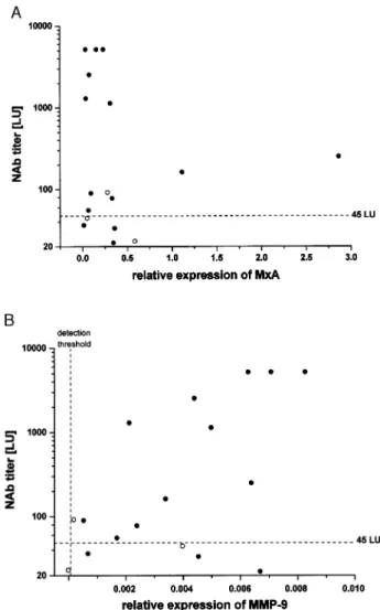
Correlation of NAb titres with MxA and MMP-9
expression levels
Earlier work has shown that NAb titres <45 LU have variable impact on the transcriptional expression of MxA, whereas titres >45 LU led to a complete suppression as measured by competitive PCR (Bertolotto et al., 2003). Accordingly, real-time PCR used here demonstrates that in presence of NAb (with the exception of two outliers), MxA levels ranged around the median value of treatment-naive patients at baseline and transcriptional induction by IFN-b was abro-gated. Fig. 5A demonstrates that this effect occurs independ-ently of the titre of NAb (be it above or below 45 LU). In contrast, there is a tendency for a correlation of MMP-9 expression levels and the whole range of NAb titres (P(n = 17) = 0.066; r = 0.46). Restricting the analysis to
titres >45LU this correlation becomes highly signi®cant (P(n = 12)= 0.011, r = 0.77) (Fig. 5B).
Discussion
There is ongoing controversy about the impact of NAb against IFN-b on its therapeutic effect in multiple sclerosis (Giovannoni et al., 2002; Polman et al., 2003). In large cohorts, up to a third of multiple sclerosis patients receiving IFN-b develop NAb, mostly after 6±18 months of treatment. Over 90% of those are reported to convert back to seronegativity after 8.5 years of treatment (Rice et al., 1999). However, the presence of NAb may be long lasting. In NAb+ patients observed for up to 67 months of IFN-b treatment, seroconversion is more likely to occur with NAb titres <200 LU, whereas titres above this threshold are associated with persistent seropositivity; in fact, 62% of NAb+ patients remained seropositive during this time period (Capobianco et al., 2003). In the PRISMS-4 trial (IFN-b1asc),
negative effects of NAb on clinical parameters were not obvious during the ®rst 2 years of treatment whereas, after 4 years, NAb+ patients had a 62% higher per year relapse rate and an almost 53 higher increase in T2 lesion load in MRI (PRISMS Study Group and University of British Columbia Multiple Sclerosis/MRI Analysis Group, 2001). Similar results were observed with IFN-b1b (Polman et al., 2003) and IFN-b1aim(Rudick et al., 1998) on these measures.
The current standard method to determine NAb in serum is indirect, i.e. via their in vitro capacity to antagonise the antiviral activity of IFN-b in a CPE assay. This test is technically dif®cult and there is no industrial standard with regard to the de®nition of seropositivity per se (detection of NAb in a single versus two consecutive samples) and the cut off level of titre considered biologically relevant (Giovannoni
Fig. 4 (A) MMP-9, (B) TIMP-1 RNA levels in NAb± (®lled circles) and NAb+ (open circles) patients in function of IFN-b treatment time. For calculation of regression analysis, values for MMP-9 (see Fig. 3A) below the detection threshold were not counted. Results of statistical evaluation of linear regression analysis for patients without (straight line) and with (dashed line) NAb were PNAb±= 0.91; r = 0.016 and PpNAb+= 0.62; r = 0.146
for MMP-9, and PNAb±= 0.055; r = 0.279 and PpNAb+= 0.79;
r = 0.077 for TIMP-1. Note different scale of y-axis for respective targets.
et al., 2002). The measurement of transcriptional or protein expression of IFN acute-response proteins such as neopterin, b-2-microglobulin and Mx proteins offers an alternative measure for IFN-b bioavailability which overcomes some of the disadvantages of the CPE assay (Vallittu et al., 2002; Bertolotto et al., 2003). However, there is no evidence for a functional role of these molecules in multiple sclerosis and data showing a direct correlation of acute-response protein induction by IFN-b with the long-term clinical course of multiple sclerosis patients with or without NAb has been published. Instead, MMPs are pre-eminent effector molecules in several crucial steps of multiple sclerosis pathogenesis (Kieseier et al., 1999; Leppert et al., 2001). A speci®c feature of multiple sclerosis, as opposed to other neuro-in¯ammatory disorders such as infectious meningitis (Leppert et al., 2001), is the induction of MMPs in the CNS and the peripheral blood
circulation without compensatory up-regulation of their endogenous tissue inhibitors, TIMPs, leading to a persistent imbalance towards excessive proteolytic activity (Lee et al., 1999; Waubant et al., 1999, 2003; Lindberg et al., 2001). Moreover, intercurrent increase of MMP-9, or of the MMP-9/ TIMP-1 ratio in serum of RR and SP multiple sclerosis predicts upcoming subclinical disease activity as detected by the development of new gadolinium-enhancing lesions in MRI (Waubant et al., 1999, 2003). There is accumulating evidence that a large part of the bene®cial effect of IFN-b in multiple sclerosis results from its capacity to suppress the expression of MMPs (for a review, see Yong et al., 2002) and, in parallel, to up-regulate TIMP-1 (Waubant et al., 1999, 2003). Interestingly, the pathophysiological link between MMPs and IFN-b in multiple sclerosis is not unidirectional. A recent study demonstrated that MMP-9 exerts proteolytic activity on IFN-b; thus the inherently increased concentra-tions of MMP-9 in serum, CSF and brain tissue of multiple sclerosis patients destroy the bioactivity of endogenous and therapeutically administrated IFN-b (Nelissen et al., 2003).
Here we show that IFN-b leads to an inverse transcriptional regulation of MxA and of MMP-2 and MMP-9 in RR multiple sclerosis patients. Conversely, the presence of NAb against IFN-b are associated with an attenuated induction of MxA and reduced suppression of MMPs. Based on these results, we propose increased MMP activity as one mechanism by which NAb abrogate the effect of IFN-b in multiple sclerosis. The response patterns of MxA and MMP-9 differ considerably; MxA is very sensitive to detect decreased bioavailability of IFN-b due to NAb on the basis of single measurements after a single injection, but lacks a dynamic response in function of NAb titres. This agrees with the results of other studies where no correlation of NAb titres with expression levels of MxA (Deisenhammer et al., 1999; Bertolotto et al., 2003) or with other IFN acute-response proteins (neopterin, b2
-micro-globulin) (Rudick et al., 1998) was demonstrated. On the other hand, the ®nding that NAb titres correlate positively with MMP-9 expression suggests that the quantitative extent of the immune response of multiple sclerosis patient against therapeutically applied IFN-b may be of biological relevance. With the caveat that treatment groups were not randomized and had different treatment durations, quantitative differences of MMP-9 suppression in NAb± patients suggest that IFN-b1ascand IFN-b1b exert a slightly higher biological activity
compared with IFN-b1aim. Although the kinetics of MMP-9
suppression by the various preparations of IFN-b are not known, it is conceivable that the higher total amount or the shorter dosage intervals of the former types of IFN-b may be responsible for this result. It is unclear, however, whether these differences are clinically relevant, vis-aÁ-vis the fact that the less suppressive activity of IFN-b1aim may be
counter-balanced by a far lower incidence of NAb induction observed in all therapeutic trials with the current formulation (for a review, see Giovannoni et al., 2002) and in longitudinal comparisons between the different types of IFN-b (Bertolotto et al., 2002), as well as in the present study cohort.
Fig. 5 Correlation of NAb titres with expression levels of MxA (A) and MMP-9 (B) in patients positive for NAb (®lled circles = pNAb+; open circles = iNAb+) at the time point of gene expression analysis. For MMP-9 and NAb titres, statistical evaluation of correlation resulted in P(n=17)= 0.066; r = 0.46 (all
patients) and P(n = 12)= 0.011; r = 0.77 (patients with titres
At ®rst glance, the present ®ndings seem to partly contradict earlier work where IFN-b was shown to increase serum protein levels of TIMP-1 after 4 months of treatment (Waubant et al., 2003). Moreover, high amounts of MMP-2 are constitutively expressed in CSF and serum, and are widely believed not to vary in the course of multiple sclerosis, speci®cally during IFN-b therapy (Trojano et al., 1999; Waubant et al., 2003). However, protein levels in serum are representative of contributions from various cellular com-partments, whereas mRNA measurements as used here allow the quanti®cation of gene usage in a speci®c cell type. Besides PBMC, endothelial cells and other cells of the vascular lining contribute to the production of MMPs and TIMPs (Bugno et al., 1999; Nelissen et al., 2002; Taraboletti et al., 2002). Quantitatively, leukocytes produce predomin-antly MMP-9, while the amounts of MMP-2 are relatively scarce as shown for transcripts (present results), as well as in the protein level in PBMC (Johnatty et al., 1997), and may be absent in resting T-cells (Leppert et al., 1996) and monocytes (Xie et al., 1994). In contrast, endothelial cells produce predominantly MMP-2, representing probably the predomin-ant source in serum, while they contribute only little MMP-9 (Nelissen et al., 2002). Thus, quantitatively subtle up-regulation of MMP-2 expression in the PBMC compartment of multiple sclerosis patients may easily escape detection in serum measurements. Conversely, IFN-b treatment ef®-ciently reduces MMP-2 transcription in PBMC of multiple sclerosis patients as observed here and by others (Galboiz et al., 2001); this is further corroborated by the changes on the protein level in experimental autoimmune encephalitis (Schmidt et al., 2001), and in vitro (Leppert et al., 1996).
Along with present results, others have found that mRNA levels of TIMP-1 and TIMP-2 from PBMC of IFN-b treated multiple sclerosis patients are unchanged or rather decreased, respectively (Lichtinghagen et al., 1999; Galboiz et al., 2001). We therefore conclude that the up-regulation of TIMP-1 protein in serum of IFN-b treated patients does not originate from PBMC, but derives most probably from endothelial cells. IFN-b does not modulate expression of MMP-2 and MMP-9 in endothelial cells (Nelissen et al., 2002) in vitro, but the effects on TIMP-1 regulation and the impact of NAb have not so far been investigated. However, there is indirect and partial evidence that IFN-b up-regulates TIMP-1 pro-duction in endothelial cells. In multiple sclerosis patients, the decreased production of the anti-in¯ammatory cytokine IL-10 by PBMC is corrected by IFN-b (Chabot et al., 2000; Ozenci et al., 2000), and this cytokine increases the production of TIMP-1 and decreases that of MMP-2 and MMP-9 in endothelial cells (Stearns et al., 1999).
Our study has some obvious limitations, as it was not designed to investigate whether NAb-induced changes of MxA and MMPs relate to decreased therapeutic effect of IFN-b and, hence, a more severe clinical course. Secondly, the interpretation of single measurements of these markers in individual patients may be of little value considering the wide range of expression levels and the overlap with NAb±
patients. A small study, which attempted to delineate the impact of NAb on the protein levels of MMP-9 (Trojano et al., 1999), showed that pNAb+ multiple sclerosis patients had higher MMP-9 serum levels during an observation time of 6± 18 months of IFN-b therapy and a greater increase of clinical disability as measured by Expanded Disability Status Score compared with NAb± patients. However, the NAb+ cohort consisted of only ®ve patients and MMP-9 measurements were performed by semi-quantitative zymography, which detects only selected species of MMP-9 (TIMP-1-free, pro-and active MMP-9). Moreover, increased MMP-9 expression in IFN-b treated multiple sclerosis patients is not speci®c for the presence of NAb, but has also been observed immediately before and during the occurrence of new gadolinium-enhancing lesions (Waubant et al., 2003). Such increased subclinical disease activity may explain why, in some patients, IFN-b treatment was not effective in lowering MMP-9 expression levels compared with controls, despite the absence of NAb. This could also be the case for MxA, as high expression levels due to intercurrent viral infection could interfere with the suppression by NAb activity. Hence, the simultaneous measurement of both targets in a longitudinal manner may be compulsory for a meaningful interpretation of NAb titres for clinical diagnostic purposes. Prospective studies are needed to determine whether measurements of MMPs, in combination with MxA and other IFN-response proteins, would provide a statistically robust array of markers to allow predicting the degree of clinical response to IFN-b on an individual basis, and to indicate decreased treatment effect due to NAb.
Acknowledgements
This work was supported by the Margarete and Walter Lichtenstein Foundation, the Swiss National Science Foundation (grants 3100±63666 and the NCCR on Neural Plasticity and Repair), the Swiss Multiple Sclerosis Society, Swiss Life Insurance, the Biomedical Research Foundation (FRB), the Cavalieri Ottolenghi Foundation and Biogen (Switzerland).
References
Antonelli G, Bagnato F, Pozzilli C, Simeoni E, Bastianelli S, Currenti M, et al. Development of neutralizing antibodies in patients with relapsing-remitting multiple sclerosis treated with IFN-b1a. J Interferon Cytokine Res 1998; 18: 345±50.
Bertolotto A, Gilli F, Sala A, Audano L, Castello A, Magliola U, et al. Evaluation of bioavailability of three types of IFNbeta in multiple sclerosis patients by a new quantitative-competitive-PCR method for MxA quanti®cation. J Immunol Methods 2001; 256: 141±52.
Bertolotto A, Malucchi S, Sala A, Ore®ce G, Carrieri PB, Capobianco M, et al. Differential effects of three interferon betas on neutralizing antibodies in patients with multiple sclerosis: a follow-up study in an independent laboratory. J Neurol Neurosurg Psychiatry 2002; 73: 148±53. Bertolotto A, Gilli F, Sala A, Capobianco M, Malucchi S, Milano E, et al. Persistent neutralizing antibodies abolish the interferon-b bioavailability in multiple sclerosis patients. Neurology 2003; 60: 634±9.
Reprogramming of TIMP-1 and TIMP-3 expression pro®les in brain microvascular endothelial cells and astrocytes in response to proin¯ammatory cytokines. FEBS Lett 1999; 448: 9±14.
Capobianco M, Sala A, Malucchi S, Di Sapio A, Gilli F, Bottero R, et al. Do neutralizing antibodies to interferon-beta disappear? [abstract]. J Neurol 2003; 250 Suppl 2: II/56.
Chabot S, Yong VW. Interferon beta-1b increases interleukin-10 in a model of T cell-microglia interaction: relevance to multiple sclerosis. Neurology 2000; 55: 1497±505.
Chiang J, Gloff CA, Yoshizawa CN, Williams GJ. Pharmacokinetics of recombinant human interferon-bserin healthy volunteers and its effect on
serum neopterin. Pharm Res 1993; 10: 567±72.
Deisenhammer F, Reindel M, Harvey J, Gasse T, Dilitz E, Berger T. Bioavailability of interferon b 1b in multiple sclerosis patients with and without neutralizing antibodies. Neurology 1999; 52: 1239±43. European Study Groupon Interferon b-1b in Secondary Progressive Multiple
Sclerosis. Placebo-controlled multicentre randomized trial of interferon b-1b in treatment of secondary progressive multiple sclerosis. Lancet 1998;352: 1491±97.
Galboiz Y, Shapiro S, Lahat N, Rawashdeh H, Miller A. Matrix metalloproteinases and their tissue inhibitors as markers of disease subtype and response to interferon-beta therapy in relapsing and secondary-progressive multiple sclerosis patients. Ann Neurol 2001; 50: 443±51.
Galboiz Y, Shapiro S, Lahat N, Miller A. Modulation of monocytes matrix metalloproteinase-2, MT1-MMP and TIMP-2 by interferon-gamma and -beta: implications to multiple sclerosis. J Neuroimmunol 2002; 131: 191± 200.
Gijbels K, Galardy RE, Steinman L. Reversal of experimental autoimmune encephalomyelitis with a hydroxamate inhibitor of matrix metallo-proteases. J Clin Invest 1994; 94: 2177±82.
Giovannoni G, Munschauer FE 3rd, Deisenhammer F. Neutralizing antibodies to interferon beta during the treatment of multiple sclerosis. J Neurol Neurosurg Psychiatry 2002; 73: 465±9.
Gniadek P, Aktas O, Wandinger KP, Bellmann-Strobl J, Wengert O, Weber A, et al. Systemic IFN-beta treatment induces apoptosis of peripheral immune cells in multiple sclerosis patients. J Neuroimmunol 2003; 137: 187±96.
IFNB Multiple Sclerosis Study Group and University of British Columbia Multiple Sclerosis/MRI Analysis Group. Interferon b-1b in the treatment of multiple sclerosis: ®nal outcome of the randomized controlled trial. Neurology 1995; 45: 1277±85.
IFNB Multiple Sclerosis Study Group and University of British Columbia Multiple Sclerosis/MRI Analysis Group. Neutralizing antibodies during treatment of multiple sclerosis with interferon b-1b: experience during the ®rst three years. Neurology 1996;47: 889±94.
Jacobs LD, Cookfair DL, Rudick RA, Herndon RM, Richert JR, Salazar AM, et al. Intramuscular interferon b-1a for disease progression in relapsing multiple sclerosis. Ann Neurol 1996; 39: 285±94.
Johnatty RN, Taub DD, Reeder SP, Turcovski-Corrales SM, Cottam DW, Stephenson TJ, et al. Cytokine and chemokine regulation of proMMP-9 and TIMP-1 production by human peripheral blood lymphocytes. J Immunol 1997; 158: 2327±33.
Kawade Y. Quantitation of neutralization of interferon by antibody. Methods Enzymol 1986; 119: 558±73.
Kieseier BC, Seifert T, Giovannoni G, Hartung HP. Matrix metalloproteinases in in¯ammatory demyelination: targets for treatment. Neurology 1999; 53: 20±5.
Lee MA, Palace J, Stabler G, Ford J, Gearing A, Miller K. Serum gelatinase B, TIMP-1 and TIMP-2 levels in multiple sclerosis. A longitudinal clinical and MRI study. Brain 1999; 122: 191±7.
Leppert D, Waubant E, Burk MR, Oksenberg JR, Hauser SL. Interferon b-1b inhibits gelatinase secretion and in vitro migration of human T cells: a possible mechanism for treatment ef®cacy in multiple sclerosis. Ann Neurol 1996; 40: 846±52.
Leppert D, Ford J, Stabler G, Grygar C, Lienert C, Huber S, et al. Matrix metalloproteinase-9 (gelatinase B) is selectively elevated in CSF during
relapses and stable phases of multiple sclerosis. Brain 1998; 121: 2327± 34.
Leppert D, Lindberg RL, Kappos L, Leib SL. Matrix metalloproteinases: multifunctional effectors of in¯ammation in multiple sclerosis and bacterial meningitis. Brain Res Brain Res Rev 2001; 36: 249±57. Lichtinghagen R, Seifert T, Kracke A, Marckmann S, Wurster U,
Heidenreich F. Expression of matrix metalloproteinase-9 and its inhibitors in mononuclear blood cells of patients with multiple sclerosis. J Neuroimmunol 1999; 99: 19±26.
Lindberg RL, De Groot CJ, Montagne L, Freitag P, van der Valk P, Kappos L, et al. The expression pro®le of matrix metalloproteinases (MMPs) and their inhibitors (TIMPs) in lesions and normal appearing white matter of multiple sclerosis. Brain 2001; 124: 1743±53.
McDonald WI, Compston A, Edan G, Goodkin D, Hartung HP, Lublin FD, et al. Recommended diagnostic criteria for multiple sclerosis: guidelines from the International Panel on the diagnosis of multiple sclerosis. Ann Neurol 2001; 50: 121±7.
Nelissen I, Ronsse I, Van Damme J, Opdenakker G. Regulation of gelatinase B in human monocytic and endothelial cells by PECAM-1 ligation and its modulation by interferon-beta. J Leukoc Biol 2002; 71: 89±98. Nelissen I, Martens E, Van den Steen PE, Proost P, Ronsse I, Opdenakker G.
Gelatinase B/matrix metalloproteinase-9 cleaves interferon-b and is a target for immunotherapy. Brain 2003; 126: 1371±81.
Ozenci V, Kouwenhoven M, Huang YM, KivisaÈkk P, Link H. Multiple sclerosis is associated with an imbalance between tumour necrosis factor-a (TNF-factor-a)- factor-and IL-10-secreting blood cells thfactor-at is corrected by interferon-b (IFN-b) treatment. Clin Exp Immunol 2000; 120: 147±53. Polman C, Kappos L, White R, Dahlke F, Beckmann K, Pozzilli C, et al.
Neutralizing antibodies during treatment of secondary progressive multiple sclerosis with interferon b-1b Neurology 2003; 60: 37±43. PRISMS Study Group and University of British Columbia Multiple
Sclerosis/MRI Analysis Group. PRISMS-4: long-term ef®cacy of interferon-b-1a in relapsing multiple sclerosis. Neurology 2001; 56: 1628±36.
Proost P, Van Damme J, Opdenakker G. Leukocyte gelatinase B cleavage releases encephalitogens from human myelin basic protein. Biochem Biophys Res Commun 1993; 192: 1175±81.
Rice GP, Paszner B, Oger J, Lesaux J, Paty D, Ebers G. The evolution of neutralizing antibodies in multiple sclerosis patients treated with interferon b-1b. Neurology 1999; 52: 1277±9.
Rudick RA, Simonian NA, Alam JA, Campion M, Scaramucci JO, Jones W, et al. Incidence and signi®cance of neutralizing antibodies to interferon b-1a in multiple sclerosis. Multiple Sclerosis Collaborative Research Group (MSCRG). Neurology 1998; 50: 1266±72.
Schmidt J, Sturzebecher S, Toyka KV, Gold R. Interferon-b treatment of experimental autoimmune encephalomyelitis leads to rapid nonapoptotic termination of T cell in®ltration. J Neurosci Res 2001; 65: 59±67. SchoÈnbeck U, Mach F, Libby P. Generation of biologically active IL-1 b by
matrix metalloproteinases: a novel caspase-1-independent pathway of IL-1 b processing. J Immunol IL-1998; IL-16IL-1: 3340±6.
Stearns ME, Rhim J, Wang M. Interleukin 10 (IL-10) inhibition of primary human prostate cell-induced angiogenesis: IL-10 stimulation of tissue inhibitor of metalloproteinase-1 and inhibition of matrix metallo-proteinase (MMP)-2/MMP-9 secretion. Clin Cancer Res 1999; 5: 189±96. StuÈve O, Dooley NP, Uhm JH, Antel JP, Francis GS, Williams G, et al. Interferon b-1b decreases the migration of T lymphocytes in vitro: effects on matrix mettalloproteinase-9. Ann Neurol 1996; 40: 853±63. Taraboletti G, D`Ascenzo S, Borsotti P, Giavazzi R, Pavan A, Dolo V.
Shedding of the matrix metalloproteinases MMP-2, MMP-9, and MT1-MMP as membrane vesicle-associated components by endothelial cells. Am J Pathol 2002; 160: 673±80.
Trojano M, Avolio C, Liuzzi GM, Ruggieri M, Defazio G, Liguori M, et al. Changes of serum sICAM-1 and MMP-9 induced by rIFN-b-1b treatment in relapsing-remitting multiple sclerosis. Neurology 1999; 53: 1402±8. Vallittu AM, Halminen M, Peltoniemi J, Ilonen J, Julkunen I, Salmi A, et al.
Neutralizing antibodies reduce MxA protein induction in interferon-b 1a-treated multiple sclerosis patients. Neurology 2002; 58: 1786±90.
Waubant E, Goodkin D, Gee L, Bacchetti P, Sloan R, Stewart T, et al. Serum MMP-9 and TIMP-1 levels are related to MRI activity in relapsing multiple sclerosis. Neurology 1999; 53: 1397±401.
Waubant E, Goodkin D, Bostrom A, Bacchetti P, Hietpas J, Lindberg R, et al. IFN-b lowers MMP-9/TIMP-1 ratio, which predicts new enhancing lesions in patients with secondary progressive multiple sclerosis. Neurology 2003; 60: 52±7.
Williams GJ, Witt PL. Comparative study of the pharmacodynamic and pharmacologic effects of Betaseron and AVONEX. J Interferon Cytokine Res 1998; 18: 967±75.
Witt PL, Storer BE, Bryan GT, Brown RR, Flashner M, Larocca AT, et al. Pharmacodynamics of biological response in vivo after single and multiple doses of interferon-b. J Immunother 1993; 13: 191±200. Xie B, Dong Z, Fidler IJ. Regulatory mechanisms for the expression of type
IV collagenases/gelatinases in murine macrophages. J Immunol 1994; 152: 3637±44.
Yong VW. Differential mechanisms of action of interferon-beta and glatiramer aetate in multiple sclerosis. Neurology 2002; 59: 802±8.