Publisher’s version / Version de l'éditeur:
Journal of Biomedical Optics, 15, 2, pp. 020501-1-020501-3, 2010-03-12
READ THESE TERMS AND CONDITIONS CAREFULLY BEFORE USING THIS WEBSITE. https://nrc-publications.canada.ca/eng/copyright
Vous avez des questions? Nous pouvons vous aider. Pour communiquer directement avec un auteur, consultez la
première page de la revue dans laquelle son article a été publié afin de trouver ses coordonnées. Si vous n’arrivez pas à les repérer, communiquez avec nous à PublicationsArchive-ArchivesPublications@nrc-cnrc.gc.ca.
Questions? Contact the NRC Publications Archive team at
PublicationsArchive-ArchivesPublications@nrc-cnrc.gc.ca. If you wish to email the authors directly, please see the first page of the publication for their contact information.
NRC Publications Archive
Archives des publications du CNRC
This publication could be one of several versions: author’s original, accepted manuscript or the publisher’s version. / La version de cette publication peut être l’une des suivantes : la version prépublication de l’auteur, la version acceptée du manuscrit ou la version de l’éditeur.
For the publisher’s version, please access the DOI link below./ Pour consulter la version de l’éditeur, utilisez le lien DOI ci-dessous.
https://doi.org/10.1117/1.3353960
Access and use of this website and the material on it are subject to the Terms and Conditions set forth at
Multimodal nonlinear optical imaging of atherosclerotic plaque
development in myocardial infarction prone rabbits
Ko, Alex C. T.; Ridsdale, Andrew; Smith, Michael S. D.; Mostaço-Guidolin,
Leila B.; Hewko, Mark D.; Pegoraro, Adrian F.; Kohlenberg, Elicia K.;
Schattka, Bernie; Shiomi, Masashi; Stolow, Albert; Sowa, Michael G.
https://publications-cnrc.canada.ca/fra/droits
L’accès à ce site Web et l’utilisation de son contenu sont assujettis aux conditions présentées dans le site LISEZ CES CONDITIONS ATTENTIVEMENT AVANT D’UTILISER CE SITE WEB.
NRC Publications Record / Notice d'Archives des publications de CNRC:
https://nrc-publications.canada.ca/eng/view/object/?id=73ee8f53-88f4-44ed-a5cc-35e09b6947ac https://publications-cnrc.canada.ca/fra/voir/objet/?id=73ee8f53-88f4-44ed-a5cc-35e09b6947acMultimodal nonlinear
optical imaging of
atherosclerotic plaque
development in myocardial
infarction-prone rabbits
Alex C. T. Ko,a,*Andrew Ridsdale,bMichael S. D. Smith,a Leila B. Mostaço-Guidolin,aMark D. Hewko,aAdrian F. Pegoraro,b Elicia K. Kohlenberg,a
Bernie Schattka,aMasashi Shiomi,cAlbert Stolow,band Michael G. Sowaa
a
National Research Council Canada, Institute for Biodiagnostics, 435 Ellice Avenue, Winnipeg, Manitoba, Canada R3B 1Y6
b
National Research Council Canada, Steacie Institute for Molecular Sciences, 100 Sussex Drive, Ottawa, Ontario, Canada K1A 0R6
c
Kobe University School of Medicine, Institute of Experimental Animals, 7-5-1, Kusunoki-cho, Chuo-ku, Kobe 650-0017, Japan
Abstract. Label-free imaging of bulk arterial tissue is
dem-onstrated using a multimodal nonlinear optical micro-scope based on a photonic crystal fiber and a single fem-tosecond oscillator operating at 800 nm. Colocalized imaging of extracellular elastin fibers, fibrillar collagen, and lipid-rich structures within aortic tissue obtained from atherosclerosis-prone myocardial infarction-prone Wa-tanabe heritable hyperlipidemic 共WHHLMI兲 rabbits is demonstrated through two-photon excited fluorescence, second harmonic generation, and coherent anti-Stokes Ra-man scattering, respectively. These images are shown to differentiate healthy arterial wall, early atherosclerotic le-sions, and advanced plaques. Clear pathological changes are observed in the extracellular matrix of the arterial wall and correlated with progression of atherosclerotic disease as represented by the age of the WHHLMI rabbits. © 2010
Society of Photo-Optical Instrumentation Engineers.
关DOI: 10.1117/1.3353960兴
Keywords: atherosclerosis; nonlinear optical microscopy; photonic crystal fiber; coherent anti-Stokes Raman scattering; two-photon ex-cited fluorescence; second harmonic generation.
Paper 09412LR received Sep. 18, 2009; revised manuscript received Jan. 27, 2010; accepted for publication Feb. 10, 2010; published on-line Mar. 12, 2010.
Atherosclerosis is the primary cause of heart disease, stroke, and lower limb amputation worldwide. It is a progres-sive disease characterized by chronic inflammation of injured intima and is associated with fatty plaque deposits in the arteries.1,2Early atherosclerosis cannot be reliably detected by current clinical methods, therefore the disease is often over-looked until at a more advanced stage. The development of new tools that provide greater sensitivity and specificity for early detection and differentiation of atherosclerotic plaques would help our understanding of early disease and help
establish preventative regimens that would slow disease pro-gression.
Recently, nonlinear optical 共NLO兲 microscopy has emerged as a powerful tool for tissue imaging. It is a label-free method with high sensitivity and specificity for major extracellular molecules. Its optical sectioning capability pre-sents a means of 3-D in vivo imaging that would be useful in the context of atherosclerosis diagnostics. Several studies have demonstrated imaging of arterial tissue using NLO microscopy,3–6 including studies imaging atherosclerotic le-sions using a multimodal coherent anti-Stokes Raman Scatter-ing 共CARS兲 microscope based on two tightly synchronized Ti:sapphire lasers and a swine animal model.6,7In our study, we demonstrate label-free visualization of the extracellular matrix of arterial lumen and atherosclerotic plaques using a photonic crystal fiber 共PCF兲-based multimodal NLO micro-scope employing only a single femtosecond oscillator. Two-photon excited autofluorescence 共TPEF兲 is able to specifically image extracellular elastin fibers, second harmonic generation 共SHG兲, type-1 collagen fibrils, and CARS lipid-rich structure or extracellular lipids droplets in unstained bulk intact tissue, indicating the methods are particularly suited to understand-ing the role and interplay between these key extracellular molecules involved in plaque development.
PCF-based CARS was recently reported as an alternative CARS imaging method in biology.8,9Because it only requires a single femtosecond laser, PCF-based CARS can be easily integrated into existing multiphoton microscopes with mini-mal reconfiguration, and provides CARS capability at a rela-tively low cost. Its portability and its future potential of being developed into an all-fiber, single-laser-based CARS imaging system are better than a conventional dual-color system. From a clinical research point of view, this CARS configuration is better adapted to being implemented in a surgical environment.
Colocalized elastin/type-1 collagen lipid imaging of bulk arterial tissues freshly harvested from WHHLMI rabbits is achieved at micron resolution using a home-built NLO micro-scope. The TPEF and SHG signals are obtained using the femtosecond pump pulses, with the CARS signal being gen-erated by spatially and temporally overlapping the pump pulse with the Stokes pulse produced in a PCF that is synchronously pumped by the same femtosecond laser. Our goals in this study are to investigate the potential of a PCF-based multimo-dal NLO imaging system for bulk tissue imaging and for ex-tracting biomorphological information related to athero-sclerotic lesion development in myocardial infarction-prone rabbits.
Figure1illustrates our PCF-based multimodal NLO imag-ing system used in this study. A green-pumped Ti:sapphire oscillator 共Tsunami, Spectra-Physics, Mountain View, Califor-nia兲 is the laser source, and the output wavelength is centered at800 nm with a pulse duration of 100 fs, an 80-MHz rep-etition rate, and average output power of1 W. The femtosec-ond pulses from the Ti:sapphire oscillator first pass through a Faraday isolator and then a pair of GTI chirp mirrors 共Lay-ertec GmbH, Germany兲. These mirrors are used to compen-sate for the large group velocity dispersion 共GVD兲 caused by the Faraday isolator. The femtosecond pulses are then split
1083-3668/2010/15共2兲/020501/3/$25.00 © 2010 SPIE *Address all correspondence to: Alex C.T. Ko. Tel: 4622; Fax:
204-984-5472; E-mail: alex.ko@nrc-cnrc.gc.ca
JBO Letters
Journal of Biomedical Optics 020501-1 March/April 2010 쎲 Vol. 15共2兲
into two beams at a beamsplitter. The reflected “pump” pulse is transmitted through a series of optical components outlined in Fig. 1, while the transmitted pulse is coupled into a PCF 共NL-1.4.775-945, NTK Photonics A/S, Denmark兲 through a 40⫻ objective lens to generate a supercontinuum 共SC兲. This SC emission is recollimated through a20⫻ objective lens and filtered through a near-infrared 共NIR兲 filter to select only the NIR portion 共950 nm⬍ ⬍ 1150 nm兲, which is then used as
the Stokes pulses for generating the CARS signal. The pump and Stokes pulses are combined at a beam combiner and sent collinearly into the microscope assembly. Epi-NLO signals are collected through an Olympus 20⫻, 0.75-NA objective lens 共UPlanSApo兲 and then detected using nondescanned PMT modules 共H-9656, Hamamatsu, Bridgewater, New Jer-sey兲. Spectral separation of TPEF, SHG, and CARS signals is achieved through an array of dichroic lenses and bandpass filters. A 40⫻, 0.8-NA, water immersion objective lens 共Olympus LUMPlanFl/lR兲 is used to collect tissue images of greater spatial and depth resolution in selected regions of in-terest. Typically, 25 mW of pump and 8 mW of Stokes are used at the sample for imaging. ScanImage software10共Cold Spring Harbor Laboratory, New York兲 is used for image ac-quisition and controlling translational stage movement during depth imaging. For images shown, total pixel dwell time is 21s from an average of four scans. ImageJ ver 1.42b 共Na-tional Institutes of Health, Bethesda, Maryland兲 is used for image viewing and post-processing.
Animal studies were approved by the local Animal Care Committee of the National Research Council of Canada. This study uses the myocardial infarction-prone Watanabe heritable hyperlipidemic 共WHHLMI兲 rabbit model11 which highly re-sembles human in lipoprotein metabolism compared to other models, and develops atherosclerotic plaques rapidly due to a hereditary defect in low-density lipoprotein processing. Seven WHHLMI rabbits were used for this study. Six rabbits were each sacrificed at 4, 10, 11, 16, 18, and22 months, whereas the seventh rabbit died naturally at the age of24 months. For each rabbit, fresh aorta was dissected from the ascending aorta to the external iliac artery, rinsed in heparinized saline,
Fig. 1 Schematic of the inhouse-built nonlinear optical laser-scanning
microscope with the Stokes pulses being generated in PCF, which is synchronously pumped by the same Ti:sapphire laser that provides the pump pulse. F-ISO: Faraday isolator; CM: chirp laser mirrors; BS: beamsplitter; NIR-F: near-IR filter; BC: beam combiner; GS: galvo scanner; DM: dichroic mirror; OBJ: objective lens; BF: bandpass filter; NDF: neutral density filter; and / 2: half waveplate.
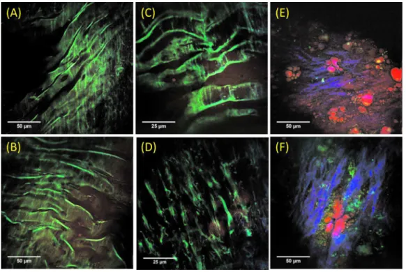
Fig. 2 Representative epi-NLO images collected from healthy arterial lumen on a 共a兲 4-month-old and 共b兲 10-month-old WHHLMI rabbit using
20⫻ air objective lens, and on a 4-month-old rabbit using 40⫻ WI lens at 共c兲 lumen surface and 共d兲 ⬃20m depth from surface. Representative epi-NLO images of arterial lumen surface obtained from 共e兲 an early atherosclerotic lesion and 共f兲 an advanced atherosclerotic lesion using 20⫻ air lens. Green:TPEF 共elastin or other fluorescent particles兲. Blue:SHG 共collagen兲. Red:CARS 共lipid-rich structure兲. 共Color online only.兲
JBO Letters
Journal of Biomedical Optics 020501-2 March/April 2010 쎲 Vol. 15共2兲
and then subdivided into ⬃20- to 30-mm sections. Each sec-tion was cut open longitudinally, exposing the luminal sur-face. The samples were placed in petri dishes with the luminal surface facing up. Phosphate buffered saline solution was ap-plied to the samples periodically to maintain hydration. Healthier arterial lumen from the4-month-old rabbit showed mostly smooth and flat surfaces with occasional raised le-sions. Plaque-covered luminal surfaces were observed in rab-bits aged 10 to 24 months with the burden increasing with age. This observation is consistent with the result reported on progressive atherosclerosis in the WHHLMI rabbits.12
Representative epi-NLO images of healthy luminal sur-faces measured on 4- and10-month-old rabbits are shown in Figs.2共a兲and2共b兲, respectively. Near the surface of the vessel wall, a layer of membrane structure giving rise to strong TPEF signal 共shown in green兲 is evident. At a depth of ap-proximately20m, a different fibril structure appears in the TPEF images with the orientation of these structural fibers running nearly perpendicular to that of the membrane layer detected closer to the surface. 关Fig. 2共d兲兴. The membrane-shaped structure shown in Figs. 2共a兲–2共c兲 is consistent with internal elastic lamina in arterial tunica intima, and the fibril structure shown in Fig.2共d兲is believed to be the bulk elastin network in arterial tunica media. A representative NLO image acquired from the surface of an early atherosclerotic lesion found on the arterial wall of the10-month-old rabbit is illus-trated in Fig.2共e兲. Unlike in the images acquired from healthy luminal surfaces, no internal elastic lamina is visible. Instead, scattered collagen fibers, accumulated lipid-rich structures, and nonfibrous fluorescent structures emerged from the im-age. Images acquired from advanced plaque reveal lumen pa-thology similar to those of early lesions, with differences in collagen fibril morphology and denser systems of nonfibrous fluorescent structures. Older, more advanced plaques show thicker and directional collagen fibrils, as seen in Fig. 2共f兲, whereas early and midstage lesions show thinner and less di-rectional collagen fibrils 关Fig. 2共e兲兴. Figure 2共e兲 also shows the accumulation of extracellular lipid aggregates and the early formation of collagen fibrils within a lesion.
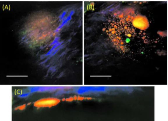
Depth scanning of an advanced plaque on a22-month-old rabbit artery shows a thin collagen fibril layer overlaying a
pool of lipid debris, a typical pathology of a rupture-prone atherosclerotic plaque. Figures 3共a兲 and 3共b兲 are two focal depth images obtained from the same location on an advanced plaque at depths approximately 10 and 60m respectively from the lumen surface. In Fig.3共c兲a reconstructed side-view image representing a depth profile of this particular plaque shows a collagen layer on top of a lipid-rich pool.
In conclusion, we demonstrated multimodal nonlinear op-tical imaging of bulk arterial tissue from WHHLMI rabbits using a cost-effective PCF-based nonlinear optical micro-scope. Epi-NLO imaging of unsectioned arteries allowed label-free visualization of extracellular components relevant to the development of atherosclerosis. Clear differences in the surface biochemical morphology were observed between healthy artery wall, early atherosclerotic lesions, and ad-vanced plaques. In particular, differences in collagen fibril structures were noted between early and advanced lesions. Such changes in collagen morphology detected by SHG may provide a clinical measure for differentiating plaque burden.
Acknowledgment
The authors acknowledge Vijay Iyer for the assistance pro-vided in using ScanImage, and Jeri Friesen for her assistance in preparing figures. This work was supported by the National Research Council Canada, Genomics and Health Initiative.
References
1. P. Libby, “Atherosclerosis: disease biology affecting the coronary vasculature,”Am. J. Cardiol.98关suppl兴, 3Q–9Q 共2006兲.
2. G. K. Hansoon, “Inflammation, atherosclerosis, and coronary artery disease,”N. Engl. J. Med.352, 1685–1695 共2005兲.
3. A. Zoumi, X. Lu, G. S. Kassab, and B. J. Tromberg, “Imaging coro-nary artery microstructure using second-harmonic and two-photon fluorescence microscopy,”Biophys. J.87共4兲, 2778–2786 共2004兲.
4. M. B. Lilledahl, O. A. Haugen, C. D. L. Davis, and L. O. Svaasand, “Characterization of vulnerable plaques by multiphoton microscopy,” J. Biomed. Opt.12共4兲, 044005 共2007兲.
5. T. T. Le, I. M. Langohr, M. J. Locker, M. Sturek, and J. X. Cheng, “Label-free molecular imaging of atherosclerotic lesions using mul-timodal nonlinear optical microscopy,” J. Biomed. Opt. 12共5兲,
054007 共2007兲.
6. H. W. Wang, T. T. Le, and J. X. Cheng, “Label-free imaging of arterial cells and extracellular matrix using a multimodal CARS mi-croscope,”Opt. Commun.281, 1813–1822 共2008兲.
7. H. W. Wang, I. M. Langohr, M. Sturek, and J. X. Cheng, “Imaging and quantitative analysis of atherosclerotic lesions by CARS-based multimodal nonlinear optical microscopy,” Arterioscler., Thromb., Vasc. Biol.29, 1342–1348 共2009兲.
8. A. F. Pegoraro, A. Ridsdale, D. J. Moffatt, Y. Jia, J. P. Pezacki, and A. Stolow, “Optimally chirped multimodal CARS microscopy based on a single Ti:sapphire oscillator,” Opt. Express 17共4兲, 2984–2996
共2009兲.
9. S. Murugkar, C. Brideau, A. Ridsdale, M. Naji, P. K. Stys, and H. Anis, “Coherent anti-Stokes Raman scattering microscopy using pho-tonic crystal fiber with two closely lying zero dispersion wave-lengths,”Opt. Express15共21兲, 14028–14037 共2007兲.
10. T. A. Pologruto, B. L. Sabatini, and K. Svoboda, “ScanImage: flex-ible software for operating laser scanning microscopes,” Biomed. Eng. Online2, 13 共2003兲.
11. M. Shiomi, T. Ito, S. Yamada, S. Kawashima, and J. Fan, “Develop-ment of an animal model for spontaneous myocardial infarction 共WHHLMI rabbit兲,” Arterioscler., Thromb., Vasc. Biol. 23, 1239
共2003兲.
12. T. Itp, S. Yamada, and M. Shiomi, “Progression of coronary athero-sclerosis relates to the onset myocardial infarction in an animal model of spontaneous myocardial infarction 共WHHLMI rabbits兲,” Exp. Anim.53, 339–346 共2004兲.
Fig. 3 Epi-NLO images of an advanced plaque obtained at 共a兲
⬃10m depth, 共b兲 ⬃60m depth from the lumen surface, and 共c兲 the side-view of the image stack showing a collagen cap overlaying lipid-rich bulk. Blue:SHG 共collagen兲, and red/orange:CARS 共lipid-rich structure兲. The scale bar is 25m. 共Color online only.兲
JBO Letters
Journal of Biomedical Optics 020501-3 March/April 2010 쎲 Vol. 15共2兲