HAL Id: tel-01295432
https://tel.archives-ouvertes.fr/tel-01295432
Submitted on 31 Mar 2016
HAL is a multi-disciplinary open access archive for the deposit and dissemination of sci-entific research documents, whether they are pub-lished or not. The documents may come from teaching and research institutions in France or abroad, or from public or private research centers.
L’archive ouverte pluridisciplinaire HAL, est destinée au dépôt et à la diffusion de documents scientifiques de niveau recherche, publiés ou non, émanant des établissements d’enseignement et de recherche français ou étrangers, des laboratoires publics ou privés.
Development of therapeutic vaccine strategies and
pre-clinical animal tumor models for head and neck
cancers
Rodney Macedo Gonzales
To cite this version:
Rodney Macedo Gonzales. Development of therapeutic vaccine strategies and pre-clinical animal tumor models for head and neck cancers. Cancer. Université Pierre et Marie Curie - Paris VI, 2015. English. �NNT : 2015PA066269�. �tel-01295432�
Université Pierre et Marie Curie
Ecole doctorale 394: Physiologie, physiopathologie et thérapeutique
CIMI-Paris - UPMC UMRS CR7, INSERM U1135, CNRS ERL 8255/Equipe Immuno-intervention et Biothérapies
Development of therapeutic vaccine strategies and
pre-clinical animal tumor models for head and neck cancers
Par Rodney Macedo Gonzales
Thèse de doctorat de Physiologie, Physiopathologie et Thérapeutique
Specialité: Immuno-oncologie
Dirigée par Pr. François Lemoine et Dr. Géraldine Lescaille
Présentée et soutenue publiquement le 28 Septembre 2015 Devant un jury composé de:
Pr. Guy GOROCHOV, Professeur, Université Paris VI Président
Pr. Claude LECLERC, Directeur de recherche, Institut Pasteur Rapporteur
Dr. Patrick MIDOUX, Directeur de recherche, INSERM Rapporteur
Pr. Eric TARTOUR, PU-PH, Université Paris V Examinateur
Dr. Pierre BRUHNS, Directeur de recherche, Institut Pasteur Examinateur
Pr. François LEMOINE, PU-PH, Université Paris VI Directeur de thèse
Acknowledgements
Pr. Claude Leclerc and Dr. Patrick Midoux, for the informed criticism and for accepting to participate of this jury as “rapporteurs”.
Pr. Eric Tartour and Dr. Pierre Bruhns, for the support, the valuable advice and for accepting to participate of this jury as “examinateurs”; and Pr. Guy Gorochov, for accepting to participate as “president of the jury”.
Dr. Christophe Combadière, for receiving me in the research unit.
Pr. François Lemoine, for the listening, the patience, the honor that he made me to have led this work, but specially for give me the opportunity to do what I like most; and Dr. Géraldine Lescaille, for the patience, guidance, and continuous encouragement.
Dr. Véronique Mateo, for the assistance and accurate advice; and Dr. Chloé Bertholus, for the assistance and her involvement with the HNSCC patients.
My team: Juliette, Maude, Aline, Kae and especially Claude, for the valuable help. Bertrand Bellier and his team, for the constructive criticism and assistance.
All people of the laboratory that contributed in many way to my PhD thesis, especially Pedro, Fabien, Audrey, Mustapha, Clara and Louis for the time, advice and friendship.
Sarah and Diego for helping me with the revision to English spelling and grammar. The Immunology Research Group (GII) from Arequipa-Peru: Dr. Jorge, Dra. Irmia, Dr. Walter, and Dr. Marcial, for the academic and moral support; Julio, for start in me, the passion for the immunology; and all members especially Juan Carlos L., Rodolfo, José Luis U., Paola, Alfredo and Nirk; for being part of this adventure.
All my friends and the Magic PSA team, who made my time living in Paris: the best; even far away from my country and my family
Finally, I would like to entirely thanks my family for trust me unconditionally; and Claudia, for supporting me and especially for “soportarme” each day, I know is not easy.
List of abbreviations
aa Amino acid
ACT Adoptive cellular immunotherapy ADCC Antibody-dependent cell mediated
cytotoxicity
AICD Activation-induced cell death APC Antigen-presenting cell AT-84 Tumor cell line B16F10 Tumor cell line BALB/C Murine strain
BALT Bronchus-associated lymphoid tissue
C3H Murine strain
C57Bl/6 Murine strain
CAF Cancer associated fibroblasts CAM Cell adhesion molecule CAR Chimeric antigen receptor
CCR C-C chemokine receptor
CD Cluster of differentiation cDC Conventional dendritic cell
CEA Carcinoembryonic antigen
CIN Cervical intraepithelial neoplasia
CMV Human citomegalovirus
CpG-ODN CpG oligodeoxynucleotide
CT Cholera toxin
CTL Cytotoxic T lymphocytes
CTLA-4 Cytotoxic T-lymphocyte-associated protein 4
CyaA Adenylate cyclase
DAMP Damage-association molecular pattern Dbait DNA repair bait
DC Dendritic cell
DC-SIGN DC-Specific Intercellular adhesion molecule-3-Grabbing Non-integrin
DNA Deoxyribonucleic acid
DNA-PK DNA-dependent protein kinase E6 E6 protein of human papillomavirus-16 E7 E7 protein of human papillomavirus-16 EGF Epidermal growth factor
EGFR Epidermal growth factor receptor
ELISA Enzyme linked immunosorbent assay ELISpot Enzyme linked immunosorbent spot FADD Fas-associated death domain protein
Fas-L Fas-ligand
Fc Fragment crystallizable region
FCS Fetal calf serum
FGF Fibroblast growth factor FoxP3 Forkhead box protein P3 Gag Group-antigen virus protein GALT Gut-associated lymphoid tissue GEMM Genetically engineered mouse model GFP Green fluorescent protein
GM-CSF Granulocyte-macrophage colony stimulating factor
HER2/Neu Human epidermal growth factor receptor 2
HBV Hepatitis B virus HCV Hepatitis C virus
HIV Human immunodeficiency virus
HNSCC Head and neck squamous cell carcinoma
HLA Human leukocyte antigen
HPV Human papillomavirus
IFN- Interferon gamma
Ig Immunoglobulin
IC Intra-cheek
ID Intradermal
iDC Interstitial dendritic cell IEL Intraepithelial lymphocyte
IL Interleukin
IN Intranasal
IP Intraperitoneal
IVIS In vivo imaging system
mAb Monoclonal antibody
MAGE Melanoma-associated antigen MALT Mucosa-associated lymphoid tissue MDSC Myeloid-derived suppressor cell MHC Major histocompatibility complex
MIP Macrophage inflammatory protein MMP Matrix metalloproteinase
mRNA Messenger ribonucleic acid
MRI Magnetic resonance imaging
MuLV Moloney murine leukemia virus NALT Nasopharyngeal-associated lymphoid
tissue
NF-B Nuclear factor-kappa B NHEJ Non-homologous end joining
NK Natural killer
NKT Natural killer T cell
NOD Non-obese diabetic
NR-S1 Tumor cell line
NSG NOD-SCID-IL2rg-/-
L1 L1 protein of human papillomavirus-16 L2 L2 protein of human papillomavirus-16 LAK Lymphokine-activated killer cells
LC Langerhans cells
Ln Langerin
Ln+iDC Langerin-expressing iDCs
LPS Lipopolysaccharide
LT Heat-labile enterotoxin OSCC Oral squamous cell carcinoma OPSCC Oropharynx squamous cell carcinoma
OVA Ovoalbumin
p53 Protein 53
PARP Poly-ADP-ribo-polymerase
PBMC Peripheral blood mononuclear cells PCR Polymerase chain reaction
PDGF Platelet-derived growth factor PDX Patient-derived xenograft
PEI Polyethylenimine
PET Positron emission tomography
pVLP Plasmo-retroVLP
PD-1 Programmed cell death 1 pDC Plasmacytoid dendritic cell PD-L1 Programmed death ligand 1
PMN Polymorphonuclear cell
RAG Recombinase activated gene
Rb Retinoblastoma protein
SC Subcutaneous
SCID Severe combined immunodeficiency SSC-VII Tumor cell line
STxB Shiga toxin B-subunit
TA Tumor antigens
TAA Tumor-associated antigen
TAM Tumor-associated macrophages
TIM-3 T 0cell immunoglobulin mucin-3 TRAIL TNF-related apoptosis inducing ligand TSA Tumor-specific antigen
TC-1 Tumor cell line
TC1-luc TC-1 tumor cell line transfected with the luciferase gene
TCM Central memory T cell
TCR T-cell receptor
TdLN Tumor draining lymph nodes TEM Effector memory T cell
TGF-β Transforming growth factor beta
Th T-helper
TNF-α Tumor necrosis factor alpha TLR Toll-like receptor
Treg T regulatory cells
VEGF Vascular endothelial growth factor VLP Virus like particle
VSV-G Vesicular stomatitis virus G protein
Table of Contents
LIST OF ABBREVIATIONS ... 3
TABLE OF CONTENTS ... 5
LIST OF TABLES AND FIGURES ... 7
PREFACE ... 8
CHAPTER I. HEAD AND NECK CANCERS ... 9
I.1 GENERALITIES ... 9
I.1.1 Heterogeneity of Head and Neck Cancers ... 9
I.1.2 Management and prognosis of Head and Neck Cancers ... 11
I.2 TUMOR MICROENVIRONMENT OF HNSCC ... 12
I.2.1 Non-immune populations in the HNSCC microenvironment ... 12
I.2.2 Immune populations in the HNSCC microenvironment ... 14
T Lymphocytes ... 15
Dendritic Cells ... 16
Natural Killers ... 16
Macrophages ... 17
Myeloid-derived suppressor cells ... 17
B Lymphocytes ... 18
Other immune cells ... 18
I.2.3 Immunosurveillance and immunoescape mechanisms in HNSCC ... 19
Evasion of immune system detection ... 20
Resistance to immune attack ... 21
Secretion or expression of inhibitory factors ... 21
Recruitment of immune inhibitory cells ... 22
I.2.4 Immune-microenvironment according to HNSCC location ... 23
Oral cavity ... 23
Oropharynx ... 25
Other sites... 25
I.3 PRE-CLINICAL MODELS FOR HNSCC ... 26
I.3.1 Ectopic pre-clinical models for HNSCC ... 27
I.3.2 Orthotopic pre-clinical models for HNSCC ... 28
I.3.4 In situ pre-clinical models for HNSCC ... 29
I.3.5 Monitoring of tumor growth in preclinical models ... 31
CHAPTER II. IMMUNOTHERAPY OF HNSCC ... 32
II.1 CANCER IMMUNOTHERAPY ... 32
II.1.1 Effector mechanisms of CD8+ T cells. ... 32
Development and activation of CD8+ T cells ... 32
Effector and memory CD8+ T cells ... 35
CTL-mediated cytotoxicity ... 35
Regulation of CD8+ T-cell activation ... 36
II.1.2 Immunotherapy and anti-tumor vaccination... 37
a. PASSIVE IMMUNOTHERAPY ... 38
b. NON-SPECIFIC ACTIVE IMMUNOTHERAPY ... 39
c. SPECIFIC ACTIVE IMMUNOTHERAPY (VACCINATION) ... 41
Peptide-based vaccines ... 41
Cell-based vaccines ... 42
Nucleic acid-based vaccines... 43
Bacteria vector-based vaccines ... 44
Viral vector-based vaccines ... 45
Non-viral vector-based vaccines ... 45
II.1.3 Virus-like particles (VLPs) and plasmo-retroVLPs (pVLPs) ... 46
II.2 MUCOSAL IMMUNIZATION ... 48
II.2.1 Organization of the mucosal immune system ... 48
DCs in the buccal mucosa (cheek) ... 49
DCs in the sublingual mucosa ... 50
DCs in the gingival mucosa ... 51
Role of T cells in oral immunity ... 51
II.2.3 Strategies for mucosal immunization ... 52
Mucosal vaccine adjuvants ... 52
Mucosal delivery systems and vectors ... 53
Mucosal immunization routes ... 54
THESIS OBJECTIVES ... 56
RESULTS ... 57
1. Efficacy of DNA vaccines forming E7 recombinant retroviral virus-like particles for the treatment of HPV-induced cancers. ... 57
2. Intra-cheek immunization as a novel vaccination route for therapeutic vaccines of head and neck squamous cell carcinomas using an orthotopic pre-clinical model. ... 70
DISCUSSION AND PERSPECTIVES ... 108
1. Improvement of vaccination strategies for HNSCC ... 110
Vaccination against HPV-negative HNSCC TAAs ... 110
Improvement in the vaccination schedule ... 111
pVLP Engineering ... 112
Improvement in vaccine delivery ... 113
2. Combination of vaccination with conventional and innovative approaches ... 114
Combination with chemotherapy ... 114
Combination with radiotherapy ... 115
Combination with DNA-repair inhibitors ... 116
Combination with targeting cancer therapies ... 117
3. Combination of vaccination with modulation of the tumor microenvironment ... 119
CONCLUSIONS ... 122
List of tables and figures
Table 1: Different clinical and biological characteristics of HNSCC ... 10
Table 2: Features of the ectopic and orthotopic models ... 28
Figure 1: The tumor microenvironment ... 12
Figure 2: Immunosurveillance and immunoescape mechanisms ... 20
Figure 3: Immune parameters linked with clinical outcome in OSCC (A) and OPSCC (B) .. 24
Figure 4: Representation of the categories of preclinical cancer models in use ... 27
Figure 5: Strategies for the generation of genetically engineered mouse models ... 30
Figure 6: Differentiation and maturation of T cells in the thymus ... 33
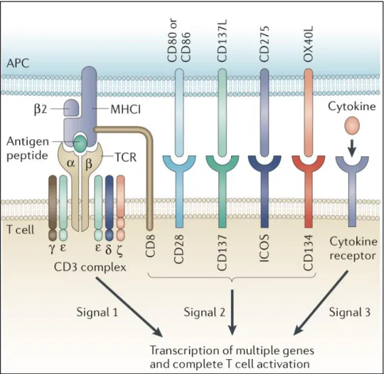
Figure 7: T-cell activation ... 34
Figure 8: Immunotherapy strategies and their action in the cancer-immunity cycle ... 37
Figure 9: Activation of innate and adaptive immunity by TLR agonists ... 41
Figure 10: Mechanisms of action of DNA vaccines ... 43
Figure 11: VLP and pVLP immunization ... 47
Figure 12: Comparison of mucosal immune system in oral mucosa (a) and gut mucosa (b) .. 49
Figure 13: Distribution and function of DCs in the murine oral mucosa. ... 50
Figure 14: Antigen delivery and presentation following sublingual or buccal vaccination .... 55
Preface
Head and neck squamous cell carcinomas (HNSCCs) represent the sixth most frequent type of cancer in the world with more than 550 000 new cases per year. It is the fourth most prevalent cancer for males in France, after prostate, lung and colon cancers. These cancers are associated with multiple risk factors; among them the most important are alcohol and/or tobacco consumption. Nevertheless, some patients develop HNSCCs without exposure to these chemical carcinogens. Recently, human papillomavirus (HPV) has been associated with the development of some types of HNSCCs. Despite standard treatment strategies for HNSCCs -including surgery, radiation and chemotherapy- HNSCCs are very challenging to treat and present high relapse rates. The prognosis of HNSCCs remains poor, with a survival rate of 10-20% at 10 years. Due to this, there is an urgent need for innovative therapies that target specific features of HNSCCs.
Currently, immunotherapy is one of the most promising strategies for cancer treatment. Many human malignancies, including HNSCC are associated with quantitative and qualitative deficiencies in the immune system. Enhanced awareness of the immune alterations present in HNSCC, as well as better understanding the basic mechanisms of the immune system in carcinogenesis rationalizes the uses of immunotherapeutic strategies for treating HNSCCs. Moreover, recent clinical trials revealed the impressive efficacy of immunological checkpoint blockade in multiple types of metastatic cancers. In addition, preclinical studies provide evidence that some cytotoxic drugs have the ability to stimulate the immune system, resulting in anti-tumor immune responses that contribute to clinical efficacy of these agents. These observations raise the hypothesis that the next step for cancer treatment is the combination of conventional treatments and immunotherapies.
In the first part of this manuscript, I will present a review of the microenvironment’s characteristics and immunoescape mechanisms of HNSCC, as well as the promising strategies of immunotherapy in this context. In the second part, I will present my work in the laboratory, whose main objective consisted in developing immunotherapeutic strategies in preclinical models of head and neck cancer. Results are discussed in the last part according to the literature, in order to present the perspectives of this contribution towards the improvement of the therapeutic care of HNSCC.
Chapter I. Head and Neck Cancers
I.1
Generalities
I.1.1 Heterogeneity of Head and Neck Cancers
Head and neck cancers represent the sixth most frequent type of cancer in the world with annual global incidence and mortality rates estimated at 680 000 and 370 000 cases, respectively (Ferlay et al., 2013). The vast majority (more than 90%) are head and neck squamous cell carcinomas (HNSCCs). HNSCCs are anatomically and clinically heterogeneous and arise from the mucosal surface of the oral cavity, oropharynx, hypopharynx, larynx, sinuses and other sites within the upper aerodigestive tract.
Tobacco and/or alcohol consumption are the predominant risk factors for the development of HNSCCs with a population attributable risk of 72%, of which 4% is due to alcohol alone, 33% is due to tobacco alone, and 35% is due to the combination of these factors (Hashibe et al., 2007; Hashibe et al., 2009). Otherwise, oral smokeless tobacco, especially when consumed in betel liquids, is a major cause of oral and oropharyngeal HNSCC in men (50%) and women (90%) in the Indian subcontinent (Barnes et al., 2005). However, in the last decades an increased incidence of HNSCCs associated with oral infection by the human type 16 papillomavirus (HPV-16) has been observed (Chaturvedi et al., 2013; Chaturvedi et al., 2011), especially among young patients. Those HNSCCs developing in the oral cavity (OSCC) and in the oropharynx (OPSCC) are both associated with tobacco and alcohol abuse, but only OPSCC is associated with HPV-16. Previously published data support a dominant role for HPV-16 in economically developed (60 to 70%) versus developing countries (less than 10%).
Tobacco is responsible for more than 30% of all cancer deaths worldwide. Tobacco smoke contains more than 4000 chemicals, of which at least 60 have been shown to be carcinogenic, these carcinogens promote tumorigenesis by inducing genetic aberrations depending on carcinogen dose and host susceptibility to HNSCC (Singh, 2008). Chronic alcohol exposure also results in increased cancer incidence; the carcinogenic effect of alcohol involves both direct (increased p450 activity, resulting in an more activation of carcinogens) and indirect (acting as solvent, facilitating the entry of other carcinogens into cells, especially in the upper aerodigestive tract (Seitz and Becker, 2007).
Human papillomaviruses are small, non-encapsulated DNA viruses that can infect epithelial cells from many organisms, including humans. A fraction of people infected with high-risk subtypes of HPV are at risk for developing squamous cell carcinoma. HPV-16 is the most frequently detected subtype in squamous cell carcinoma and was found in up to 90% of HPV-positive tumors (Gillison et al., 2000; Wiest et al., 2002). HPV DNA replicates to a high copy number in well-differentiated cells near the epithelial surface through the action of the E6 and E7 proteins and induces cell-cycle progression and viral DNA replication in differentiated keratinocytes. The human papillomavirus encodes up to 10 proteins, including the E6 protein, a 150 amino-acid (aa) protein containing two zinc-like fingers joined by an interdomain linker, which binds to and induces the degradation of the p53 tumor suppressor protein (Werness et al., 1990), and the E7 protein, a 98 aa zinc-binding phosphoprotein that binds and destabilizes the retinoblastoma (Rb) tumor suppressor protein (Dyson et al., 1989) leading to increased cell cycling and decreased apoptosis.
Even if the majority of head and neck cancers are squamous cell carcinomas, recent insight has revealed that this type of cancer is not homogeneous (Leemans et al., 2011). Various subclasses of HNSCCs can be distinguished at the histological (Woolgar and Triantafyllou, 2009), molecular (Chung et al., 2004) and genetic (Smeets et al., 2009) level. Genome-wide sequencing projects have identified a number of recurrently mutated genes in HNSCC, including TP53, CDKN2A, EGFR, PIK3CA, FAT1, NOTCH1, and chromatin related genes, among others (Riaz et al., 2014). Furthermore, around 20% are HPV positive and 80% are HPV-negative. In addition, about 20% of HNSCC cases that are not caused by HPV seem to have only a few copy-number alterations, the rest presents high chromosome instability and TP53 mutations. These data underline head and neck cancers as a heterogeneous disease, and HPV-positive tumors as a specific subclass of HNSCCs due to the differences at the molecular level and clinical outcome (Chung and Gillison, 2009).
Feature HPV-negative HNSCC HPV-positive HNSCC
Incidence Decreasing Increasing
Etiology Smoking, excessive alcohol use Oral sex
Age Above 60 years Under 60 years
Field cancerization Yes Unknown
TP53 mutations Frequent Infrequent
Predilection site None Oropharynx
Prognosis Poor Favorable
I.1.2 Management and prognosis of Head and Neck Cancers
The standard of care for HNSCC is the use of surgery in association with radiotherapy and/or chemotherapy (Choong and Vokes, 2008). However, the choice of treatment for a patient must to be deliberated on a multidisciplinary board, taking into account individual parameters from the patient (general condition) and the tumor (localization, stage, etc.).
Despite current treatment, head and neck cancers have a survival rate of no more than 20% at ten years (Chaturvedi et al., 2011). The five year relative survival rates reported from 21 countries in the European Cancer Registry-based Study of Survival and Care of Cancer Patients (EUROCARE-4) were 44.4%, 48.5% and 45.42% for oral cavity cancer and 31.0%, 39.8% and 38.71% for oropharyngeal cancer, during the periods of 1990–1994, 1995–1999 (Karim-Kos et al., 2008), and 2000-2007 (De Angelis et al., 2014) respectively. The five year relative survival rate reported by the SEER program for the period 1996–2004 was 59.7% for oral cavity and pharyngeal cancers (Ries et al., 2007).
Several characteristics of HNSCCs patients have been linked with favorable prognosis, including non-smoker, minimum exposure to alcohol, good performance status, and no comorbid disorders, all of which are related to HPV-positive tumor status (Marur et al., 2010). Furthermore, overall survival and free-disease survival of HPV-positive OPSCC patients is significantly better than that for HPV-negative OPSCCs patients as showed by several retrospective and prospective studies in the United States, Australia and Western Europe (Ang et al., 2010; Chaturvedi et al., 2011; Posner et al., 2011).
The improvement in survival may be a result of one or more of several reasons. Augmented sensitivity to chemotherapy and radiotherapy has been attributed to absence of exposure to tobacco and presence of functional unmutated TP53 (Bristow et al., 1996). Increased survival of patients with HPV-positive cancer might also be attributable in part to absence of field cancerization related to tobacco and alcohol exposure (Gillison et al., 2000). Nonetheless, HPV-positive OPSCCs patients have significantly better survival compared to HPV-negative patients even after adjustment for differences in favorable prognostic factors often observed among HPV-positive patients (younger age, better performance status, fewer co-morbidities, and less exposure to tobacco smoking) (Ang et al., 2010). HPV-positive HNSCCs are in general, more sensitive to chemotherapy and radiation than HPV-negative tumors. Thus leading to some improvement in prognosis and therefore to longer survival.
12
I.2
Tumor microenvironment of HNSCC
Like other cancers, HNSCCs arise from the accumulation of genetic and epigenetic changes and abnormalities in cancer-associated signaling pathways, causing the acquisition of cancer-related phenotypes. However, the biology of a tumor can only be understood by studying tumor cells as well as the tumor microenvironment in which malignant cells subsist. Cancer cells initiate tumors and drive tumor progression forward, carrying mutations that define cancer as a genetic disease. They have been portrayed as homogeneous cell populations until relatively late in the course of tumor progression. Many human tumors are histopathologically diverse, containing regions with various degrees of differentiation, proliferation, vascularity, inflammation, and invasiveness (Hanahan and Weinberg, 2011). The tumor microenvironment consists of extracellular matrix and diverse types of non-malignant cells, including endothelial cells, cancer-associated fibroblasts (CAFs), pericytes and a variety of immune cells and their precursors (Lee et al., 2015).
Figure 1: The tumor microenvironment (Koontongkaew, 2013)
I.2.1 Non-immune populations in the HNSCC microenvironment
HNSCC non-malignant stromal and parenchymal cells largely contribute to cancer progression through their crosstalk with cancer cells, extracellular matrix and other non-cancer cells, using growth factors, proteases and other signaling molecules (Koontongkaew, 2013). These cell-to-cell communications promote tumor growth (Whiteside, 2008), angiogenesis and metastatic invasion (Watnick, 2012).
Journal of Cancer 2013, Vol. 4 70
tem to reject the tumor. However, this observation remained largely neglected for over a century until it was demonstrated that innate immune cells, in par-ticular phagocytes, play an active role in promoting the tumorigenesis. In addition to leukocyte
infiltra-tion, angiogenesis is now being recognized as another stromal reaction promoting cancer progression. Therefore, chronic inflammatory and neovasculariza-tion are critical, if not essential, for cancer progression [62-64].
Figure 2. The tumor microenvironment (TME). TME comprises different stromal cells in addition to tumor cells. These include vascular or lymphatic endothelial cells, supporting pericytes, fibroblasts, and both innate and adaptive infiltrating immune cells. Moreover, TME contains non-cellular components, including extracellular matrixes, growth factors, proteases, protease inhibitors and other signaling molecules that play important roles in stromal reactions in TME.
The cellular microenvironment of H N SCC
Studies have demonstrated a higher incidence of malignancy in acquired or iatrogenic immunodefi-cient hosts, which suggest an important role for im-munosurveillance in HNSCC [65-68]. In those with a competent immune system, this is an additional bar-rier to malignancy, which must be overcome and which shapes both the tumor and its microenviron-ment [69]. Although effective antitumor immune re-sponses likely involve many components of the im-mune system, T-cells continue to be considered as the critical immune cells involved in antitumor immuni-ty. T lymphocytes are considered an essential com-ponent of antitumor immunity, with CD8+ T cells serving as cytotoxic effector cells and CD4+ Th1 cells serving to ‘help’ and enhance the magnitude and du-ration of the antitumor responses. However, CD4+
Th2 cells and CD4+ T regulatory cells are capable of suppressing effective CD8+ antitumor responses. In fact, several investigators have found dysfunctional circulating and tumor-infiltrating T cells in HNSCC patients, with functional assays identifying multiple defects in T-cell activation and effector function, sug-gesting that the tumor has successfully suppressed an otherwise robust lymphocytic response [70-72].
Patterns of tumor-related leukocyte infiltration varies between primary tumors and metastatic lymph nodes in HNSCC with a local decrease in the number of CD8+ T-cells and increase in CD20+ B-cells being the most relevant findings. This indicated that sup-pression of local cellular immunity might be a mech-anism by which tumor cells evade host immunity [73]. Patients with tumors expressing HPV16 had an in-creased frequency of CD8+ T cells specific for pep-tides derived from the oncogenic HPV E7 proteins, compared with those patients with tumor negative for
Journal of Cancer 2013, Vol. 4
http://www.jcancer.org
70 tem to reject the tumor. However, this observation
remained largely neglected for over a century until it was demonstrated that innate immune cells, in par-ticular phagocytes, play an active role in promoting the tumorigenesis. In addition to leukocyte
infiltra-tion, angiogenesis is now being recognized as another stromal reaction promoting cancer progression. Therefore, chronic inflammatory and neovasculariza-tion are critical, if not essential, for cancer progression [62-64].
Figure 2. The tumor microenvironment (TME). TME comprises different stromal cells in addition to tumor cells. These include vascular or lymphatic endothelial cells, supporting pericytes, fibroblasts, and both innate and adaptive infiltrating immune cells. Moreover, TME contains non-cellular components, including extracellular matrixes, growth factors, proteases, protease inhibitors and other signaling molecules that play important roles in stromal reactions in TME.
The cellular microenvironment of H N SCC
Studies have demonstrated a higher incidence of malignancy in acquired or iatrogenic immunodefi-cient hosts, which suggest an important role for im-munosurveillance in HNSCC [65-68]. In those with a competent immune system, this is an additional bar-rier to malignancy, which must be overcome and which shapes both the tumor and its microenviron-ment [69]. Although effective antitumor immune re-sponses likely involve many components of the im-mune system, T-cells continue to be considered as the critical immune cells involved in antitumor immuni-ty. T lymphocytes are considered an essential com-ponent of antitumor immunity, with CD8+ T cells serving as cytotoxic effector cells and CD4+ Th1 cells serving to ‘help’ and enhance the magnitude and du-ration of the antitumor responses. However, CD4+
Th2 cells and CD4+ T regulatory cells are capable of suppressing effective CD8+ antitumor responses. In fact, several investigators have found dysfunctional circulating and tumor-infiltrating T cells in HNSCC patients, with functional assays identifying multiple defects in T-cell activation and effector function, sug-gesting that the tumor has successfully suppressed an otherwise robust lymphocytic response [70-72].
Patterns of tumor-related leukocyte infiltration varies between primary tumors and metastatic lymph nodes in HNSCC with a local decrease in the number of CD8+ T-cells and increase in CD20+ B-cells being the most relevant findings. This indicated that sup-pression of local cellular immunity might be a mech-anism by which tumor cells evade host immunity [73]. Patients with tumors expressing HPV16 had an in-creased frequency of CD8+ T cells specific for pep-tides derived from the oncogenic HPV E7 proteins, compared with those patients with tumor negative for
Endothelial cells represent one of the more important non-immune components of the tumor stroma, forming the tumor-associated vasculature. They have a significant impact on the progression of HNSCC through secretion of factors involved in tumor proliferation and angiogenesis, including vascular endothelial growth factor (VEGF), platelet-derived growth factor (PDGF) and IL-8 (Campos et al., 2012; Li et al., 2005); and through the activation of MAPK, Notch-1 and STAT3/Akt/ERK signaling (Neiva et al., 2009; Zeng et al., 2005). In addition to blood vessels, HNSCCs are typically infiltrated by lymphatic vessels, which are distributed throughout the tumor and the peritumoral regions (Cao, 2005; Zhao et al., 2008). Increased tumor lymphatic vessel density correlates with metastasis to lymph nodes in HNSCC (Frech et al., 2009). Another component, the pericyte, is a specialized mesenchymal cell that wraps around the endothelial tubing of blood vessels. They provide paracrine signals to the endothelium and synthesize the vascular basement membrane that helps vessel walls to withstand the pressure of blood flow (Dvorak et al., 2011). Abnormal pericyte integration into tumor endothelium vessels destabilizes their integrity leading to cancer cell intravasation into the circulatory system; thus, enabling dissemination and metastasis (Raza et al., 2010). In HNSCC, immunohistochemical analysis has shown high activity of new structurally abnormal vessel formation, indicated by non-homogeneous patterns of endothelial cells and loosely attached pericytes to the endothelium, together with precursor cells being incorporated into these structures (Hollemann et al., 2012).
CAFs are the preponderant cell population of the tumor stroma with similarities to normal fibroblasts and myofibroblasts, but different biological roles and properties (Xouri and Christian, 2010). Two dominant patterns of distribution have been described in HNSCC: the ‘network’ pattern where CAFs occupy almost the entire tumor stroma and the ‘spindle’ pattern where CAFs are observed at the periphery of a tumor island (Thode et al., 2011). CAFs can enhance cancer cell proliferation, neoangiogenesis, and invasion and metastasis through the increased expression of various growth factors, cytokines and extracellular matrix proteins (Shimoda et al., 2010). In primary and metastatic HNSCC, they produce invasion-promoting signals that encourage production of TNF-α and IL-1α by tumor cells (Koontongkaew et al., 2009; Leef and Thomas, 2013). These signals include a brain-derived neurotrophic factor, that promotes epithelial-mesenchymal transition (MET) facilitating metastasis in this type of cancers (Dudas et al., 2011), the hepatocyte growth factor, that binds to the MET receptor on HNSCC cells triggering invasion through the basement membrane (Knowles et al., 2009), and the insulin-like growth factor 2 and CCL7, that promotes HNSCC invasion (Jung et al., 2010).
I.2.2 Immune populations in the HNSCC microenvironment
In addition to cancer cells and their surrounding stroma, the tumor microenvironment contains infiltrating cells of the innate and adaptive immune system. Currently, tumor immune-microenvironment is known to be crucial for understanding the tumor development and its response to treatment. Furthermore, tumors occurring in different anatomical sites differ in their immunecontexture and vary in their response to immunotherapy, suggesting that the tissue surrounding the tumor site can have a decisive role in determining its composition (Devaud et al., 2014). Indeed, the density and the composition of the immune microenvironment are heterogeneous between tumor types, and are very diverse from patient to patient (Angell and Galon, 2013).
These tumor infiltrating immune cells and their immune mediators and modulators can shape the tumor growth in two conflicting ways: tumor-promoting inflammation or anti-tumor immunity (Grivennikov et al., 2010). During tumor promoting inflammation, infiltrating immune cells supply direct and indirect mitogenic growth mediators, including the epidermal growth factor (EGF), transforming growth factor- (TGF-), tumor necrosis factor-a (TNF-), fibroblast growth factors (FGFs), and various chemokines, and cytokines (Hanahan and Coussens, 2012). In addition, these cells may produce proangiogenic and proinvasive molecules such as VEGF, PDGF, matrix-degrading enzymes, including MMP-9 and other matrix metalloproteinases, cysteine cathepsin proteases, and heparanase (Kitamura et al., 2015; Qian and Pollard, 2010). On the other hand, tumor-infiltrating immune cells support anti-tumor immunity by immunosurveillance and effector mechanism that leads, in the best case, to the elimination of cancer cells. This attribute raises the possibility that recruitment of certain immune cells may be a double-edge sword, by directly promoting angiogenesis and tumor progression while at the same time affording a means to tumor destruction.
Different leukocytes are present within the tumor-microenvironment granulocytes, mast cells, macrophages, and myeloid-derived suppressor cells (MDSC) are often present within the tumor mass, whereas natural killer (NK) cells are principally found in the stroma. CD8+ T cells congregate around the invasive margin and memory T cells are found in adjacent lymph tissue. Immature dendritic cells (DC) are more common within the core of the tumor as opposed to the stroma, whereas mature DCs congregate in tertiary lymphoid structures (Fridman et al., 2012; Gajewski et al., 2013).
T Lymphocytes
Immune cells recruited to the tumor include T cells (CD3+ TCR+); which can be categorized according to their effector functions including CD8+ cytotoxic T lymphocytes (CTLs) and CD4+ T-helper (Th) cells. CTLs are thought to be the major effector immune cells directed against tumor cells, having the ability to recognize and kill malignant cells (Boissonnas et al., 2007). CD4+ T-helper cells can be further subcategorized as Th1, Th2, Th17 and T regulatory (Treg) cells, they can promote or suppress anti-tumor immunity, as determined by their function (Kim and Cantor, 2014). Th1 cells, determined by the T-bet transcription factor and the secretion of interferon gamma (IFN-), TNF-, monocyte
chemotactic protein-1 (MCP-1) and macrophage inflammatory protein-1 (MIP-1);
mediates antitumor immunity by enhancing priming and expansion of CD8+ T cells, and by
recruiting NK and type I macrophages to tumor sites. Th2 cells, determined by the GATA-3 transcription factor and the secretion of IL-4, IL-5 and IL-13, may contribute to antitumor immunity by recruiting eosinophils and macrophages.
High densities of CTLs and Th1 cells, correlate with better survival in many different tumor types, including melanoma, HNSCCs, breast cancer, colorectal cancer, lung cancer, among others (Fridman et al., 2012; Gooden et al., 2011). Indeed, increased amounts of intratumoral CD8+Ki67+ cells have been linked with improved disease outcome in colorectal tumors (Galon et al., 2006). Moreover, the presence of CD3+CD8+ cells in HNSCCs, correlated with better clinical outcome and response to chemoradiotherapy (Balermpas et al., 2014a). However, recent findings in renal cell cancer, showed that the infiltration of CD8+ T cells was correlated with poor prognosis, modulated by the expression of immune checkpoints and the localization of DCs (Giraldo et al., 2015). Treg cells, characterized by the expression of the FoxP3 (forkhead box protein P3) transcription factor, are important for immunologic homeostasis. Intratumoral Tregs impede effective immunity against cancer and their presence correlates differently depending of the tumor (deLeeuw et al., 2012). They were associated with poor prognosis in hepatocellular cancer, breast cancer, and melanoma; and good prognosis in colorectal and ovarian cancer. In HNSCC, Tregs positively correlates with loco-regional control, possibly through downregulation of the pro-tumoral inflammatory reaction (Badoual et al., 2006). However, the prognostic value of Treg may be different, depending on the tumor site (deLeeuw et al., 2012; Weller et al., 2014).
The Th17 subset depends of the expression of the STAT3 and RORt transcription factors and the production of IL-17A and IL-17F. They have been recovered from multiple human tumors including ovarian, gastric, prostate, colon, renal and pancreatic cancer where Th17 cells have shown both anti-tumorigenic and pro-tumorigenic functions (Zou and Restifo, 2010). Furthermore, it have been shown that HNSCC and tumor draining lymph nodes are infiltrated with Th17 T cells in response to cytokines present in the HNSCC tumor microenvironment. Moreover, Th17 cells inhibit the proliferation and compromise the angiogenesis of HNSCC (Kesselring et al., 2010).
Dendritic Cells
Dendritic cells are antigen-presenting cells responsible for the uptake, processing and cross-presentation of tumor antigens (TAs) to T cells; which are, in turn, activated to proliferate and secrete cytokines, forming a key part of the adaptive immune response (Benencia et al., 2012). Multiple subsets of DCs have been described in human and mice. They can be classified into two main categories: plasmacytoid DCs (pDCs) and conventional DCs (cDCs), cDCs can be further divided into lymphoid resident DCs and migratory DCs, which are present in peripheral tissues and non-lymphoid organs (Segura and Amigorena, 2013). Elevated intratumoral DCs have often, but not always, been associated with improved clinical outcome in HNSCC most likely due to the fact that DCs subsets cannot be appropriately discriminated (Senovilla et al., 2012). However, the infiltration of Langerhans cells (LCs), a type of DC present in the epithelium of the mucosa, is associated with longer disease-free survival and decreased recurrence in HNSCC patients (Yilmaz et al., 2015). Additionally, a low number of intratumoral S-100+ DCs predicts poor survival in patients with OSCCs (Reichert et al., 2001).
Natural Killers
NK cells are lymphocytes that mediate innate immunity and recognize and kill virally infected or malignant cells. They are capable of eliminating tumors with reduced or absent major histocompatibility complex (MHC) class I expression that evade CD8+ T cell-mediated control. In a variety of solid tumor such as gastric, renal, HNSCC and colorectal cancers the presence of high numbers of tumor-infiltrating NK cells correlates with improved prognosis of patients (Moretta et al., 2014). Interestingly, the presence of NK cells were not correlated with the clinical outcome in lung cancer, suggesting that tumor microenvironment renders NK less
tumoricidal by reducing NK receptor expression and IFN- secretion (Platonova et al., 2011). In HNSCC, it has been observed that NK cell numbers are diminished in peripheral blood of patients and did not correlate with the tumor site (Accomando et al., 2012; Wulff et al., 2009). However, aggressive infiltration in the peritumoral stroma by CD57+ inflammatory cells (30-60% of mature NKs), may contribute to an ineffective loco-regional anti-tumoral response (Fraga et al., 2012).
Macrophages
Tumor-associated macrophages (TAMs) could exert phagocytic properties and can also present antigens to stimulate the adaptive pathway. In a classical and functional description, macrophages referred as ‘M1’ are activated with lipopolysaccharide (LPS) or IFN- to express pro-inflammatory cytokines (IL-12) and priming anti-tumor immune responses, whereas macrophages referred as ‘M2’ are IL-4-activated to express regulatory cytokines (IL-10) leading to anti-inflammatory responses and promoting tumor angiogenesis (Ostuni et al., 2015). In addition, it was showed that different tumor microenvironments could contain functionally distinct subsets of monocytes-derived TAMs that are poor antigen presenters and could suppress T-cell activation by using different mechanisms (Movahedi et al., 2010). Increased numbers of TAMs correlates with poor prognosis in many cancers, including breast, bladder, prostate and colorectal cancer; but worse overall survival in patients with HNSCC, gastric, and urogenital cancer (Biswas et al., 2013). Indeed, in HNSCC, TAMs were associated with angiogenesis and high histopathological grade malignancy (El-Rouby, 2010), and the expression of the macrophage inflammatory protein-3a (MIP-3a) that was shown to promote oral cancer cell migration and invasion, and was correlated with poorer prognosis for patient survival (Chang et al., 2011).
Myeloid-derived suppressor cells
MDSCs are immature myeloid cells that are precursors of DC, macrophages, and granulocytes. Their accumulation has been documented in most patients and mice with cancer, where they were induced by various tumor-derived factors produced in the tumor microenvironment (Ostrand-Rosenberg and Sinha, 2009). MDSCs can suppress the effector functions of NK, and T cells by the production of reactive oxygen species, reactive nitrogen species and cytokines as well as interactions with other suppressor cells like Tregs (Ostrand-Rosenberg, 2010). Low levels of circulating MDSC have been reported as a good prognosis
factor in patients with B-cell lymphoma, lung cancer, melanoma, gastrointestinal neoplasms and bladder carcinoma (Senovilla et al., 2012). In HNSCC, MDSCs can be defined as CD33+IL-4+CD14+HLADRint/negCD11b+ cells. They are able to suppress T-cell proliferation and their intratumoral accumulation correlates with tumor recurrence. Moreover, the daily treatment with taladafil modifies the tumor microenvironment and reduces the number of MDSCs, increasing anti-tumor immunity in HNSCC patients (Weed et al., 2015). Also, it has been shown that HNSCC intratumoral CD34+ cells can suppress immune functions by secreting granulocyte-macrophage colony-stimulating factor (GM-CSF) (Pak et al., 1995).
B Lymphocytes
B cells are lymphocytes with antigen-presenting properties and when activated differentiates into an antibody-secreting effector cell. They have also an immunoregulatory role in tumor microenvironment by the production of cytokines and chemokines to promote T-cell responses (Nelson, 2010). Infiltrating B cells are associated with good prognosis in other squamous cell cancers such as non-small cell lung cancer (Germain et al., 2015). In HNSCC, increased numbers of peritumoral B cells in lymph node metastasis were associated with favorable outcome (Pretscher et al., 2009).
Other immune cells
Polymorphonuclear (PMN) leukocytes like granulocytes, eosinophils and mast cells have also been found in tumor microenvironment. High levels of granulocytes have been associated with bad prognosis in tumors such as hepatocellular carcinoma (Kuang et al., 2011) and melanoma (Jensen et al., 2012). Analysis of HNSCC tumors, exhibited considerable infiltration by polymorphonuclear granulocytes, and strong infiltration was associated with poorer survival in advanced disease. Furthermore, the serum concentration of cytokines and chemokines that modulates PMN functions, such as IL-8, MIP-1 and RANTES, were significantly higher (Trellakis et al., 2011). Tumor infiltration by eosinophils has been reported to be a positive prognostic indicator for gastric and lung cancer, still it gives no clear prognostic information in head and neck cancer (Senovilla et al., 2012). Otherwise, robust tumor infiltration of mast cells has been linked to good prognosis in various cancers including HNSCC (Khazaie et al., 2011). However, as HNSCC progresses, there is an increase in mast cell numbers, which is correlated with new vascular tube formation suggesting a role in angiogenesis (Iamaroon et al., 2003).
I.2.3 Immunosurveillance and immunoescape mechanisms in HNSCC
The interactions between the host and the tumor have been referred to as “immunosurveillance”, where the immune system is able to spot, recognize and eliminate tumor cells. A new concept introduced in 2002, refers this interaction as “immunoediting”, where the host immune system recognize and destroy sensitive tumor cells, but also can edits for survival of tumors that become resistant. This “immunoediting” process is composed of three phases: elimination, equilibrium and escape (Schreiber et al., 2011).
Immunosurveillance occurs during the elimination phase, in which the innate and adaptive system works together to detect a developing tumor and destroy it. In HNSCCs, tumors invoke a host immune response to the over expression of tumor-associated antigens (TAAs) and the secretion of cytokines and chemokines by the tumor, causing a leukocytic infiltrate into the tumor microenvironment (Junker et al., 2012). Among TAAs, p53 is the most expressed mutated gene in HNSCC (about 62%) (Stransky et al., 2011). Also we can find TAAs from the melanoma-associated antigens (MAGE) group like MAGE-A3 (51%) and MAGE-A4 (60%), and other antigens from the cancer-testis group like NY-ESO-1 (Cuffel et al., 2011). Importantly, in HPV-positive HNSCC patients, CD8+ T cells against HPV oncogenic proteins like E7 can be found (Heusinkveld et al., 2012).
DC activation due to TAAs and damage-association molecular patterns (DAMPs) is believed to be a crucial step in initiating immune responses against tumors in HNSCCs (Kacani et al., 2005). An effective anti-tumor response would involve primed DCs migrating to regional lymph nodes, where they could present processed tumor antigens on HLA-I and II molecules to CD8+ and CD4+ T cells, and Th1 cells activation by cytokine secretion (IL-12 and TNF-) leading to secretion of IL-2 and IFN- to generate CTLs with specific cytotoxic activity against tumor cells bearing TAAs (Allen et al., 2012).
However, tumor cell variants may show resistance to persistent CTLs and NKs attack and gradually survive and proliferate. Thus, entering the equilibrium phase in which the adaptive immune system sculpts tumor immunogenicity and prevents tumor cell growth (Matsushita et al., 2012). Finally, in the escape phase, edited cancer cells efficiently overcome the immune recognition and destruction, so they expand and become a tumor clinically evident (Schreiber et al., 2011). HNSCCs cells escape can occur through many different mechanisms at different levels, as described below.
Figure 2: Immunosurveillance and immunoescape mechanisms (Schreiber et al., 2011)
Evasion of immune system detection
At the HNSCC tumor cell level, alterations leading to reduced immune recognition, such as loss of antigens, can be achieved through emergence during immunoediting of tumor cells that lack expression of immunodominant epitopes (Badoual et al., 2010), loss or decreasing the expression of surface MHC class I molecules (Ferris et al., 2006) or through loss of function of their antigen processing machinery (Meissner et al., 2005). The end result is the generation of poorly immunogenic tumor cell variants that become “invisible” to the immune system and thus acquire the capacity to grow progressively. However, even if 15% of primary and 40% of
metastatic HNSCCs presents MHC class I molecules loss, this alteration is not correlated with clinical outcome (Hasmim et al., 2013). Furthermore, patients TAA-specific CD8+ T cells that are expanded in vitro and exposed to autologous tumor cells can lyse the tumor cells when incubated with stimulatory factors like IFN- (Lopez-Albaitero et al., 2006).
Resistance to immune attack
HNSCC tumors may increase their resistance to cytotoxic effects of immune cells, through induction of anti-apoptotic mechanisms involving receptors and transcription factors. One receptor is toll-like receptor 4 (TLR-4), which normally binds to LPS; their overexpression is correlated with tumor grade and short survival (Ren et al., 2014). LPS binding to TLR-4 on HNSCC tumor cells enhanced proliferation and activated the nuclear factor-kappa B (NF-B) and PI3K/AKT anti-apoptotic ways (Szczepanski et al., 2009). Moreover, the expression in CD8+ T cells of another receptor implicated in the activation of PI3K/AKT anti-apoptotic way, the chemokine receptor 7 (CCR7), was associated with disease recurrence in HNSCCs patients (Czystowska et al., 2013).
Secretion or expression of inhibitory factors
Head and neck cancer cells can promote the development of an immunosuppressive microenvironment by producing regulatory cytokines and expressing negative costimulatory molecules. Among these cytokines IL-10, TGF- and prostaglandin E2 were described to interfere in immune reactivity to HNSCCs. Additionally, secretion by these cancer cells of GM-CSF and VEGF could obstruct the maturation of fully functional DCs (Pries and Wollenberg, 2006). A number of apoptosis-promoting factors have been identified in HNSCC including Galectin-1, Fas-L, TNF-related apoptosis inducing ligand (TRAIL), and programmed death-ligand 1 (PD-L1). Galectin-1 inhibits T-cell effector functioning by promoting T-cell apoptosis, blocking T-cell activation and inhibiting the secretion of proinflammatory cytokines (Saussez et al., 2007). Tumor-derived cells and plasma microvesicles from HNSCCs patients express Fas-L and trigger apoptotic death of activated T cells expressing Fas (Bergmann et al., 2009). Furthermore, OSCC may be capable to induce apoptosis in tumor-infiltrating lymphocytes (TILs) using the alternative TRAIL and
TNF- pathways (Kassouf and Thornhill, 2008). In HPV-positive HNSCCs, PD-L1 is
commonly expressed and it was showed that they could promote anergy, exhaustion or apoptosis in programmed cell death-1 (PD-1) expressing T cells (Lyford-Pike et al., 2013).
Recruitment of immune inhibitory cells
HNSCCs, also can promote immunosuppressive microenvironment by recruiting regulatory immune cells. Treg cells, MDSCs and TAMs are main leukocyte populations that play key roles in inhibiting host-protective anti-tumor responses (Schreiber et al., 2011).
Treg cells are CD4+ T cells that constitutively express CD25 and the transcription factor Foxp3. HNSCC intratumoral Tregs inhibit T-cell anti-tumor activity via a number of mechanisms including the production of immunosuppressive cytokines IL-10 and TGF- (Strauss et al., 2007b), the expression of negative costimulatory molecules like cytotoxic T-lymphocyte-associated protein 4 (CTLA-4), T cell immunoglobulin mucin-3 (TIM-3) and PD-1 (Jie et al., 2013), and by consuming IL-2 a cytokine that is critical for maintaining CTL function. Importantly, CD4+CD25highFoxP3+ are increased in the peripheral blood and tumor tissue (Strauss et al., 2007a). As described above, Tregs positively correlates with loco-regional control in HNSCC patients, possibly through downregulation of the pro-tumoral inflammatory reaction (Badoual et al., 2006; Weed et al., 2013). However, while overall FoxP3 expression in tumor infiltrating CD4+ T cells does not correlate with tumor recurrence, nuclear FoxP3 localization is associated with recurrence of oral HNSCC within 3 years (Weed et al., 2013). Interestingly, PD-1+ T cells where positively correlated with a favorable clinical outcome in HPV-associated HNSCC (Badoual et al., 2013).
MDSCs and TAMs are intratumoral myeloid cells that can be converted into immunosuppressive cells by the tumor microenvironment (Gabrilovich et al., 2012). In HNSCCs, MDSCs inhibit lymphocyte function by inducing Treg cells, producing TGF-, removing L-arginine required for T-cell function, or nitrating T-cell receptors (Chikamatsu et al., 2012; Pak et al., 1995). CD163+ TAMs are correlated with an unfavorable clinical
outcome in HNSCC patients after definitive chemo-radiotherapy; and CD11b+ TAMs
I.2.4 Immune-microenvironment according to HNSCC location
Head and neck cancers, in common with many other malignancies, are inflammatory foci by nature. It was previously shown that the presence of tumor infiltrating lymphocytes subsets in mixed populations of patients with HNSCCs of various sites were associated with a more favorable prognosis (Badoual et al., 2006; Balermpas et al., 2014a), and infiltrates of TAMs were associated with nodal metastases (Marcus et al., 2004). However, the degree of leukocyte infiltration appears to be specific depending on the site (OSCC or OPSCC) and is likely to be influenced by the differing microenvironments and the stage of the tumor (Green et al., 2013; Wansom et al., 2012; Wolf et al., 2015). Better understanding of the immunologic characteristics of the microenvironment including numbers, location and function of tumor infiltrating lymphocytes and macrophages is necessary in order to explore and test strategies that might be beneficial to HNSCCs patients (Badoual et al., 2010; Wallis et al., 2015).
Oral cavity
In oral cavity squamous cell carcinoma, the density of lymphocytic infiltrate at the tumor/host interface have been linked with good prognosis, while a lower overall density was associated with worse overall survival and local recurrence (Brandwein-Gensler et al., 2005). Immunohistochemical analysis of resected specimens of OSCC showed that CD8+ T cells infiltrated the stroma and cancer nest, whereas most Tregs only occupied the stroma. Also, greater CD8+ cell counts and CD8+/CCR4+ Treg ratio were associated with better survival (Watanabe et al., 2010). This finding suggests that Tregs may be detrimental within the tumor microenvironment in OSCC. However, in a further study of Tregs investigating the relationship between CD4, CD25, CTLA-4, and FoxP3 staining and survival, they found that high CD4+ cell counts showed a correlation with decreased survival, whereas high CD25+ cell counts were associated with better overall survival in OSCC patients (Moreira et al., 2010).
In addition to lymphocytic populations in the microenvironment, some other factors have demonstrated prognostic significance in OSCC. The expression of Granzyme B, a protein secreted by CTLs and NK cells, which induces apoptosis in abnormally proliferating cells, was associated with longer survival in OSCC (Costa et al., 2010). There are cytokines and cell receptors, which also seem to be of prognostic significance, including IL-6 and TGF-1, both linked with worse overall survival (Chen et al., 2012), and IL-10 linked with
HPV-positive OSCC, and correlated with bad prognosis (Chuang et al., 2012). The ability of OSCC tumor cells to express PD-L1, a surface glycoprotein that causes T-lymphocyte dysfunction, has been linked to decreased numbers of TIL in the peritumoral region, but did not affect patients survival (Cho et al., 2011). Besides, a low number of DCs was predictive for poor survival in OSCCs (Reichert et al., 2001).
Oropharynx
In OPSCCs, improved outcomes are associated with increased TILs independent of HPV status, suggesting that the local immune response may be more related with other factors such as tumor size or Karnofsky performance status (score from 100 = no evidence of disease to 0 = dead) (Wansom et al., 2012). High CD8+ T-cell infiltration and CD8+ T cells/Treg ratio were significantly positively correlated to a good clinical outcome in both HPV-positive and HPV-negative OPSCC. Moreover, HPV-positive status was associated with higher numbers of infiltrating CD8+ T cells and FoxP3+ T cells (Nasman et al., 2012). CD20+ infiltration was associated with better survival rate in low risk OPSCC, but bad prognosis in high rate OPSCC patients, revealing that the impact of TILs on prognosis in these patients may be affected by type of treatment and the stage of disease (Distel et al., 2009). Additionally, increased numbers of intraepithelial CD8+ TILs in metastatic tumors and peritumoral B cells in lymph node metastasis were associated with favorable outcome (Pretscher et al., 2009).
Other sites
Higher density intratumoral and peritumoral TAMs predicted poorer survival in laryngeal SCC (Lin et al., 2011). However, numbers of CD43+ and CD45+RO T cells and DCs have been linked with improved survival in laryngeal SCC (Esteban et al., 2012; Gabriel et al., 1999). In nasopharyngeal SCC, higher Treg/CD8+ ratios were seen in keratinizing type compared with non-keratinizing and undifferentiated carcinomas (Yip et al., 2009). In addition, CD8+ T cells expressing PD-1 were associated with poorer survival and loco-regional control (Hsu et al., 2010).
I.3
Pre-clinical models for HNSCC
To better understand the tumor and their microenvironment, and in order to develop and evaluate novel anticancer agents, appropriate animal pre-clinical models that can accurately recapitulate the disease process are required. The mice (Mus musculus) is the most frequently used animal species in laboratories because they are small in size, relatively inexpensive to maintain, reproduce rapidly, and can be genetically maintained. In addition, there are many available human and murine immortalized cell lines that have been tested for tumorigenicity in mice (Schuh, 2004). Several strains are used for the development of tumor preclinical models including immunocompetent strains such as BALB/c, C57Bl/6 and C3H, and immunodeficient strains like nude athymic (T-cell deficient), SCID or severe combined immunodeficiency (T and B-cell deficient), NOD-SCID or non-obese diabetic SCID (T and B-cell deficient, low NK), and NSG or NOD-SCID-IL2rg-/- (T, B and NK deficient) mice (Zhou et al., 2014). However, other animal species are also used in oncology research including the laboratory rat (Rattus norvegicus) which immunocompetent strains (Sprague-Dawley and Wistar) and some immunodeficient nude strains represent the most used among them (Festing, 2006), the syrian hamster (Mesocricetus auratus) and the Chinese hamster (Cricetulus griseus), used as preclinical models for pancreatic cancer and oral carcinoma (Vairaktaris et al., 2008), the rabbit (Oryctolagus cuniculus) used for more invasive strategies requiring bigger organisms (Kreuter et al., 2008), the zebrafish (Danio rerio) an animal model used for embryogenesis and oncology research that allows a direct follow-up of tumor growth because of their ability to present transparent skin during their embryo stage (Feitsma and Cuppen, 2008), and dogs and other bigger animals (Khanna et al., 2006).
Preclinical cancer models can be organized in different categories depending to the modality of tumor implantation and/or the nature and origin of tumor cells (McConville et al., 2007). Tumor models can be obtained by transplanting into the animal solid tumors or tumor cell lines in the original tumor site (orthotopic models) or a site that does not correspond to the original one (ectopic models). The nature of this transplant varies depending if it comes from the same animal species (syngeneic models) or another species, like human (xenogeneic models). In addition, tumors can be induced in situ by using different types of carcinogens (spontaneous and autochthonous models) or by the introduction of somatic mutations that are implicated in neoplastic transformation (transgenic models).
Figure 4: Representation of the categories of preclinical cancer models in use (McConville et al., 2007)
I.3.1 Ectopic pre-clinical models for HNSCC
Ectopic models are established when tumor grafts are performed on a site that does not match that of origin. Subcutaneous ectopic tumor grafts were used since the beginning of the 20th century as the standard for carcinogenesis research (Levin, 1912). HNSCC tumor models were developed in ectopic sites using human-derived cell lines injected in immunodeficient mice (Langdon et al., 1994; Shimosato et al., 1976), but also using syngeneic oral SCC murine-derived cell lines such as NR-S1 (Tsushima et al., 2006) and SCC-VII (Strome et al., 2003). Interestingly, HPV-positive HNSCC and HPV-negative ectopic preclinical models where established using E6/E7 transduced primary mouse tonsil epithelial cells (Williams et al., 2009). Another approach involves the direct implantation, serial implantation and propagation of freshly excised primary human tumors into immunodeficient mice to create a HNSCC primary tumorgraft that preserves genotypic and phenotypic features of the original tumor. Using nude and NSG mice this approach have shown to maintain molecular and histologic characteristics (Peng et al., 2013a) and to be useful for preclinical testing of therapeutic response of HNSCC tumorgrafts to radiation and chemotherapy (Kimple et al., 2013), and methylation alterations in this type of cancers (Hennessey et al., 2011). However, the main disadvantage of tumorgrafts is the unfeasibility for studying immune responses.
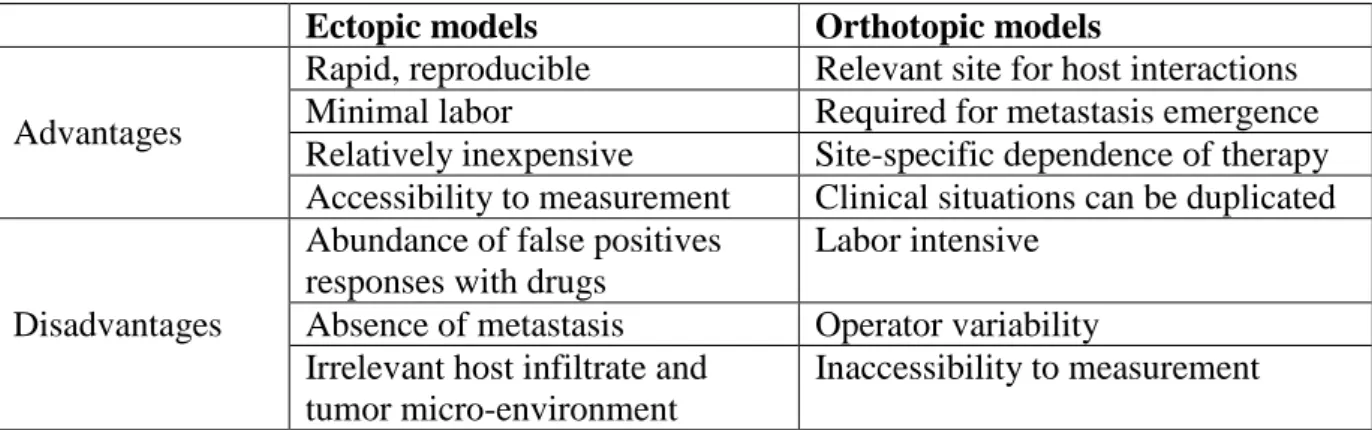
Ectopic tumor models are advantageous because of the ease of tumor establishment, measurement and reproducibility. However, ectopic site does not reproduce the primary tumor microenvironment as well as an orthotopic site does (Killion et al., 1998). Other disadvantages are the abundance of false-positive responses with drugs and the absence of metastasis, which can explain the differences in effectiveness of certain drugs in preclinical models and clinical trials (Ruggeri et al., 2014).
Ectopic models Orthotopic models
Advantages
Rapid, reproducible Relevant site for host interactions
Minimal labor Required for metastasis emergence
Relatively inexpensive Site-specific dependence of therapy
Accessibility to measurement Clinical situations can be duplicated
Disadvantages
Abundance of false positives responses with drugs
Labor intensive
Absence of metastasis Operator variability
Irrelevant host infiltrate and tumor micro-environment
Inaccessibility to measurement
Table 2: Features of the ectopic and orthotopic models (Killion et al., 1998)
I.3.2 Orthotopic pre-clinical models for HNSCC
Orthotopic models are established when tumorgrafts are performed on a site that matches that of origin. They were described since 1970s as models of various tumor diseases (Bibby, 1999; Tan et al., 1977). Although ectopic tumor models are often used in preclinical studies, these models lack the specific interactions that exist between the tumor cells and their native environment; the establishment of tumors at orthotopic sites may restore these distinct patterns of interactions and more closely simulates the natural tumor microenvironment with intact pathological, immunological responses and much higher metastatic rates, which recapitulates the human clinical course of tumor disease (Ruggeri et al., 2014).
In HNSCCs, orthotopic models have been reported since late 1980s by injecting human cancer cells into the tongue or into the mouth’s floor of nude mice (Dinesman et al., 1990) and more recently by injecting into immunocompetent mice, syngeneic murine cell lines like SCC-VII cells (Cui et al., 2005), HPV-16 E7 expressing TC-1 cells (Sandoval et al., 2013; Wu et al., 2011), and HPV-16 E7 expressing AT-84 oral SCC cells (Paolini et al., 2013). Mouth’s floor or tongue implantation allows to reproduce some clinical signs like dysphagia and weight loss, however mice have to be euthanized in the first two weeks after tumor cell implantation because of tumor growth preventing correct feeding by the animals, resulting in unethical suffering and
death (Bozec et al., 2009; Myers et al., 2002). Orthotopic models are very useful to evaluate anti-tumor therapeutic approaches; in several studies these models can predict the clinical activity of certain molecules (Bibby, 2004; Bozec et al., 2008). In addition, there are many studies describing metastasis of orthotopic tumors to clinically relevant sites (Cabanillas et al., 2005; Kubota, 1994). Although the improvements over ectopic tumors, orthotopic models have some limitations, these models are technically challenging to establish and to reproduce, and it can be difficult to evaluate tumor growth depending on the site (Sano and Myers, 2009).
Few studies have been published in orthotopic sites other than the mouth or tongue. One study reported an orthotopic model developed by implanting tumor fragments in the inner aspect of the mouse cheek. Importantly, this tumor model allowed a survival time of 30 days (Atallah et al., 2014). Additionally, in a model of sinonasal malignancy where tumor cells were injected in the right maxillary sinus or soft palate in mice, it was shown that this model recapitulates the malignant behavior of the tumor types seen in these patients (Gelbard et al., 2008). Finally, two other studies established an orthotopic model of salivary cancer in the parotid glands suitable for anti-tumor strategies (Choi et al., 2008; Younes et al., 2006).
I.3.4 In situ pre-clinical models for HNSCC
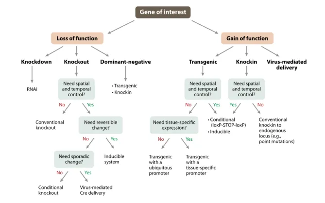
In situ tumor models including transgenic and genetically engineered mouse model
(GEMM), and carcinogen induced tumor models, have been developed with increased complexity but also clinical relevance in certain cases.
Transgenic and GEMM models offer the possibility to study relationships between molecular changes and cancer development, as well as prevention and early interventions therapies. The most common way to generate this type of mouse models of cancers are to activate oncogenes or inactivate tumor-suppressor genes (or both) in vivo through the use of different techniques of genetic manipulations such as transgenic approaches created by microinjection of foreign DNA into the pronuclei of fertilized zygotes, gene-targeting approaches involving multiple steps that result in either deletions of the coding sequence of a gene (knockouts) or the introduction of exogenous sequences into the specific locus (knockins), and conditional and inducible systems that allow the induction of somatic mutations in a tissue-specific and time-controlled manner. Loss-of-function studies typically employ knockout or conditional knockout alleles, whereas gain-of-function studies use transgenic, conditional transgenic, and knockin approaches (Cheon and Orsulic, 2011).