Publisher’s version / Version de l'éditeur:
ACS Nano, 2, 5, pp. 1051-1057, 2008-04-01
READ THESE TERMS AND CONDITIONS CAREFULLY BEFORE USING THIS WEBSITE. https://nrc-publications.canada.ca/eng/copyright
Vous avez des questions? Nous pouvons vous aider. Pour communiquer directement avec un auteur, consultez la première page de la revue dans laquelle son article a été publié afin de trouver ses coordonnées. Si vous n’arrivez pas à les repérer, communiquez avec nous à PublicationsArchive-ArchivesPublications@nrc-cnrc.gc.ca.
Questions? Contact the NRC Publications Archive team at
PublicationsArchive-ArchivesPublications@nrc-cnrc.gc.ca. If you wish to email the authors directly, please see the first page of the publication for their contact information.
NRC Publications Archive
Archives des publications du CNRC
This publication could be one of several versions: author’s original, accepted manuscript or the publisher’s version. / La version de cette publication peut être l’une des suivantes : la version prépublication de l’auteur, la version acceptée du manuscrit ou la version de l’éditeur.
For the publisher’s version, please access the DOI link below./ Pour consulter la version de l’éditeur, utilisez le lien DOI ci-dessous.
https://doi.org/10.1021/nn8000774
Access and use of this website and the material on it are subject to the Terms and Conditions set forth at
Picomolar detection of protease using peptide/single walled carbon
nanotube/gold nanoparticle-modified electrode
Mahmoud, Khaled A.; Hrapovic, Sabahudin; Luong, John H. T.
https://publications-cnrc.canada.ca/fra/droits
L’accès à ce site Web et l’utilisation de son contenu sont assujettis aux conditions présentées dans le site LISEZ CES CONDITIONS ATTENTIVEMENT AVANT D’UTILISER CE SITE WEB.
NRC Publications Record / Notice d'Archives des publications de CNRC:
https://nrc-publications.canada.ca/eng/view/object/?id=dd837ce9-b257-4968-8029-a81522855e51
https://publications-cnrc.canada.ca/fra/voir/objet/?id=dd837ce9-b257-4968-8029-a81522855e51
Picomolar Detection of Protease Using
Peptide/Single Walled Carbon
Nanotube/Gold Nanoparticle-Modified
Electrode
Khaled A. Mahmoud,
†Sabahudin Hrapovic,
†and John H. T. Luong
†,‡,*
†
Biotechnology Research Institute, National Research Council Canada, Montreal, Canada H4P2R2, and
‡Department of Chemistry, University College Cork, Cork, Ireland
P
rotease, a critical component in
mammalian cells and tissues, occurs
naturally in all organisms and
consti-tute 1%⫺5% of the gene content for
vari-ous metabolic processes. This important
en-zyme is involved in a multitude of
physiological reactions from simple
diges-tion of food proteins to highly regulated
cascades. Proteases have been used in
vari-ous forms of therapy: oncology,
inflamma-tory conditions, blood rheology control, and
immune regulation. Proteases are also
im-plicated in many viral and infectious
dis-eases, for example, HIV-1 protease (HIV-1
PR), an aspartic protease that functions to
cleave the nascent polyproteins synthesized
during the viral replication cycle.
1–3Conse-quently, intensive efforts have been
di-rected toward the search for novel
pro-tease inhibitors with improved
bioavailability, antiviral potency, and
activ-ity toward drug-resistant mutant viruses.
4–6Protease inhibition renders the virus
nonin-fectious as the body rapidly clears immature
virions. However, owing to the high viral
mutation rates, a single amino acid change
within HIV-1 PR can render it invisible to an
inhibitor, the active site of this enzyme can
change rapidly when under the selective
pressure of replication-inhibiting drugs.
Thus, there is a critical need for
high-throughput and sensitive protocols for
de-tecting protease activity and/or screening
potential protease inhibitors. In addition,
the effect of binding various inhibitors on
the protease structure is currently the focus
of intensive research.
7–9Although a
plethora of different methods has been
de-veloped for detecting protease activity,
10–17fluorescent methods are predominant.
18–23However, fluorogenic methods require
laboratory-based equipment, whereas
other techniques require overnight
incuba-tion times. Recently, peptide-modified
opti-cal filters for detecting protease activity
with a detection limit of 37 nM have been
reported.
24Integration of metallic nanoparticles
(NPs) with carbon nanotubes (CNTs) has
at-tracted much interest because of the low
re-sistance ohmic contacts of these
composites.
25,26Of particular interest is the
attachment of gold NPs to the side walls of
CNTs to fabricate highly efficient sensor
devices.
27,28CNT and gold NP-modified
electrodes have many advantages such as
high surface area, favorable electronic
prop-erties, ease of biomolecule attachment,
and electrocatalytic effects.
29–32Ferrocence
(Fc)-peptide conjugates bound to gold
elec-trodes,
33allows for identifying and
screen-ing of HIV-1 PR in physiological conditions
with a detection limit (LOD) of 1 nM.
34This paper describes an ultrasensitive
electrochemical procedure that is more
amenable to applications in the field and
ar-ray format. A new strategy is engineered
*Address correspondence to John.Luong@cnrc-nrc.gc.ca. Received for review February 7, 2008 and accepted April 01, 2008.
Published online April 18, 2008.
10.1021/nn8000774 CCC: $40.75
© 2008 American Chemical Society
ABSTRACT
Picomolar electrochemical detection of human immunodeficiency virus type-1 protease (HIV-1
PR) using ferrocene (Fc)-pepstatin-modified surfaces has been presented. Gold electrode surface was modified
with gold nanoparticles (AuNP) or thiolated single walled carbon nanotubes/gold nanoparticles (SWCNT/AuNP).
Thiol-terminated Fc-pepstatin was then self-assembled on such surfaces as confirmed by Raman spectroscopy and
scanning electron microscope. The interaction between the Fc-pepstatin-modified substrates and HIV-1 PR was
studied by cyclic voltammetry and electrochemical impedance spectroscopy. Both electrode materials showed
enhanced electrochemical responses to increasing concentrations of HIV-1 PR with shifting to higher potentials
as well as decrease in the overall signal intensity. However, the sensing electrode modified with thiolated SWCNTs/
AuNPs showed remarkable detection sensitivity with an estimated detection limit of 0.8 pM.
KEYWORDS:protease · ferrocene-pepstatin inhibitor · carbon nanotubes · gold nanoparticles · biosensor
ARTICLE
Downloaded by CANADA INSTITUTE FOR STI on September 29, 2009 | http://pubs.acs.org
for the detection of HIV-1 protease (HIV-1 PR) by
utiliz-ing a gold NP-CNT-modified electrode to attain
unprec-edented detection sensitivity below the picomolar
(pM) level (Figure 1). We advocate the use of
pepsta-tin, a well-known potent inhibitor of aspartyl proteases.
This hexa-peptide (Iva-Val-Val-Sta-Ala-Sta) has an
un-usual amino acid, statine or
(3S,4S)-4-amino-3-hydroxy-6-methylheptanoic acid. It was first isolated from
Acti-nomyces
35because of its ability to inhibit pepsin at low
concentrations.
36Indeed, it inhibits nearly all acid
pro-teases with high potency and has become a valuable
re-search tool as well as a common constituent of
pro-tease inhibitor cocktails.
RESULTS AND DISCUSSION
Initially, we attempted to introduce gold NPs to the
gold electrode surface (Figure 1) aiming for high
detec-tion sensitivity, the approach described by Li et al.
37for DNA-binding cancer drugs. Gold NPs were
depos-ited from a 0.5 M H
2SO
4solution containing 5 mM
HAuCl
4using chronoamperometry at E
r⫽
0.2 V versus
Ag/AgCl for 10 s. Under this condition, AuCl
4⫺was
re-duced to AuNPs. The cystamine containing Fc-pepstatin
conjugate was self-assembled on the gold surface via
Au⫺S bonding
38,39and followed by the binding of
in-creasing concentrations of HIV-1 PR as described in the
Experimental Section. This setup allows Fc to be in
close proximity to the electrode surface and thus
unaf-fected by diffusive processes. Moreover, the
communi-cation between the recognition sequence and
elec-trode surface would not be blocked by the binding
event.
33The electrochemical response of the films was
evaluated by cyclic voltammetry (CV) measurements in
an HIV-1 PR activation buffer at pH 4.7 containing 2 M
NaClO
4as the supporting electrolyte.
Figure 2 illustrates the formal potential (E°) change
of the Fc probe as a function of the enzyme
concentra-tion. The films displayed a reversible one-electron
re-dox peak with E° ⫽ 0.486 ⫾ 0.005 at 0.1 V/s versus Ag/
AgCl. A linear relationship between the peak current i
pcand the scan rate indicated an adsorption controlled
process (Figure S1, Supporting Information). Upon the
selective binding of HIV-1 PR to the surface bound
in-hibitory peptide (Fc-pepstatin), a shift to higher
poten-tial together with a decrease in the peak current
inten-sity was observed; indicating that the oxidation of the
Fc group became increasingly difficult. Evidently, the
bound enzyme was capable of blocking the
penetra-tion of the supporting electrolyte to the electrode
sur-face, thereby decreasing its ability to oxidize Fc. At 0.5
nM enzyme concentration, only a slight decrease of the
current intensity and a negligible shift in the formal
po-tential was observed. This could be attributed to the
limitation of the biosensor to register any binding
activ-ity at such a low concentration. ⌬E° versus enzyme
con-centration was linear over the 0.5⫺10 nM range.
Be-yond the upper enzyme level, the response became
nonlinear, indicating the saturation of the enzyme
bind-ing to the maximum protease
inhibi-tor that could be loaded on the
elec-trode surface. The LOD was 0.08 nM,
estimated from 3(S
b/m), where S
bis the
standard deviation of the
measure-ment signal for the blank and m is the
slope of the analytical curve in the
lin-ear region.
40This detection limit was
over 10-fold lower than the previously
reported value (1 nM).
34A series of experiments was
con-ducted to improve the LOD by
intro-ducing aligned carbon nanotubes
(ACNTs) as electrode material. Gold
nanoparticles were deposited
electro-chemically on the outer walls of ACNTs
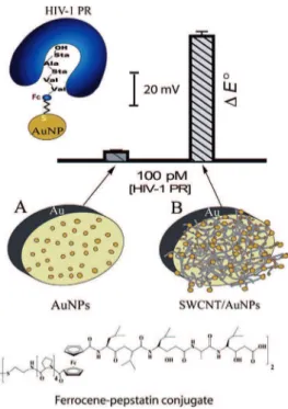
Figure 1. Schematic representation of the two detection protocols for HIV-1 PR using Fc-pepstatin modified surfaces: (A) AuNP and (B) SWCNT/AuNP-modified gold electrodes at 100 pM of the enzyme.
Figure 2. (A) Cyclic voltammogram of AuNP-modified gold electrode modified with Fc-pepstatin conjugate in the presence of increasing concentrations of HIV-1 PR: 0 (black, control), 0.5 (green), 4 (blue), and 8 nM (red); (B) Linear relationship between HIV-1 PR concentration and the formal potential E°. The assay buffer consisted of 0.1 M sodium
ac-etate, 2 M NaClO4, 1 mM EDTA, 1 mM DTT, 10% DMSO, pH 4.7. The E° of the Fc/Fc
ⴙcouple
under the experimental conditions is 448 (ⴞ5) mV.
ARTICLE
Downloaded by CANADA INSTITUTE FOR STI on September 29, 2009 | http://pubs.acs.org
to facilitate the self-assembly of the peptide probe.
The Fc-pepstatin film was self-assembled as described
above and allowed to interact with HIV-1 PR. The
bind-ing of HIV-1 PR to the peptide modified electrode was
confirmed by SEM micrographs. Upon binding with the
HIV-1 PR, the morphology of ACNTs changed from
aligned strands (Figure 3A) to ordered aggregates of
several tubes (Figure 3D). This was not observed by
washing the tubes with the assay buffer in the
ab-sence of HIV-1PR. The electrochemical response was
monitored by CV (data not shown). However, the LOD
of this system (⬃0.1 nM) was only comparable to the
gold nanoparticle-modified electrode as discussed
ear-lier. Notice also that when 20 m length-ACNTs arrays
were used, AuNPs were deposited mainly on the tips of
the nanotubes (inset, Figure 3D). In this case,
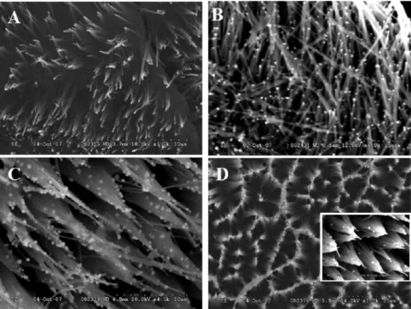
Fc-Figure 3. SEM images of (A) pristine aligned CNTs (5000ⴛ), (B) AuNP modified aligned CNTs (6000ⴛ), (C) peptide/AuNP/ aligned CNTs (4500ⴛ), and (D) protein/peptide/AuNP/ aligned CNTs (1200ⴛ). The inset in D shows AuNPs observable only at higher magnification (7000ⴛ). Only minimum bending of aligned CNTs was observed upon their rinsing under mild wa-ter stream (A) and drying under an argon stream. The Au nanoparticles deposition process (B) introduced additional stress but the nanotube array still consisted of individual tubes although with more bended morphology. Peptide binding (C) and protein adsorption step (D) exerted an additional pressure on the array and tubes were grouped together forming interest-ing patterns (D).
Figure 4. SEM images of (A) SWCNTs layer (2500ⴛ) dried upon 20 min of sonication in DMF and (B) SWCNTs layer (5000ⴛ) dried upon acid/peroxide treatment. SWCNTs after sonication in DMF treatment preserved their integrity. Bundled together
they were about 5 m long and distinct (A). In the second case, prolonged sonication in H2SO4/HNO3followed by H2O2/
H2SO4treatment, neutralization, and filtering, led to significantly shorter SWCNTs with almost a granular structure.
Electrode-posited AuNPs were not easily observable in the network of short SWCNTs.
ARTICLE
Downloaded by CANADA INSTITUTE FOR STI on September 29, 2009 | http://pubs.acs.org
pepstatin self-assembled on AuNPs, was distant from
the electrode surface, resulting in poor
electrochemi-cal communication. The inset in Figure 3D shows AuNPs
observable only at higher magnification (7000⫻). Only
minimum bending of aligned CNTs was observed upon
their rinsing under mild water stream (Figure 3B) and
drying under an argon stream. The Au nanoparticle
deposition process introduced additional stress but the
nanotube array still consisted of individual tubes
al-though with more bended morphology (Figure 3C).
Peptide binding and protein adsorption exerted an
ad-ditional pressure on the array and in turn, tubes were
grouped together forming such an interesting pattern
as shown in Figure 3D.
Significant improvement of the detection sensitivity
was achieved by modification of the sensing probe with
thiolated SWCNTs and gold nanoparticles. SWCNTs can
be easily functionalized and decorated with a variety of
inorganic nanomaterials including gold
nanoparti-cles.
26Thus, the resulting modified electrode was
antici-pated to improve the response to the binding event
due to its higher exposed surface area. As compared
with the only CNT modified surface (Figure 4A), the
syn-ergistic effect between AuNPs and the
SWCNT integrated network (Figure 4B)
facilitated electron transfer with the
substrate. Functionalization of purified
SWCNTs with thiol groups was realized,
following the literature procedures
41with slight modification as described
in Experimental Section. Predispersed
SWCNTs (10 L) were coated on the
gold electrode surface prior to the
deposition of AuNPs. The high density
of ⫺SH groups allowed for
electro-chemical reduction of AuCl
4⫺to AuNPs
at the side walls of SWCNTs. The
forma-tion of the SWCNT/AuNP composite
was confirmed by SEM and Raman spectroscopy
(Fig-ure S2, Supporting Information). Fig(Fig-ure 5 shows the
change of the formal potential E° as a function of HIV-1
PR concentration. Under the optimized conditions, a
lin-ear range was obtained for all tested enzyme
concen-trations (C), starting from 0.5 pM. The linear regression
equation was E (mV) ⫽ 0.458 ⫹ 0.00247C (C in pM) with
a correlation coefficient of 0.998. The LOD for the
tar-get HIV-1 PR under the optimal conditions was 0.8 pM.
This was ⬃1000-fold more sensitive than the level using
the screen-printed electrode.
34Detection
reproducibil-ity was determined from five different electrodes
pre-pared under the same experimental conditions, with a
relative standard deviation of 3.1% (n ⫽ 5). Hence, the
signal amplification of the SWCNT-AuNP matrix resulted
in a remarkable low detection limit where only SWCNT/
AuNP could register the binding event at picomolar
level (Figure 6A). In contrast, interactions with the
Fc-pepstatin modified AuNP electrode composite were
pronounced only with 1 nM of HIV 1-PR or higher.
Therefore, this method might significantly improve the
development of the inhibitory drug amount needed to
suppress 50% of the PR activity in vivo (IC
50), which is
still in the range of 10⫺30 nM.
42,43Cur-rent efforts to design and screen for
therapeutic protease inhibitors are
be-ing accelerated by the availability of
structural information and several
as-say formats to screen, especially in the
area of anticoagulation,
neurodegener-ative conditions, osteoporosis, type II
diabetes, and rheumatoid arthritis.
Therefore, our proposed detection
scheme is expected to open up a new
strategy for screening potent drugs for
AIDS therapy or the detection of
HIV-activity in infected T4 cells.
Conceptu-ally, protease when forming a complex
with a potent inhibitor will no longer
bind to the pepstatin-modified
elec-trode and provoke no signal
response.
Figure 5. (A) Cyclic voltammograms of SWCNT/AuNP-modified gold electrodes: 0 pM (black, control), 20 pM (red), 40 pM (blue), 60 pM (brown), and 80 pM (cyan). Ag/AgCl was used as ref-erence at 100 mV/s. (B) The linear relationship between HIV-1 PR concentration and the for-mal potential E°. Other conditions were as described in Figure 2.
Figure 6. (A) The column chart showing the formal potential shift as a function of HIV-1 PR concentration for the two modified gold electrodes with SWCNT/ AuNP (light gray) and AuNP
(dark gray). Error bars indicate the standard deviation (n ⴝ 5). (B) The Nyquist plot (Zimvs
Z
re) for electrochemical impedance measurement in the buffer medium, pH 7.4 containing
5.0 mM Fe(CN)63ⴚ/4ⴚ(1:1 mixture) as the redox probe at 0.24 V for (a) bare Au electrode, (b)
the Fc-pepstatin-modified AuNP electrode, (c) the Au NP modified electrode after incubation with 1 nM HIV-1 PR, and (d) the peptide SWCNT/AuNP electrode after incubation with 10 pM HIV-1 PR. Frequency range: 0.1ⴚ100 kHz; ac amplitude, 5 mV. Other conditions are same as de-scribed in Figure 2.
ARTICLE
Downloaded by CANADA INSTITUTE FOR STI on September 29, 2009 | http://pubs.acs.org
A preliminary impedance analysis of the interfacial
process (Figure 6B) demonstrated the simultaneous
binding of HIV-1 PR with Fc-pepstatin that had bound
to the SWCNT/AuNP or AuNP electrodes, as an
indica-tion of the surface binding events. The incubaindica-tion of
HIV-1 PR with the SWCNT/AuNP modified gold
elec-trode drastically increased the electrochemical
imped-ance, indicating an increased amount of HIV-1 PR was
allowed to interact with the surface. This provides
im-mediate evidence for the surface confinement of the
SWCNTs, resulting in higher accessibility to the target
enzyme. Notice that the experimental data of
imped-ance can be further analyzed by using a model
equiva-lent circuit, comprising a solution resistor, a
charge-transfer resistance, a surface capacitance, and the
impedance due to mass transfer of the redox species
to the electrode described by the conventional
War-burg.
37This is a topic of future endeavors and beyond
the scope of this work, that is, the development of
simple cyclic voltammetry for detection of HIV-1 PR.
To assay the specificity of the proposed method,
the electrochemical recognition of HIV-1 PR was
tested in the presence of a large excess (5.1 mg/ml)
of human serum albumin (HSA), the most abundant
protein in human blood plasma. HSA normally
con-stitutes about 60% of human plasma protein, and its
concentration in blood plasma ranges from 3.5 to
5.0 mg/ml.
44HSA only provoked a small decrease in
i
paand there was no change in E°. In contrast, a
sig-nificant shift of E° was observed only when 10 nM
HIV-1 PR was spiked to the measurement solution
(Fig-ure 7). Similarly, human serum did not provoke any
ap-preciable change in i
pawhereas 10 mM spiked human
serum effected a comparable shift of E° as observed
for HSA (data not shown).
We have demonstrated for the first time the
combi-nation of ferrocene-pepstatin with thiolated SWCNT/
AuNP toward the construction of an extremely
sensi-tive electrochemical detection method for HIV-1 PR at
low picomolar levels. This proof of concept paves the
way toward the development of an important tool for
screening about 475 known putative proteases in the
human genome or many disease states which manifest
as altered protease expression and substrate
proteo-lyzis are targets for therapeutic intervention. The
gener-ality of this approach can be easily extended to other
protease/inhibitor pairs to expand the scope of using
nanomaterials in biosensing.
EXPERIMENTAL SECITON
Materials.Human serum albumin (HSA), human male serum (AB), and HIV-1 PR recombinant expressed in Escherichia coli (25 g) were purchased from Sigma. DTT (⫾threo-2,3-dihydroxy-1,4-butanedithiol) was obtained from Fluka. Fc-conjugated pepsta-tin (Cys-(NH-Pro4-C(O)-Fc-C(O)-Val2-Sta-Ala-Sta-OH)2was
pre-pared as described in the literature.34Single walled carbon
nanotubes (SWCNTs) (diameter of 1 nm, purity of CNTs ⬎ 90 wt %; length, 5⫺30 m; specific surface area, 407 m2/g; electrical
conductivity, ⬎ 10⫺2S/cm) were purchased from Carbon
Nano-technology (Houston, TX). Aligned carbon nanotube (ACNT) ar-rays (grown on Cr⫺Pt substrate, diameter 100⫺200 nm, length 20 m, and density 109/cm2) were obtained from
Nano-Laboratory (Brighton, MA).
Preparation of ACNT/AuNP Electrodes.The plate containing the ACNT array was cut into 5 mm ⫻ 5 mm squares that were at-tached on a double-sided adhesive carbon tape. The other side of the C-tape was glued on plastic stripes (80 mm ⫻ 8 mm) to en-sure the electrode rigidity. The 3 M (St. Paul, MN) insulating ep-oxy resin was carefully applied on the carbon so that only the ACNT array surface was exposed to the electrolyte for the elec-trochemical measurement. The epoxy layer solidified completely after 12 h in the oven at 40 °C. Electrodeposition of gold nano-particles (NPs) on CNT arrays was performed by the potential-step technique from acidic solution of 0.5 M H2SO4containing
5 mM HAuCl4. The applied potential was stepped from 0.80 V (vs
Ag/AgCl) to 0.20 V during 10 s. Such prepared electrodes were ready for electrochemical measurement and SEM analysis.
Pretreatment of SWCNTs.SWCNTs (30 mg) were first treated with a H2SO4/HNO3(3:1) mixture (10 mL) and sonicated for 10 h at
35 ⫾ 5 °C. The resulting suspension was diluted with distilled wa-ter and filwa-tered using a polycarbonate membrane with a pore size of 200 nm. The pH was adjusted to above 6 by subsequent rinsing with water. SWCNTs were suspended in a H2O2(30 wt %,
aq)/H2SO4(4:1) mixture and refluxed for 3 h at 70 °C followed
by sonication for 7 h to introduce carboxylic acid functional groups to the tubes. The same neutralization and filtration steps were followed. The oxidized and shortened SWCNTs were oven-dried and the powder was dissolved into absolute ethanol and treated with excess NaBH4, and the resulting suspension was
re-fluxed at 80 °C to reduce the carboxylic groups to hydroxyl groups. The reaction was then quenched by adding concentrated HCl dropwise until the excess of NaBH4disappeared. The suspension
was then refluxed in thionyl chloride at 70 °C for 12 h followed by reflux in 3 M aq NaOH at 100 °C for 5 h. After dilution with distilled water, 1 M HCl was added dropwise until the neutral thiolated form was obtained, and the suspension was then filtered using a 200 nm pore-sized Teflon membrane filter. The remaining powder was washed three times with ethanol and oven-dried.
Electrode Preparation (AuNP-Modified Gold Electrode). Electrochemi-cal deposition of gold nanoparticles on the gold disk electrode (3 mm in diameter, BAS) was performed in 0.5 M H2SO4solution
con-taining 5 mM HAuCl4using chronoamperometry at a constant
po-tential. The electrode potential was stepped from an initial
poten-Figure 7. Cyclic voltammograms of the
SWCNT/AuNP/Fc-pepstatin modified gold electrode in the presence of (______)
assay buffer medium, (ⴚ ⴚ ⴚ) 75 m HSA in the buffer
so-lution, (... ... ...) 10 nM HIV-1 PR spiked in HSA, at 100 mV sⴚ1.
Other conditions were as described in Figure 2.
ARTICLE
Downloaded by CANADA INSTITUTE FOR STI on September 29, 2009 | http://pubs.acs.org
tial (Ei) of ⫹0.8 V, where no reaction occurred, to ⫹0.20 V, at which
potential AuCl4⫺was reduced to Au nanocrystals. The NP-modified
electrode was then removed and rinsed with water and placed in the solution of 0.5 M H2SO4for further measurements.
SWCNT/AuNP-Modified Gold Electrode.A 1 mg portion of the pre-treated SWCNTs was redispersed into 2 mL of absolute ethanol and sonicated for 2 h to generate a black suspension. The prepol-ished gold electrode was coated with 10 L of SWCNTs and al-lowed to dry under an argon stream. After rinsing, gold nanoparti-cles were then deposited electrochemically onto the SWCNT-modified electrode by following the same procedure as mentioned above. This would allow the thiolated SWCNTs to conjugate to gold-NPs through the stable Au⫺S bond. The modified electrodes were then rinsed thoroughly to remove unattached gold NPs and scanned in 0.5 M H2SO4in the range of 0.1⫺1.2 V. The resulting electrodes were stable and could be stored for several days at room temperature without loosing their initial activity.
The peptide films were immobilized on SWCNT/NP elec-trodes using the published procedure.33In brief, the electrodes
were immersed in ⬃1 mM thiol-terminated Fc-pepstatin conju-gate prepared in 5% (by volume) acetic acid in ethanol for 12 h. A 20 nM HIV-1 PR stock solution was prepared in 0.1 M sodium acetate, pH 4.7, containing 2 mM EDTA, 1 mM DTT, and 10% DMSO was incubated at 23 °C for 1 h prior to the measurement. The activated enzyme was kept on ice. Subsequent dilutions of the enzyme were prepared by using the assay buffer. The peptide-modified electrodes were incubated with different HIV-1 PR concentrations for 1 h and washed twice with the assay buffer and deionized water. Control experiments were carried out with 75 m HSA with and without HIV-1 PR.
Instrumentation.Cyclic voltammetry (CV), electrochemical im-pedance (EIS), and amperometric measurements were per-formed using an electrochemical analyzer coupled with a pico-amp booster and Faraday cage (CHI 760B, CH Instruments, Austin, TX). A Pt wire (Aldrich, 99.9% purity, 1 mm diameter) and an Ag/AgCl, 3 M NaCl (BAS, West Lafayette, IN) electrode were used as counter and reference electrodes, respectively. Gold disk electrodes were polished with polishing paper (grid 2000) and subsequently with alumina until a mirror finish was obtained. The electrodes were sonicated for 5 min to remove the alumina residues followed by thorough rinsing with water and ethanol. The electrodes were then cleaned by cyclic voltamme-try between 0 and ⫹1.4 V versus Ag/AgCl at 100 mV s⫺1in 0.5 M
H2SO4until a stable CV profile was obtained.
Scanning electron microscope (SEM) (Hitachi, S-2600 N, To-kyo, Japan) operating in high vacuum mode with an accelera-tion voltage of 10⫺24 kV and a working distance of 3⫺15 mm, depending on the sample, was used to analyze the morphology of CNT arrays and SWCNTs before and after different modifica-tion steps. Raman spectra were obtained by a Horiba/Jobin Yvon Laser Raman Analyzer (LabRAM HR800, Longjumeau, France) equipped with a frequency doubled argon ion 514 nm laser (Lexel 95-SHG) operating at 100 mW.
Acknowledgment. The authors are grateful to Prof. H.-B. Kraatz,
University Western Ontario, London, ON, Canada, for his generous donation of the Fc-peptide conjugate, Y. Liu for obtaining Raman data, and K. B. Male of the Biotechnology Research Institute, Na-tional Research Council, Canada, for valuable discussion.
Supporting Information Available: SEM micrographs, Raman
spectroscopy, and electrochemistry data. This material is avail-able free of charge via the Internet at http://pubs.cas.org.
REFERENCES AND NOTES
1. Brik, A.; Wong, C. HIV-1 Protease: Mechanism and Drug Discovery. Org. Biomol. Chem. 2003, 1⫺14, 5–14. 2. Davies, D. R. T. The Structure and Function of the Aspartic
Proteinases. Annu. Rev. Biophys. Biophys. Chem. 1990, 19, 189–215.
3. Jaskolski, M.; Tomasselli, A. G.; Sawyer, T. K.; Staples, D. G.; Heinrikson, R. L.; Schneider, J.; Kent, S. B. H.; Wlodawer, A. A. Structure at 2.5 Å Resolution of Chemically Synthesized Human Immunodeficiency Virus Type 1
Protease Complexed with a Hydroxyethylene-based Inhibitor. Biochem. 1991, 30, 1600–1609.
4. Nalam, M. N. L.; Ali, A.; Reddy, K.; Altman, M.; Chellappan, S.; Bandaranayake, R.; Anjum, S. G.; Rana, T. M.; Gilson, M. K.; Tidor, B.; et al. Circumventing Drug Resistance: Using the Substrate Envelope Hypothesis to Develop Robust Novel HIV-1 Protease Inhibitors. Antiviral Ther. 2007, 12, S138⫺S138.
5. Nalam, M. N. L.; Peeters, A.; Jonckers, T. H. M.; Dierynck, I.; Schiffer, C. A. Crystal Structure of Lysine Sulfonamide Inhibitor Reveals the Displacement of the Conserved Flap Water Molecule in Human Immunodeficiency Virus Type 1 Protease. J. Virology 2007, 81, 9512–9518.
6. Wainberg, M. A.; Clotet, B. Immunologic Response to Protease Inhibitor-Based Highly Active Antiretroviral Therapy: A Review. Aids Patient Care Stand. 2007, 21, 609– 620.
7. Jayatilleke, P. R.; Nair, A. C.; Zauhar, R.; Welsh, W. J. Computational Studies on HIV-1 Protease Inhibitors: Influence of Calculated Inhibitor-Enzyme Binding Affinities on the Statistical Quality of 3D-QSAR CoMFA Models.
J. Med. Chem. 2000, 43, 4446–4451.
8. Sham, Y. Y.; Chu, Z. T.; Tao, H.; Warshel, A. Examining Methods for Calculations of Binding Free Energies: LRA, LIE, PDLD-LRA, and PDLD/S-LRA Calculations of Ligands Binding to an HIV Protease. Proteins 2000, 39, 393–407. 9. Zhu, J.; Fan, H.; H, H. L.; Shi, Y. Structure-based Ligand
Design for Flexible Proteins: Application of New F-Dyco Block. J. Comput. Aided Mol. Des. 2001, 15, 979–996. 10. Blair, I. S.; McDowell, D. A. Detection of Extracellular Proteinase of Pseudomonas fragi by Enzyme-Linked Immunosorbent Assay. Curr. Microbiol. 1995, 31, 180–185. 11. Frederiks, W. M.; Mook, O. R. F. Metabolic Mapping of
Proteinase Activity with Emphasis on in situ Zymography of Gelatinases: Review and Protocols. J. Histochem.
Cytochem. 2004, 52, 711–722.
12. Grant, S. A.; Weilbaecher, C.; Lichlyter, D. Development of a Protease Biosensor Utilizing Silica Nanobeads. Sens.
Actuators B 2007, B121, 482–489.
13. Hook, V. Y. H.; Schiller, M. R.; Nguyen, C.; Yasothornsrikul, S. Production of Radiolabeled Neuropeptide Precursors by
in vitro Transcription and Translation. Pept. Res. 1996, 9,
183–187.
14. Ionescu, R. E.; Cosnier, S.; Marks, R. S. Protease
Amperometric Sensor. Anal. Chem. 2006, 78, 6327–6331. 15. Orosco, M. M.; Pacholski, C.; Miskelly, G. M.; Sailor, M. J.
Protein-Coated Porous Silicon Photonic Crystals for Amplified Optical Detection of Protease Activity. Adv.
Mater. 2006, 18, 1393–1396.
16. Wegner, G. J.; Wark, A. W.; Lee, H. J.; Codner, E.; Saeki, T.; Fang, S.; Corn, R. M. Real-Time Surface Plasmon Resonance Imaging Measurements for the Multiplexed Determination of Protein Adsorption/Desorption Kinetics and Surface Enzymatic Reactions on Peptide Microarrays. Anal. Chem.
2004, 76, 5677–5684.
17. Williams, B. A.; Toone, E. J. Calorimetric Evaluation of Enzyme Kinetic Parameters. J. Org. Chem. 1993, 58, 3507– 3510.
18. Eggeling, C.; Jaeger, S.; Winkler, D.; Kask, P. Comparison of Different Fluorescence Fluctuation Methods for Their Use in FRET Assays: Monitoring a Protease Reaction. Curr.
Pharm. Biotechnol. 2005, 6, 351–371.
19. Eggeling, C.; Kask, P.; Winkler, D.; Jaeger, S. Rapid Analysis of Forster Resonance Energy Transfer by Two-Color Global Fluorescence Correlation Spectroscopy: Trypsin Proteinase Reaction. Biophys. J. 2005, 89, 605–618.
20. Gosalia, D. N.; Denney, W. S.; Salisbury, C. M.; Ellman, J. A.; Diamond, S. L. Functional Phenotyping of Human Plasma using a 361-Fluorogenic Substrate Biosensing Microarray.
Biotechnol. Bioeng. 2006, 94, 1099–1110.
21. Kaufmann, S. H.; Mesner, P. W., Jr.; Martins, L. M.; Kottke, T. J. Methods used to Study Protease Activation during Apoptosis. In Apoptosis in Neurobiology, Hannun, Y. A., Boustany, R.-M., Eds.; CRC Press: Boca Raton, FL, 1999; pp 205⫺232.
ARTICLE
Downloaded by CANADA INSTITUTE FOR STI on September 29, 2009 | http://pubs.acs.org
22. Kohl, T.; Heinze, K. G.; Kuhlemann, R.; Koltermann, A.; Schwille, P. A. Protease Assay for Two-Photon Crosscorrelation and FRET Analysis based Solely on Fluorescent Proteins. Proc. Natl. Acad. Sci. U.S.A. 2002, 99, 12161–12166.
23. Pinto, M. R.; Schanze, K. S. Amplified Fluorescence Sensing of Protease Activity with Conjugated Polyelectrolytes.
Proc. Natl. Acad. Sci. U.S.A. 2004, 101, 7505–7510.
24. Kilian, K. A.; Bo¨cking, T.; Gaus, K.; Gal, M.; Gooding, J. J. Peptide-Modified Optical Filters for Detecting Protease Activity. ACS Nano 2007, 1, 355–361.
25. Azamian, B. R.; Coleman, K. S.; Davis, J. J.; Hanson, N.; Green, M. L. H. Directly Observed Covalent Coupling of Quantum Dots to Single-Wall Carbon Nanotubes. Chem.
Commun. 2002, 4, 366–367.
26. Hrapovic, S.; Liu, Y.; Male, K. B.; Luong, J. H. T. Electrochemical Biosensing Platforms using Platinum Nanoparticles and Carbon Nanotubes. Anal. Chem. 2004,
76, 1083–1088.
27. Ellis, A. V.; Vijayamohanan, K.; Goswami, R.; Chakrapani, N.; Ramanathan, L. S.; Ajayan, P. M.; Ramanath, G.
Hydrophobic Anchoring of Monolayer-Protected Gold Nanoclusters to Carbon Nanotubes. Nano Lett. 2003, 3, 279–282.
28. Wu, B.; Hou, S.; Yin, F.; Zhao, Z.; Wang, Y.; Wang, X.; Chen, Q. Amperometric Glucose Biosensor based on Multilayer Films via Layer-by-Layer Self-Assembly of Multi-wall Carbon Nanotubes, Gold Nanoparticles and Glucose Oxidase on the Pt Electrode. Biosens. Bioelectron. 2007, 22, 2854–2860.
29. Valentini, F.; Amine, A.; Orlanducci, S.; Terranova, M. L.; Palleschi, G. Nanotube Purification: Preparation and Characterization of Carbon Nnanotube Paste Electrodes.
Anal. Chem. 2003, 75, 5413–5421.
30. Wang, J. Carbon Nanotube Based Electrochemical Biosensors: A Review. Electroanalysis 2005, 17, 7–14. 31. Castaneda, M. T.; Merkoic, A.; Pumera, M.; Alegret, S.
Electrochemical Genosensors for Biomedical Applications based on Gold Nanoparticles. Biosens. Bioelectron. 2007,
22, 1961–1967.
32. Pumera, M.; Sanchez, S.; Ichinose, I.; Tang, J.
Electrochemical Nanobiosensors. Sens. Actuators B 2007,
123, 1195–1205.
33. Mahmoud, K. A.; Kraatz, H.-B. A Bioorganometallic Approach for the Electrochemical Detection of Proteins: a Study on the Interaction of Ferrocene-Peptide Conjugates with Papain in Solution and on Au Surfaces. Chem. Eur. J.
2007, 13, 5885–5895.
34. Kerman, K.; Mahmoud, K. A.; Kraatz, H.-B. An Electrochemical Approach for the Detection of HIV-1 Protease. Chem. Commun. 2007, 3829, 3831. 35. Umezawa, H.; Aoyagi, T.; Morishima, H.; Matsuzaki, M.;
Hamada, M. Pepstatin, a New Pepsin Inhibitor Produced by Actinomycetes. J. Antibiot. 1970, 23, 259–62. 36. Marciniszyn, J.; Hartsuck, J. A.; Tang, J. Mode of Inhibition
of Acid Proteases by Pepstatin. J. Biol. Chem. 1976, 251, 7088–7094.
37. Li, C.-Z.; Liu, Y.; Luong, J. H. T. Impedance sensing of DNA binding drugs using gold substrates modified with gold nanoparticles. Anal. Chem. 2005, 77, 478–485. 38. Biebuyck, H. A.; Bian, C. D.; Whitesides, G. M.
organization of Organic Liquids on Patterned Self-assembled Monolayers of Alkanethiolates on Gold.
Langmuir 1994, 10, 1825–1831.
39. Willey, T. M.; Vance, A. L.; Bostedt, C.; van Buuren, T.; Meulenberg, R. W.; Terminello, L. J.; Fadley, C. S. Surface Structure and Chemical Switching of Thioctic Acid Adsorbed on Au(111) as Observed using Near-Edge X-Ray Absorption Fine Structure. Langmuir 2004, 20, 4939–4944. 40. Miller, J. C.; Miller, J. N., Statistics for Analytical Chemistry.
In Ellis Horwood Series in Analytical Chemistry, Chalmers, R. A., Masson, M., Eds.; Chichester, U.K., 1993; p 119. 41. Lim, J. K.; Yun, W. S.; Yoon, M.-H.; Lee, S. K.; Kim, C. H.; Kim,
K.; Kim, S. K. Selective Thiolation of Single-walled Carbon Nanotubes. Synth. Met. 2003, 139, 521–527.
42. Cheng, Y.-S.; Lo, K.-H.; Hsu, H.-H.; Shao, Y.-M.; Yang, W.-B.; Lin, C.-H.; Wong, C.-H. Screening for HIV Protease Inhibitors by Protection against Activity-Mediated Cytotoxicity in E. coli. J. Virol Methods 2006, 137, 82–87. 43. Fuse, T.; Watanabe, K.; Kitazato, K.; Kobayashi, N.
Establishment of a New Cell Line Inducibly Expressing HIV-1 Protease for Performing Safe and Highly Sensitive Screening of HIV Protease Inhibitors. Microb. Infec. 2006, 8, 1783–1789.
44. Peters, T. All About Albumin: Biochemistry, Genetics, and
Medical Applications; Academic Press: San Diego, CA, 1996.
ARTICLE
Downloaded by CANADA INSTITUTE FOR STI on September 29, 2009 | http://pubs.acs.org