Peptides reproducibly released by in vivo digestion of beef meat
and trout flesh in pigs
Caroline Bauchart
1,2, Martine Morzel
2, Christophe Chambon
2, Philippe Patureau Mirand
1,
Christelle Reyne`s
3, Caroline Buffie`re
1and Didier Re´mond
1*
1INRA, UMR1019 Unite´ de Nutrition Humaine, F-63122 St Gene`s Champanelle, France
2INRA, UR370 Qualite´ des Produits Animaux, F-63122 St Gene`s Champanelle, France
3Laboratoire de Physique Mole´culaire et Structurale, Faculte´ de Pharmacie, F-34060 Montpellier, France
(Received 18 December 2006 – Revised 18 April 2007 – Accepted 27 April 2007)
Characterisation and identification of peptides (800 to 5000 Da) generated by intestinal digestion of fish or meat were performed using MS analyses (matrix-assisted laser desorption ionisation time of flight and nano-liquid chromatography electrospray-ionisation ion trap MS/MS). Four pigs fitted with cannulas at the duodenum and jejunum received a meal exclusively made of cooked Pectoralis profundus beef meat or cooked trout fillets. A protein-free meal, made of free amino acids, starch and fat, was used to identify peptides of endogenous origin. Peptides reproducibly detected in digesta (i.e. from at least three pigs) were evidenced predominantly in the first 3 h after the meal. In the duodenum, most of the fish- and meat-derived peptides were characteristic of a peptic digestion. In the jejunum, the majority of peptides appeared to result from digestion by chymo-trypsin and chymo-trypsin. Despite slight differences in gastric emptying kinetics and overall peptide production, possibly in relation to food structure and texture, six and four similar peptides were released after ingestion of fish or meat in the duodenum and jejunum. A total of twenty-six different peptides were identified in digesta. All were fragments of major structural (actin, myosin) or sarcoplasmic (creatine kinase, glyceraldehyde-3-phos-phate dehydrogenase and myoglobin) muscle proteins. Peptides were short (, 2000 Da) and particularly rich in proline residues. Nineteen of them contained bioactive sequences corresponding mainly to an antihypertensive activity. The present work showed that after fish or meat ingestion, among the wide variety of peptides produced by enzymic digestion, some of them can be reproducibly observed in intestinal digesta.
Peptides: Meat: Fish: Digestion: Mass spectrometry
The concept of food-derived peptides possessing biological activity, or bioactive peptides, brings a completely new dimension that should be considered in the description of diet-ary protein quality. Bioactive peptides have been defined as protein fragments with specific sequences that have, in addition to their nutritional value, a positive impact on body
functions and may ultimately influence health1. Once liberated
in the body, they can exert different beneficial effects such as antihypertensive, antithrombotic, opioid, immunomodulating,
antimicrobial and antioxidant activities1,2. Most dietary
pro-teins contain bioactive sequences, but the corresponding pep-tides need to be released by proteolysis, during food processing or gastrointestinal digestion, in order to show a bio-logical activity.
Many bioactive peptides have been identified in milk
pro-ducts3,4; in comparison, less information is available for
meat and fish products. Such products may nonetheless rep-resent a valuable source of peptides with biological
activi-ties5,6. For example, two angiotensin-converting enzyme
(ACE) inhibitory peptides, obtained by in vitro thermolysin
digestion of porcine skeletal muscle7, were shown to have
an in vivo antihypertensive activity when administered
orally to spontaneously hypertensive rats8. However, there
is evidence that peptides produced by in vitro digestion yield different peptides compared with those generated in vivo. For example, b-casomorphin fragments isolated
from intestinal contents following ingestion of casein9 are
larger fragments than those found in in vitro digesta. To our knowledge, studies on the occurrence of bioactive pep-tides in digesta after ingestion of meat or fish have not been reported. More generally, and despite the central role of digestive events in the supply of nutrients or biologically active compounds to the organism, almost no information is available on peptides resulting from in vivo digestion of meat or fish, in particular their sizes and sequences, let alone the random or reproducible manner in which they are generated.
In this context, and following description of peptides
pre-sent in ready-to-eat beef meat or trout10,11, the present study
aimed at characterising and identifying peptides (, 5 kDa) generated by intestinal digestion of such food products, using the pig as an animal model for man.
* Corresponding author: Dr Didier Re´mond, fax þ 33 4 73 62 47 55, email dremond@clermont.inra.fr
Abbreviations:ACE, angiotensin-converting enzyme; MALDI-ToF, matrix-assisted laser desorption ionisation time of flight; nano-LC-ESI-IT MS/MS, nano-liquid chromatography electrospray-ionisation ion trap MS/MS; PP1, first 3 h period after meal; PP2, second 3 h period after meal.
qThe Authors 2007
British
Journal
of
Nutrition
. IP address: 118.70.52.165 , on 24 Jun 2021 at 12:11:44, subject to the Cambridge Core terms of use, available at
https://www.cambridge.org/core/terms
.
Materials and methods Animals
The study involved four 3-month-old female pigs (40·5 (SD
5·2) kg). At 3 weeks before the experimentation, pigs were surgically fitted with T-shaped cannulas (silicone rubber; 12 mm internal diameter, 17 mm outer diameter) in the duo-denum (10 cm downstream from the pylorus) and the mid-jejunum (about 4 m downstream from the pylorus), and a catheter (polyvinyl chloride; 1·1 mm internal diameter, 1·9 mm outer diameter) in the cava vein. Surgical procedures and post-surgical care were conducted in accordance with national legislation on the care and use of laboratory animals. Pigs were housed in individual pens (1 £ 1·5 m) in a ventilated room with controlled temperature (19 – 238C). They were given 800 g/d of a concentrate feed containing 18 % protein, 2 % fat, 5 % cellulose and 6 % ash (Porcyprima; Sanders Centre Auvergne, Aigueperse, France) distributed in two equal portions at 8.00 and 16.00 hours, and had free access to water. The daily ration was adjusted to cover 150 % of
the maintenance requirements12.
Test meals
Three types of foods (beef, trout, protein-free food) were pre-pared. Beef meat was Pectoralis profundus from a 15-month-old Charolais bull, aged for 14 d, and cooked under vacuum for 90 min at 758C in a water-bath. Trout was the dorsal part of rainbow trout (Oncorhynchus mykiss) fillets, stored for 7 d on ice, and cooked under-vacuum for 5 min at 708C in a water-bath. Both types of muscle foods were frozen at 2 208C until use. The protein-free food, used for the purpose of monitoring the endogenous peptide composition of digesta in a postprandial situation, was made of 47 % free amino acids (in proportion close to the amino acid composition of muscle), 23 % wheat starch, 12 % fat and 18 % water. Each test meal provided 40 g crude protein, or the equivalent in amino acids, and was exclusively made of either beef (115 g; 44 g DM), trout (174 g; 44 g DM) or the control protein-free food mixture (85 g; 75 g DM). Meals were offered for 15 min and were always consumed entirely within that time. Experimental procedures
The three types of foods were tested on each animal. Test meals were separated by at least 2 d. Duodenum and jejunum digesta sampling was performed on two distinct test meals. The test meal was given at 09.00 hours. Digesta were continu-ously collected from 08.00 to 15.00 hours in cold plastic bot-tles replaced each hour. Water intake was restricted to 250 ml/ h. To prevent dehydration, 1 litre Ringer-lactate solution was intravenously infused at a rate of 150 ml/h throughout the sampling session.
Hourly collected digesta were immediately homogenised in a Waring Blender (VWR International SAS, Fontenay-sous-Bois, France) (twice 5 s at high speed, separated by 5 s). A first fraction of the homogenate (about 10 g) was used for DM determination (24 h at 1048C). A second fraction (about
100 g) was treated with perchloric acid at 170 mM (final
con-centration). After vigorous shaking, samples were kept on ice for 15 min, and then centrifuged at 10 000 g for 20 min at
48C. For the postprandial period, fractions of supernatant fractions (2 % of hourly collected digesta) were pooled to yield two samples per animal: a first pool representative of the 3 h following the meal (PP1), and a second one represen-tative of the period spanning from 3 to 6 h after the meal (PP2). Post-absorptive (sample collected before the meal), PP1 and PP2 supernatant fractions were filtered through 5000 Da cut-off filters (Vivaspin 15; Vivascience, Hanover, Germany) at 2000 g for 8 to 15 h at 48C. Filtrates were stored at 2 808C until analysis.
Analytical methods
Chromatographic analysis of peptides in extracts was carried out using an HPLC system (Kontron Instruments, Bletchley, Bucks,
UK). Separation was done in a NucleodurwC18 pyramid (110 A˚ ;
4·6 £ 250 mm) with 5 mm particles (Macherey-Nagel, Du¨ren, Germany) at 308C in a column oven and at a flow rate of 0·9 ml/min. The gradient was performed using two solvents (A, 0·1 % trifluoroacetic acid in water; B, 0·1 % trifluoroacetic acid in 100 % acetonitrile) and formed as follows: 0 % B up to 5 min, 0 – 60 % B in 5 – 35 min and 60 – 100 % B in 35 – 38 min. Detection of peptide bonds was done at 220 nm.
Matrix-assisted laser desorption ionisation time-of-flight (MALDI-ToF) analysis and nano-liquid chromatography elec-trospray-ionisation ion trap MS/MS (nano-LC-ESI-IT MS/ MS) analysis were performed as previously described by
Bau-chart et al.10. For MALDI-ToF analysis, masses were recorded
in the m/z range 800 to 5000. Below 800, m/z may not be reliably monitored by MALDI-ToF because of interference with matrix-generated peaks.
Statistical analyses
DM flux, at each site, during the first 3 h after feeding (PP1) was examined by a two-way randomised-block ANOVA with food as factor and animal as block using SAS version 8.10 (SAS Institute Inc., Cary, NC, USA).
A statistical procedure, previously described10, was applied
to MALDI-ToF data to determine whether peaks, at close m/z but originating from different spectra, were likely to corre-spond to the same compound. For one type of food and one sampling time (PP1, PP2), only the peaks gathered in a group consisting of data originating from at least three animals were considered. A similar procedure was subsequently applied to determine which groups of peaks corresponded to the same compound, across sampling times.
Results
Intestinal transit kinetics
DM collected at the duodenum after ingestion of protein-free food, trout or beef during the three first 3 h after feeding (PP1)
accounted for 88·5 (SD4·0), 90 (SD1·2) and 74 (SD10·2) % of
the total DM collected in the whole postprandial period (data not shown). This proportion was lower for beef than for pro-tein-free food and trout (P, 0·05). At the jejunum level, the proportion of DM collected in PP1 was not affected
by test-meal composition, and averaged 77 (SD 14) % (data
not shown).
British
Journal
of
Nutrition
. IP address: 118.70.52.165 , on 24 Jun 2021 at 12:11:44, subject to the Cambridge Core terms of use, available at
https://www.cambridge.org/core/terms
.
Chromatographic profiles of digesta
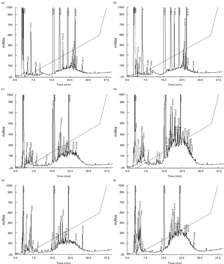
Reverse-phase HPLC analysis of digesta extracts collected after the ingestion of protein-free food (Fig. 1(a) and (b)) revealed the presence of numerous endogenous peptides. Chromatographic
profiles showed very few qualitative differences between duode-num and jejuduode-num, with common peaks at retention times of about 5·0, 6·2, 8·3, 9·9, 15·3, 18·0, 19·5, 21·3, 23·4, 23·9, 24·2, 24·5, 24·9 and 27·4 min. These endogenous peptides were still observed after ingestion of trout (Fig. 1(c) and (d)) or beef (Fig. 1(e) and (f)) but
Fig. 1. Reverse-phase HPLC chromatograms of digesta extracts from the duodenum (a, c and e) and the jejunum (b, d and f) collected in one pig over the first 3 h following ingestion of control protein-free food (a and b), trout (c and d) and beef (e and f).
British
Journal
of
Nutrition
. IP address: 118.70.52.165 , on 24 Jun 2021 at 12:11:44, subject to the Cambridge Core terms of use, available at
https://www.cambridge.org/core/terms
.
in addition, absorbance at 220 nm globally increased across the retention times 15 to 25 min. This increase was more prominent for jejunum digesta than for duodenum digesta, and generally larger after a meal of trout than after a meal of beef.
Peptides evidenced by matrix-assisted laser desorption ionisation time of flight analysis
Although peptides up to 5000 Da should be extracted by the procedure we used (perchloric acid precipitation of proteins
and ultrafiltration), MALDI-ToF analysis evidenced very few compounds between 2000 and 5000 m/z in the digesta (Fig. 2). Masses of peptides detected by MALDI-ToF in at least three of the four pigs (later called ‘reproducibly’ detected) in digesta extracts after ingestion of beef or trout are listed in Table 1. All endogenous compounds, i.e. detected at least once in the intestinal contents of the post-absorptive period or after a protein-free meal, were removed from the list in order to retain only peptides of an alimentary origin. The time of sampling had a major effect on the number of
Fig. 2. Representative matrix-assisted laser desorption ionisation time of flight spectra of duodenum (a and c) and jejunum (b and d) digesta collected from one pig during the first 3 h after ingestion of cooked trout flesh (a and b) or cooked beef Pectoralis profundus (c and d).
British
Journal
of
Nutrition
. IP address: 118.70.52.165 , on 24 Jun 2021 at 12:11:44, subject to the Cambridge Core terms of use, available at
https://www.cambridge.org/core/terms
.
reproducibly detected peptides, since in the second 3 h period after the meal (PP2) only one peptide (m/z 1018·50) was listed, in the duodenum after ingestion of beef. Compounds listed in PP1 were more numerous in the duodenum than in jejunum (eight and seven for trout; nine and five for beef). Some food-derived peptides were common to the duodenum and jejunum digesta, four for trout (m/z 1115·89, 1148·44, 1182·44 and 1434·17) and two for beef (m/z 1115·89 and 1198·75). In PP1, there was a striking similarity in the masses of peptides reproducibly generated by digestion of either trout or beef. At the duodenum level, out of eight and nine reproducibly detected peptides, seven were common to trout and beef. At the jejunum level, five were common to trout and beef out of seven and five.
Identification of reproducibly generated food-derived peptides Fish- and meat-derived peptides identified by nano-LC-ESI-IT MS/MS analysis in digesta extracts collected over PP1 are listed in Table 2. The identified peptides came from abundant muscle proteins, namely actin, myosins, creatine kinase and glyceraldehyde 3-phosphate dehydrogenase, and myoglobin specifically for beef. Again, there were some similarities between the intestinal contents after a meal of trout or beef, with six and four food-derived peptides common to the two types of muscle foods in the duodenum and jejunum, respect-ively. Overall, a particularity of the sequences was the high proportion of proline, accounting for approximately 12·0 % (fifteen of 125) and 13·1 % (fourteen of 107) of all residues in peptides identified at the duodenum level for trout and beef, respectively. At the jejunum level, the proportion was even higher and reached 15·0 % (twelve of eighty) and 19 % (eleven of fifty-eight) for trout and beef, respectively. After the protein-free meal (data not shown) several fragments of pepsin A, trypsin precursor, pancreatic a-amylase and salivary proline-rich proteins were identified in duodenum digesta. Fragments of trypsin (IKLSSPATL and LIKLSSPATL) and salivary proline-rich protein (ARPLPGPPPPGPPP) were common to duodenum and jejunum digesta. The jejunum
content was also characterised by the presence of many more fragments of salivary proline-rich proteins.
A search of potential biological activity of identified
pep-tides was carried out with the BioPep database13and by
com-parison with sequences either reviewed in Vercruysse et al.6or
described in Sentandreu & Toldra´14. Some sequences with
potential biological activity were found within nineteen of the identified peptide sequences (Table 3). They were mainly short sequences of two or three residues, except for the sequences IVGRPR, YALPHA and ALPHA, all contained in actin fragments. The most frequently found potential bio-logical activity was an antihypertensive effect.
Discussion
The present study intended to characterise and identify pep-tides reproducibly generated by in vivo gastroduodenal diges-tion of fish or meat, focusing on peptides ranging from 800 to 5000 Da. The lower limit was dictated by technical consider-ations while the upper limit was chosen in relation to the gen-erally reported size of bioactive peptides. Overall, we have identified nineteen and fifteen different food-derived peptides in digestive contents of pigs after a meal of fish or beef, respectively. Many of the identified peptides contained bio-active sequences.
DM flux measured at the duodenal level illustrates the rapidity of gastric emptying after the meal, even more for trout than for beef. Food texture has been recognised as a
factor influencing gastric emptying15. Here, in the cooking
conditions used for beef, meat was dry and compact. The looser structure and moister texture of trout is a plausible explanation for the accelerated kinetics. Compositional fac-tors, in particular the high proportion of n-3 PUFA in trout, may also contribute to a faster gastric emptying as described
by Robertson et al.16. Values in the jejunum also evidence
that transit throughout the anterior intestine proceeded rapidly. After a meal of fish or meat, only one peptide was reproduci-bly detected in PP2 digesta, suggesting that peptides earlier gen-erated by digestion were rapidly further processed by
Table 1. List of compounds (m/z 800 to 5000) detected by matrix-assisted laser desorption ionisation time of flight analysis in pig duodenum and jejunum digesta, collected during the first 3 h (PP1) and the subsequent 3 h (PP2) after ingestion of trout or beef*
Duodenum Jejunum
PP1 PP2 PP1 PP2
Trout Beef Trout Beef Trout Beef Trout Beef
976·50 976·50 1018·45 1018·45 1018·45 1035·34 1115·89 1115·89 1115·89 1115·89 1148·44 1148·44 1148·44 1158·59 1158·59 1182·44 1182·44 1182·44 1184·41 1184·41 1198·75 1198·75 1198·75 1261·70 1261·70 1434·17 1434·17 1434·17 1520·77 1520·77
* Listed compounds had an alimentary origin and were found in at least three of the four animals.
British
Journal
of
Nutrition
. IP address: 118.70.52.165 , on 24 Jun 2021 at 12:11:44, subject to the Cambridge Core terms of use, available at
https://www.cambridge.org/core/terms
.
transformation or absorption. For these reasons, peptide identi-fication was limited to samples collected in the first 3 h following the meal. Based on masses of peptides present in the two types of
muscle foods constituting the meals10,11, it appears that none of
those dietary oligopeptides were found in digesta. This suggests that food-borne peptides were either degraded and/or that they were present in too low quantity to be detected. The latter is sup-ported by the complex chromatographic profiles of digesta com-pared with the MS results, suggesting that only a fraction of the peptides was characterised and identified. It can reasonably be assumed that, within the size range studied, the identified pep-tides were those most important quantitatively, which is further reinforced by the fact that all identified peptides were fragments of abundant muscle proteins. Therefore, the relative contribution of peptides brought directly as constituents of cooked fish or meat to the peptide composition in digesta is likely to be very limited.
The size characteristics and identification of reproducibly detected peptides provided several types of information on digestive events and peptide release in digesta. First, a note-worthy characteristic of the detected peptides was their rela-tively small size, as indicated by MALDI-ToF spectra where very few compounds at m/z . 2000 were found. Data on the size of peptides generated in vivo during digestion of proteins by gastric or intestinal enzymes are scarce. In human subjects, several peptides of six to twelve amino acids were identified in the stomach and in the duodenum after ingestion of milk or
yogurt17. This peptide length corresponds well to sizes we
observed. In an in vitro study focusing on bonito muscle treated
by artificial gastric juice18, all proteins were converted into
pep-tides (, 10 kDa) after 5 min. Some peppep-tides were identified in samples collected after 1 h of artificial digestion and, again, their lengths (eight to fifteen amino acids) were consistent with sizes we observed in vivo. In both studies, the analytical Table 2. Peptides identified by nano-liquid chromatography-ion trap tandem mass spectrometry in duodenum and jejunum digesta
extracts collected in pigs over the first 3 h following ingestion of trout or beef*
Protein fragment m/z (M þ Hþ) Sequence Accession number of the parent protein Trout – duodenum
Actin, skeletal muscle f(24 – 33) 1018·50 AGDDAPRAVF gi 10953948
Actin, skeletal muscle f(96 – 106) 1261·70 LRVAPEEHPTL gi 10953948
Actin, skeletal muscle f(97 – 106) 1148·61 RVAPEEHPTL gi 10953948
Actin, skeletal muscle f(171 – 178) 915·48 YALPHAIM gi 10953948
Actin, skeletal muscle f(269 – 278) 1043·48 IGMESAGIHE gi 10953948
Actin, skeletal muscle f(328 – 339) 1455·91 KIKIIAPPERKY gi 10953948
Myosin heavy chain f(109 – 115) 985·51 YNLKERY gi 21623523
Myosin heavy chain f(835 – 842) 1021·65 YFKIKPLL gi 21623523
Myosin light chain 2 f(106 – 115) 1209·62 FKVLDPDATGF gi 7960275
Myosin light chain 3 f(153 – 160) 928·56 FVKHVLSV gi 14335431
Creatine kinase f(195 – 204) 1128·67 LFDKPVSPLL gi 64315
GA3PDH f(232 – 241) 1115·62 FRVPTPNVSV gi 15010816
GA3PDH f(245 – 254) 1163·64 TVRLEKPASY gi 15010816
Trout – jejunum
Actin, skeletal muscle f(21 – 30) 976·45 AGFAGDDAPR gi 10953948
Actin, skeletal muscle f(31 – 41) 1198·71 AVFPSIVGRPR gi 10953948
Actin, skeletal muscle f(34 – 41) 881·53 PSIVGRPR gi 10953948
Actin, skeletal muscle f(96 – 105) 1148·61 LRVAPEEHPT gi 10953948
Actin, skeletal muscle f(241 – 252) 1334·68 SYELPDGQVITI gi 10953948
Myosin heavy chain f(1488 – 1497) 1190·53 NSYEEALDHL gi 21623523
Creatine kinase f(195 – 203) 1015·58 LFDKPVSPL gi 64315
GA3PDH f(232 – 241) 1115·62 FRVPTPNVSV gi 15010816
Beef – duodenum
Actin, skeletal muscle f(24 – 33) 1018·50 AGDDAPRAVF gi 27819614
Actin, skeletal muscle f(31 – 41) 1198·71 AVFPSIVGRPR gi 27819614
Actin, skeletal muscle f(96 – 106) 1261·70 LRVAPEEHPTL gi 27819614
Actin, skeletal muscle f(96 – 105) 1148·61 LRVAPEEHPT gi 27819614
Actin, skeletal muscle f(171 – 180) 1184·66 YALPHAIMRL gi 27819614
Actin, skeletal muscle f(171 – 178) 915·48 YALPHAIM gi 27819614
Actin, skeletal muscle f(181 – 191) 1251·62 DLAGRDLTDYL gi 27819614
Myosin heavy chain polypeptide 4 f(326 – 333) 1021·65 YFKIKPLL gi 76643989
Creatine kinase f(193 – 202) 1128·67 LFDKPVSPLL gi 60097925
GA3PDH f(231 – 240) 1115·62 FRVPTPNVSV gi 77404273
Myoglobin f(147 – 154) 920·50 YKVLGFHG gi 73586735
Beef – jejunum
Actin, skeletal muscle f(21 – 30) 976·45 AGFAGDDAPR gi 27819614
Actin, skeletal muscle f(31 – 41) 1198·71 AVFPSIVGRPR gi 27819614
Actin, skeletal muscle f(32 – 41) 1127·67 VFPSIVGRPR gi 27819614
Creatine kinase f(193 – 201) 1015·58 LFDKPVSPL gi 60097925
Creatine kinase f(194 – 201) 902·50 FDKPVSPL gi 60097925
GA3PDH f(231 – 240) 1115·62 FRVPTPNVSV gi 77404273
GA3PDH, glyceraldehyde 3-phosphate dehydrogenase.
* Peptides were identified by searching the National Center for Biotechnology Information nr protein database. Listed peptides had an alimentary origin and were found in at least three of the four animals.
British
Journal
of
Nutrition
. IP address: 118.70.52.165 , on 24 Jun 2021 at 12:11:44, subject to the Cambridge Core terms of use, available at
https://www.cambridge.org/core/terms
.
procedure involved separation of compounds by reverse-phase HPLC, which might create a bias in the size of separated and identified peptides. Nevertheless, the present results corroborate that, in the molecular weight range 0 – 5 kDa, mainly short pep-tides were released by digestive proteolytic enzymes. In other words, the first steps of protein intestinal digestion were fast and efficient enough to prevent accumulation of intermediary degradation products.
Another indication on digestive proteolytic functions resides in the number of reproducibly detected peptides. The larger number in duodenum digesta, i.e. after gastric digestion mainly performed by pepsin, results most probably from a more homogeneous digestion process. In the jejunum, where also several pancreatic enzymes intervene, the chromato-graphic profiles suggest that more peptides were produced, but the shorter list of reproducible peptides indicates a higher composition diversity.
Peptides identified in duodenum digesta were characteristic of pepsin hydrolysis: when combining results for trout and beef, out of eighteen different peptides, twelve possessed an N-terminal Y, F or L and nine peptides possessed a C-terminal residue M, F, or L, the most frequent amino acids after
diges-tion by pepsin19. In the luminal phase of the small intestine,
products of peptic degradation are further cleaved by pancrea-tic proteases (trypsin, a-chymotrypsin, elastase and carboxy-peptidase A and B) at more alkaline pH. We confirmed in the present study that reproducibly detected peptides in the jejunum were a mixture of peptides generated upstream and withstanding proteolysis entirely (for example, FRVPTP-NVSV) or partly (for example, LFDKPVSPL, most probably issued from LFDKPVSPLL), and of newly generated peptides. Excluding FRVPTPNVSV which was not affected by pancrea-tic enzymes, out of nine peptides, three had a C-terminal leu-cine which is characteristics of hydrolysis by porleu-cine
chymo-trypsin C20 and four had a C-terminal arginine which is
characteristics of tryptic hydrolysis21.
More generally, detected or identified peptides were prob-ably among the most resistant to proteolysis. This is supported by the high proportion of proline in their sequences. Proline-containing peptides were reported to be generally resistant to
degradation by digestive enzymes22 – 24. Accordingly, the
pro-portion of proline in identified peptides was well above the
average abundance in fish or meat proteins25.
Finally, the last information drawn from characterisation of peptides in digesta after a meal of trout or beef is a cer-tain similarity regarding peptides release (six and four Table 3. Bioactive sequences contained within peptides identified in digesta in the first 3 h following
ingestion of beef or trout
Bioactive sequence Peptide containing the sequence Food ingested Biological activity
PR* AGFAGDDAPR Trout, beef Antihypertensive
AGDDAPRAVF Trout, beef
AVFPSIVGRPR Trout, beef
VFPSIVGRPR Beef
PSIVGRPR Trout
VF*† AGDDAPRAVF Trout, beef Antihypertensive
AVFPSIVGRPR Trout, beef
VFPSIVGRPR Trout, beef
RP‡ AVFPSIVGRPR Trout, beef Antihypertensive
VFPSIVGRPR Trout, beef
PSIVGRPR Trout
FP* AVFPSIVGRPR Trout, beef Antihypertensive
VFPSIVGRPR Trout, beef
GRP*† AVFPSIVGRPR Trout, beef Antihypertensive
VFPSIVGRPR Trout, beef
PSIVGRPR Trout
IVGRPR† AVFPSIVGRPR Trout, beef Antihypertensive
VFPSIVGRPR Trout, beef
PSIVGRPR Trout
VAP* LRVAPEEHPTL Trout, beef Antihypertensive
LRVAPEEHPT Trout, beef
RVAPEEHPTL Trout
YALPHA*† YALPHAIMRL Beef Antihypertensive
YALPHAIM Trout, beef
ALPHA*† YALPHAIMRL Beef Antihypertensive
YALPHAIM Trout, beef
RL* YALPHAIMRL Beef Antihypertensive
TVRLEKPASY Trout
YL*† DLAGRDLTDYL Beef Antihypertensive, opioid
KY† KIKIIAPPERKY Trout Antihypertensive
RY*† YNLKERY Trout Antihypertensive
IKP*† YFKIKPLL Trout, beef Antihypertensive
HL* NSYEEALDHL Trout Antioxidative
VSP* LFDKPVSPLL Trout, beef Antihypertensive
FDKPVSPL Beef
LF*† LFDKPVSPL Trout, beef Antihypertensive
* BioPep database13.
† Vercruysse et al.6.
‡ Sentandreu & Toldra´14.
British
Journal
of
Nutrition
. IP address: 118.70.52.165 , on 24 Jun 2021 at 12:11:44, subject to the Cambridge Core terms of use, available at
https://www.cambridge.org/core/terms
.
common peptides in the duodenum and jejunum, respect-ively). This is all the more remarkable since the two types of foods were chosen and prepared especially to show large differences in terms of collagen content, texture, struc-ture etc. The difference between the two types of foods con-cerned the quantity of peptides released, which seemed slightly higher for trout as indicated by all methods, reverse-phase HPLC, MALDI-ToF and nano-LC-ESI-IT MS/MS. Similarly to DM flux, the difference between the two types of foods may be linked to the looser structure of trout, improving accessibility of proteolytic enzymes to their targets. However, resemblance in peptide release was in line with the previously mentioned efficiency of the digestive hydrolytic systems, able to some extent to function similarly on foods of different structures.
Most of bioactive sequences found within detected peptides in digesta corresponded to antihypertensive activity. Many muscle-derived ACE inhibitory peptides have been described. For example, LKPNM has been identified as an ACE inhibitor
in thermolysin-digested dried bonito26. Katayama et al.27
iso-lated an ACE inhibitory peptide (RMLGQTPTK) from porcine troponin C hydrolysed with pepsin. In thermolysin digest of
por-cine skeletal muscle, Arihara et al.7 evidenced two myosin
heavy chain fragments (MNPPK and ITTNP) with ACE
inhibi-tory activity. More recently, Jang & Lee28purified an ACE
inhibitory peptide (VLAQYK) from the hydrolysate of sarco-plasmic protein extracts from beef rump by using the combi-nation of proteinases A and thermolysin. None of these frag-ments were identified in the digesta in the present study. Never-theless, out of twenty-six identified peptides, nineteen contained sequences with potential bioactivity. ACE inhibitory sequences may be released by peptidases associated with the brush-border
membrane of the enterocytes or to intracellular peptidases29.
Under these conditions, our findings would confirm a previous suggestion that ACE inhibitory peptides may be generated in
the gastrointestinal tract after ingestion of muscle foods7. To
exert their biological activity, the ACE inhibitory peptides con-tained within oligopeptides we have described also have to reach the bloodstream. The possibility of absorption into the circula-tory blood system of an antihypertensive dipeptide has been
demonstrated in human subjects30. For larger peptides, although
mechanisms of absorption have not been thoroughly studied, an ultimate impact on blood pressure has been demonstrated after oral administration to spontaneously hypertensive rats. This applies, for example, to peptides derived from porcine skeletal
muscle myosin8or to the bonito-derived peptide LKPNM31.
In conclusion, the present study showed that after fish or meat ingestion, among the wide range of peptides produced by enzymic digestion, some of them can be reproducibly observed in intestinal digesta. Some peptides have been
shown to have a local action in the intestine2. However, the
biological activity of peptides is generally peripheral, thereby implying peptide absorption. Although absorption has been occasionally proven for particular peptides, this topic deserves further studies.
Acknowledgements
The authors thank D. Durand for surgical preparation of the animals, and C. Lafarge for animal care.
References
1. Kitts DD & Weiler K (2003) Bioactive proteins and peptides from food sources. Applications of bioprocesses used in iso-lation and recovery. Curr Pharm Des 9, 1309 – 1323.
2. Korhonen H & Pihlanto A (2006) Bioactive peptides: pro-duction and functionality. Int Dairy J 16, 945 – 960.
3. Clare DA & Swaisgood HE (2000) Bioactive milk peptides: a prospectus. J Dairy Sci 83, 1187 – 1195.
4. Rutherfurd-Markwick KJ & Moughan PJ (2005) Bioactive pep-tides derived from food. J AOAC Int 88, 955 – 966.
5. Arihara K (2006) Functional properties of bioactive peptides derived from meat proteins. In Advanced Technologies for Meat Processing, pp. 245 – 273 [LML Nollet and F Toldra´, edi-tors]. New York: CRC Taylor & Francis Group.
6. Vercruysse L, Van Camp J & Smagghe G (2005) ACE inhibi-tory peptides derived from enzymatic hydrolysates of animal muscle protein: a review. J Agric Food Chem 53, 8106 – 8115. 7. Arihara K, Nakashima Y, Mukai T, Ishikawa S & Itoh M (2001) Peptide inhibitors for angiotensin I-converting enzyme from enzymatic hydrolysates of porcine skeletal muscle proteins. Meat Sci 57, 319 – 324.
8. Nakashima Y, Arihara K, Sasaki A, Mio H, Ishikawa S & Itoh M (2002) Antihypertensive activities of peptides derived from porcine skeletal muscle myosin in spontaneously hypertensive rats. J Food Sci 67, 434 – 437.
9. Meisel H (1986) Chemical characterization and opioid activity of an exorphin isolated from in vivo digests of casein. FEBS Lett 196, 223 – 227.
10. Bauchart C, Re´mond D, Chambon C, Patureau Mirand P, Savary-Auzeloux I, Reyne`s C & Morzel M (2006) Small peptides (, 5 kDa) found in ready-to-eat beef meat. Meat Sci 74, 658 – 666. 11. Bauchart C, Chambon C, Patureau Mirand P, Savary-Auzeloux I, Re´mond D & Morzel M (2007) Peptides in rainbow trout (Oncorhynchus mykiss) muscle subjected to ice storage and cooking. Food Chem 100, 1566 – 1572.
12. Se`ve B (1994) Alimentation du porc en croissance: inte´gration des concepts de prote´ine ide´ale, de disponibilite´ digestive des acides amine´s et d’e´nergie nette (Feeding of growing pigs: inte-gration of concepts of ideal protein, of digestive availability of amino acids and of net energy). INRA Prod Anim 7, 275 – 291. 13. Dziuba J, Minkiewicz P, Nalecz D & Iwaniak A (1999) Data-base of biologically active peptide sequences. Nahrung-Food 43, 190 – 195.
14. Sentandreu MA & Toldra´ F (2007) Evaluation of ACE inhibi-tory activity of dipeptides generated by the action of porcine muscle dipeptidyl peptidases. Food Chem 102, 511 – 515. 15. Santangelo A, Perrachi M, Conte D, Fraquelli M & Porrini M
(1998) Physical state of meal affects gastric emptying, chole-cystokinin release and satiety. Br J Nutr 80, 521 – 527. 16. Robertson MD, Jackson KG, Fielding BA, Morgan LM,
Wil-liams CM & Frayn KN (2002) Acute ingestion of a meal rich in n-3 polyunsaturated fatty acids results in rapid gastric empty-ing in humans. Am J Clin Nutr 46, 232 – 238.
17. Chabance B, Marteau P, Rambaud JC, Migliore-Samour D, Boynard M, Perrotin P, Guillet R, Jolles P & Fiat AM (1998) Casein peptide release and passage to the blood in humans during digestion of milk or yogurt. Biochimie 80, 155 – 165. 18. Hasan F, Kitagawa M, Kumada Y, Hashimoto N, Shiiba M,
Katoh S & Terashima M (2006) Production kinetics of ACE inhibitory peptides from bonito meat in artificial gastric juices. Process Biochem 41, 505 – 511.
19. Kageyama T (2002) Pepsinogens, progastricsins, and prochymo-sins: structure, function, evolution, and development. Cell Mol Life Sci 59, 288 – 306.
20. Folk JE & Schirmer EW (1965) Chymoptrypsin C – I. Isolation of the zymogen and the active enzyme:
prelim-British
Journal
of
Nutrition
. IP address: 118.70.52.165 , on 24 Jun 2021 at 12:11:44, subject to the Cambridge Core terms of use, available at
https://www.cambridge.org/core/terms
.
inary structure and specificity studies. J Biol Chem 240, 181 – 192.
21. Gray GM & Cooper HL (1971) Protein digestion and absorp-tion. Gastroenterology 61, 535 – 544.
22. Hausch F, Shan L, Santiago NA, Gray GM & Khosla C (2002) Intestinal digestive resistance of immunodominant gliadin pep-tides. Am J Physiol 283, G996 – G1003.
23. Kaspari A, Diefenthal T, Grosche G, Schierhorn A & Demuth HU (1996) Substrates containing phosphorylated residues adjacent to proline decrease the cleavage by proline-specific peptidases. Biochim Biophys Acta 1293, 147 – 153.
24. Vanhoof G, Goosens F, De Meester I, Hendriks D & Scharpe´ S (1995) Proline motifs in peptides and their biological proces-sing. FASEB J 9, 736 – 744.
25. Sosulski FW & Imafidon GI (1990) Amino acid composition and nitrogen-to-protein conversion factors for animal and plant foods. J Agric Food Chem 38, 1351 – 1356.
26. Fujita H & Yoshikawa M (1999) LKPNM: a prodrug-type ACE-inhibitory peptide derived from fish protein. Immunopharmacology 44, 123–127.
27. Katayama K, Tomatsu M, Fuchu H, Sugiyama M, Kawahara S, Yamaguchi K, Kawamura Y & Muguruma M (2003) Puri-fication and characterization of an angiotensin I-converting enzyme inhibitory peptide derived from porcine troponin C. Anim Sci J 74, 53 – 58.
28. Jang A & Lee M (2005) Purification and identification of angiotensin converting enzyme inhibitory peptides from beef hydrolysates. Meat Sci 69, 653 – 661.
29. Vermeirssen V, Van Camp J & Verstraete W (2004) Bioavail-ability of angiotensin I converting enzyme inhibitory peptides. Br J Nutr 92, 357 – 366.
30. Matsui T, Tamaya K, Seki E, Osajima K, Matsumoto K & Kawasaki T (2002) Val-Tyr as a natural antihyper-tensive dipeptide can be absorbed into the human circul-atory blood system. Clin Exp Pharmacol Physiol 29, 204 – 208.
31. Fujita H, Yamagami T & Ohshima K (2001) Effects of an ACE-inhibitory agent, katsuobushi oligopeptide, in the spontaneously hypertensive rat and in borderline and mildly hypertensive subjects. Nutr Res 21, 1149 – 1158.
British
Journal
of
Nutrition
. IP address: 118.70.52.165 , on 24 Jun 2021 at 12:11:44, subject to the Cambridge Core terms of use, available at
https://www.cambridge.org/core/terms
.