HAL Id: hal-02196317
https://hal.archives-ouvertes.fr/hal-02196317
Submitted on 27 Jul 2019
HAL is a multi-disciplinary open access
archive for the deposit and dissemination of
sci-entific research documents, whether they are
pub-lished or not. The documents may come from
teaching and research institutions in France or
abroad, or from public or private research centers.
L’archive ouverte pluridisciplinaire HAL, est
destinée au dépôt et à la diffusion de documents
scientifiques de niveau recherche, publiés ou non,
émanant des établissements d’enseignement et de
recherche français ou étrangers, des laboratoires
publics ou privés.
Chronic Exposure to Low Doses of Dioxin Promotes
Liver Fibrosis Development in the C57BL/6J
Diet-Induced Obesity Mouse Model
Caroline Duval, Fatima Teixeira-Clerc, Alix Leblanc, Sothea Touch, Claude
Emond, Michèle Guerre-Millo, Sophie Lotersztajn, Robert Barouki, Martine
Aggerbeck, Xavier Coumoul
To cite this version:
Caroline Duval, Fatima Teixeira-Clerc, Alix Leblanc, Sothea Touch, Claude Emond, et al.. Chronic
Exposure to Low Doses of Dioxin Promotes Liver Fibrosis Development in the C57BL/6J Diet-Induced
Obesity Mouse Model.
Environmental Health Perspectives, National Institute of Environmental
Introduction
Non-alcoholic fatty liver disease (NAFLD) is associated strongly with obesity and has become the most common cause of chronic liver diseases in Western countries due to the increasing prevalence of obesity and comor-bidities worldwide (Loomba and Sanyal 2013). NAFLD includes a wide spectrum of hepatic histological abnormalities ranging from benign steatosis to pathological non-alcoholic steatohepatitis (NASH) and its fibrotic complications that can progress to life-threatening liver cirrhosis and hepato-cellular carcinoma (Angulo 2002). The progression from simple steatosis to NASH is a key concern as it is not fully understood why up to 30% of the obese patients with steatosis will develop aggressive NASH (Vernon et al. 2011). According to the “two-hit hypothesis” model, the “first hit” (insulin resistance, obesity, genetic factors) causes accumulation of excess triglycerides in the liver and increases the vulnerability of the liver to the “second hit” (oxidative stress, proinflammatory cytokines, adipokines, mitochondrial dysfunction) that triggers
hepatic inflammation and fibrogenesis (Marra and Lotersztajn 2013). Although the exact cause of the inflammation is still difficult to pinpoint, recent studies suggest that the accu-mulation of triglycerides in the liver (“first hit”) might actually prevent further hepatic damage. Instead, the interruption of triglyc-eride synthesis could be the initiating event for free fatty acid (FA)-mediated lipotoxicity that leads to NASH and fibrosis (Choi and Diehl 2008; Trauner et al. 2010).
Increasing epidemiological evidence suggests that exposure to environmental pollutants could contribute to the progres-sion of chronic liver diseases by accelerating the progression of fibrosis, particularly in NAFLD patients (Marrero et al. 2005; Zein et al. 2011). The populations of both indus-trialized and developing countries are exposed commonly to numerous organic pollutants present in the air or in food and several accidents, such as at Seveso (Consonni et al. 2008; CDC 1988), have led to high exposure to such molecules. Among these pollutants, the persistent organic pollutants (POPs), characterized by a long half-life, accumulate
life-long due to their storage in the adipose tissue and the liver of exposed organisms (La Merrill et al. 2013; Van den Berg et al. 1994). The toxicity of the POPs depends upon several factors, among which are the molecular structures and the mechanisms of action of these compounds.
The POP 2,3,7,8-tetrachlorodibenzo-p-dioxin (TCDD) is the most toxic congener of the dioxin family and is also one of the most potent activators of the aryl hydrocarbon receptor (AhR) (Barouki et al. 2012). Upon ligand binding, the AhR transcriptionally activates enzymatic and transport machinery that allows the elimination of xenobiotics through detoxification processes. However, these processes also can lead to toxicity, due to undesirable chemical reactions, such as oxidative stress (Wilson and Safe 1998). It has been proposed that environmental factors trigger the progression of NAFLD to NASH through the enhanced production of reactive oxygen and/or nitrogen species (Begriche et al. 2011; He et al. 2013). In
*These authors contributed equally to this work. Address correspondence to X. Coumoul, INSERM UMR-S1124, Université Paris Descartes, 45 rue des Saints-Pères, 75006 Paris, France. Telephone: 33142863359. E-mail: xavier.coumoul@ parisdescartes.fr
Supplemental Material is available online (http:// dx.doi.org/10.1289/EHP316).
We thank L. Aggerbeck for the critical reading of this manuscript.
This work was supported by INCa [Institut National du Cancer, postdoctoral fellowship to C.D., ANR (Agence Nationale de la Recherche) – CESA (Contaminants Ecosystèmes SAnté) n°201101]; Ministère de l’Enseignement Supérieur et de la Recherche (doctoral fellowship to A.F.L., S.T.); INSERM; CNRS (Centre National de la Recherche Scientifique); Université Paris Descartes; AP-HP; Université de Montréal.
The authors declare they have no actual or potential competing financial interests.
Received: 8 April 2016; Revised: 11 July 2016; Accepted: 19 August 2016; Published: 7 October 2016.
Note to readers with disabilities: EHP strives to ensure that all journal content is accessible to all readers. However, some figures and Supplemental Material published in EHP articles may not conform to
508 standards due to the complexity of the information being presented. If you need assistance accessing journal content, please contact ehponline@niehs.nih.gov. Our staff will work with you to assess and meet your accessibility needs within 3 working days.
Chronic Exposure to Low Doses of Dioxin Promotes Liver Fibrosis
Development in the C57BL/6J Diet-Induced Obesity Mouse Model
Caroline Duval,1,2 Fatima Teixeira-Clerc,3,4 Alix F. Leblanc,1,2 Sothea Touch,5,6 Claude Emond,7 Michèle Guerre-Millo,5,6
Sophie Lotersztajn,3,4 Robert Barouki,1,2,8 Martine Aggerbeck,1,2* and Xavier Coumoul1,2*
1INSERM UMR (Institut National de la Santé et de la Recherche Médicale Unité Mixte de Recherche)-S1124, Paris, France; 2Université
Paris Descartes, ComUE (Communauté d’universités et d’établissements), Sorbonne Paris Cité, Paris, France; 3INSERM UMR-S955,
Hôpital Henri Mondor, Créteil, France; 4Université Paris-Est, Créteil, France; 5INSERM UMR-S1166, Paris, France; 6Université Pierre et
Marie Curie, Paris, France; 7Department of Environmental and Occupational Health, School of Public Health, Université de Montréal,
Montreal, Quebec, Canada; 8AP-HP (Assistance Publique - Hôpitaux de Paris), Hôpital Necker-Enfants Malades, Paris, France
Background: Exposure to persistent organic pollutants (POPs) has been associated with the progression of chronic liver diseases, yet the contribution of POPs to the development of fibrosis in non-alcoholic fatty liver disease (NAFLD), a condition closely linked to obesity, remains poorly documented.
Objectives: We investigated the effects of subchronic exposure to low doses of the POP 2,3,7,8-tetrachlorodibenzo-p-dioxin (TCDD), an aryl hydrocarbon receptor ligand, on NAFLD progression in diet-induced obese C57BL/6J mice.
MethOds: Male C57BL/6J mice were fed either a 10% low-fat (LFD) or a 45% high-fat (HFD) purified diet for 14 weeks and TCDD-exposed groups were injected once a week with 5 μg/kg TCDD or the vehicle for the last 6 weeks of the diet.
Results: Liver histology and triglyceride levels showed that exposure of HFD fed mice to TCDD worsened hepatic steatosis, as compared to either HFD alone or LFD plus TCDD and the mRNA levels of key genes of hepatic lipid metabolism were strongly altered in co-treated mice. Further, increased liver collagen staining and serum transaminase levels showed that TCDD induced liver fibrosis in the HFD fed mice. TCDD in LFD fed mice increased the expression of several inflam-mation and fibrosis marker genes with no additional effect from a HFD.
cOnclusiOns: Exposure to TCDD amplifies the impairment of liver functions observed in mice fed an enriched fat diet as compared to a low fat diet. The results provide new evidence that environmental pollutants promote the development of liver fibrosis in obesity-related NAFLD in C57BL/6J mice.
citatiOn: Duval C, Teixeira-Clerc F, Leblanc AF, Touch S, Emond C, Guerre-Millo M, Lotersztajn S, Barouki R, Aggerbeck M, Coumoul X. 2017. Chronic exposure to low doses of dioxin promotes liver fibrosis development in the C57BL/6J diet-induced obesity mouse model. Environ Health Perspect 125:428–436; http://dx.doi.org/10.1289/EHP316
Dioxin exposure and chronic liver diseases
addition to its role in detoxification, the AhR has been found to affect lipid metabolism and to participate in the development of hepatic steatosis. In rodents, TCDD induces fatty liver via an AhR-dependent mechanism increasing free FA uptake while inhibiting FA β-oxidation, de novo lipogenesis and very low-density lipoprotein (VLDL) secre-tion (Angrish et al. 2012; Lee et al. 2010). Furthermore, our own work (Pierre et al. 2014) and that of others (He et al. 2013) have shown that exposure to a high dose of TCDD leads to hepatic inflammation and liver fibrosis in mice.
Our aim was to investigate the effect of subchronic exposure to a low dose of TCDD on NAFLD progression in the C57BL/6J mouse diet-induced obesity experimental model. We hypothesized that an exposure to 5 μg/kg of TCDD for 6 weeks, combined with the consumption of a moderately high- fat diet (HFD; 45% energy from fat) for 14 weeks, may alter hepatic lipid metabolism and increase inflammation that could aggra-vate the steatosis that arises following either treatment alone and promote the development of fibrosis in the obese mice.
Methods
Animal Experiments
Mice were housed in temperature- and humidity-controlled rooms, kept on a 12-hr light-dark cycle, and provided unrestricted amounts of food and water. Body weight and food intake were monitored weekly throughout the experiment. The animal treat-ment protocol was approved by the bioethics committee of the Paris Descartes University (authorization no. CEEA34.MA.003.12.) and all of the animals received humane care in accordance with the Guide for the Care and the Use of Laboratory Animals (NRC 2011).
Upon arrival, 60 male C57BL/6J mice (Janvier Laboratories) of 7 weeks of age (about 22 g body weight) were fed a purified low-fat diet (LFD; 10% energy from fat) (D12450B, Research Diets, Brogaarden). After 1 week of acclimatization, the mice were divided into two weight-matched groups (n = 30). One group was maintained on the LFD whereas the other one was switched to a HFD (D12451, Research Diets), which contained 45% energy from fat, for 14 weeks. During the last 6 weeks of the diet intervention, the mice from each group were injected intra-peritoneally (200 μL/25 g) once a week with either 5 μg/kg TCDD (LGC Standards) diluted in corn oil (Sigma) (n = 16) or the vehicle (nonane diluted in corn oil, Sigma) (n = 14). C57BL/6J mice display high inter-individual variability characterized by the presence of low and high weight gain indi-viduals (Koza et al. 2006), that could impact
their liver functions, particularly under HFD (Duval et al. 2010). Therefore, on the basis of the leptin and body weight gain measures at week 5, potential low and high weight gain individuals in the LFD and HFD groups were equally distributed into the sub-groups destined for treatment or not with TCDD in order to avoid a biased TCDD effect (see Table S1 and Figure S1). At week 5 and week 13, a few drops of blood were collected as described below, after food was removed between 0800 and 1400 hours to allow the consistent determination of metabolic param-eters (referred to as “fasted” measurements in the text). Five days after the last injection,
ad libitum fed mice were anesthetized with
isoflurane and blood was drawn through retro-orbital sinus puncture prior to sacrifice of the mice by decapitation. The liver and white adipose tissues (epididymal and inguinal) were removed, weighed, and either snap-frozen in liquid nitrogen or, for histology, fixed in buffered formalin and processed for paraffin embedding. Serum and plasma samples were obtained after centrifugation of the blood. All samples were stored at –80°C until use.
At the end of the experiment, two mice (from the LFD subgroups) displayed abnor-malities (ex: immobility, tremors) and were excluded from the analyses [final group sizes: LFD-fed mice (LF-ctrl, n = 13); LFD-fed mice exposed to TCDD (LF-tcdd, n = 15); HFD-fed mice (HF-ctrl, n = 14); and HFD-fed mice exposed to TCDD, (HF-tcdd, n = 16)].
Blood Measurements
Blood glucose levels were determined using a glucose meter (Accu-Chek performa, Roche). Serum aspartate aminotransferase and alanine aminotransferase activities were measured on an automated analyzer in the Biochemistry Department of the Henri Mondor Hospital. Plasma leptin and insulin levels were quanti-fied by ELISA (R&D Systems and Alpco, Eurobio Laboratories, respectively).
Quantification of Triglycerides
Lipids were extracted with acetone from 80 mg of liver using a TissueLyser LT (Qiagen) and triglycerides were determined enzymatically (DiaSys), as previously described (Louvet et al. 2011).
RNA Extraction and Quantitative Real-Time PCR (qPCR)
Total RNA was isolated from the liver with TRIzol reagent (Invitrogen) and purified using the RNeasy minikit (Qiagen), according to the manufacturer’s instructions. RNA reverse-transcription and qPCR were performed as described in Pierre et al. (2014). PCR primer sequences (see Table S2) were ordered from Eurogentec. The relative mRNA levels were estimated using the delta-delta Ct method with
the geometric mean of Gapdh, Ppia/cyclophilin and Hprt as the reference.
Histology
Liver paraffin sections (5 μm) were stained with hematoxylin-eosin or picro-sirius red by standard procedures. Slides were examined by brightfield microscopy. Picro-sirius red stained areas from two fields (200× magnifica-tion) per mouse were quantified with ImageJ software (http://imagej.net/Downloads).
Statistical Analyses
The results are expressed as the mean ± standard error of the mean (SEM) and were analyzed by the Kruskal-Wallis test of the agri-colae pack in the R software (version 3.0; R Project for Statistical Computing). A p-value < 0.05 was considered to be significant.
Results
TCDD Dose to Combine with the Diet Intervention
In a preliminary part of the study (see “Supplemental Results: Determination of the threshold dose of TCDD that induces liver fibrosis” in the Supplemental Material), we first established, using dose-response experiments (see Figure S2), that the threshold subchronic dose of TCDD, which induces liver fibrosis in the mice, was between 1 and 10 μg/kg TCDD. We thus chose to use 5 μg/kg TCDD in the following experiments. With a physi-ologically based pharmacokinetic model, the intra- peritoneal injection of 5 μg/kg TCDD is predicted to give a final concentration of TCDD in the serum of mice that is below 70 ppt, which is coherent with values for highly exposed human populations (see Figure S3).
TCDD Does Not Affect HFD-Induced Obesity
To test the hypothesis that subchronic exposure to low doses of AhR ligands could be a cofactor in the development of fibrosis in obesity related-NAFLD, male mice were fed either a LFD or a HFD for 14 weeks and were injected weekly with either 5 μg/kg TCDD (LF-tcdd and HF-tcdd, respectively) or the vehicle (LF-ctrl and HF-ctrl, respectively) for the last 6 weeks of the diet intervention. After 14 weeks of diet intervention, the four groups of mice had received an isocaloric energy intake as based on the estimate of food intake (data not shown). The HFD led to significant increases in body weight, in inguinal and epididymal white adipose tissue weight, as well as in epididymal white adipose tissue leptin mRNA levels and plasma fasted-leptin concentrations in the mice, as compared to the LFD, with no difference between TCDD-treated and control groups (Figure 1A,B). Fasted glycemia and fasted insulinemia were not altered significantly by
the experimental protocol (data not shown). These results confirm that the HFD interven-tion induced the first signs of obesity and that TCDD had no significant effect on these obesity-related parameters.
TCDD Worsens HFD-Induced Hepatic Steatosis
In contrast, subchronic exposure to TCDD was associated with a moderate but signifi-cant increase in liver weight, whatever the
diet (Figure 1C). Hematoxylin-eosin staining of liver sections (Figure 1D) showed that, in control mice, the HFD led to steatosis with no sign of inflammation as compared to LF-ctrl mice, which displayed a normal
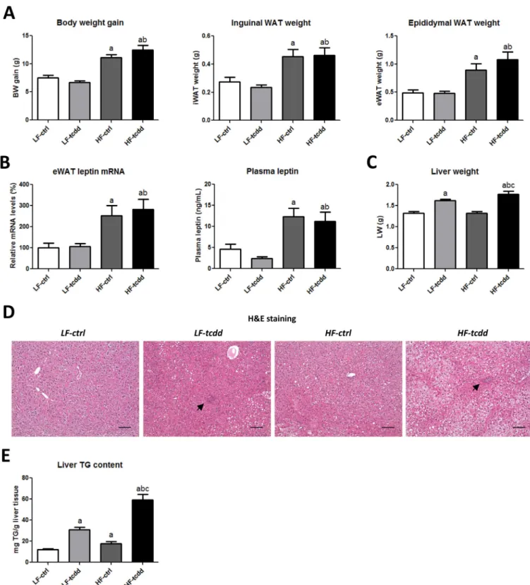
Figure 1. Effect of TCDD on HFD-induced obesity and hepatic steatosis. Mice fed either a LFD or a HFD for a total of 14 weeks were injected with 5 μg/kg of TCDD
(LF-tcdd and HF-tcdd, respectively) or the vehicle (LF-ctrl and HF-ctrl, respectively) during the last 6 weeks. (A) Body weight (BW) gain, inguinal and epididymal white adipose tissue (WAT) weight. (B) Leptin mRNA levels in epididymal WAT (eWAT) measured at 14 weeks and plasma fasted-leptin concentrations at 13 weeks. (C) Liver weight. (D) Hematoxylin-eosin staining (H&E) of representative liver sections of the different groups, black arrows indicate the islets of infiltrated inflammatory cells (bar = 150 μm). (E) Hepatic triglyceride content measured at 14 weeks. Note: Data are expressed as mean ± SEM; a, versus LF-ctrl; b, versus LF-tcdd; c, versus HF-ctrl; p < 0.05.
Dioxin exposure and chronic liver diseases
liver histology. In contrast, TCDD injec-tions in LFD mice led to steatosis together with the infiltration of inflammatory cells grouped in islets. Strikingly, in HFD mice, TCDD dramatically worsened the steatosis, which was accompanied by the infiltration of inflammatory cells. The quantification of the hepatic triglyceride content demonstrated a cumulative effect of HFD and TCDD on lipid accumulation in the liver (4.9-fold increase), as compared to each parameter alone (1.4-fold or 2.5-fold increases for HFD or TCDD, respectively) (Figure 1E). These observations suggest that a moderate HFD combined with a subchronic exposure to TCDD leads to a worsening of a NAFL-like phenotype towards NASH.
TCDD Impairs HFD Adaptative Molecular Mechanisms
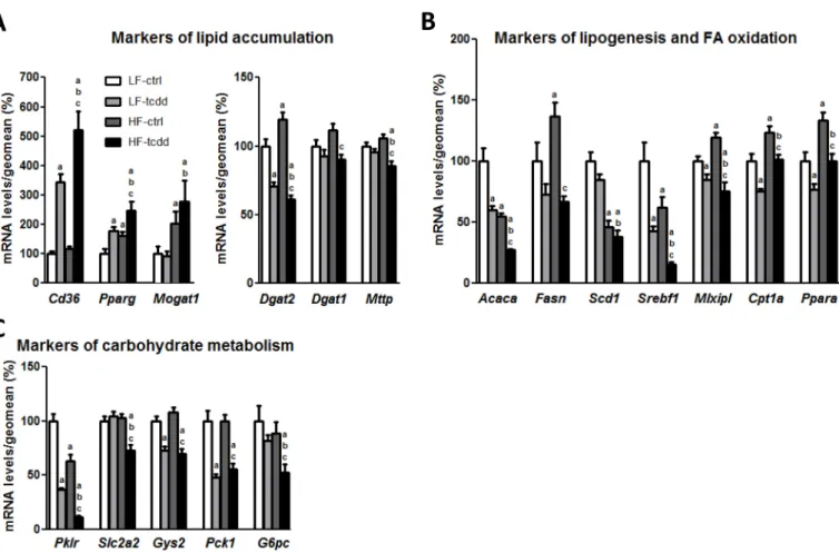
To decipher the mechanisms involved in the aggravation of steatosis in HF-tcdd mice, the hepatic levels of mRNAs of several genes that are markers of lipid metabolism were quantified by qPCR (Figure 2). HF-tcdd mice exhibit further increases in the expres-sion of the fatty acid transporter Cd36 and of
the nuclear receptor Pparg, involved in lipid storage, as compared to LF-tcdd or HF-ctrl mice. Moreover, the expression of Mttp, a crucial enzyme for very low-density lipo-protein secretion, was significantly decreased in the HF-tcdd mice although there was no effect in HF-ctrl or LF-tcdd mice compared to LF-ctrl animals (Figure 2A). However, exposure to TCDD counteracted the HFD-induced increase in expression of Dgat2, a major enzyme of triglyceride synthesis and the co-treatment also decreased the expression of Dgat1, whereas either treatment alone had no effect on this gene. In contrast, the expres-sion of Mogat1, the upstream enzyme of the monoacylglycerol pathway, was increased by HFD, without any additional effect of TCDD treatment (Figure 2A). These results suggest an inhibition of triglyceride synthesis in HF-tcdd mice, potentially due to an adaptive feedback mechanism related to triglyceride accumu-lation. Furthermore, TCDD, together with the HFD, further decreased the expression of both Srebf1/Srebp1c, a central regulator of lipogenesis, and its target gene, Acaca, the rate-limiting enzyme of de novo FA synthesis as compared to LF-tcdd and HF-ctrl mice.
In addition, TCDD exposure counteracted the HFD-induced increase in expression of Mlxipl/Chrebp, another major transcrip-tion factor involved in lipid synthesis and its target gene, Fasn, an enzyme crucial for palmitate synthesis. In contrast, the expression of the stearoyl coA desaturase-1, Scd1, was decreased only by the HFD, with no effect of TCDD (Figure 2B). Moreover, the expres-sion of Ppara, a nuclear receptor regulating FA catabolism, and its target gene Cpt1a, the rate-limiting enzyme of mitochondrial β-oxidation, were decreased in HF-tcdd mice as compared to HF-ctrl mice (Figure 2B). This suggests that TCDD prevented a physiological adaptative up-regulation of FA catabolism in HFD mice. Finally, the expression of several genes involved in carbohydrate metabolism, such as the key enzyme of glycolysis Pklr and the hepatic glucose transporter Slc2a2/
Glut2 were diminished in HF-tcdd mice
(Figure 2C), which indicates additional disruption of energy homeostasis. Together, these results suggest that exposure to TCDD worsens the effects of the HFD by counter-acting the adaptative mechanisms triggered by the diet. This is coherent with the dramatic
Figure 2. Effect of the co-exposure to TCDD and HFD on the hepatic mRNA levels of markers of lipid and carbohydrate metabolism. Mice fed either a LFD or a
HFD for a total of 14 weeks were injected with 5 μg/kg of TCDD (LF-tcdd and HF-tcdd, respectively) or the vehicle (LF-ctrl and HF-ctrl, respectively) during the last 6 weeks. The mRNA levels of hepatic genes were measured by qPCR. Mean expression in the LF-ctrl group is set at 100%. (A) Markers of lipid accumulation. (B) Markers of lipogenesis and FA oxidation. (C) Markers of carbohydrate metabolism. Note: Data are expressed as mean ± SEM; a, versus LF-ctrl; b, versus LF-tcdd; c, versus HF-ctrl; p < 0.05.
increase in the accumulation of triglycerides in the liver.
TCDD Promotes Liver Fibrosis Development in Obese Mice
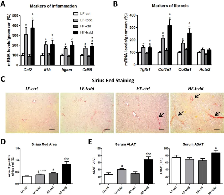
To further characterize the impairment of liver function in HF-tcdd mice, the levels of the mRNAs of genes involved in inflam-mation and fibrosis were analyzed by qPCR. TCDD induced the gene expression of inflammatory markers, such as the chemo-kine Ccl2/Mcp1, the interleukin Il1b and the macrophage Itgam/Cd11b integrin and
Cd68 glycoprotein (Figure 3A), to the same
extent in the LF-tcdd and HF-tcdd groups, consistent with the histological observations
(Figure 1E). Similarly, the mRNA levels of fibrotic markers, such as the pro-fibrogenic cytokine Tgfb1, and the two major collagen fiber components Col1a1 and Col3a1, were increased by the treatment with TCDD inde-pendently of the diet and there was no modi-fication in the level of Acta2/aSma mRNA among the four groups of mice (Figure 3B). However, picro-sirius red staining of liver sections showed that fibrosis (assessed as the percentage of picro-sirius red stained areas) was significantly greater in HF-tcdd mice than in LF-tcdd or HF-ctrl mice (Figure 3C,D). This was associated with an increase in the alanine and aspartate amino-transferase activities in HF-tcdd mice, which
reflects liver injury (Figure 3E). These results suggest that subchronic exposure to low doses of TCDD is sufficient to increase the number of liver fibrotic scars in obese mice.
Discussion
Epidemiological studies suggest that there is an increased risk of liver pathologies when individuals are exposed to POPs (Cave et al. 2010; Consonni et al. 2008; Yi et al. 2014; Yu et al. 1997). In particular, an increased incidence of liver cirrhosis has been reported in individuals from the Seveso population and the Korean Vietnam Veterans cohort, who were highly exposed to TCDD (CDC 1988; Consonni et a. 2008; Yi et al. 2014).
Figure 3. Effect of the combined exposure to TCDD and HFD on the development of hepatic fibrosis. Mice fed either a LFD or a HFD for a total of 14 weeks were
injected with 5 μg/kg of TCDD (LF-tcdd and HF-tcdd, respectively) or the vehicle (LF-ctrl and HF-ctrl, respectively) during the last 6 weeks. The hepatic mRNA levels of markers of (A) inflammation and (B) fibrosis were measured by qPCR. Mean expression in the LF-ctrl group is set at 100%. (C) Picro-sirius red staining shows fibrotic scars of collagen I and III (large black arrows, bar = 150 μm). (D) Quantification of picro-sirius red staining. (E) Serum alanine (ALAT) and aspartate (ASAT) aminotransferase activities. Note: Data are expressed as mean ± SEM; a, versus LF-ctrl; b, versus LF-tcdd; c, versus HF-ctrl; p < 0.05.
Dioxin exposure and chronic liver diseases
Even though the prevalence of NAFLD is increasing worldwide along with obesity (Loomba and Sanyal 2013), there are very few studies that have examined the asso-ciation between exposure to POPs and the development of chronic liver diseases in obese NAFLD patients (Marrero et al. 2005; Zein et al. 2011). However, these studies have focused on AhR ligands (cigarette smoke), other than TCDD. The present study provides evidence that TCDD acts as a cofactor for liver fibrosis progression in a background of obesity in mice.
The preliminary objective of our study was to define experimental conditions to study the combination of TCDD treatment and an obesogenic diet in mouse liver. We performed dose-response experiments to establish that 5 μg/kg TCDD is the threshold dose that leads, after repeated exposure during 6 weeks, to the first signs of liver impairment in C57BL/6J mice. Physiologically based pharmacokinetic modeling of a subchronic exposure of mice to 5 μg/kg TCDD predicted final concentrations of 67 ppt TCDD in blood and 57,000 ppt in liver (wet weight), which are consistent with the concentrations previ-ously described in mouse liver (Boverhof et al. 2005; Vezina et al. 2004). Even if caution must be applied when extrapolating results from mice to humans due to species differ-ences, the concentrations predicted by the model in the blood of our mice (11,551 ppt lipid adjusted) are within the same range as those measured in the blood of the population close to the Seveso industrial accident (zone A, range 15–56,000 ppt) (Eskenazi et al. 2004) or of the Ranch Hand cohort of U.S. veterans exposed to the herbicide Agent Orange (range 318–40,376 ppt) at the time of discharge from Vietnam (Emond et al. 2005). These concentrations range far above the background TCDD levels found in the general popula-tion. A large study of dioxin blood levels in the United States [the University of Michigan Dioxin Exposure Studies (https://sph.umich. edu/dioxin/)] reported average TEQ blood levels of 23.9 ppt (lipid adjusted) in adults who were 18 years old or older. In contrast to blood, the concentration of TCDD has been measured only rarely in human liver (Leung et al. 1990) due to a limited access to biopsies. A physiologically based pharma-cokinetic model of the Ranch Hand cohort predicts TCDD levels of 5,535 ppt in liver for individuals with 162 ppt TCDD in the blood (Emond et al. 2005). These concentra-tions suggest that, for highly exposed Seveso residents or U.S. veterans with TCDD concen-trations above 40,000 ppt in their blood, their level of TCDD in the liver might be within the same range or even higher than the ones predicted in our rodent model (1,360,000 ppt lipid adjusted). Considering that the
C57BL/6J mouse model of diet-induced obesity allows a physiological approach to study the metabolic syndrome and related disorders, such as NAFLD (Duval et al. 2010; Larter and Yeh 2008), we combined the subchronic administration of the threshold dose of TCDD (5 μg/kg) for fibrosis with a 14-week HFD nutritional intervention.
We found that 14 weeks of HFD (45% energy from fat) induced early stages of obesity and subchronic exposure to TCDD did not influence the obese phenotype in contrast to exposure to high doses of TCDD that have been related to cachexia (wasting syndrome) (Kelling et al. 1985). The dose of 5 μg/kg used in this study impaired neither weight gain nor leptin levels (mRNA and hormone) in the LFD and HFD groups. On the contrary, the combination of exposure to TCDD and HFD led to the striking impair-ment of several liver functions, as shown by the drastic increase in the amount of steatosis, and the accumulation of fibrotic scars.
Gene expression analysis suggested that the exposure to TCDD interfered with the metabolic adaptation to the HFD. Whereas exposure to either HFD or TCDD, alone, is known to induce the accumulation of lipid through distinct alterations of lipid and carbo-hydrate metabolism (Angrish et al. 2012; Duval et al. 2010; Lee et al. 2010; Patsouris et al. 2006), their combination led to a unique gene expression signature. For example, the addition of TCDD to HFD altered the expres-sion of key genes of lipid metabolism such as
Ppara, Mlxipl/Chrebp, Cpt1a, Fasn and Dgat2
in a direction that is opposite to that exerted by HFD alone (decrease instead of increase) whereas TCDD exerts its effects in the same direction as HFD on Pparg and Cd36 (increase) or Srebf1/Srebp1c, Acaca and Pklr (decrease). In contrast, exposure to TCDD did not alter the HFD-induced modifica-tions of the expression of Scd1 (decrease) and
Mogat1 (increase). In addition, only HFD and
exposure to TCDD in combination decreased the expression of Dgat1, Mttp, Slc2a2/Glut2 as compared to the three other conditions.
Our results suggest that the molecular mechanisms that explain the effects of TCDD and HFD on lipid metabolism are complex and could implicate i) a direct regulation of AhR target genes such as Cd36 (Lee et al. 2010) and ii) an interference between the AhR and other signaling pathways. Indeed, the addition of TCDD to the HFD impacted the expression level of crucial regulators of lipid and carbohydrate metabolism during an HFD metabolic adaptation. For example, TCDD down-regulates Ppara and up-regulates Pparg, which is consistent with the compensatory effects described by Patsouris et al. (2006) in PPARa KO mice receiving a HFD. This is consistent also with an inter action of the
AhR with the PPARa signaling pathway, as it has been proposed (Lee et al. 2010; Shaban et al. 2004; Wang et al. 2011). Moreover, our moderate HFD intervention increases
Mlxipl/Chrebp and decreases Srebf1/Srebp1c
mRNA levels. These results are in agreement with those of a study by Benhamed et al. (2012) that showed that transgenic over-expression of Chrebp was associated with a decrease of Srebp1c and a “good steatosis” profile. The levels of Chrebp and Srebp1c mRNAs are decreased by TCDD, even when mice receive a HFD. A physical interac-tion has been described between SREBP1c and the AhR that leads to the disruption of SREBP1c signaling after AhR activation (Cui et al. 2011). This interaction could be the underlying mechanism that explains the drastic decrease of Srebp1c in mice exposed to TCDD. Finally, the down-regulations of both Ppara and Srebp1c also are consistent with a disruption of both carbohydrate and lipid metabolism and may be at the origin, partially, of the unique profile associated with HFD and TCDD (“bad steatosis”).
The effect of TCDD on the expression of
Cd36, Cpt1a, Acaca, Fasn and Pklr is in
accor-dance with the literature (Angrish et al. 2012; Boverhof et al. 2005; Lee et al. 2010; Sato et al. 2008) and the lack of effect of TCDD on Mogat1 and Scd1 mRNA expression in our diet experiment is probably due to the lower dose of TCDD that we used as compared to those found in the literature (Angrish et al. 2011, 2012). Cd36 is a direct transcriptional target of the AhR and the PPARg receptors. TCDD increases Cd36 mRNA expression but this effect is potentiated by the high-fat diet. This might be due to the simultaneous stimulation of the PPARg by TCDD and the HFD, whereas the increase in Pparg mRNA expression after 14 weeks of HFD is not suffi-cient to induce Cd36 by itself. Interactions between the AhR and the PPAR family might also explain the decreased expression of Cpt1a. Indeed, the AhR interacts negatively with PPARalpha that stimulates the expres-sion of Cpt1a. Similarly, Srebp1c and Chrebp signaling are counteracted by the AhR, and this could explain the expression profiles of their target genes that are involved in fatty acid synthesis such as Fas or Acaca.
However, our results are surprising for
Dgat2 (down-regulation). Angrish et al.
(2012), who used an acute exposure to a high dose of TCDD in young mice, reported that
Dgat2 was induced by TCDD. This result,
together with the increase in expression of
Mogat1/2, Cd36, and Fabp and the decrease
in very low-density lipoprotein secretion, was suggested to explain the accumulation of triglycerides. Nevertheless, in a human hepatic cell line, HepaRG, treated with TCDD, we found a similar decrease of Dgat2
mRNA (Ambolet-Camoit et al. 2015) as in the present work. Although the regulation of this gene is poorly characterized (Postic and Girard 2008), the knock-down of Dgat2 has been associated with both an improvement of steatosis (Shi and Cheng 2009) and an aggravation of hepatic lesions (Yamaguchi et al. 2007). The down-regulation of Dgat2 that we observe in the TCDD-injected mice, whatever the diet, might, therefore, indicate poorly regulated triglyceride storage
and lipotoxicity. The decrease of Dgat1 in the HF-tcdd group compared to the HF-ctrl group, although modest, indicates that this isoform does not compensate for the decrease in Dgat2 gene expression. Finally, Mogat1, which is markedly induced by the combined exposure, might also possess a DGAT activity and could have a compensatory activity (Shi and Cheng 2009).
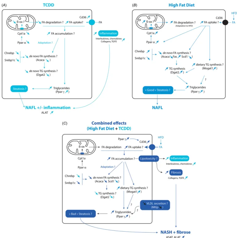
The effects of TCDD, HFD, and their combination on multiple end points of
interest are presented in graphic form in Figure 4. Overall, as compared to LF-ctrl mice, our study suggests that the combined effects of TCDD and HFD are a) to increase the uptake of FA (Cd36), b) to normalize FA β-oxidation (Cpt1a), c) to decrease de novo lipogenesis (Acaca, Fasn, Scd1) and carbohy-drate metabolism (Glut2, Pklr), d) to decrease
de novo triglyceride synthesis (Dgat2/1) while
increasing the monoacylglycerol pathway (Mogat1) and e) to decrease very low-density
Figure 4. Schema of the effects of TCDD, high fat diet and their combination on liver gene expression and various end points. The effects of TCDD are shown in
A, for the high-fat diet in B, and for their combination in C as compared to untreated mice (LF-ctrl). Question marks indicate end points for which the possible modifications have not been measured in the present study.
Dioxin exposure and chronic liver diseases
lipoprotein secretion (Mttp). As previously described, the accumulation of triglycerides in the liver might protect against further hepatic damage and the interruption of triglyceride synthesis is proposed as an initiating event for free FA-mediated lipotoxicity that leads to fibrosis (Choi and Diehl 2008; Trauner et al. 2010).
Importantly, HFD combined with exposure to TCDD led to liver collagen accumulation and increased transaminase levels whereas each treatment alone did not lead to robust changes of these parameters. Consistent with the histological observations, TCDD induced the levels of mRNA of genes that are markers for inflammation, as previ-ously reported (He et al. 2013; Pierre et al. 2014), although there was no further effect with HFD, probably due to the shorter length of our protocol, as opposed to the results of another study (Duval et al. 2010). In contrast, although collagen staining was increased in the co-treated mice (2.6-fold increase) compared to each treatment alone (1.4-fold or 1.2-fold increases for the HF-ctrl or the LF-tcdd groups respectively), the mRNA markers of fibrosis that were tested here were not differentially regulated in the LF-tcdd and HF-tcdd groups. This suggests that other molecular targets underlie the accumulation of collagen such as the regulation of extracellular matrix degra-dation. For example, both AhR and PPARg modify the expression of the matrix metallo-proteases MMP2 and MMP9 that play roles in the development of fibrosis (Pierre et al. 2014; Duval et al. 2002).
In summary, the HFD could lead to an increase of TG synthesis but also to an adapta-tive increase of fatty acid oxidation. This could limit the steatosis as compared to TCDD that would otherwise lead to a decrease of fatty acid oxidation and TG synthesis but also to an increase of fatty acid uptake. We believe that the combined effects of TCDD and the HFD reflect, mostly, the effects of TCDD when large amounts of fat (through the diet) lead to accumulation of TG in the liver. In addition, TCDD, per se, increases liver inflammation, which, together with the disruption of lipid metabolism that may lead to lipotoxicity, might contribute to the occurrence of fibrosis when combined with HFD. The HFD also might potentiate the effects of TCDD. The HFD might increase the availability of endog-enous ligands of AhR, such as tryptophan and its derivatives (Denison and Nagy 2003) that could contribute to the development of obesity-related NAFLD in mice (Moyer et al. 2016). The affinity of AhR for endogenous ligands might be lower than the affinity of exogenous ligands such as TCDD but these endogenous ligands might contribute, never-theless, to the occurence of TG accumulation with the HFD.
Other studies have reported on the rela-tionship between pollutants, liver abnormali-ties and diet in mice. Acute exposure to a high dose of TCDD was found to sensitize mice to the development of a NASH with fibrosis following a methionine- and choline-deficient diet (He et al. 2013). The effects of other potential AhR ligands (or mixtures containing AhR ligands) such as polychlori-nated biphenyls (Wahlang et al. 2013, 2014; Shan et al. 2015), cigarette smoke (Mallat and Lotersztajn 2009), and diesel particles (Arciello et al. 2013) have been tested on different mouse models of obesity. All these studies suggest that pollutants could be co-factors in the progression of NAFLD in mice. Our study reveals unique features that concern the regulation of gene expression and the development of fibrosis in the livers of obese mice exposed to TCDD.
Conclusions
We believe that our study helps to unravel the effects of pollutants (TCDD and other AhR ligands) following subchronic exposure in obese mice. It furthers our understanding of the molecular mechanisms that underlie the impairment of liver functions and the development of the final steps of chronic liver diseases, which is crucial for developing preventive measures.
RefeRences
Ambolet-Camoit A, Ottolenghi C, Leblanc A, Kim MJ, Letourneur F, Jacques S, et al. 2015. Two persistent organic pollutants which act through different xenosensors (alpha-endosulfan and 2,3,7,8 tetrachlorodi benzo-p-dioxin) interact in a mixture and downregulate multiple genes involved in human hepatocyte lipid and glucose metabolism. Biochimie 116:79–91, doi: 10.1016/j. biochi.2015.07.003.
Angrish MM, Jones AD, Harkema JR, Zacharewski TR. 2011. Aryl hydrocarbon receptor-mediated induc-tion of stearoyl-CoA desaturase 1 alters hepatic fatty acid composition in TCDD-elicited steatosis. Toxicol Sci 124:299–310, doi: 10.1093/toxsci/kfr226. Angrish MM, Mets BD, Jones AD, Zacharewski TR.
2012. Dietary fat is a lipid source in 2,3,7,8-tetra-chloro dibenzo-ρ-dioxin-elicited hepatic steatosis in C57BL/6 mice. Toxicol Sci 128:377–386, doi: 10.1093/toxsci/kfs155.
Angulo P. 2002. Nonalcoholic fatty liver disease. N Engl J Med 346:1221–1231, doi: 10.1056/NEJMra011775. Arciello M, Gori M, Maggio R, Barbaro B, Tarocchi M,
Galli A, et al. 2013. Environmental pollution: a tangible risk for NAFLD pathogenesis. Int J Mol Sci 14:22052–22066, doi: 10.3390/ijms141122052. Barouki R, Aggerbeck M, Aggerbeck L, Coumoul X.
2012. The aryl hydrocarbon receptor system. Drug Metabol Drug Interact 27:3–8, doi: 10.1515/ dmdi-2011-0035.
Begriche K, Massart J, Robin MA, Borgne-Sanchez A, Fromenty B. 2011. Drug-induced toxicity on mitochondria and lipid metabolism: mechanistic diversity and deleterious consequences for the liver. J Hepatol 54:773–794, doi: 10.1016/j. jhep.2010.11.006.
Benhamed F, Denechaud PD, Lemoine M, Robichon C, Moldes M, Bertrand-Michel J, et al. 2012. The lipogenic transcription factor ChREBP dissoci-ates hepatic steatosis from insulin resistance in mice and humans. J Clin Invest 122:2176–2194, doi: 10.1172/JCI41636.
Boverhof DR, Burgoon LD, Tashiro C, Chittim B, Harkema JR, Jump DB, et al. 2005. Temporal and dose-dependent hepatic gene expression patterns in mice provide new insights into TCDD-mediated hepatotoxicity. Toxicol Sci 85:1048–1063, doi: 10.1093/toxsci/kfi162.
Cave M, Appana S, Patel M, Falkner KC, McClain CJ, Brock G. 2010. Polychlorinated biphenyls, lead, and mercury are associated with liver disease in American adults: NHANES 2003–2004. Environ Health Perspect 118:1735–1742, doi: 10.1289/ ehp.1002720.
CDC (Centers for Disease Control). 1988. Preliminary report: 2,3,7,8-tetrachlorodibenzo-p-dioxin exposure to humans—Seveso, Italy. MMWR Morb Mortal Wkly Rep 37(48):733–736.
Choi SS, Diehl AM. 2008. Hepatic triglyceride synthesis and nonalcoholic fatty liver disease. Curr Opin Lipidol 19:295–300, doi: 10.1097/ MOL.0b013e3282ff5e55.
Consonni D, Pesatori AC, Zocchetti C, Sindaco R, D’Oro LC, Rubagotti M, et al. 2008. Mortality in a population exposed to dioxin after the Seveso, Italy, accident in 1976: 25 years of follow-up. Am J Epidemiol 167:847–858, doi: 10.1093/aje/kwm371. Cui G, Qin X, Wu L, Zhang Y, Sheng X, Yu Q, et al. 2011.
Liver X receptor (LXR) mediates negative regula-tion of mouse and human Th17 differentiaregula-tion. J Clin Invest 121:658–670, doi: 10.1172/JCI42974. Denison MS, Nagy SR. 2003. Activation of the aryl
hydrocarbon receptor by structurally diverse exogenous and endogenous chemicals. Annu Rev Pharmacol Toxicol 43:309–334, doi: 10.1146/ annurev.pharmtox.43.100901.135828.
Duval C, Chinetti G, Trottein F, Fruchart JC, Staels B. 2002. The role of PPARs in atherosclerosis. Trends Mol Med 8:422–430, doi: 10.1016/ S1471-4914(02)02385-7.
Duval C, Thissen U, Keshtkar S, Accart B, Stienstra R, Boekschoten MV, et al. 2010. Adipose tissue dysfunction signals progression of hepatic steatosis towards nonalcoholic steatohepatitis in C57Bl/6 mice. Diabetes 59:3181–3191, doi: 10.2337/db10-0224. Emond C, Michalek JE, Birnbaum LS, DeVito MJ. 2005.
Comparison of the use of a physiologically based pharmacokinetic model and a classical pharma-cokinetic model for dioxin exposure assessments. Environ Health Perspect 113:1666–1668, doi: 10.1289/ ehp.8016.
Eskenazi B, Mocarelli P, Warner M, Needham L, Patterson DG Jr, Samuels S, et al. 2004. Relationship of serum TCDD concentrations and age at exposure of female residents of Seveso, Italy. Environ Health Perspect 112:22–27, doi: 10.1289/ehp.6573.
He J, Hu B, Shi X, Weidert ER, Lu P, Xu M, et al. 2013. Activation of the aryl hydrocarbon receptor sensi-tizes mice to nonalcoholic steatohepatitis by deacti-vating mitochondrial sirtuin deacetylase 3 Sirt3. Mol Cell Biol 33:2047–2055, doi: 10.1128/MCB.01658-12. Kelling CK, Christian BJ, Inhorn SL, Peterson RE. 1985.
Hypophagia-induced weight loss in mice, rats, and guinea pigs treated with 2,3,7,8-tetrachloro-dibenzo-p-dioxin. Fundam Appl Toxicol 5:700–712, doi: 10.1016/0272-0590(85)90194-0.
Koza RA, Nikonova L, Hogan J, Rim JS, Mendoza T, Faulk C, et al. 2006. Changes in gene expression foreshadow diet-induced obesity in genetically identical mice. PLoS Genet 2:e81, doi: 10.1371/ journal.pgen.0020081.
La Merrill M, Emond C, Kim MJ, Antignac JP, Le Bizec B, Clément K, et al. 2013. Toxicological function of adipose tissue: focus on persistent organic pollutants. Environ Health Perspect 121:162–169, doi: 10.1289/ehp.1205485.
Larter CZ, Yeh MM. 2008. Animal models of NASH: getting both pathology and metabolic context right. J Gastroenterol Hepatol 23:1635–1648, doi: 10.1111/j.1440-1746.2008.05543.x.
Lee JH, Wada T, Febbraio M, He J, Matsubara T, Lee MJ, et al. 2010. A novel role for the dioxin receptor in fatty acid metabolism and hepatic steatosis. Gastroenterology 139:653–663, doi: 10.1053/j.gastro.2010.03.033.
Leung HW, Wendling JM, Orth R, Hileman F, Paustenbach DJ. 1990. Relative distribu-tion of 2,3,7,8 tetrachlorodi benzo-p-dioxin in human hepatic and adipose tissues. Toxicol Lett 50:275–282.
Loomba R, Sanyal AJ. 2013. The global NAFLD epidemic. Nat Rev Gastroenterol Hepatol 10:686–690, doi: 10.1038/nrgastro.2013.171. Louvet A, Teixeira-Clerc F, Chobert MN, Deveaux V,
Pavoine C, Zimmer A, et al. 2011. Cannabinoid CB2 receptors protect against alcoholic liver disease by regulating Kupffer cell polarization in mice. Hepatology 54:1217–1226, doi: 10.1002/hep.24524. Mallat A, Lotersztajn S. 2009. Cigarette smoke exposure:
a novel cofactor of NAFLD progression? J Hepatol 51:430–432, doi: 10.1016/j.jhep.2009.05.021. Marra F, Lotersztajn S. 2013. Pathophysiology of NASH:
perspectives for a targeted treatment. Curr Pharm Des 19:5250–5269, doi: 10.2174/13816128113199990344. Marrero JA, Fontana RJ, Fu S, Conjeevaram HS,
Su GL, Lok AS. 2005. Alcohol, tobacco and obesity are synergistic risk factors for hepatocellular carcinoma. J Hepatol 42:218–224, doi: 10.1016/j. jhep.2004.10.005.
Moyer BJ, Rojas IY, Kerley-Hamilton JS, Hazlett HF, Nemani KV, Trask HW, et al. 2016. Inhibition of the aryl hydrocarbon receptor prevents Western diet-induced obesity. Model for AHR activation by kynurenine via oxidized-LDL, TLR2/4, TGFβ, and IDO1. Toxicol Appl Pharmacol 300:13–24, doi: 10.1016/j.taap.2016.03.011.
NRC (National Research Council (US) Committee for the Update of the Guide for the Care and Use of
Laboratory Animals). 2011. Guide for the Care and Use of Laboratory Animals. 8th ed. Washington, DC:National Academies Press.
Patsouris D, Reddy JK, Müller M, Kersten S. 2006. Peroxisome proliferator-activated receptor α mediates the effects of high-fat diet on hepatic gene expression. Endocrinology 147:1508–1516, doi: 10.1210/en.2005-1132.
Pierre S, Chevallier A, Teixeira-Clerc F, Ambolet-Camoit A, Bui LC, Bats AS, et al. 2014. Aryl hydro-carbon receptor-dependent induction of liver fibrosis by dioxin. Toxicol Sci 137:114–124, doi: 10.1093/toxsci/kft236.
Postic C, Girard J. 2008. Contribution of de novo fatty acid synthesis to hepatic steatosis and insulin resis-tance: lessons from genetically engineered mice. J Clin Invest 118:829–838, doi: 10.1172/JCI34275. Sato S, Shirakawa H, Tomita S, Ohsaki Y, Haketa K,
Tooi O, et al. 2008. Low-dose dioxins alter gene expression related to cholesterol biosynthesis, lipogenesis, and glucose metabolism through the aryl hydrocarbon receptor-mediated pathway in mouse liver. Toxicol Appl Pharmacol 229:10–19, doi: 10.1016/j.taap.2007.12.029.
Shaban Z, El-Shazly S, Abdelhady S, Fattouh I, Muzandu K, Ishizuka M, et al. 2004. Down regulation of hepatic PPARα function by AhR ligand. J Vet Med Sci 66:1377–1386, doi: 10.1292/jvms.66.1377. Shan Q, Huang F, Wang J, Du Y. 2015. Effects of
co-exposure to 2,3,7,8-tetrachlorodibenzo-p-dioxin and polychlorinated biphenyls on nonalco-holic fatty liver disease in mice. Environ Toxicol 30:1364–1374, doi: 10.1002/tox.22006.
Shi Y, Cheng D. 2009. Beyond triglyceride synthesis: the dynamic functional roles of MGAT and DGAT enzymes in energy metabolism. Am J Physiol Endocrinol Metab 297:E10–E18, doi: 10.1152/ ajpendo.90949.2008.
Trauner M, Arrese M, Wagner M. 2010. Fatty liver and lipotoxicity. Biochim Biophys Acta 1801:299–310, doi: 10.1016/j.bbalip.2009.10.007.
Van den Berg M, De Jongh J, Poiger H, Olson JR. 1994. The toxicokinetics and metabolism of polychlori-nated dibenzo-p-dioxins (PCDDs) and dibenzofu-rans (PCDFs) and their relevance for toxicity. Crit Rev Toxicol 24:1–74, doi: 10.3109/10408449409017919. Vernon G, Baranova A, Younossi ZM. 2011. Systematic
review: the epidemiology and natural history of non-alcoholic fatty liver disease and non-alcoholic steatohepatitis in adults. Aliment Pharmacol Ther 34:274–285, doi: 10.1111/j.1365-2036.2011.04724.x. Vezina CM, Walker NJ, Olson JR. 2004. Subchronic
exposure to TCDD, PeCDF, PCB126, and PCB153: effect on hepatic gene expression. Environ Health Perspect 112:1636–1644, doi: 10.1289/txg.7253. Wahlang B, Falkner KC, Gregory B, Ansert D, Young D,
Conklin DJ, et al. 2013. Polychlorinated biphenyl 153 is a diet-dependent obesogen that worsens non-alcoholic fatty liver disease in male C57BL6/J mice. J Nutr Biochem 24:1587–1595, doi: 10.1016/j. jnutbio.2013.01.009.
Wahlang B, Song M, Beier JI, Cameron Falkner K, Al-Eryani L, Clair HB, et al. 2014. Evaluation of Aroclor 1260 exposure in a mouse model of diet-induced obesity and non-alcoholic fatty liver disease. Toxicol Appl Pharmacol 279:380–390, doi: 10.1016/j.taap.2014.06.019.
Wang C, Xu CX, Krager SL, Bottum KM, Liao DF, Tischkau SA. 2011. Aryl hydrocarbon receptor defi-ciency enhances insulin sensitivity and reduces PPAR-α pathway activity in mice. Environ Health Perspect 119:1739–1744, doi: 10.1289/ehp.1103593. Wilson CL, Safe S. 1998. Mechanisms of ligand-induced
aryl hydrocarbon receptor-mediated biochemical and toxic responses. Toxicol Pathol 26:657–671. Yamaguchi K, Yang L, McCall S, Huang J, Yu XX,
Pandey SK, et al. 2007. Inhibiting triglyceride synthesis improves hepatic steatosis but exac-erbates liver damage and fibrosis in obese mice with nonalcoholic steatohepatitis. Hepatology 45:1366–1374, doi: 10.1002/hep.21655.
Yi SW, Hong JS, Ohrr H, Yi JJ. 2014. Agent Orange exposure and disease prevalence in Korean Vietnam veterans: the Korean Veterans Health Study. Environ Res 133:56–65, doi: 10.1016/j.envres.2014.04.027. Yu ML, Guo YL, Hsu CC, Rogan WJ. 1997. Increased
mortality from chronic liver disease and cirrhosis 13 years after the Taiwan “yucheng” (“oil disease”) incident. Am J Ind Med 31:172–175.
Zein CO, Unalp A, Colvin R, Liu YC, McCullough AJ, Nonalcoholic Steatohepatitis Clinical Research Network. 2011. Smoking and severity of hepatic fibrosis in nonalcoholic fatty liver disease. J Hepatol 54:753–759, doi: 10.1016/j.jhep.2010.07.040.