HAL Id: hal-01959016
https://hal.umontpellier.fr/hal-01959016
Submitted on 17 Feb 2021
HAL is a multi-disciplinary open access
archive for the deposit and dissemination of
sci-entific research documents, whether they are
pub-lished or not. The documents may come from
teaching and research institutions in France or
abroad, or from public or private research centers.
L’archive ouverte pluridisciplinaire HAL, est
destinée au dépôt et à la diffusion de documents
scientifiques de niveau recherche, publiés ou non,
émanant des établissements d’enseignement et de
recherche français ou étrangers, des laboratoires
publics ou privés.
Distributed under a Creative Commons Attribution| 4.0 International License
Full-Thickness Porcine and Human Skin Using an In
Vitro Approach
Hinda Dabboue, Nicolas Builles, Eric Frouin, Dan Scott, Jeanne Ramos,
Gilberte Marti-Mestres
To cite this version:
Hinda Dabboue, Nicolas Builles, Eric Frouin, Dan Scott, Jeanne Ramos, et al.. Assessing the
Im-pact of Mechanical Damage on Full-Thickness Porcine and Human Skin Using an In Vitro
Ap-proach. BioMed Research International , Hindawi Publishing Corporation, 2015, 2015, pp.434623.
�10.1155/2015/434623�. �hal-01959016�
Research Article
Assessing the Impact of Mechanical Damage on Full-Thickness
Porcine and Human Skin Using an
In Vitro Approach
Hinda Dabboue,
1Nicolas Builles,
2Éric Frouin,
3Dan Scott,
1Jeanne Ramos,
4and Gilberte Marti-Mestres
11Faculty of Pharmacy, IBMM-UMR 5247, University of Montpellier, France 2Tissue Bank and CCBHM, Saint Eloi Hospital, Montpellier, France 3Pathology Department, University Hospital of Poitiers, France
4Service of Anatomy and Cytopathology, Gui de Chauliac Hospital, CHU, University of Montpellier, France
Correspondence should be addressed to Hinda Dabboue; hinda.dabboue@univ-montp1.fr Received 16 March 2015; Accepted 28 April 2015
Academic Editor: Maxim E. Darvin
Copyright © 2015 Hinda Dabboue et al. This is an open access article distributed under the Creative Commons Attribution License, which permits unrestricted use, distribution, and reproduction in any medium, provided the original work is properly cited. For most xenobiotics, the rates of percutaneous absorption are limited by diffusion through the horny layer of skin. However, percutaneous absorption of chemicals may seriously increase when the skin is damaged. The aim of this work was to develop an
in vitro representative model of mechanically damaged skins. The epidermal barrier was examined following exposure to a razor,
a rotating brush, and a microneedle system in comparison to tape-stripping which acted as a reference. Excised full-thickness skins were mounted on a diffusion chamber in order to evaluate the effect of injuries and to mimic physiological conditions. The transepidermal water loss (TEWL) was greatly increased when the barrier function was compromised. Measurements were made for all the damaged biopsies and observed histologically by microscopy. On human and porcine skins, the tape-stripping application (0 to 40 times) showed a proportional increase in TEWL which highlights the destruction of the stratum corneum. Similar results were obtained for all cosmetic instruments. This is reflected in our study by the nonsignificant difference of the mean TEWL scores between 30 strips and mechanical damage. For a specific appreciation, damaged skins were then selected to qualitatively evaluate the absorption of a chlorogenic acid solution using fluorescence microscopy.
1. Introduction
The primary property of the skin is to act as a barrier function. The outermost epidermal layer, the stratum corneum (SC), is an effective barrier that protects against external aggression and prevents the delivery of xenobiotic molecules across the skin [1–5].
Due to skin barrier properties, a chemical must exhibit specific physicochemical traits, that is, a low molecular weight, a low melting point, and a logP (octanol-water parti-tion coefficient) from 1 to 4, in order to be a candidate for passive transepidermal delivery [6–8]. To overcome signif-icant barrier properties of the stratum corneum, numerous approaches were conducted in the pharmaceutical domain to enhance percutaneous penetration of drugs, such as nano-formulations [9, 10] and by production of temporary [11] or permanent holes [12] in the skin. The use of these
techniques has now advanced to the field of cosmetics. A large number of instruments, apparatus, and devices are now marketed as “high-tech beauty gadgets” that are claimed to smooth wrinkles as well as renew and temporarily alter the appearance of the face and skin [13]. Cosmetics and cosmetic devices are used to improve appearance and should not impart any health benefits or permeate past the epidermal layer; otherwise they would be classified as a medicine. The skin as an outer organ is naturally susceptible to mechanical damage from its environment which can impair its barrier function, and this must be factored into the development and design of cosmetic gadgetry.
The aim of our study was to establish an in vitro model of acute barrier disruption, using Franz cell with full-thickness porcine and human skin [14–16], to investigate various types of skin damage, based on Fick’s law of diffusion [17,18].
Hindawi Publishing Corporation BioMed Research International Volume 2015, Article ID 434623, 10 pages http://dx.doi.org/10.1155/2015/434623
Tape-stripping, first described by Fritsch et al. [19], is a robust method in SC physiology research. Adhesive films are pressed onto the surface of the skin with a fixed amount of pressure before removal [20]. The superficial layers of the SC adhere to the film, are stripped from the SC, and are then accessible for further investigation. At the same time, repeated tape-stripping may be an effective comparative model for impaired skin barrier function [21–23]. Transepi-dermal water loss (TEWL) was used as the unit of SC damage between models, measured in grams per centimeter squared per hour (g/cm2/h). The TEWL is widely used in skin integrity tests with a large historical dataset [24–28]. Many studies have suggested that high TEWL is associated with various skin diseases, including atopic dermatitis, psoriasis, contact dermatitis, and ichthyosis [29–32]. Thus, TEWL is thought to be a useful parameter that characterizes skin barrier function in man. In vitro experiments were performed using Franz cells with full-thickness porcine and human skin. Healthy pig ear skin was compared to healthy human skin with and without stretch marks because they are an excellent surrogate to human skin, due to physiological similarity and availability. The effects of selected 5 to 30 or 40 repeated tape-strippings were then compared to the other types of induced skin injury.
The impact of two new cosmetic “gadgets,” micronee-dles [33] and rotating brush [34], was studied in order to evaluate skin damage after their application. Influence of a conventional razor [35] was also investigated. Microneedles of 1 mm length disposed on a roller were studied with respect to the efficiency of skin perforation. Microneedles were initially used for skin disruption to facilitate transdermal drug delivery until recently. This device was then introduced in the cosmetic domain to treat scars, wrinkles, and stretch marks. The impact of a rotating brush used for face cleansing was also investigated via application to a fresh biopsy with a cosmetic gel containing salicylic acid. A manual razor was also applied three times on a biopsy in the same direction.
The purpose of the current work was to investigate the suitability of different skin integrity tests to differentiate impaired from intact human skin.
2. Materials and Methods
2.1. Skin Preparation. Porcine ears were obtained from
freshly killed animals in a local slaughterhouse (P´ezenas, France). After cleaning with cold tap water, full-thickness skin was removed with a scalpel from the cartilage of the outer region. Human skin was obtained from “Centre des Collections Biologiques Hospitali`eres, CHU (Central Univer-sity Hospital), Montpellier” (biobank identification number BB-0033-00031) following official agreement compliant with French regulation and full written consent from donors. Human skin was retrieved from the plastic surgery unit (abdominoplasty), treated with povidone iodine antiseptic (PVP-I, Betadine) prior to extraction, harvested by a surgeon in a medical grade sterile pot system (Cryokit, Verreries Talanconnaises, France) with sterile NaCl 0.9% at +4∘C (Sodium Chloride 0.9%, B/Braun, Melsungen, Germany), sent to the tissue bank where subcutaneous fat was removed
up from the dermal layer, and conditioned within a Cryokit with NaCl 0.9% system. The skin was sent to the laboratory at +4∘C up to 4 hours from retrieval, ensuring an optimal skin quality. All skins were inspected for visible skin lesions prior to use. Only intact healthy looking skin was used for experiments.
All skins were cut using a punch-biopsy in the lab-oratory (2 cm2 diameter) to fit Franz cells, and thickness was measured in each case using Mitutoyo 2050S apparatus (ranging between 1.0 and 1.4 mm) prior to labelling, freezing in aluminium foil, and storing at −20∘C for a period not exceeding 4 months. Skin from different donors was used to demonstrate reproducibility of the study. A minimum of 4 different subjects was assigned to each group in order to minimize any individual variance which would interfere with overall outcome.
2.2. Types of Skin Damage
2.2.1. Tape-Stripping. Standard sized D-Squame Skin
Sam-pling Discs (22 mm2 diameter, Monaderm, Monaco) were applied to skin biopsies prior to application of 225 g/cm2 pressure during 3 seconds, provided by the D-Squame D500 apparatus applicator (Monaderm, Monaco). This process was subjected to affected skin samples, 5 to 40 times using fresh discs each time. The D-Squame tapes were peeled from different directions (90∘ each time) in rotation until the process was completed.
2.2.2. Microneedles. A titanium Micro Needle Roller System
(RoHS, CE) composed of 540 needles of 1 mm length was used. The instrument was rolled firmly onto the biopsies ten times vertically followed by ten times horizontally.
2.2.3. Razor. Wilkinson Sword Extra Beauty 3 razors were
used. They contained an aloe vera adjuvant alongside physio-logic solution (NaCl 0.9%, Versylene, Fresinius Kabi, Sevres, France). The skin biopsies were shaved 3 times in the same direction without shaving formulation.
2.2.4. Rotating Brush. Pureo Sonic Brush (Elle by Beurer,
France) was applied to designated biopsies at the highest speed and rotated firmly around the skin for one minute at the highest speed. Integrity of the skin was further challenged following application of a cleansing gel. A pure active gel (20𝜇L) containing salicylic acid and zinc gluconate was applied to previously moistened skin with saline and rubbed gently before being washed off 60 seconds after.
2.3. In Vitro Model. Glass Franz diffusion cells with average
capacity of 9 mL± 0.35 mL were used with a surface area of 1 cm2. Each cell was filled with saline solution (0.9% NaCl) representing the thermodynamic equivalent of fluid beneath the epidermis in vivo. Franz cells were thermostated at 37.2∘C ± 1∘C (Polystat CC1, Huber, Offenburg, Germany) with recep-tors stirred at 600 rpm/min with a magnetic bar throughout the experiment. Skin surface temperature was then mea-sured at 32∘C ± 1∘C in order to confirm correlation to in
BioMed Research International 3
and before TEWL measurements. The complete model was held in place by clamps (Rotulex, Pyrex, SciLabware, Clichy, France).
2.4. TEWL Measurement. A Tewameter TM 300 was used
(Monaderm, Monaco). A minimum of one hour was allowed for samples to equilibrate following direct application to Franz cell receptor temperature from a frozen state. After this time, the TEWL results were obtained (g⋅m2⋅h−1). In the case of deliberately damaged skin of deliberately damaged skin, TEWL measurements were taken on an intact biopsy and compared to the same biopsy 30 minutes after injuries were applied. Care was taken prior to measurement to ensure that there was an absence of air bubbles lying under the dermis in contact with the receptor fluid. TEWL measurements were conducted on average three times per sample from the top of the donor cell.
2.5. Histological Analysis
2.5.1. Optical Microscopy. After mechanical damage, biopsies
were fixed in 4% paraformaldehyde (Sakura Society, Tokyo, Japan) solution for up to 48 hours. Thereafter, skin samples were embedded in paraffin (Leica Society, Germany) and cross sections of 5𝜇m were cut. After drying and deparaf-fining blades, cut sections were automatically colored by haematoxylin and eosin (Dako Society, Les Ulis, France). Slides (Superfrost Plus, VWR International, Fontenay-sous-Bois, France) that contained histological sections were placed on a motorized support under the Nanozoomer Slide Scanner (Hamamatsu). Then, the entire surface of the sample section was analyzed with NDP Nanozoomer software.
2.5.2. Scanning Electron Microscopy. Skin biopsies were
washed in PBS and fixed in a 2.5% glutaraldehyde (Elec-tron Microscopy Sciences, Hatfield, PA, USA) and Sorensen phosphate buffer (0.133 M, Electron Microscopy Sciences, Hatfield, PA, USA) solution, pH 7.2, for an hour at room temperature and rinsed in Sorensen buffer. Samples were then dehydrated using a gradient ethanol series (30–100%), followed by critical point drying with CO2. Subsequently, samples were sputter-coated with an approximative 10 nm thick gold film and then examined under a scanning electron microscope (Hitachi S4000, at CRIC, Montpellier, France) using a lens detector with an acceleration voltage of 10 kV at calibrated magnifications.
2.5.3. Fluorescence Microscopy. 1% of chlorogenic acid
(Sigma-Aldrich, St. Louis, USA) was solubilized in a mixture (2/8, v/v) of PEG-400 (Cooper, Melun, France) and methanol (Sigma-Aldrich, St. Louis, USA). 20𝜇L of this solution were added to skin samples and incubated in a thermostated Franz cell for 24 hours. The donor compartment of the cell was covered with parafilm to prevent evaporation of the applied compounds. After 24 hours, skin samples were dried; surfaces were gently dabbed with methanol (Sigma Aldrich) using gauze prior to embedding in OTC matrix (CellPath, UK) and frozen in liquid nitrogen. The samples were then stored at−80∘C until preparation of microscope slides using
StretchM.HSkin IntactHSkin IntactPigS. Type of skin 40 30 20 10 0 TE WL (g/m 2/h)
Figure 1: Box and whisker-plot, TEWL analysis, and comparison between healthy human (IntactHSkin) and porcine (IntactPigS.) skin and human stretch marks biopsies (StretchM.HSkin) (KW is equal to 105.55,𝑃 value = 0.00). +: mean.
a cryostat. Six-micron cryo-cross-sections were observed by fluorescence microscopy (Leica DMR-Camera Leica DFC 310 FX, Nanterre, France) with a DAPI filter (excitation 350 nm and 450–490 nm emission) with few drops of Neu reagent [36] (1% of 2-aminoethyl-diphenylborinate) (Sigma-Aldrich, St. Louis, USA) in methanol (Sigma-Aldrich, St. Louis, USA), which potentiates the fluorescence of chlorogenic acid.
2.6. Statistical Analysis. Statistical calculations were
per-formed by means of the PC program, Statgraphics-Centurion XVII [37]. A nonparametric Kruskal-Wallis (KW) was used with a box and whisker (BaW) representation. Notches are useful in offering a guide to significance of difference of medians, in the case that the notches of two boxes do not overlap. Bonferroni test was then used to show pairwise comparison between the average ranks of each group.
3. Results and Discussion
3.1. Comparison of Healthy Human and Pig Skins. As shown
inFigure 1, intact pig and human skins were differentiated between biopsies with stretch marks. A KW nonparamet-ric test was used, whereby this test does not require the assumption that all the samples were drawn from normally distributed populations with equal variance. The KW test for TEWL by type of skin is equal to 105.55 with a𝑃 value = 0.00, confirming that there was a statistically significant difference amongst the medians at a 95% confidence level. For the intact pig skin (𝑛 = 154), the average rank was 166.68 and for the intact human skin (𝑛 = 106) the average rank was 77.92. Finally, for the stretch mark human skin (𝑛 = 8), average rank was 264.5. All data were reported in
Table 1. Graphically, the box and whisker procedure denoted a statistically significant difference between the 3 types of skin, which was corroborated by the Bonferroni procedure. We have demonstrated an important increase in the mean of TEWL for the stretch mark human skin (13.6 g/m2/h) compared to the two others, where the mean TEWL is 3 times greater than that of healthy human skin (4.2 g/m2/h) and almost twice as large as healthy pig skin (6.7 g/m2/h).
Table 1: TEWL data (number of experimentations with mean and median) obtained from healthy human (IntactHSkin) and porcine (IntactPigS.) skin biopsies and human stretch marks biopsies (StretchM.HSkin).
Type of skins 𝑛 Average rank Median (TEWL) g/m2/h Mean (TEWL) g/m2/h
Intact pig skins 154 166.68 6.60 6.70± 0.23
Intact human skins 106 77.92 3.95 4.23± 0.28
Stretch marks skins 8 264.5 13.55 13.6± 0.58
One hypothesis is that stretch marks are induced by excessive mechanical stretching of skin to the point of rupturing dermal elastic fibers and that local fibroblasts are unable to adequately repair or replace those extracel-lular matrix components that are solely responsible for the resilience of skin [38]. The presence of stretch marks on the human skin is equivalent to the presence of a lesion when comparing TEWL results. Therefore as human skin with stretch marks already corresponds to endogenous lesions that could increase TEWL, we decided not to use them in the following experiments. In contrast, excised porcine ear skin has been shown to be a suitable skin substitute model for human skin, based on morphological and functional data [39,40]. However, sources for excised human skin for in vitro studies are limited.
3.2. Stripped Skin as Model. A control group with undamaged
skin was compared with a group where epidermis of the biopsies was stripped 5, 10, 20, 30, and 40 times. The maximal number of adhesive tapes used was fixed to 40 for human and porcine skin. For comparison, two protocols were implemented in the study: in the first one (protocol 1) tape-stripping was applied 5 to 40 times successively on the same biopsy, while in the second one (protocol 2), a new skin biopsy was used each time.
Netzlaff et al. [41] have proven that TEWL measurement cannot detect small changes in the stratum corneum, but a clear increase in TEWL induced by the impairment of the SC barrier was expected.
With both protocols, there was a strong correlation between the number of stripping times and TEWL. And the removal of 30–40 tape-strips formed a plateau corresponding to removal of the last stratum corneum layers. At these steps, a 4-fold loss of barrier function for pig skin and a 6-fold loss of barrier function for human skin were observed.Figure 2
illustrates the increase in TEWL as SC width decreases. The KW test for TEWL comparing protocol and skin is equal to 0.88 with a 𝑃 value = 0.82, and there are no statistically significant differences amongst the medians at 95% confidence level (all data are reported inTable 2). This provides important information and all data obtained from protocols 1 and 2 were then pooled for neatest tests. This crucial step was corroborated by Rubio et al. who have shown that 20 and 35 strips cause, respectively, minor and major increases in TEWL and that more strips have nonsignificant effect [42].
In order to visualize the destruction of the stratum corneum in porcine ear and human skins models, histological sections were investigated as shown in Figures 3 and 4, respectively. The stratum corneum is progressively removed
0 5 10 15 20 25 30 35 40 0 5 10 15 20 25 30 35 40 45 Number of tape-strips
Pig skin—protocol 2 Pig skin—protocol 1
Human skin—protocol 1 Human skin—protocol 2
TE
WL (g/m
2 /h)
Figure 2: TEWL analysis and comparison between two protocols of tape-stripping and healthy porcine and human skin; protocol 1: tape-stripping followed in the same skin, protocol 2: tape-stripping on different biopsies.
by serial adhesive tape-stripping. With 40 strips drastic damage is observed on the epidermis. Thus 30 tape-strips were used as a realistic reference to damaged skin for further comparative studies.
Compared to intact skin, smoother skin surface was observed by scanning electron microscopy (SEM) (Figure
3(c)) in porcine samples. Such findings have explained the very small thickness of the stratum corneum after 30 tape-strips observed on optical microscopy images (Figure 3(d)). The same observations were made with human skin samples when 30-tape-strip damage was applied (Figure 4(b)). Thus we have herein developed a standardized model based on 30 tape-strips for evaluating skin injuries when the stratum corneum is impaired homogeneously. This method of tape-stripping application has often been used as a model of skin lesions in order to study the penetration of xenobiotics [43–
48].
3.3. Mechanical Damage by Devices. With recent
develop-ments in the cosmetic industry with regard to device models, one trend is towards home use. But are these practices safe if cosmetic products are applied after their use? The Margin of Safety (MoS) of substances in a finished cosmetic product is derived by dividing the nonobserved adverse effect level (NOAEL) by the systemic exposure dosage (SED). Exposure scenario is based to an extent on the amount of substance that may be absorbed through the skin in order to calculate the SED [49]. Numerous complications may arise because SED is usually calculated with data of absorption obtained from
BioMed Research International 5
Table 2: TEWL data (number of experimentations with mean and median) of stripped skin for 30 times.
Type of skins 𝑛 Average rank Median (TEWL) g/m2/h Mean (TEWL) g/m2/h Pig skins protocol 1—30 times 4 26.5 26.05 27.20± 2.77 Pig skins protocol 2—30 times 23 20.91 24.9 25.30± 1.15 Human skins protocol 1—30 times 10 23.75 27.3 26.03± 1.75 Human skins protocol 2—30 times 7 23.64 24.9 26.80± 2.09
(a) SC Ep. De. (b) (c) (d)
Figure 3: Histological analysis, comparison between porcine skin tape-stripped and no treatment by scanning electron microscopy (a, c), and optical microscopy of haematoxylin and eosin stained section (b, d). (a, b) Control porcine skin; (c, d) tape-stripping (×30) porcine skin. Scale bar (b, d): 100𝜇m. SC: stratum corneum, Ep.: epidermis, and De.: dermis.
chemical applications on healthy skin. In a risk assessment, the toxicity of the chemical is considered in conjunction with anticipated exposure levels for the target population. But with the use of cosmetics devices levels of SED are underestimated and will not represent the worst case compared to exposure.
A statistically significant difference was determined (Table 3) between intact skin and punctured, brushed, shaved, and 30-stripped skins (KW was equal to 256.59, 𝑃 value = 0.00). However, no difference could be detected between the skin samples when compared to differing sub-groups of mechanical damage (Figure 5) following TEWL analysis. Histological findings of skin samples are shown in Figures6and7. We have demonstrated that the lesions and functional changes induced by the 30-stripped skin model of barrier disruption are similar to those observed with devices examined.
Compared to intact skin, porcine biopsy holes (micro-lancing) are observed on the skin surface by SEM (Figure
6(a)). They are explained by a fracture from the stratum corneum into the dermis after the microneedles application (Figure 6(b), denoted by ∗). Moreover, the needles were
soaked in black ink in order to avoid an artifact observation due to sample preparation. The same observation was made on human skin (Figure 7(b), denoted by ∗). Microneedles were used initially in the biopharmaceutical field for trans-dermal drug delivery in order to overcome the skin barrier by formation of mechanically produced conduits through the stratum corneum by the use of small needles [50–54].
Traditional methods of removing unwanted hair include shaving [55]; this method has a temporary impact on skin barrier function. This process removes the hair shaft very close to the surface of the skin as observed inFigure 6(c)
(porcine biopsy). In contrast, no collateral damage to the softer skin surface was noticed for porcine and human skins (resp., in Figures6(d)and7(c)).
Microscopic examination of porcine skin after a rotative brush treatment depicts a very slight disturbance of epider-mal tissue (Figures6(e)and6(f)). The same observation was made on human skin with rotating brush (Figure 7(d)). Con-sumers often combined the rotating brush with a cleansing gel as stimulated by our investigations. This technique is well known by dermatologists in the treatment of skin surface
SC Ep. De.
(a) (b)
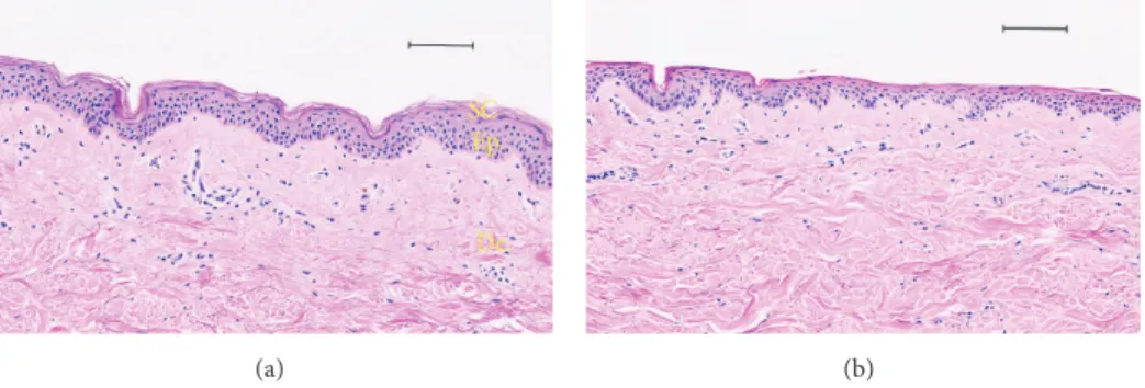
Figure 4: Histological analysis and comparison between a human skin tape-stripped and no treatment by optical microscopy (haematoxylin and eosin staining). (a) Human healthy skin; tape-stripping (×30) human skin (b). Scale bar: 100 𝜇m.
Table 3: TEWL data (number of experimentations with mean and median) obtained with healthy human (IntactHSkin) and porcine (IntactPigS.) skins and human stretch marks biopsies (StretchM.HSkin).
Type of skins 𝑛 Average rank Median (TEWL) g/m2/h Mean (TEWL) g/m2/h
Intact pig skin 154 166.68 6.60 6.70± 0.23
30-strip pig skin (pooled protocols 1-2) 27 302.01 25.70 25.58± 0.56
Brush gel pig skin 9 290.00 23.60 23.53± 0.87
Razor pig skin 6 340.50 35.35 33.96± 1.18
Microneedle pig skin 14 300.35 24.40 25.45± 0.77
Intact human skin 106 77.92 3.95 4.23± 0.28
30-strip human skin (pooled protocols 1-2) 17 306.29 26.90 26.34± 0.70 Brush gel human skin 5 319.10 29.50 28.78± 1.30
Razor human skin 8 328.68 30.08 30.82± 1.02
Microneedle human skin 79 312.83 30.00 27.67± 0.97
In ta ct PigS. 30S tr ip PigS. Br ushGP igS. R azo rP igS. M icr oNP igS. Int ac tH Sk in 30S tr ip HS kin Br ushG.HS kin R azo rHS kin M icr oN.HS kin Type of skin 40 30 20 10 0 TE WL (g/m 2 /h)
Figure 5: Box and whisker-plot, TEWL analysis with the different mechanical injuries, and comparison with the control biopsy in porcine and human skin (KW is equal to 256.59,𝑃 value = 0.00, +: mean, and◻: outliers points).
troubles like acne, scars, and other skin blemishes. It involves direct removal or disruption of the upper layer of the skin to enhance the penetration of topically applied xenobiotic [56]. From our observations, daily application of a cleansing gel with a device could be deleterious and could enhance percutaneous penetration of other chemicals applied onto the
skin. In conclusion, based on data presented here, 30 tape-strips are necessary to obtain a model of realistic damage.
Numerous studies of TEWL or electrical resistance have compared healthy and damaged skin, but in each report a maximum of 10 or 20 tape-strippings were implemented to reenact disturbed skin. In contrast to our study, these tape-stripping models were not compared with other mechanical damage [48,57,58]. Although we observed a link between increases of TEWL absorption with numerous damage mod-els, our results need to be further investigated in a quantitative fashion to appreciate the potential real life impact.
In our study, nonsignificant differences between mechan-ical damage and 30 tape-strips were demonstrated with reproducible data. This last procedure appeared to be a more realistic model in order to mimic human skin with impaired SC due to various mechanical reasons. We recommend a standardized method with 30 adhesive discs pressed onto the surface during 3 seconds using a 225 g/cm2applicator to evaluate skin absorption for risk assessment.
With such a great enhancement of the TEWL found for all skin injuries, one can reasonably expect that skin absorption of chemicals would be similar following injury. A solution of 1% chlorogenic acid, a compound used as reference for skin absorption [59], was deposited on intact skin and two different types of damaged skins: 30 tape-strips and microneedles treatment. Absorption was qualitatively analyzed 24 hours after using fluorescent microscopy.
BioMed Research International 7 ∗ (a) ∗ (b) (c) (d) (e) (f)
Figure 6: Histological analysis of damaged porcine skin. Scanning electron microscopy (a, c, and e) and optical microscopy haematoxylin and eosin staining (b, d, and f). (a, b) Microneedles application (∗microlancing); (c, d) razor and shaving; and (e, f) rotative brush application with the gel. Scale bar (b, d, and f): 100𝜇m.
A bright high intensity fluorescence was clearly visible in upper layers of the intact skin, while a more diffuse signal was present at deeper skin layers (Figure 8). On the opposite, a wide area of fluorescence was observed deeper in the skin (Figures8(b)and8(c)) for both stripping method and microneedles. But, with the use of sharp microneedles, the diffusion of the fluorescent molecule through the conduits over time seemed to deeply penetrate the epidermis and the dermis. These results also suggested that a wide amount of chemicals could be absorbed, but more absorption studies are necessary to confirm our results.
4. Conclusion
Our objective was to determine a realistic and practical in
vitro model of barrier impairment using a stepwise approach
of sequential tape-stripping of pig and human skins in com-parison to much mechanical damage currently encountered
in the cosmetic field. TEWL was used to compare skin barrier function in human or pig skins. A dramatic increase of the TEWL value was observed with human skin with stretch marks compared to intact human skin. The experimental work presented herein has shown that the removal of stratum corneum by 30 tape-strips is the most relevant procedure in order to make a standardized model of injured skin in
vitro. Skin exposed to microneedles, a razor, or a rotating
brush was strongly disturbed and all the features of the damage were comparable to the 30 tape-strippings procedure in TEWL analysis, but we observe different kinds of skin barrier disruption. Results obtained in this work support the need for new absorption studies on damaged skin. Further perspectives are needed to answer further questions created in this study: how deep is the skin penetration for different compounds and what lies between healthy and damaged skins in this regard. This leads to the opening of investigations for the future, which questions the safety of advances in the
SC Ep. De. (a) ∗ (b) (c) (d)
Figure 7: Histological analysis by optical microscopy (haematoxylin and eosin stain), comparison between a healthy human skin (a) and microneedles application with black ink (b), shaving human skin with a razor (c), and rotating brush and a gel (d). Scale bar: 100𝜇m; SC: stratum corneum, Ep.: epidermis, and De.: dermis.
100 𝜇m (a) 100 𝜇m (b) 100 𝜇m (c)
Figure 8: Fluorescence microscopy analysis: evaluation of penetration by chlorogenic acid (1% in solution), on healthy and damaged porcine skin. (a) Healthy porcine skin, (b) 30-time tape-stripped porcine skin, and (c) microneedles application. Scale bar: 100𝜇m.
development of topical formulations and cosmetic gadgetry in years to come.
Conflict of Interests
The authors have no conflict of interests to disclose.
Acknowledgments
The ANSM (project ThroughSKIN) is acknowledged for financial support. The authors would like to thank the slaughterhouse of P´ezenas, France, Kocel Kouril SARL, Mireval, France, and also Mrs. Chantal Cazevieille in the Electron Microscopy Service, Montpellier Rio Imaging, Saint Eloi Hospital, Montpellier, France. This work used samples of human skin from living donors and, under expertise
of Dr S. Domergue, samples were treated at Montpellier CHU Tissue Bank part of Centre des Collections Biolo-giques Hospitali`eres (CCBHM), CHU, Montpellier (http:// www.chu-montpellier.fr/fr/plateformes/) (Biobank no. BB-0033-00031).
References
[1] F. N. Marzulli, “Barriers to skin penetration,” The Journal of
Investigative Dermatology, vol. 39, pp. 387–393, 1962.
[2] A. Vieille-Petit, N. Blickenstaff, G. Coman, and H. Maibach, “Metrics and clinical relevance of percutaneous penetration and lateral spreading,” Skin Pharmacology and Physiology, vol. 28, no. 2, pp. 57–64, 2015.
[3] W. Smith, “Stratum corneum barrier integrity controls skin homeostasis,” International Journal of Cosmetic Science, vol. 21, no. 2, pp. 99–106, 1999.
BioMed Research International 9
[4] R. J. Scheuplein, “A personal view of skin permeation (1960– 2013),” Skin Pharmacology and Physiology, vol. 26, no. 4-6, pp. 199–212, 2013.
[5] L. Norl´en, “Current understanding of skin barrier morphology,”
Skin Pharmacology and Physiology, vol. 26, no. 4–6, pp. 213–216,
2013.
[6] J. Hadgraft, “Passive enhancement strategies in topical and transdermal drug delivery,” International Journal of
Pharmaceu-tics, vol. 184, no. 1, pp. 1–6, 1999.
[7] S. Singh and J. Singh, “Transdermal drug delivery by passive diffusion and iontophoresis: a review,” Medicinal Research
Reviews, vol. 13, no. 5, pp. 569–621, 1993.
[8] H. E. Junginger, “Formulation aspects on dermatological prepa-rations and transdermal drug delivery systems,” Acta
Pharma-ceutica Nordica, vol. 4, no. 2, p. 117, 1992.
[9] C. Caddeo, M. Chessa, A. Vassallo et al., “Extraction, purifi-cation and nanoformulation of natural phycocyanin (from
Klamath algae) for dermal and deeper soft tissue delivery,” Journal of Biomedical Nanotechnology, vol. 9, no. 11, pp. 1929–
1938, 2013.
[10] S. Ghanbarzadeh and S. Arami, “Enhanced transdermal deliv-ery of diclofenac sodium via conventional liposomes, etho-somes, and transferetho-somes,” BioMed Research International, vol. 2013, Article ID 616810, 7 pages, 2013.
[11] A. Alexander, S. Dwivedi, T. K. Giri, S. Saraf, and D. K. Tripathi, “Approaches for breaking the barriers of drug perme-ation through transdermal drug delivery,” Journal of Controlled
Release, vol. 164, no. 1, pp. 26–40, 2012.
[12] J.-H. Park, S.-O. Choi, S. Seo, Y. B. Choy, and M. R. Prausnitz, “A microneedle roller for transdermal drug delivery,” European
Journal of Pharmaceutics and Biopharmaceutics, vol. 76, no. 2,
pp. 282–289, 2010.
[13] O. Andre, M. Paye, and H. I. Maibach, Handbook of Cosmetic
Science and Technology, Informa Healthcare, New York, NY,
USA, 3rd edition, 2009.
[14] OECD Guideline for the Testing of Chemicals, Test No. 428: Skin
Absorption: In Vitro Method, OECD Publishing, 2004.
[15] U. Jacobi, M. Kaiser, R. Toll et al., “Porcine ear skin: an in vitro model for human skin,” Skin Research and Technology, vol. 13, no. 1, pp. 19–24, 2007.
[16] M. E. Darvin, H. Richter, Y. J. Zhu et al., “Comparison of in
vivo and ex vivo laser scanning microscopy and multiphoton
tomography application for human and porcine skin imaging,”
Quantum Electronics, vol. 44, no. 7, pp. 646–651, 2014.
[17] E. L. Roetman and R. E. Barr, “The mechanical basis for Fick’s law and its generalizations,” Advances in Experimental Medicine
and Biology, vol. 75, pp. 261–265, 1976.
[18] A. Couto, R. Fernandes, M. N. S. Cordeiro, S. S. Reis, R. T. Ribeiro, and A. M. Pessoa, “Dermic diffusion and stratum corneum: a state of the art review of mathematical models,”
Journal of Controlled Release, vol. 177, no. 1, pp. 74–83, 2014.
[19] P. O. Fritsch, F. Gschnait, G. Kaaserer et al., “PUVA suppresses the proliferative stimulus produced by stripping on hairless mice,” Journal of Investigative Dermatology, vol. 73, no. 2, pp. 188–190, 1979.
[20] M. Breternitz, M. Flach, J. Pr¨assler, P. Elsner, and J. W. Fluhr, “Acute barrier disruption by adhesive tapes is influenced by pressure, time and anatomical location: integrity and cohesion assessed by sequential tape stripping; a randomized, controlled study,” British Journal of Dermatology, vol. 156, no. 2, pp. 231– 240, 2007.
[21] L. E. Berrutti, A. J. Singer, and S. A. McClain, “Histopathologic effects of cutaneous tape stripping in pigs,” Academic Emergency
Medicine, vol. 7, no. 12, pp. 1349–1353, 2000.
[22] H. Dickel, A. Goulioumis, T. Gambichler et al., “Standardized tape stripping: a practical and reproducible protocol to uni-formly reduce the stratum corneum,” Skin Pharmacology and
Physiology, vol. 23, no. 5, pp. 259–265, 2010.
[23] M. Peppelman, W. A. van den Eijnde, E. J. Jaspers, M. P. Gerritsen, and P. E. van Erp, “Combining tape stripping and non-invasive reflectance confocal microscopy: an in vivo model to study skin damage,” Skin Research and Technology, 2015. [24] T. Yamamura and T. Tezuka, “A new technique for measuring
trans-epidermal water loss (TEWL),” Nihon Hifuka Gakkai
Zasshi, vol. 98, no. 4, pp. 439–442, 1988.
[25] J. Pinnagoda, R. A. Tupker, T. Agner, and J. Serup, “Guidelines for transepidermal water loss (TEWL) measurement. A report from the Standardization Group of the European Society of Contact Dermatitis,” Contact Dermatitis, vol. 22, no. 3, pp. 164– 178, 1990.
[26] J. Serup and J. J. Guilhou, “TEWL measurement standardiza-tion,” Acta Dermato-Venereologica, vol. 75, no. 1, pp. 91–92, 1995. [27] J. Hattingh, “A comparative study of transepidermal water loss through the skin of various animals,” Comparative Biochemistry
and Physiology A: Molecular & Integrative Physiology, vol. 43,
no. 4, pp. 715–718, 1972.
[28] K. Guth, M. Sch¨afer-Korting, E. Fabian, R. Landsiedel, and B. van Ravenzwaay, “Suitability of skin integrity tests for dermal absorption studies in vitro,” Toxicology in Vitro, vol. 29, no. 1, pp. 113–123, 2015.
[29] M. Lod´en, “Role of topical emollients and moisturizers in the treatment of dry skin barrier disorders,” American Journal of
Clinical Dermatology, vol. 4, no. 11, pp. 771–788, 2003.
[30] Y. L. V. A. Werner and M. Lindberg, “Transepidermal water loss in dry and clinically normal skin in patients with atopic dermatitis,” Acta Dermato-Venereologica, vol. 65, no. 2, pp. 102– 105, 1985.
[31] K. Grice, H. Sattar, H. Baker, and M. Sharratt, “The relationship of transepidermal water loss to skin temperature in psoriasis and eczema,” Journal of Investigative Dermatology, vol. 64, no. 5, pp. 313–315, 1975.
[32] C. M. Blattner, G. Coman, N. R. Blickenstaff, and H. I. Maibach, “Percutaneous absorption of water in skin: a review,” Reviews on
Environmental Health, vol. 29, no. 3, pp. 175–180, 2014.
[33] J. S. Kochhar, T. C. Quek, W. J. Soon, J. Choi, S. Zou, and L. Kang, “Effect of microneedle geometry and supporting substrate on microneedle array penetration into skin,” Journal
of Pharmaceutical Sciences, vol. 102, no. 11, pp. 4100–4108, 2013.
[34] F. K. Akomeah, G. P. Martin, A. G. Muddle, and M. B. Brown, “Effect of abrasion induced by a rotating brush on the skin per-meation of solutes with varying physicochemical properties,”
European Journal of Pharmaceutics and Biopharmaceutics, vol.
68, no. 3, pp. 724–734, 2008.
[35] L. H. Kircik, “A study to assess the occlusivity and moisturiza-tion potential of three topical corticosteroid products using the skin trauma after razor shaving (STARS) bioassay,” Journal of
Drugs in Dermatology, vol. 13, no. 5, pp. 582–585, 2014.
[36] R. Neu, “A new reagent for differentiating and determining flavones on paper chromatograms,” Naturwissenschaften, vol. 43, pp. 147–156, 1957.
[37] Manugistics, Statgraphics Plus for Windows, Manugistics, Rockville, Md, USA, 2014.
[38] T. F. Mitts, F. Jimenez, and A. Hinek, “Skin biopsy analysis reveals predisposition to stretch mark formation,” Aesthetic
Surgery Journal, vol. 25, no. 6, pp. 593–600, 2005.
[39] N. Sekkat, Y. N. Kalia, and R. H. Guy, “Biophysical study of porcine ear skin in vitro and its comparison to human skin in vivo,” Journal of Pharmaceutical Sciences, vol. 91, no. 11, pp. 2376–2381, 2002.
[40] S. Debeer, J. B. Le Luduec, D. Kaiserlian et al., “Comparative histology and immunohistochemistry of porcine versus human skin,” European Journal of Dermatology, vol. 23, no. 4, pp. 456– 466, 2013.
[41] F. Netzlaff, K.-H. Kostka, C.-M. Lehr, and U. F. Schaefer, “TEWL measurements as a routine method for evaluating the integrity of epidermis sheets in static Franz type diffusion cells in vitro. Limitations shown by transport data testing,” European Journal
of Pharmaceutics and Biopharmaceutics, vol. 63, no. 1, pp. 44–50,
2006.
[42] L. Rubio, C. Alonso, O. L´opez et al., “Barrier function of intact and impaired skin: percutaneous penetration of caffeine and salicylic acid,” International Journal of Dermatology, vol. 50, no. 7, pp. 881–889, 2011.
[43] K. L. Trebilcock, J. R. Heylings, and M. F. Wilks, “In vitro tape stripping as a model for in vivo skin stripping,” Toxicology in
Vitro, vol. 8, no. 4, pp. 665–667, 1994.
[44] Y. Gao, X. Wang, S. Chen, S. Li, and X. Liu, “Acute skin barrier disruption with repeated tape stripping: an in vivo model for damage skin barrier,” Skin Research and Technology, vol. 19, no. 2, pp. 162–168, 2013.
[45] J. J. Escobar-Ch´avez, V. Merino-Sanju´an, M. L´opez-Cervantes et al., “The tape-stripping technique as a method for drug quantification in skin,” Journal of Pharmacy and Pharmaceutical
Sciences, vol. 11, no. 1, pp. 104–130, 2008.
[46] C. Fernandez, F. Nielloud, R. Fortun´e, L. Vian, and G. Marti-Mestres, “Benzophenone-3: rapid prediction and evaluation using non-invasive methods of in vivo human penetration,”
Journal of Pharmaceutical and Biomedical Analysis, vol. 28, no.
1, pp. 57–63, 2002.
[47] C. C. Bettoni, C. C. Felippi, C. De Andrade et al., “Isotretinoin-loaded nanocapsules: stability and cutaneous penetration by tape stripping in human and pig skin,” Journal of Biomedical
Nanotechnology, vol. 8, no. 2, pp. 258–271, 2012.
[48] D. J. Davies, J. R. Heylings, T. J. McCarthy, and C. M. Correa, “Development of an in vitro model for studying the penetration of chemicals through compromised skin,” Toxicology in Vitro, vol. 29, no. 1, pp. 176–181, 2015.
[49] SCCS/1501/12, The SCCS’s notes of guidance for the testing of cosmetic substances and their safety evaluation-8th Revision, Scientific Committee on Consumer Safety, 2012.
[50] T.-M. Tuan-Mahmood, M. T. C. McCrudden, B. M. Torrisi et al., “Microneedles for intradermal and transdermal drug delivery,”
European Journal of Pharmaceutical Sciences, vol. 50, no. 5, pp.
623–637, 2013.
[51] S. Henry, D. V. McAllister, M. G. Allen, and M. R. Prausnitz, “Microfabricated microneedles: a novel approach to transder-mal drug delivery,” Journal of Pharmaceutical Sciences, vol. 87, no. 8, pp. 922–925, 1998.
[52] M. R. Prausnitz, “Microneedles for transdermal drug delivery,”
Advanced Drug Delivery Reviews, vol. 56, no. 5, pp. 581–587,
2004.
[53] M. S. Gerstel and V. A. Place, “Drug delivery device,” US Patent 3, p. 964, 1976.
[54] F. J. Verbaan, S. M. Bal, D. J. van den Berg et al., “Assem-bled microneedle arrays enhance the transport of compounds varying over a large range of molecular weight across human dermatomed skin,” Journal of Controlled Release, vol. 117, no. 2, pp. 238–245, 2007.
[55] K. Cowley and K. Vanoosthuyze, “Insights into shaving and its impact on skin,” British Journal of Dermatology, vol. 166, supplement 1, pp. 6–12, 2012.
[56] M. B. Brown, M. J. Traynor, G. P. Martin, and F. K. Akomeah, “Transdermal drug delivery systems: skin perturba-tion devices,” Methods in Molecular Biology, vol. 437, pp. 119–139, 2008.
[57] H. Zhai, E. Dika, M. Goldovsky, and H. I. Maibach, “Tape-stripping method in man: comparison of evaporimetric meth-ods,” Skin Research and Technology, vol. 13, no. 2, pp. 207–210, 2007.
[58] V. Klang, J. C. Schwarz, B. Lenobel et al., “In vitro vs. in
vivo tape stripping: validation of the porcine ear model and
penetration assessment of novel sucrose stearate emulsions,”
European Journal of Pharmaceutics and Biopharmaceutics, vol.
80, no. 3, pp. 604–614, 2012.
[59] G. Marti-Mestres, J. P. Mestres, J. Bres, S. Martin, J. Ramos, and L. Vian, “The ‘in vitro’ percutaneous penetration of three antiox-idant compounds,” International Journal of Pharmaceutics, vol. 331, no. 1, pp. 139–144, 2007.