UNIVERSITY OF NEUCHÂTEL
-
F
ACULTY OF
B
IOLOGY
-
Laboratory of Plant Physiology
Ph.D. Thesis
F
UNCTIONAL CHARACTERIZATION OF THREE
ABC1
-LIKE KINASES FOUND IN THE
PLASTOGLOBULE PROTEOME
PhD Candidate:
Jacopo Martinis
Supervisor:
Prof. Felix Kessler
Thesis committee: Prof. Michel Goldschmidt-Clermont
Prof. Jean-Marc Neuhaus
Prof. Samuel Zeeman
•
Ull
UNIVERSITé DE NEUCHÂTEL Faculté des sciences Secrétariat-décanat de Faculté Rue EmHe-Argand 11 2000 Neuchatel - Suisse Tél:+ 41 (0)32 718 2100 E-mail: [email protected]IMPRIMATUR POUR THESE DE DOCTORAT
La Faculté des sciences de l'Université de Neuchâtel
autorise l'impression de la présente thèse soutenue par
M
onsieur Jacopo
M
ARTINIS
Titre:
Functional characterization of three ABC1-Iike kinases
found in the plastoglobule proteome
sur le rapport des membres du jury:
• Prof. Felix Kessler, Université de Neuchâtel, directeur de thèse
• Prof. Jean-Marc Neuhaus, Université de Neuchâtel
• Prof. Michel Goldschmidt-Ciermont, Université de Genève
• Dr Samuel C. Zeeman, ETH Zürich
Neuchâtel, le 22 janvier 2013 Le Doyen, Prof. P. Kropf
Functional characterization of three ABC1-like proteins identified in the plastoglobule proteome 5
A
BSTRACTIn bacteria and mitochondria, the members of the ABC1/ADCK/UbiB family regulate ubiquinone synthesis, and their mutations cause severe respiration defects, including progressive neurological disorders in humans. Little is known about plant ABC1-like kinases: in Arabidopsis thaliana five are predicted in mitochondria, but surprisingly six are located at lipid droplets (plastoglobules) in chloroplasts. These are a known site of carotenoid (β-carotene, lutein) and prenylquinone (including Vitamin E, K and plastoquinone) metabolism and contain a large proportion of the tocopherol cyclase (VTE1) required for Vitamin E synthesis and Redox recycling. Although the key enzymes involved in carotenoid and prenylquinone biosynthesis are mostly known, the regulation of these pathways is still poorly understood. Therefore, ABC1-like kinases may be suitable candidates for such regulators and be involved in the modulation of chloroplast lipid metabolism.
Using a non-targeted lipidomics approach we demonstrate that plants lacking either of the plastoglobule kinase ABC1I (At1g79600) or ABC1k3 (At4g31390) are defective both in the production of tocopherols and plastochromanol-8 (a plastoquinone-derived lipid antioxidant) as well as in the Redox recycling of α-tocopherol (Vitamin E). All of these pathways require tocopherol cyclase (VTE1) activity. However, in both mutants VTE1 levels are strongly reduced post-transcriptionally. Our results strongly indicate that both kinases are directly involved in the regulation of the activity of the tocopherol cyclase VTE1, likely by phosphorylation. This may stabilize VTE1 levels at plastoglobules or influence its activity. At the same time, we demonstrate that the plastoglobule kinase ABC1k3 is allelic to the photosynthetic mutant pgr6, identified in a screening of A. thaliana plants with low NPQ, possibly because of a low carotenoid content. The abc1k3/pgr6 mutant is also characterized by a defect in Fv/Fm and ETR after short high light treatment. Remarkably however, mutant plants are able to acclimate to high light, concurrently with a recovery in the cellular content of the xanthophylls lutein and β-carotene and a drastic alteration in the starch-to-sucrose ratio. A knock-down mutant line for a third kinase, ABC1V (At5g05200), was also produced and subjected to preliminary characterization.
In conclusion, our results indicate that plastoglobule ABC1-like kinases may regulate prenylquinone, carotenoid and sugar metabolism and that VTE1 is a highly probable kinase substrate. However, the ABC1-like kinases may also have other targets and thereby act as major regulators in a wide chloroplast regulatory network.
Functional characterization of three ABC1-like proteins identified in the plastoglobule proteome 7
K
EYWORDSABC1-like kinases, Plastoglobules, Carotenoids, Tocopherols, Plastochromanol, Photosynthesis, VTE1.
A
CKNOWLEDGMENTSI would like to thank Prof. Dr. Felix Kessler for giving me the opportunity to perform my Ph.D. thesis in his group at the Laboratory of Plant Physiology, University of Neuchâtel. I would also like to acknowledge Dr. Gaétan Glauser of the Chemical Analytical Service of the Swiss Plant Science Web, University of Neuchâtel, for his valuable help in the development of the method for the prenylquinone profiling by UHPLC-MS and for performing the lipidomics measurements. At the same time, my thanks go to Dr. Michaela Stettler and Prof. Dr. Samuel Zeeman of the Institute of Agricultural Sciences, ETH Zurich, for their precious contribution to the understanding of the possible role of the plastoglobule ABC1-like kinases in the regulation of sugar and starch metabolism.
I would also like to thank Prof. Dr. Toshiharu Shikanai of the Department of Botany, Graduate School of Science, Kyoto University (Japan), for sharing with us his previous experience on the pgr6 mutant, which put us on the good path for better understanding the biological role of the kinase ABC1k3/PGR6 in the regulation of plant photoacclimation. I’m also grateful to Dr. Birgit Agne and Prof. Dr. Sasha Baginsky of the Institute of Biochemistry and Biotechnology, Halle University (Germany), for the effort they put in the preliminary analysis of the plastoglobule proteome, hoping that the collaboration between our groups can lead to significant advances in the understanding of the role of plastoglobules in chloroplasts. Last but not least, I would like to thank Dr. Michel Havaux and Dr. Dominique Rumeau of the CEA Cadarache (France) for their initial involvement in the characterization of the plastoglobule ABC1-like kinases, and Michèle Vlimant of the University of Neuchâtel for introducing me to electron microscopy.
Functional characterization of three ABC1-like proteins identified in the plastoglobule proteome 9
T
ABLE OF
C
ONTENTS
Abstract
5Keywords
7Acknowledgments
7CHAPTER 1 - Introduction
15 1.1 - Plastoglobules 161.1.1 - Overview of plastid lipid droplets 16
1.1.1.1 - Plastoglobules are highly dynamic structures in plastids 16 1.1.1.2 - Functional association with thylakoid membranes in chloroplasts 16
1.1.1.3 - The algal eyespot apparatus 17
1.1.1.4 - Plastoglobules as lipoprotein particles 17
1.1.2 - Lipid composition 19
1.1.3 - Protein composition 20
1.1.3.1 - Plastoglobule structural proteins (PAP/fibrillins/plastoglobulins) 22
1.1.3.2 - Chloroplast metabolic proteins 25
1.1.4 - The plastoglobule gene coexpression network 28
1.2 - ABC1-like kinases 29
1.3 - Role of ABC1 homologs in chloroplast metabolism 32
1.3.1 - Lipid metabolism 32
1.3.1.1 - Carotenoids 33
1.3.1.2 - Prenylquinones 38
1.3.2 - Photosynthesis and primary metabolism 43
1.3.2.1 - Photosynthetic efficiency and the pgr phenotypes 44
1.3.2.2 - Sugar and starch metabolism 47
1.4 - Aim of this work 52
CHAPTER 2 - Results
482.1 - Bioinformatic analyses and predictions 56
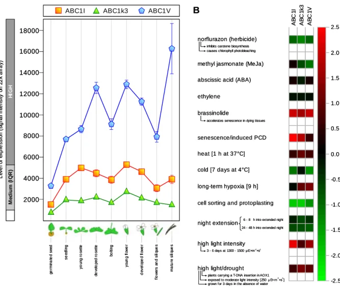
2.1.1 - The ABC1-like kinase family in Arabidopsis thaliana 56 2.1.2 - Genetic regulation of the plastoglobule ABC1-like kinases 63 2.1.2.1 - Expression profiles and response to stimuli 63
Functional characterization of three ABC1-like proteins identified in the plastoglobule proteome
10
2.1.2.2 - The three kinases are part of a common gene coexpression network 64
2.2 - Experimental setup 69
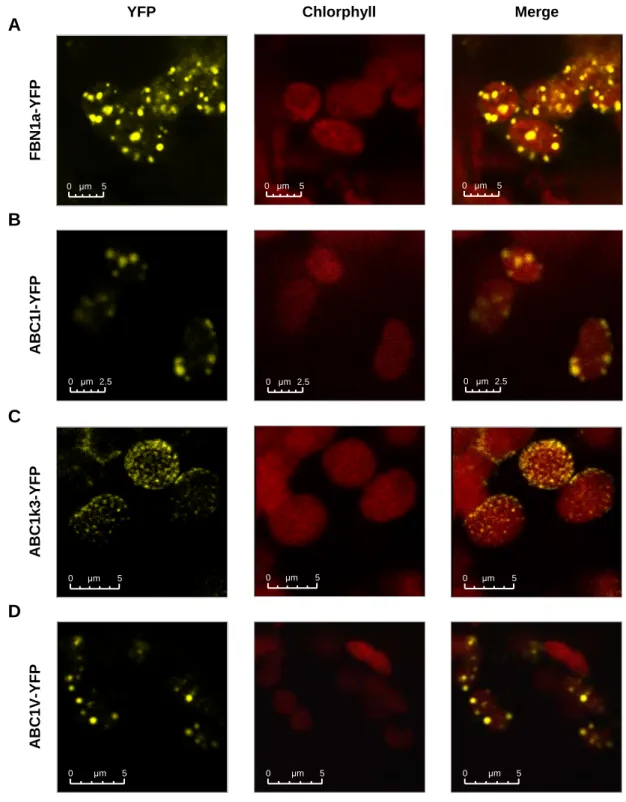
2.2.1 - The three ABC1-like kinases localize to plastoglobules in vivo 69
2.2.2 - Production of polyclonal antibodies 70
2.2.3 - Development of a novel method for prenylquinone and carotenoid profiling
in plant tissues 72
2.3 - Mutant characterization 75
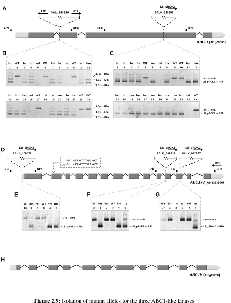
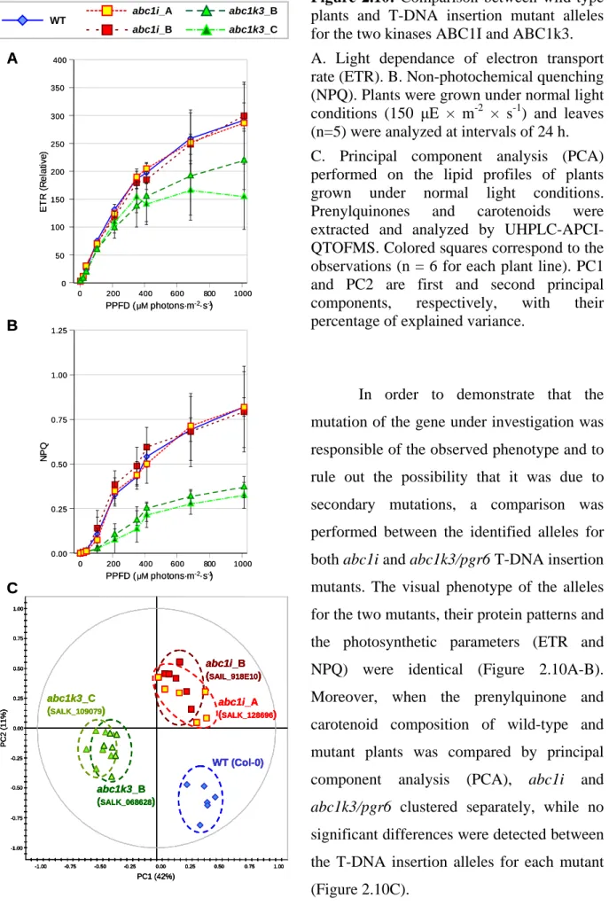
2.3.1 - Selection of mutant plant lines for the ABC1-like kinases 75 2.3.1.1 - Isolation of T-DNA insertion mutants 75 2.3.1.2 - ABC1V downregulation by RNA-mediated gene silencing 79 2.3.2 - abc1k3/pgr6 affects growth rate and pigment content 81 2.3.3 - Chloroplast ultrastructure is severely affected in the two mutants 83 2.3.4 - Photosynthetic activity and the pgr6 phenotype 85 2.3.5 - Starch and sugar metabolism are affected in abc1k3/pgr6 88 2.3.6 - ABC1I and ABC1k3 have a major effect on prenylquinone and carotenoid
composition of membranes 90
2.3.6.1 - Global effect on the lipid composition of mutant plants 91 2.3.6.2 - abc1i and abc1k3/pgr6 mutations affect carotenoid and
prenylquinone content 92
2.3.6.3 - Prenylquinone sub-organellar distribution 97 2.3.7 - The plastoglobule proteome is directly affected in both abc1i and
abc1k3/pgr6 mutant plants 98
2.3.7.1 - Plastoglobule analysis by immunoblotting 99 2.3.7.2 - Preliminary PG proteome profiling by mass spectrometry 104
2.3.8 - ABC1I and ABC1k3 phosphorylate VTE1 108
2.3.8.1 - Phosphorylation assays on purified chloroplast fractions 108 2.3.8.2 - The two kinases phosphorylates the recombinant VTE1 in vitro 108
CHAPTER 3 - Discussion
1043.1 - The ABC1 plastoglobule homologs 112
3.1.1 - ABC1-like kinases are both in chloroplasts and mitochondria 112 3.1.2 - ABC1I, ABC1k3 and ABC1V localize to plastoglobules in vivo 112 3.1.3 - Chloroplast ABC1 homologs are involved in the regulation of diverse
Functional characterization of three ABC1-like proteins identified in the plastoglobule proteome 11
3.2 - Initial remarks 115
3.2.1 - Selection of the experimental conditions 115 3.2.2 - A new method was required for rapid lipid profiling 116
3.3 - Functional characterization of ABC1I and ABC1k3 117
3.3.1 - ABC1I and ABC1k3 modulate VTE1 activity 117 3.3.1.1 - ABC1I specifically affects PC-8 synthesis and α-TQ recycling 117 3.3.1.2 - ABC1k3 activity is required both for the synthesis of tocopherols and
PC-8 and for the recycling of α-TQ 118
3.3.1.3 - ABC1I and ABC1k3 phosphorylate the tocopherol cyclase, possibly regulating its activity and its sub-organellar localization 118 3.3.2 - Effects on primary photosynthetic metabolism 120
3.3.2.1 - ABC1k3 affects photosynthetic activity via the modulation of
chloroplast lipid metabolism 120
3.3.2.2 - The lack of PC-8 and Vitamin E alone does not explain the photosynthetic phenotype of abc1k3/pgr6 mutants 122 3.3.2.3 - Starch and sugar metabolism are affected in the abc1k3/pgr6 mutant 123 3.3.3 - Other effects of the activity of the two kinases 126 3.3.3.1 - Pigment content is affected in the abc1k3/pgr6 mutant under HL stress 126 3.3.3.2 - Effects on chloroplast ultrastructure 126 3.3.3.3 - The plastoglobule proteome is severely affected in the two mutants 127
3.4 - Preliminary results on ABC1V 129
3.5 - Future perspectives 130
3.5.1 - Future characterization of ABC1I and ABC1k3 130 3.5.1.1 - Identification of other targets of the kinases 130 3.5.1.2 - Effects of the overexpression of the two kinases 130
3.5.2 - ABC1V functional characterization 131
3.5.3 - Lipid profiling 131
3.5.3.1 - Identification of carotenoids by UHPLC-APCI-QTOFMS 131 3.5.3.2 - Determine the lipid composition of chloroplast sub-fractions 132
3.5.4 - (Phospho)proteomics 133
3.5.4.1 - Extended profiling of the plastoglobule proteome 133 3.5.4.2 - Screening for phosphoproteins in plastoglobules 135 3.5.2.3 - Implementation of the in vitro phosphorylation assays 135
Functional characterization of three ABC1-like proteins identified in the plastoglobule proteome
12
CHAPTER 4 - Materials and Methods
1304.1 - Materials 138 4.1.1 - Plants 138 4.1.2 - Micro-organisms 138 4.1.3 - Oligonucleotides 138 4.1.4 - cDNA clones 139 4.1.5 - Plasmids 139 4.1.6 - Antibodies 140
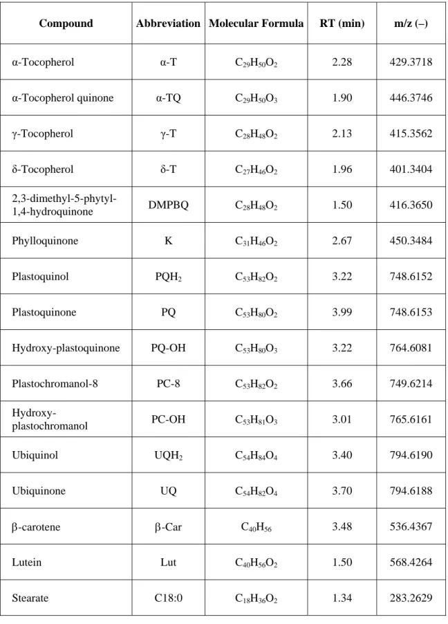
4.1.7 - Purified standards for Lipidomic analysis 140
4.1.8 - Chemicals 141
4.2 - Methods 142
4.2.1 - Plant Growth 142
4.2.1.1 - Growing A. thaliana on Murashige and Skoog medium 142 4.2.1.2 - Growing A. thaliana and N.benthamiana on soil 142
4.2.1.3 - High Light (HL) treatment 143
4.2.1.4 - Plastoglobule purification from A. thaliana 143
4.2.2 - Molecular biology techniques 144
4.2.2.1 - Molecular cloning 144
4.2.2.2 - Plasmid isolation and purification 145 4.2.2.3 - Genomic DNA extraction from A. thaliana leaves 145 4.2.2.4 - Isolation of knock-out mutant plants 145 4.2.2.5 - ABC1V knock-down by gene silencing. 147 4.2.2.6 - RNA extraction from A. thaliana leaves and cDNA preparation. 147 4.2.2.7 - Determination of gene expression rate by quantitative PCR. 147 4.2.2.8 - Protein extraction from A. thaliana total leaves. 154 4.2.2.9 - Protein separation and Western blot analysis 154
4.2.3 - Microbiology techniques 155
4.2.3.1 - Agrobacterium tumefaciens transformation by electroporation 155 4.2.3.2.- Transient transformation of tobacco leaf cells. 156 4.2.3.3 - Arabidopsis stable transformation. 156
4.2.4 - Analytical techniques 156
4.2.4.1 - Protein sub-cellular localization. 156 4.2.4.2 - Cloning for protein overexpression in plants. 157 4.2.4.3 - Cloning for Tandem Affinity Purification. 157 4.2.4.4 - Production of polyclonal antibodies. 158
Functional characterization of three ABC1-like proteins identified in the plastoglobule proteome 13 4.2.4.6 - Determination of the photosynthetic parameters. 158 4.2.4.8 - Arabidopsis chloroplast ultrastructural analysis. 159 4.2.4.10 - Arabidopsis membrane prenylquinone and carotenoid profiling. 161 4.2.4.11 - Plastoglobule proteome profiling. 162 4.2.4.12 - In vitro production of recombinant proteins. 163
4.2.4.13 - Phosphorylation assays. 164
4.2.4.13 - Bioinformatics. 165
Bibliography
159Appendix -
A novel method for prenylquinone profiling in plant tissues by ultra-high pressure liquid chromatography-mass spectrometry 190Functional characterization of three ABC1-like proteins identified in the plastoglobule proteome 15
C
HAPTER
1
Introduction
Functional characterization of three ABC1-like proteins identified in the plastoglobule proteome
16
1.1
-
P
LASTOGLOBULES1.1.1 - Overview of plastid lipid droplets
1.1.1.1 - Plastoglobules are highly dynamic structures in plastids
Early observations of different plastid types in transmission electron microscopy revealed the presence of “osmiophilic globules”(1-3), small lipid-filled vesicles which carried similarities to the lipid bodies formed at the endoplasmic reticulum (ER) in both animal and plant cells(4).
The average diameter of these lipid bodies, later termed plastoglobules, ranges from 30 to 100 nm in chloroplasts of vegetative leaf cells, but their number and size can significantly change (up to several μm) depending to the developmental stage and in response to particular environmental conditions(5). In particular, etioplasts with poorly developed thylakoids are characterized by a high number of plastoglobules which decreases during the light-induced conversion to chloroplasts(5-7), while an increased accumulation of plastoglobules was observed in mutants defective in thylakoid biogenesis(8,9). On the contrary, in senescing chloroplasts plastoglobules enlarge and increase in number as thylakoid membranes are disassembled(5,10-12) It has been also demonstrated that plastoglobules play a role in the formation of the carotenoid-rich colored fibrils during the chloroplast to chromoplasts transition in flowers and fruits(10,13,14). At the same time, both the size and the number of plastoglobules have been reported to increase in response to environmental conditions that aggravate oxidative stress on the photosynthetic apparatus. These include abiotic stress like the exposure to high light intensities(15), increased CO2 concentrations(16), drought(17) and
salinity(18), as well as in plants growing under nitrate starvation(19) and in soils containing high levels of heavy metals(20,21).
1.1.1.2 - Functional association with thylakoid membranes in chloroplasts
Plastoglobules consist of a neutral lipid core surrounded by a polar lipid monolayer that has been demonstrated to originate from the outer thylakoid membrane leaflet(22). Electron tomography analysis showed that plastoglobules “blister” from the thylakoid surface and are physically attached to the thylakoid membrane, which is consistent with reports that
Introduction
Functional characterization of three ABC1-like proteins identified in the plastoglobule proteome 17
plastoglobules are part of the thylakoid network(23,24) and is in disagreement with previous hypotheses proposing a plastoglobule origin at the inner chloroplast membrane(25) or their presence as independent plastid entities which float freely in the stroma(26). Moreover, the observation that plastoglobules are associated with highly curved regions of the thylakoids suggests that membrane curvature may be an important factor for their formation. It was also observed that in young chloroplasts most plastoglobules occur as single but they appear interconnected in small groups under stress conditions or during senescence, suggesting that new globules may also blister from previously-existing plastoglobules and form clusters not directly connected to thylakoids but linked to each other(22). Similarly, it was observed that fibrils elongate from plastoglobule surface during chloroplast transition to chromoplast(13).
1.1.1.3 - The algal eyespot apparatus
A particular case related to plastoglobules is represented by the Chlamydomonas
reinhardtii eyespot apparatus, a primordial visual system responsible for sensing light
direction, quality and intensity in unicellular green algae, thus representing an important biological model in the study of the evolution of photoreceptors(27).
Interestingly, a key component of this chloroplast light-sensing complex is represented by an highly organized lens system formed by one to several layers of carotenoid-rich lipid globules separated by single stroma thylakoid lamellae and which functionally correspond to plastoglobules. Moreover, early electron microscopy observations by thin sectioning and freeze fracturing revealed the presence of protein particles exactly outlining the shape of green algal eyespot globules, strongly suggesting the existence of a specialized protein interface towards the stroma(28-30).
1.1.1.4 - Plastoglobules as lipoprotein particles
Similarly to the lipid globules of the algal eyespot apparatus, plastoglobules from higher plants are characterized by the presence of proteins in addition to the lipid component. In particular, the detection of nitrogen in early biochemical experiments performed on purified plastoglobules suggested the presence of proteins associated with the lipid
Introduction
Functional characterization of three ABC1-like proteins identified in the plastoglobule proteome
18
globules(1,10), while later experiments demonstrated that these proteins were not contaminants from thylakoid membranes but were specific of plastoglobules(13,25,31,32).
Among these, both the fibrillin/plastoglobulin FBN1a/PGL35 and the tocopherol cyclase VTE1 were initially localized to plastoglobules by immunogold labelling of high-pressure frozen and freeze-substituted chloroplasts, thus confirming that these proteins are plastoglobule specific and do not occur elsewhere in the chloroplasts(22). Besides, these labelling experiments suggested that both proteins partially extends across the membrane monolayer that surround plastoglobules and into the neutral lipid core.
Owing to their structure and composition, plastoglobules are easily separated from thylakoid membranes by sonication or mechanical homogenization and can be conveniently isolated by flotation density centrifugation because of their low density that is due to the high lipid content. Highly pure plastoglobules allowed to investigate their lipid and protein composition while minimizing the possible contaminations from other chloroplast fractions, the thylakoids in particular.
Neutral lipid core
Polar lipid monolayer ATP synthetase Photosystem I Photosystem II Structural proteins (Fibrillins/Plastoglobulins) Other proteins
Neutral lipid core
Polar lipid monolayer ATP synthetase Photosystem I Photosystem II Structural proteins (Fibrillins/Plastoglobulins) Other proteins ATP synthetase Photosystem I Photosystem II Structural proteins (Fibrillins/Plastoglobulins) Other proteins
Introduction
Functional characterization of three ABC1-like proteins identified in the plastoglobule proteome 19
1.1.2 - Lipid composition
The lipid composition of plastoglobules extracted from chloroplasts is markedly different from that of non-green plastids. It has been demonstrated that plastoglobules contain prenylquinones like plastoquinone (PQ-9)(5,10,33,34), its derivate plastochromanol (PC-8)(34,35), tocopherols (Vitamin E)(36) and phylloquinone (Vitamin K)(33,34). In particular, experimental evidence suggested that plastoglobules have a role as a sink for the deposit of excess K that does not associate with PSI(33) and for a significant amount of the PQ-9 pool in chloroplast membranes(35).
Traces of carotenoids (β-carotene, lutein) and chlorophylls, as well as phospho- and galactolipids have also been detected in lipid globules purified from chloroplast fractions(2), although their presence in plastoglobule preparations at the time was attributed to contamination by thylakoids(10). Conversely, a significant carotenoid accumulation is observed in correlation with the chloroplast to chromoplast transition, during which carotenoids localize primarily in plastoglobules(10). Moreover, it was observed that carotenoids within plastoglobules exhibit much higher stability than those within chloroplast membranes, possibly because of the presence of other lipid-soluble antioxidants. In turn, their accumulation could also contribute to the protection from spontaneous photo-oxidation of triacylglycerols, unsaturated lipids and other light sensitive molecules(37).
Plastoglobules extracted from gerontoplasts (“old” chloroplasts) in senescing leaves accumulate free fatty acids, oxidised prenylquinones and carotenoid esters(10), the latter of which accumulate in the hydrophobic core of chromoplast plastoglobules and fibrils, contributing to color development in fruits and petals(13). Moreover, under stress conditions like nitrogen starvation membrane-destabilizing compounds are released that include phytol from chlorophyll degradation and acyl groups from membrane lipids. However, their incorporation in fatty acid phytyl esters (FAPE) inside plastoglobules prevents plastid membrane damage(19).
Plastoglobules also have a major role as lipid deposits in elaioplasts, particular types of leucoplasts enriched in oil bodies and present notably in tapetum cells, in which lipids are deposited in maturing pollen(32).
Introduction
Functional characterization of three ABC1-like proteins identified in the plastoglobule proteome
20
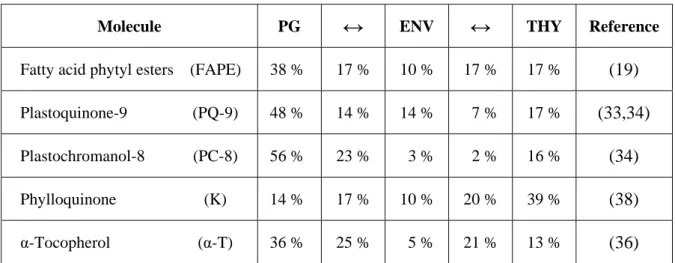
Molecule PG
↔
ENV↔
THY ReferenceFatty acid phytyl esters (FAPE) 38 % 17 % 10 % 17 % 17 % (19)
Plastoquinone-9 (PQ-9) 48 % 14 % 14 % 7 % 17 % (33,34) Plastochromanol-8 (PC-8) 56 % 23 % 3 % 2 % 16 % (34) Phylloquinone (K) 14 % 17 % 10 % 20 % 39 % (38) α-Tocopherol (α-T) 36 % 25 % 5 % 21 % 13 % (36)
Table 1.1: Reported relative distribution of selected neutral lipids between chloroplast
sub-fractions. PG. Plastoglobules. ENV. Envelope. THY. Thylakoids.
1.1.3 - Protein composition
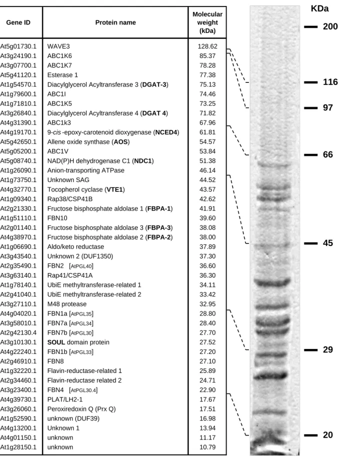
In an effort to precisely determine the protein composition of plastoglobules, several independent proteome studies were performed on highly purified fractions by mass spectrometry analysis(36,39,40). These resulted in the identification of about forty proteins that constitute the Arabidopsis plastoglobule proteome (Figure 1.2 on page 17).
More recently, in an attempt to identify the proteins highly enriched in plastoglobules and likely specific for these sub-organellar compartments, an in silico statistical filtering was applied to distinguish the “PG core” proteins from potential contaminants from outside plastids or localized primarily in other sub-organellar compartments(40). As a consequence, some of the proteins previously included in the plastoglobule proteome were removed, leaving only about thirty proteins in the bona fide “PG core”. However, this does not imply that the excluded proteins are absent from plastoglobules and simply correspond to contaminants but it strongly suggests that their primary localization is elsewhere inside chloroplasts, suggesting that their presence in plastoglobules might not be necessary to carry out their primary biological role.
Independently from the number of candidate proteins included in each study, plastoglobule proteins were further divided into three main groups: structural proteins, known metabolic enzymes and proteins of unknown function.
Functional characterization of three ABC1-like proteins identified in the plastoglobule proteome 21 Introduction KDa 200 116 97 66 45 29 20
Gene ID Protein name
Molecular weight (kDa) At5g01730.1 WAVE3 128.62 At3g24190.1 ABC1K6 85.37 At3g07700.1 ABC1K7 78.28 At5g41120.1 Esterase 1 77.38
At1g54570.1 Diacylglycerol Acyltransferase 3 (DGAT-3) 75.13
At1g79600.1 ABC1I 74.46
At1g71810.1 ABC1K5 73.25
At3g26840.1 Diacylglycerol Acyltransferase 4 (DGAT 4) 71.82
At4g31390.1 ABC1k3 67.96
At4g19170.1 9-cis -epoxy-carotenoid dioxygenase (NCED4) 61.81
At5g42650.1 Allene oxide synthase (AOS) 54.57
At5g05200.1 ABC1V 53.84
At5g08740.1 NAD(P)H dehydrogenase C1 (NDC1) 51.38
At1g26090.1 Anion-transporting ATPase 46.14
At1g73750.1 Unknown SAG 44.52
At4g32770.1 Tocopherol cyclase (VTE1) 43.57
At1g09340.1 Rap38/CSP41B 42.62
At2g21330.1 Fructose bisphosphate aldolase 1 (FBPA-1) 41.91
At1g51110.1 FBN10 39.60
At2g01140.1 Fructose bisphosphate aldolase 3 (FBPA-3) 38.08
At4g38970.1 Fructose bisphosphate aldolase 2 (FBPA-2) 38.00
At1g06690.1 Aldo/keto reductase 37.89
At3g43540.1 Unknown 2 (DUF1350) 37.30
At2g35490.1 FBN2 [AtPGL40] 36.60
At3g63140.1 Rap41/CSP41A 36.30
At1g78140.1 UbiE methyltransferase-related 1 34.11
At2g41040.1 UbiE methyltransferase-related 2 33.42
At3g27110.1 M48 protease 32.95
At4g04020.1 FBN1a [AtPGL35] 28.80
At3g58010.1 FBN7a [AtPGL34] 28.40
At2g42130.4 FBN7b [AtPGL30] 27.70
At3g10130.1 SOUL domain protein 27.52
At4g22240.1 FBN1b [AtPGL33] 27.20
At2g46910.1 FBN8 27.10
At1g32220.1 Flavin-reductase-related 1 25.89
At2g34460.1 Flavin-reductase related 2 24.71
At3g23400.1 FBN4 [AtPGL30.4] 22.90
At4g39730.1 PLAT/LH2-1 17.67
At3g26060.1 Peroxiredoxin Q (Prx Q) 17.51
At1g52590.1 unknown (DUF39) 16.98
At4g13200.1 Unknown 1 13.94
At4g01150.1 unknown 11.17
At1g28150.1 unknown 10.79
Figure 1.2: The plastoglobule proteome.
List of all the proteins currently identified in plastoglobule preparations. The predicted molecular weight and the relative position range after separation by SDS-PAGE are also indicated.
Introduction
Functional characterization of three ABC1-like proteins identified in the plastoglobule proteome
22
In particular, this last group includes members with sequence similarities with known proteins having annotated enzymatic or metabolic functions but for which no precise enzymatic or regulatory activity has been reported yet. Among these, this thesis gives particular attention to the description of the members of the ABC1/ADCK/UbiB kinase family (see Section 1.2).
1.1.3.1 - Plastoglobule structural proteins (PAP/fibrillins/plastoglobulins)
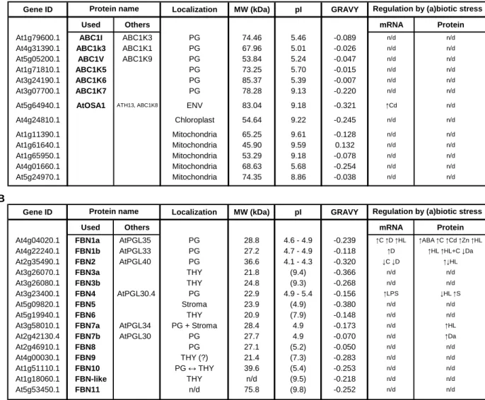
The PAP/fibrillin/plastoglobulin family in Arabidopsis consists of twelve fibrillin (FBN) proteins as well as an FBN-like homolog(41). Interestingly, seven FBNs were identified principally in highly purified plastoglobules, while another member of this family (FBN10) is predicted to have a dual localization between plastoglobules and the stroma-exposed thylakoid surface(36,39,40) (see Figure 2.1 on page 54).
Fibrillins (FBN) are named after fibrils, the sub-organellar structures in chromoplasts in which the proteins were first identified. In chromoplasts, fibrils are tube- or thread-like structures of varying shape and thickness related to plastoglobules and correspond to the main site of pigment accumulation, in particular carotenoids as well as glyco- and phospho-lipids(42).Alternatively, considering their strong association with plastoglobules in other plastid types, other names have been proposed such as “Plastid lipid-Associated Proteins” (PAP) and “Plastoglobulins” (PGL)(43), although members of this family have been identified in thylakoids as well. For reference, the alternative names for the members of the PAP/fibrillin/plastoglobulin family are reported in Table 2.1 on page 53.
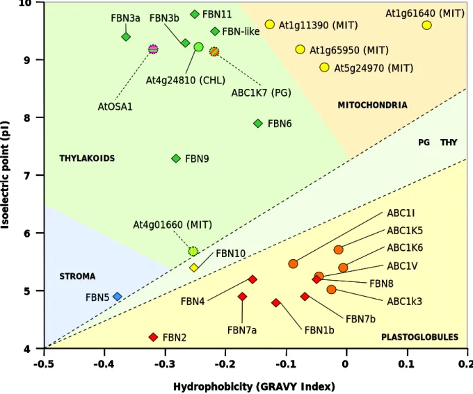
Fibrillins include proteins with different physical-chemical properties, having molecular weights that usually range from 20 to 40 kDa and isoelectric points (pI) comprised between 4 and 9(41) (see Table 2.1B on page 53), thus suggesting possible variations in the biological functions of each member of the fibrillin family. Furthermore, despite the plastid nature of all fibrillins, their selective distribution between different sub-organellar compartments suggests a specialization in their functions according to the plastid type and the precise localizations in specific plastid structures.
As previously discussed, fibrillins were first identified as protein components of chromoplast fibrils(13,42), and their presence is sufficient for the self-assembly of fibril-like structures in the presence of lipid droplets enriched in carotenoids(13). Besides, eight of the C. reinhardtii
Introduction
Functional characterization of three ABC1-like proteins identified in the plastoglobule proteome 23
fibrillins are associated with the carotenoid-enriched globules of the eyespot apparatus, and their activity is necessary to stabilize the interactions between globule clusters during the formation of the highly ordered eyespot complex.(30,44). Similarly, the degree of plastoglobule clustering in higher plants strongly correlates with the relative abundance of fibrillins, indicating that these proteins mediate plastoglobule clustering by preventing their coalescence and possibly mediating cross-links between lipoprotein particles(45,46).
In chloroplasts, several fibrillins are located on the surface of chloroplast plastoglobules and are required for their assembly as well as for the accumulation of lipids inside them(47,48). On the other hand, fibrillins have also been identified in locations other than plastoglobules. In addition to the previously discussed FBN10, at least six fibrillins are also associated with thylakoid membranes, while only one FBN protein is located in the stroma.
Therefore, fibrillin sub-organellar localization is not fixed but appears to be highly dynamic. In particular, the two plastoglobule-predicted fibrillins FBN1a and FBN4 were found to be components of the Light-harvesting complex II (LHCII) in Arabidopsis plants exposed to light stress(49,50).
The differential regulation of the members of the fibrillin family by a variety of biological and environmental factors, as well as during different plant growth stages, strongly suggests the existence of a complex regulatory system for fibrillins both at the transcript and protein levels(51). In particular, in various plant species the expression rate of the family member FBN1 is enhanced in correlation with the chloroplast to chromoplast transition, during fruit ripening(13,31), as well as in senescent leaves, while it is lowest in young leaves(52,53). Similarly, treatment with abscissic acid (ABA) accelerates FBN1 accumulation in plants, while high auxin (IAA) levels significantly delays it(13).
At the same time, fibrillin gene expression is affected in response to stress, both biotic such as viruses, bacteria and pathogenic fungi, and to a larger extent abiotic(51). In particular, complex and varied patterns in the regulation of both fibrillin transcript and protein levels are observed in response to heat, cold, drought, salinity, high light intensity and after exposure to herbicides or heavy metals such as Cd2+ and Zn2+.(39, 51,53-56) (see Table 2.1B on page 53).
Introduction
Functional characterization of three ABC1-like proteins identified in the plastoglobule proteome
24
Despite the substantial amount of information relative to the expression and post-transcriptional regulation of fibrillins in plants, relatively little is known about their biological functions and mechanisms of action.
In view of the wide range of molecules with known antioxidant activity accumulating inside plastoglobules, it is not surprising that fibrillins have previously been implicated in plant resistance to oxidative stress, their mutation significantly affecting plastoglobule formation and lipid content(47,48). However, functional studies have been carried out only for three out of the twelve fibrillins identified in Arabidopsis (FBN1a/b, FBN2 and FBN4).
Other functions have been proposed for these fibrillins though. In particular, indications of a possible involvement in chloroplast development come from the increase in FBN1a and FBN1b levels in Arabidopsis mutants defective for the ClpPRS protease complex required in chloroplast biogenesis and characterized by a delayed development, a pale green phenotype, and an increased numbers of plastoglobules(9,57). At the same time, the increase in plastoglobule size and number during senescence has been associated with the disassembly of thylakoid membranes and the accumulation of the catabolites originating from membrane lipid degradation(58). As previously discussed, plastoglobules have been reported to contain triacylglycerol (TAG) but few galactolipids(10,19). The increase in TAG levels in plastoglobules during senescence has been proposed to correlate with galactolipid mobilization from thylakoids, concomitantly with the conversion of thylakoid fatty acids to phloem-mobile sucrose(59). Indications come from the identification of a putative TAG lipase localized in plastoglobules and which can mobilize the TAG accumulating there. Knock-down mutant plants for this protein have been reported to be delayed in the initiation of a senescence program and are characterized by smaller but more numerous plastoglobules and fewer and deformed thylakoids, which would be consistent with a delay in the onset of senescence in rosette leaves(60).
Besides, the observed increase in plastoglobule size in older chloroplasts has been also proposed to represent a response to the accumulation of reactive oxygen species (ROS) concentration during plant aging(17).
On the other hand, the analysis of fibrillin structure highlighted the presence of a conserved domain which is highly similar to the “motif 1” of lipocalins(61), a large protein family present in prokaryotes, insects, vertebrates, and plants whose members are characterized by the ability to bind small hydrophobic ligands and often work as part of protein complexes(62). This would be consistent with the predicted biological activity of fibrillins, in particular with the
Introduction
Functional characterization of three ABC1-like proteins identified in the plastoglobule proteome 25
plastoglobule-based trafficking of small hydrophobic molecules such as carotenoids, plastoquinone and tocopherols(10). Furthermore, the accumulation of FBN1a in Arabidopsis plants treated with ABA has also been reported to stabilize Photosystem II (PSII) and enhance its protection from photoinhibition(63).
1.1.3.2 - Chloroplast metabolic proteins
For at least seven among the proteins of the chloroplast plastoglobule proteome by mass spectrometry analysis(36,39,40) an enzymatic activity was previously known or demonstrated after further characterization.
The tocopherol cyclase (VTE1) was previously thought to associate exclusively with the chloroplast inner envelope membrane. However, the specific association with plastoglobules of VTE1 was independently confirmed by electron tomography(22). In chloroplasts, VTE1 is a key enzyme not only for tocopherol synthesis(64,65) and Redox recycling(66) but it is also responsible for the synthesis of plastochromanol-8 (PC-8), resulting from the cyclization of PQ-9(34,35,67). Furthermore, the overexpression of VTE1 in transgenic plant lines resulted in the over-accumulation of PC-8, while tocopherol levels were only slightly affected(34,68), thus suggesting that VTE1 activity is a limiting factor for PC-8 rather than Vitamin E synthesis. At the same time, Northern Blot analysis revealed that VTE1 expression is up-regulated in response to the exposure of plants to high light irradiance, further suggesting a connection of VTE1 activity with the response to oxidative stress in plants(34,69). The activity and regulation of VTE1 will be discussed further with regard to its multiple roles in prenylquinone metabolism (see Section 1.3.1.2).
The precise sub-organellar localization of VTE1 in chloroplasts became the object of intense investigation. Interestingly, despite the fact that most of the enzymes involved in tocopherol metabolism have previously been localized to the inner envelope membrane(70), several independent screenings by mass spectrometry and immunolabeling strongly indicate that plastoglobules are the major site of VTE1 localization in chloroplasts(22,36,39,40). Moreover, observations performed at different developmental stages demonstrated that VTE1 localizes to plastoglobules throughout the whole Arabidopsis life cycle(36). At the same time, the
Introduction
Functional characterization of three ABC1-like proteins identified in the plastoglobule proteome
26
intersection of PC-8 and Vitamin E synthesis in chloroplasts appears to be consistent with a plastoglobule localization of VTE1 activity(34).
Another protein whose involvement in prenylquinone metabolism has been recently investigated is the NAD(P)H dehydrogenase C1 (NDC1), the only member of the type II NAD(P)H quinone oxidoreductase (NDH) gene family that localizes not only to mitochondria but to chloroplasts as well, and more specifically to plastoglobules(71-73). In chloroplasts, the NDH complex is known to be involved in the cyclic electron flow around Photosystem I (PSI) by oxidizing ferredoxin using the PQ-9 pool as electron acceptor(74), similarly to the PGR5 complex(75). However, the sub-organellar localization of the family member NDC1 and its ability to oxidize NADH in the presence of purified plastoglobules suggests that the enzyme may rely on the plastoglobule PQ-9 pool rather than that in the thylakoids(76).
At the same time, the ndc1 mutant is characterized by the lack of phylloquinone (Vitamin K) and plants accumulate its direct precursor 2-phytyl-1,4-naphthoquinone. Furthermore, a moderate reduction in PC-8 content was also detected in mutant plants. Interestingly, the enzyme required for the last methylation step in K biosynthesis (MenG, At1g23360) does not appear to be present. In addition, MenG expression levels did not significantly change between wild-type and ndc1 plants. It has been proposed that MenG activity may be dependent on the Redox state of the PQ-9 plastoglobule pool(76,77). Most strikingly however, both ndc1 and menG mutant had no visible phenotype. It has been previously demonstrated that in the absence of K, PS I recruits PQ-9 into the A1 site, where it functions as an efficient cofactor in electron transfer from A0 to the iron-sulfur clusters(78,79), while it has been also suggested that the 2-phytyl-1,4-naphthoquinone precursor might functionally replace K in PSI(76).
At the same time, the reduction in PC-8 levels was attributed to the selectivity of VTE1 for the reduced form of PQ-9 (PQH2) as substrate for PC-8 synthesis(77). For instance, a
significant accumulation of the oxidized form of PQ-9 (PQ) was reported in ndc1 plants extracted with MeOH(76). However, the use of a solvent with relatively high polarity such as MeOH was found to be unsuitable for the extraction of apolar prenylquinones such as PQ-9 and PC-8, as well as in maintaining the original PQ/PQH2 ratio, as previously reported(80).
Introduction
Functional characterization of three ABC1-like proteins identified in the plastoglobule proteome 27
With regard to the involvement of plastoglobules in carotenoid metabolism, the identification of a 9-cis-epoxy-carotenoid dioxygenase (NCED4) in chloroplast plastoglobules needs to be discussed.
In plants, NCEDs are a subgroup of the carotenoid cleavage dioxygenases (CCD) family, responsible for the first enzymatic step in the formation of apocarotenoids, a class of terpenoid compounds generated by the oxidative cleavage of a carotenoid molecule at one of its double bonds(81-86). The CCD gene family consists of nine putative members in A. thaliana, five of which encode chloroplast-localized proteins that differ in their sub-organellar location(84) and are believed to be involved in the synthesis of apocarotenoids such as the phytohormone ABA. In particular, the significant accumulation of NCED4 in plastoglobules from leaves kept in darkness suggests an active role of the protein in the dark- and probably senescence-induced breakdown of carotenoids(39). Moreover, NCED4 has been reported to interact with the zinc-finger protein VAR3 (At5g17790). In A. thaliana, the variegated 3 (var3) mutant is characterized by somatic areas lacking or containing developmentally-retarded chloroplasts and by a significant reduction in carotenoid levels as well as a minor reduction in chlorophyll content(87).
Interestingly, a significant accumulation of other enzymes involved in carotenoid metabolism was detected during the chloroplast to chromoplast transition in red bell pepper fruits(39). In particular, the increase in the levels of enzymes required for the synthesis of bicyclic carotenoids (α/β-carotene and xanthophylls) such as the ζ-carotene desaturase (ZDS), the lycopene-β-cyclase (β-Lyc) and two β-carotene-β-hydroxylases (LUT5 or CrtR-β) further suggests an active role of plastoglobules in carotenoid synthesis.
Although its localization in plastoglobules is not exclusive and it is also present in envelope and thylakoid membranes, the identification of the allene oxide synthase (AOS) in chloroplast lipid globules is indicative of their possible involvement in plant response to abiotic stress involving jasmonic acid (JA) and it is also consistent with their predicted role during thylakoid membrane disassembly.(40). During senescence, the thylakoid membrane is dismantled, resulting in the release of mono- and di-galactosyl glycerols, as well as free fatty acids. These can become the substrate of AOS, which is one of the first enzymes acting in the lipoxygenase pathway which leads to the formation of JA and it is responsible for the conversion of 13-hydroperoxylinolenic acid into an unstable epoxide. Considering the presence of high concentrations of potential fatty acid-containing substrates, it is possible that
Introduction
Functional characterization of three ABC1-like proteins identified in the plastoglobule proteome
28
the localization of AOS activity at plastoglobules might contribute to the massive production of JA during senescence. It is also possible that the free fatty acids released during thylakoid disassembling might become substrate for other putative enzymes identified in the plastoglobule proteome, such as two diacylglycerol acyltransferases (DGAT-3/4) and one putative esterase/lipase/thioesterase, although these proteins have not been characterized yet.
Most surprisingly, three stromal fructose-bisphosphate aldolase (FBPA-1 to 3) were highly abundant in plastoglobule preparations. Considering their high stroma:plastoglobule abundance ratio, it is doubtful whether these proteins should be considered bona fide plastoglobule proteins(40). On the other hand, the certified presence of the three aldolases in plastoglobules (even higher than many “PG core” proteins) together with the absence of other abundant chloroplast enzymes involved in glycolysis and in the Calvin cycle strongly suggest that the three FBPAs may have a specific function in association with plastoglobules(39).
1.1.4 - The plastoglobule gene coexpression network
A genome-wide gene coexpression network has been recently proposed based on the mRNA expression data available for all the proteins specifically localizing to plastoglobules(40). In particular, most of the “PG core” proteins had strong associations with the expression of other plastoglobule proteins, as well as that of chloroplast proteins not located in plastoglobules. Based on these in silico predictions, the existence of four distinct functional modules was highlighted, each of them enriched in specific biological, biochemical and physiological functions. In particular, one module was characterized by genes involved in protein and chlorophyll degradation during senescence, while another one contained proteins affecting plastid biogenesis and several enzymes involved in the Calvin cycle. On the other hand, a third module included Redox regulators, plastid proteases and phosphatases as well as nonlinear electron flow components involved in plant photoacclimation. However, the largest module was predicted to have a central role in plastoglobule metabolism, being characterized by genes required for carotenoid and prenylquinone metabolism, as well as regulators of photosynthetic activity and state transition.
Moreover, each module contained at least one fibrillin and one ABC1-like kinase, suggesting a specific role of each of these proteins in different metabolic and regulatory pathways.
Introduction
Functional characterization of three ABC1-like proteins identified in the plastoglobule proteome 29
1.2
-
ABC1-
LIKE KINASESAmong the about thirty proteins identified in plastoglobules by mass spectrometry analysis(36,39,40), only a few belonged to the fibrillin family of structural proteins or were known enzymes with an established biochemical function such as the tocopherol cyclase (VTE1) or the allene oxide synthase (AOS). Conversely, the biological role of most of the proteins predicted to localize to plastoglobules was still largely unknown. However, based on sequence similarities with known proteins, many of these putative enzymes have annotated enzymatic or metabolic functions.
The case of interest for this thesis is represented by the members of the ABC1/ADCK/UbiB family of atypical kinases, which is completely unrelated to the ABC (ATP Binding Cassette) family of transport proteins(88). The ABC1/ADCK/UbiB family consists of putative kinases identified by sequence alignment methods (Psi-Blast and HMMs) and which have a domain that is similar to the eukaryotic protein kinase (ePK) domain. This includes the most conserved metal binding residues and catalytic motifs. An attempt to further define the ABC1 kinase domain has recently been carried out by multiple sequence alignment of ABC1-like homologs from different plant species as well as by comparison with model kinases belonging to the ePK superfamily(89).
Members of the ABC1 family were found both in bacteria and eukaryotes. These findings suggest that the family evolved in bacteria and entered early eukaryotes by horizontal transfer(90). Moreover, the phylogenetic analysis of ABC1-like homologs in diverse species of eukaryotes, bacteria and Archaea further highlighted a common evolutionary origin preceding that of the endosymbiotic events leading to the formation of mitochondria and chloroplasts(89).
The ABC1/ADCK/UbiB family includes ABC1 (YGL119W) from yeast, YigR from
E. coli and AarF from Providencia stuartii. The prototypical family member, S. cerevisiae
ABC1/Coq8 (Activity of bc1 complex/ COenzyme Q biosynthesis), is defective in aerobic
respiration because of a reduction in the activity of the mitochondrial bc1 complex which can
be overcome by exogenously supplied decylubiquinol(91,92). Similarly, mutations of the human ABC1 homolog (CABC1 or ADCK3) cause ubiquinone deficiency and defects in the respiratory chain, resulting in a progressive neurological disorder with cerebellar atrophy, developmental delay, and hyperlactatemia(93). To summarize, the inactivation of the ABC1
Introduction
Functional characterization of three ABC1-like proteins identified in the plastoglobule proteome
30
gene does not abolish the synthesis of cytochromes b, c1, c and aa3 but strongly decreases the
in situ endogenous reduction of cytochromes cl and c. The loss of NADH and succinate Cyt. c
oxidoreductase activities seems to be due to the inability to reduce Cyt. c, suggesting a deficiency in the activity of the bc1 segment.
Studies performed on bacteria showed that YigR, the E. coli homologue of ABC1, corresponds to UbiB, a gene required for the first monooxygenase step in ubiquinone biosynthesis(94). Both the P. stuartii aarF and E. coli ubiB (yigR) disruption mutant strains lack ubiquinone and accumulate octaprenylphenol, an intermediate in ubiquinone biosynthesis pathway. Moreover, ABC1 has been shown to be able to complement the yeast mutant coq8-1, defective in ubiquinone synthesis(95).
However, the data do not suggest an enzymatic function for ABC1 but point to a regulatory role in ubiquinone biosynthesis. Prior experiments identified a pool of octaprenylphenol that remained bound to a protein complex until it was activated in the presence of oxygen to continue the ring modifications required in ubiquinone biosynthesis(96). The rapid conversion of octaprenylphenol to UQ-8 upon transfer from anaerobic to aerobic growth conditions is consistent with a mechanism that may require kinase regulation. In support to this hypothesis, it has been demonstrated in S. cerevisiae that Coq3, Coq5, and Coq7 polypeptides are phosphorylated in a ABC1/Coq8-dependent manner and that the expression of the human homolog ADCK3/CABC1 rescued the growth of yeast coq8 mutants as well as the phosphorylation state of several of the Coq polypeptides, suggesting the existence of multiple enzyme targets of the kinase in the ubiquinone biosynthesis pathway(97).
Until recently, ABC1-like kinases have been mainly studied in mitochondria and bacteria, while little evidence was obtained on the role of their chloroplast homologs.
One of these putative kinases, AtOSA1 (A. thaliana Oxidative Stress-related ABC1-like protein) was identified in the inner envelope membrane of A. thaliana chloroplasts(98). The analysis of gene expression showed that AtOSA1 transcription is induced by Cd2+ treatment and oxidative stress conditions, and knock-out plants for this gene permanently suffer from oxidative stress. However, AtOSA1 was unable to complement yeast strains lacking the endogenous ABC1 gene, and this suggests a different function for this chloroplast kinase when compared to mitochondrial ABC1. Similarly, the expression of the maize homolog of AtOSA1 was enhanced in response to Cd2+, while its mRNA levels were lower when plants were grown in darkness and after treatment with ABA and H2O2(99).
Introduction
Functional characterization of three ABC1-like proteins identified in the plastoglobule proteome 31
Another kinase belonging to the ABC1 family, the C. reinhardtii serine/threonine protein kinase EYE3, has been recently demonstrated to localize to the pigment granule arrays in the eyespot apparatus and to be required for their assembly(100) (see Section 1.1.1.3). Furthermore, a strong correlation in the expression of the A. thaliana homolog of EYE3 (ABC1K6, At3g24190) with the zeaxanthin epoxidase (ZEP) has been predicted as well, suggesting a possible involvement in the regulation of the xanthophyll cycle(40).
Interestingly, six out of the eight ABC1-like kinases currently identified in A. thaliana chloroplasts were highly enriched in the plastoglobule proteome(36,39,40). Considering the coincident presence of several enzymes involved in prenylquinone metabolism in plastoglobules, it appears possible that the ABC1-like kinases are involved in the regulation of prenylquinone biosynthesis comparable to ABC1 in bacteria and mitochondria. Moreover, indications of a possible regulatory role of ABC1-like kinases in carotenoid biosynthesis, photoacclimation, plastid biogenesis and senescence come from the previously-reported genome-wide coexpression analysis(40), suggesting the existence of a wide plastoglobule regulatory network affecting multiple metabolic pathways.
In particular, the research project carried out during this PhD thesis was aimed at the functional characterization of three out of the six ABC1-like kinases located in plastoglobules: ABC1I (At1g79600), ABC1k3 (At4g31390) and ABC1V (At5g05200).
A change in the nomenclature of ABC1 homologs has been recently proposed(40) but it was not embraced in this report to avoid inconsistencies with the experimental material produced during the PhD project and presented in this thesis. For reference, the alternative names for the three kinases are reported in Table 2.1 on page 53.
Relatively to ABC1k3, it has been recently demonstrated that this kinase is required to maintain the number of chlorophyll-binding proteins in the photosynthetic complexes and that knock-down plants accumulate chlorophyll degradation products, have a lower anthocyanin content and are more sensitive to photooxidative stress under high light conditions(101). Moreover, it is here demonstrated that ABC1k3 corresponds to the photosynthetic mutant
pgr6 (proton gradient regulation 6), previously identified in a screening of Arabidopsis
mutants showing a reduced quenching of chlorophyll fluorescence(102) and which will be described in details in the following sections (see Section 1.3.2.1).
Introduction
Functional characterization of three ABC1-like proteins identified in the plastoglobule proteome
32
1.3
-
R
OLE OFABC1
HOMOLOGS IN CHLOROPLAST METABOLISM1.3.1 - Lipid metabolism
Although plastoglobules were initially considered passive lipid storage sites, increasing evidence demonstrated that they play a fundamental role not only in chloroplast development, being directly involved in thylakoid membrane formation as well as in its disassembly during senescence, but also in the transition to chromoplast and the accumulation of carotenoid pigments. Most strikingly however, the identification in the plastoglobule proteome of several enzymes belonging to prenylquinone and carotenoid biosynthetic pathways demonstrated that plastoglobules play an active role not only in the accumulation but also in the synthesis of these molecules. Considering their strategic position at the intersection of different pathways in plastid lipid metabolism, it is then conceivable that plastoglobule activity is highly regulated, suggesting the existence of a dynamic regulatory network. Although, on the one hand, the key enzymes involved in carotenoid and prenylquinone biosynthesis are known for the best part, the regulation of these pathways is still poorly understood. On the other hand, the identification of six ABC1-like kinases in chloroplast lipid globules makes them suitable candidates for such regulators.
In order to highlight possible key regulatory nodes shared between biochemical pathways apparently different but being part of a common metabolic network, an understanding of the biosynthesis of both carotenoids and prenylquinones is required. At the same time, a particular reference will be given in the following sections to the intersection of plastoquinone and tocopherol synthetic pathways in plastoglobules, which both represent a potential target of the activity of the chloroplast ABC1-like kinases, comparable to ubiquinone synthesis regulation by ABC1 in bacteria and mitochondria.
Introduction
Functional characterization of three ABC1-like proteins identified in the plastoglobule proteome 33
1.3.1.1 - Carotenoids
Carotenoids are an assorted class of pigments naturally found in bacteria, fungi, algae and plants, in which they play various roles in development, photosynthesis as well as the synthesis of phytohormones such as strigolactones and abscisic acid. On the contrary, animals are unable to synthesize carotenoids but they can absorb them through their diet. The beneficial effects of carotenoids on human and animal health have been extensively reviewed, with particular reference to their antioxidant activity, their valuable effect on the immune system and their role as precursors of retinol (Vitamin A).(103-105).
Carotenoid biosynthesis is a highly regulated process throughout the life cycle of the plant and it is characterized by dynamic changes to match plant developmental requirements during germination, photomorphogenesis, photosynthesis, fruit development as well as in response to external environmental stimuli. A general model of the carotenoid biosynthetic pathway is reported in Figure 1.3.
Like all isoprenoids, carotenoids are assembled from the condensation of monomers of the isopentenyl diphosphate (IPP) and of its double-bond isomer dimethylallyl diphosphate (DMAPP). Although two independent pathways exist in plant cells for the synthesis of these prenyl diphosphate precursors, one in cytoplasm via mevalonate and one in plastids, carotenoids are mainly derived from the 2-C-methyl-D-erythritol 4-phosphate/1-deoxy-D-xylulose 5-phosphate (MEP/DOXP) pathway of terpenoid biosynthesis. This pathway is entirely localized in plastids and leads to the formation of geranylgeranyl diphosphate (GGPP)(106-108). Defects in isoprenoid biosynthesis or changes in other metabolic pathways using GGPP as substrate can significantly affect carotenoid synthesis as well. Furthermore, the availability of isoprenoid precursors can be significantly influenced by abiotic factors such as light and oscillations of the circardian rhythm as well as biotic factors such as the colonization of plant roots by arbuscular mycorrhizal fungi(109).
Figure 1.3: Model of carotenoids, xanthophylls and abscissic acid biosynthetic pathways and
their regulation in Arabidopsis. The modifications introduced in each enzymatic step are indicated with grey triangles.
Introduction
Functional characterization of three ABC1-like proteins identified in the plastoglobule proteome
34 HO OH O O O P O OH O P OH OH O HO OH HO OH HO OH O HO OH C O OH O O OH OH HO OH O O O P O OH O P OH OH O HO OH HO OH HO OH O HO OH C O OH O O OH OH Abscissic acid (ABA) Geranylderanyl-diphosphate (GGDP) 15-cis-Phytoene 9,15,9’-tricis-ζ-Carotene 7,9,7’,9’-tetra-cis-Lycopene α-Carotene β-Carotene Lutein Zeaxanthin Antheraxanthin Violaxanthin 9-cis-Neoxanthin all-trans-Lycopene SDR PSY PDS PQ PQH2 PQ PQH2 ZDS PQ PQH2 PQ PQH2 CRTISO (CCR2) ε-LyC (LUT2) β-LyC (LYC) LUT5 LUT5 β-LyC (LYC) LUT1 ZEP (ABA1) VDE (NPQ1) ZEP (ABA1) VDE (NPQ1) NXS NCED4 NCED1 ABA2 CCDs Chlorophylls Giberellins Phytyl-PP Octaprenyl-PP Polyprenyl-PP Solanesyl-PP chlorophyll degradation Apocarotenoids (CCD1, CCD4) Strigolactones (CCD7, CCD8) Sugars SDG8 Epigenetic regulation
Primary carbon metabolism
? SnRk1 metabolic feedback MET/DOXP pathway Terpenoid biosynthesis Figure 1.3
Introduction
Functional characterization of three ABC1-like proteins identified in the plastoglobule proteome 35
The condensation of two GGPP molecules by the phytoene synthase (PSY) results in the synthesis of phytoene, the precursor molecule for all the other carotenoids. PSY regulation is probably the most relevant rate-limiting step in carotenoid biosynthesis. In particular, PSY expression is enhanced by salinity, drought and high ABA levels(110,111). Despite this, it has been demonstrated that stress-induced variations in PSY transcript abundance do not always result in changes in carotenoid flux(112), and that PSY mRNA levels are also upregulated during photomorphogenesis by both red and far-red light treatments via a phytochrome-mediated pathway(113). Interestingly, the transcription of the maize homolog
ZmPSY2 can be induced by blue photoreceptors (phototrophins and cryptochromes) activity as
well(110). Moreover, a cis-acting motif (ATCTA) identified in the PSY promoter region is also present in other photosynthesis-related genes and it is believed to be important in mediating their transcriptional regulation(114).
The conversion of phytoene to lycopene consists of a two-step desaturation requiring the sequential activity of the phytoene desaturase (PDS) and the ζ-carotene desaturase (ZDS), followed by an isomerization mediated by the carotenoid isomerase (CRTISO), which catalyses cis-trans reactions conversion of the four cis-bonds introduced by the desaturases
(115-118)
. Moreover, the characterization of CRTISO mutants such as ccr2 demonstrated that the biosynthetic pathway can proceed anyway via a spontaneous photoisomerization, although at lower efficiency, resulting in a delayed plant greening during plant de-etiolation as well as a significant reduction in lutein content(115).
Indications of a possible PDS regulation via a phytochrome-mediated pathway come from the transcriptional analysis of genes involved in carotenoid, chlorophyll and isoprenoid biosynthesis during photomorphogenesis(113,119).
At the same time, both of the desaturases require PQ-9 as electron acceptor(120-122), further suggesting a functional relationship between carotenoid synthesis and prenylquinone metabolism in chloroplasts(123). A defect in the activity of the plastid terminal oxidase (PTOX), required for the oxidation of the PQ-9 pool, has been demonstrated to severely impair both PDS and ZDS activity(123) and to be responsible for the immutans (im) variegation mutant in Arabidopsis(124). In particular, im leaves are characterized not only by the presence of white sectors in which carotenoid synthesis is impaired and that accumulate the non-colored phytoene, but also by severe defects in chloroplast development, suggesting that the Redox state of the PQ-9 pool may also affect other pathways than carotenoid biogenesis(125).
Introduction
Functional characterization of three ABC1-like proteins identified in the plastoglobule proteome
36
Regarding a possible epigenetic regulation of lycopene synthesis, the chromatin-modifying histone methyltransferase SDG8 (Set Domain Group 8) was demonstrated to be required for CRTISO expression, mutations leading to alterations in the methylation of the chromatin associated with the CRTISO gene and resulting in a reduction in its expression(126).
The branch point in carotenoid synthesis after lycopene, leading either to α- or
β-carotene, represents a key step in the modulation of the ratio between α- and β-xanthophylls,
the oxygenated derivatives of carotenes, and specifically between lutein, the most abundant xanthophyll, and the members of the xanthophyll cycle (zeaxanthin, antheraxanthin and
violaxanthin).
Theoretically and considering that the β-lycopene cyclase (β-Lyc) is shared by both biosynthetic branches, the activity of the ε-lycopene cyclase (ε-Lyc) would appear to be critical for the synthesis of α-carotene and lutein. However, it was observed that the downregulation of ε-Lyc lead to an increase in total carotene levels, both β-carotene and its derivates and, most surprisingly, lutein(127). Conversely, in plants lacking β-Lyc, β-carotene and its derivates are absent and mutants accumulate a number of unusual carotenes produced by ε-Lyc including δ-carotene, ε-carotene and lactucaxanthin (ε,ε-carotene-3,3’-diol)(128). Lutein content appears to be affected in mutants defective for the previous isomerization step catalyzed by CRTISO(115), suggesting either a metabolite feedback regulation or a differential affinity of the two cyclases for the lycopene substrate(129,130). At the same time, it has recently been demonstrated that ε-LYC transcript levels are affected in both the crtiso and sdg8 mutants previously discussed, indicating a possible epigenetic regulation of ε-Lyc as well.
Lutein is the most abundant xanthophyll in the photosynthetic apparatus of higher plants and the only ligand for the L1 site of all Lhc proteins, where its presence is required to successfully quench chlorophyll triplets(131). Similarly, β-carotene is bound to the Lhc complex of PSI(132), while the conversion between zeaxanthin, antheraxanthin and violaxanthin under different light conditions constitute the xanthophyll cycle.
Considering its critical role in the generation of non photochemical quenching (NPQ) and in plant photoprotective response to excess light, xanthophyll cycle will be treated in detail in the following sections (see Section 1.3.2.1). Regarding the effect of light intensity on the modulation of the two enzymes involved in this cycle, it has been demonstrated that under low or limiting light zeaxanthin epoxidase (ZEP) converts zeaxanthin to violaxanthin via the
Introduction
Functional characterization of three ABC1-like proteins identified in the plastoglobule proteome 37
intermediate antheraxanthin, while under excessive light and after the acidification of the thylakoid lumen reaches a critical threshold, violaxanthin deepoxidase (VDE) is activated and converts violaxanthin back to antheraxanthin and then zeaxanthin(133-137). Moreover, it has also been demonstrated that the expression of ZEP and VDE is induced and repressed by high light, respectively(138), and that VDE activity is modulated by pH(139)
In addition to carotenoid role as antioxidants and in plant photoprotection, an essential biological function has also been demonstrated for their oxidative cleavage products,
apocarotenoids. In animals, retinol is derived from the enzymatic cleavage of pro-vitamin A,
while in plants a large spectrum of molecules is obtained from the carotenoid pool. Examples are plant hormones such as ABA(140), strigolactones(141) and mycorradicin, an apocarotenoid synthesized by plant roots during symbiosis with arbuscular mycorrhyza(142). Moreover, other carotenoid derivates accumulating in fruits and flowers are involved in the formation of colour (α-crocin, the main pigment of the spice saffron)(83) and aroma (α/β-ionone, two major component of the aroma of roses and largely used in perfumery)(143).
While the oxidative cleavage of a carotenoid molecule at one of its double bonds can occur nonenzymatically in plants, the predominant mechanism requires the activity of the carotenoid cleavage dioxygenases (CCD) family(81-86). The A. thaliana genome contains nine members of the CCD family, among which the four enzymes CCD1, -4, -7, and -8 as well as five members of the 9-cis-epoxy-carotenoid dioxygenase subfamily (NCED2, 3, 5, 6, and 9). NCEDs are a group of enzymes that show a specificity for 9-cis-epoxycarotenoids as substrates. In particular, the family members NCED1 and NCED4 cleave the 11,12 and 11’,12’ double bonds of the 9-cis isomers of violaxanthin and neoxanthin, respectively, and both lead to the synthesis of xanthoxin, the precursor of ABA. Interestingly, the identification of NCED4 in plastoglobules and its accumulation during the dark-induced breakdown of carotenoids(36,39,40) strongly suggests an involvement of plastid lipid globules in carotenoid turnover and apocarotenoid synthesis, as previously discussed (see Section 1.1.3.2).
The remaining members of the CDD family have low sequence homologies to the NCEDs and are also characterized by a different substrate specificity and enzymatic activity. In particular, CCD7 and -8 are expressed predominantly in the root, where they act in concert leading to the synthesis of strigolactones(85,86), a group of compounds originally identified in root exudates for their ability to trigger the seed germination of parasitic weeds of the genera