HAL Id: hal-01272661
https://hal-univ-rennes1.archives-ouvertes.fr/hal-01272661
Submitted on 11 Feb 2016HAL is a multi-disciplinary open access archive for the deposit and dissemination of sci-entific research documents, whether they are pub-lished or not. The documents may come from teaching and research institutions in France or abroad, or from public or private research centers.
L’archive ouverte pluridisciplinaire HAL, est destinée au dépôt et à la diffusion de documents scientifiques de niveau recherche, publiés ou non, émanant des établissements d’enseignement et de recherche français ou étrangers, des laboratoires publics ou privés.
To cite this version:
Raluca Ion, Catalin Luculescu, Anisoara Cimpean, Philippe Marx, Doina-Margareta Gordin, et al.. Nitride coating enhances endothelialization on biomedical NiTi shape memory alloy. Materials Science and Engineering: C, Elsevier, 2016, 62, pp.686-691. �10.1016/j.msec.2016.02.031�. �hal-01272661�
ACCEPTED MANUSCRIPT
1
Nitride coating enhances endothelialization on biomedical
NiTi shape memory alloy
Raluca Iona, Catalin Luculescub, Anisoara Cimpeana,*,
Philippe Marxc, Doina-Margareta Gordind, Thierry Gloriantd
a
University of Bucharest, Department of Biochemistry and Molecular Biology, 91-95 Spl. Independentei, 050095, Bucharest, Romania
b
National Institute for Laser, Plasma and Radiation Physics, 409 Atomistilor, PO Box MG-36, 077125, Magurele-Bucharest, Romania
c
AMF Company, Route de Quincy, 18120 Lury-sur-Arnon, France
d
INSA Rennes, UMR CNRS 6226 ISCR, 20 avenue des Buttes de Coësmes, 35708 Rennes cedex 7, France
*Corresponding author:
E-mail address: [email protected]
Tel.: +40 21 3181575/106; Fax: +40 21 3181575/ 102
University of Bucharest, Department of Biochemistry and Molecular Biology, 91-95 Spl.
ACCEPTED MANUSCRIPT
2
Surface nitriding was demonstrated to be an effective process for improving the biocompatibility
of implantable devices. In this study, we investigated the benefits of nitriding the NiTi shape
memory alloy for vascular stent applications. Results from cell experiments indicated that,
compared to untreated NiTi, a superficial gas nitriding treatment enhanced the adhesion of
human umbilical vein endothelial cells (HUVECs), cell spreading and proliferation. This
investigation provides data to demonstrate the possibility of improving the rate of
endothelialization on NiTi by means of nitride coating.
ACCEPTED MANUSCRIPT
3 1. Introduction
In the last two decades, the NiTi alloy (also known as NitinolTM) has been extensively used as
vascular stent material due to its shape-memory, super-elastic properties and good
biocompatibility. Moreover, the mechanical behavior of NiTi stented vessels is more similar with
that of non-stented vessel areas, if compared with that of other metallic materials commonly used
for biomedical devices, such as stainless steel 316L and cobalt-chromium alloys [1]. However,
NiTi shape memory alloy has several unfavorable clinical shortcomings when implanted in the
human body that prevent it from being deemed as ideal material for stent devices. One concern
with NiTi is related to the release of nickel ions which have very recently been shown to
generate inhibition of the pathways associated with actin cytoskeleton, focal adhesion, energy
metabolism, inflammation, and amino acid metabolism in endothelial cells [2]. Also, thrombotic
complications frequently occur when NiTi alloy is used for vascular implants in small arteries
[3]. Rapid re-endothelialization on the stent has been gradually considered as a feasible approach
to prevent the occurrence of thrombosis, making the rate of endothelialization an important
factor in characterizing the performance of a stent material. Consequently, a variety of strategies
has been used in modifying NiTi to to provide the engineered devices with the desired properties
such as increased corrosion resistance and improved endothelial cell coverage, which are
important for fast endothelialization [4-7].
In the present study, the application of a nitride coating on NiTi alloy is proposed as an
easy and economical surface treatment aimed to increase its biocompatibility. Based on reports
in the literature, the nitride coating generally indicated a very good in vitro cell response [8-14].
ACCEPTED MANUSCRIPT
4
maintain functional characteristics of endothelial cells for long-term culture, human umbilical
vein endothelial cells (HUVECs) were cultured on untreated and nitrided NiTi alloy, and the
effects of both materials on the cellular responses, including cellular morphology, viability,
proliferation and functional markers expression, were examined.
2. Materials and methods
2.1. Materials and superficial gas nitriding treatment
The NiTi alloy was provided by AMF Company (Lury-sur-Arnon, France) in sheet of 1 mm in
thickness, with a composition of 55.8%wt of Ni and 44.2%wt of Ti. From the sheet, disc-shaped
samples (diameter: 13 mm, thickness: 1 mm) were cut. All samples were mechanically polished
first on silicon carbide abrasive papers (up to 4000 grit) and then on polishing felt disc
containing finely abrasive corundum powder (1 Pm). They were finally ultrasonically cleaned in acetone, thoroughly washed with ethanol and dried in air. Part of the samples was superficially
treated by gas nitriding. For that, samples were first introduced in a silica tube and a
high-vacuum pumping (10−6 mbar) was realized. Then, one atmosphere of high purity nitrogen gas
(N2 > 99.99%) was introduced in the tube. The gas nitriding treatment of the samples was carried
out at 950°C for 2 h in a conventional furnace. Once the gas nitriding treatment was realized,
nitrided NiTi samples (nNiTi) were observed by atomic force microscopy (AFM, CSM
ACCEPTED MANUSCRIPT
5 2.2. Cell culture
Human umbilical vein endothelial cells, purchased from American Type Culture Collection
(ATCC) were maintained in F-12K Medium (Kaighn's Modification of Ham's F-12 Medium)
supplemented with 10% fetal bovine serum (Gibco), 1% penicillin – streptomycin (Gibco) and
30 μg·mL-1 endothelial cell growth supplement (Sigma-Aldrich Co.) at 37°C in 5% CO2
atmosphere. HUVECs (passages 3-7) were used in this study. For the experiments, HUVECs
were seeded onto the samples at a density of 104 cells·cm-2 and maintained for up to 5 days in
standard culture conditions. Prior to cell seeding, samples were sterilized by soaking in 70 %
ethanol for 30 min. Then, the samples were rinsed twice for 30 min in sterile-filtered MilliQ
water, air dried and exposed to ultraviolet light in a sterile tissue culture hood, for 30 minutes on
each side.
2.3. Assessment of cell adhesion and spreading
HUVEC cell attachment and spreading on each sample were examined using Scanning electron
microscopy (SEM). Cells that had been incubated on substrates for 2 h were processed for SEM
visualization according to the procedure previously presented [15]. Cell imaging investigation
was conducted on a field emission scanning electron microscope (Inspect S Electron Scanning
Microscope, FEI Company). The attached cells in three randomly selected fields were captured
and the cell area was measured using ImageJ software.
ACCEPTED MANUSCRIPT
6
Cell morphology was assessed by double fluorescent staining of vinculin and actin at 2 h and 24
h post-seeding. Briefly, at the end of each incubation time, the cells were washed with PBS
(phosphate buffered saline), fixed with 4% paraformaldehyde in PBS and permeabilized with
0.1% Triton X-100 in PBS. For immunostaining, cells were blocked in PBS containing 2%
bovine serum albumin (BSA) and then incubated with anti-vinculin antibody (Santa Cruz
Biotechnology, dilution 1:50 in 1.2% BSA in PBS) for 2 h at room temperature, washed again
with PBS and incubated for 1 h with Alexa-Fluor 546- conjugated specific secondary antibody
(Invitrogen, dilution 1:200 in 1.2% BSA in PBS). For double-staining, Alexa Fluor 488
Phalloidin (20 μg·mL-1, Invitrogen) was added, and the samples were incubated at room
temperature for 15 min. The labeled cells were washed three times with PBS and examined by
using Olympus IX71 microscope equipped for epifluorescence. The images were captured by
means of Cell F image acquiring system.
2.5. Assay of cell viability and proliferation
Cell proliferation was quantified by MTT [3-(4,5-dimethylthiazol-2-yl)-2,5-diphenyltetrazolium
bromide)] colorimetric assay at 1-, 3- and 5- days post-seeding. Briefly, cell cultures were
incubated with MTT solution (1 mg·mL-1 in serum free culture medium) for 3 h at 37 °C. Then,
the MTT solution was decanted and formazan crystals were solubilized with dimethyl sulfoxide.
Absorbance of the dye was measured at a wavelength of 550 nm and recorded using a microplate
ACCEPTED MANUSCRIPT
7
The LIVE/DEAD Viability/Cytotoxicity Assay Kit (L-3224, Invitrogen) was also used to
image and assess cellular viability on the tested substrates. After 1-, 3- and 5- days of culture,
samples were rinsed with PBS and then stained with a calcein acetoxymethyl ester (calcein AM):
ethidium homodimer 1 (EthD-1) solution (2 M: 4 μM) for 10 min in the dark, at room
temperature. Following incubation, the cell populated materials were rinsed with PBS and,
subsequently, visualized using an inverted fluorescence microscope Olympus IX71. Images of
representative microscopic fields were captured by means of Cell F image acquiring system.
2.6. Evaluation of functional markers expression
HUVECs were seeded on the samples at a density of 10 000 cells·cm-2 and cultured for 5 days.
Immunofluorescence staining was performed to detect the expression of endothelial-specific
markers in endothelial cells grown on the samples. Briefly, the samples were rinsed with PBS,
fixed with 4% paraformaldehyde, permeabilized with 0.1% Triton X-100 in PBS, and blocked in
PBS containing 2% BSA. The samples were then incubated with mouse anti-human von
Willebrand factor (vWf) monoclonal antibody (Santa Cruz Biotechnology) and rabbit
anti-human endothelial nitric oxide synthase (eNOS) polyclonal antibody (Santa Cruz Biotechnology)
in PBS containing 1.2% BSA. After washes with PBS, they were further incubated with Alexa
Fluor 546-conjugated goat anti-mouse IgG antibody (Invitrogen) and Alexa Fluor
488-conjugated goat anti-rabbit IgG antibody (Invitrogen), respectively, in PBS containing 1.2%
BSA, followed by washing with PBS. A 2 μg·mL-1 DAPI (4′6-diamidino-2- phenylindole)
solution was used to stain cell nuclei. Fluorescent images were taken with an Olympus IX71
ACCEPTED MANUSCRIPT
8 2.7. Statistical analysis
Statistical analysis was performed with GraphPad Prism software using one-way ANOVA with
Bonferroni's multiple comparison tests. Triplicate samples were used in MTT assay to ensure the
reproducibility of the results. The data are presented as means ± SD (standard deviation). The
p-values ᦪ 0.05 were considered to be statistically significant.
3. Results and discussion
3.1. Surface observations
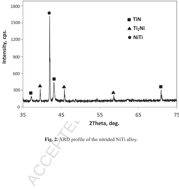
A photograph of the untreated NiTi and the nNiTi surfaces has been taken and is presented in
Fig. 1. A clear difference of the superficial aspect can be observed. The untreated NiTi presents
the classical metallic grey color of the alloy (A) while the nitrided NiTi presents a golden yellow
color (B). This golden yellow color is typically that which one observes when theface-centered
cubic δ-TiN nitride (space group: Fm3 m) was formed on the surface. This observation is in
good agreement with the literature since whatever the superficial nitriding treatment used on
NiTi (gas nitriding, laser gas nitriding...), this TiN nitride constitutes the main component of the
coating obtained [16, 17]. The formation of the δ-TiN nitride on surface was confirmed by X-ray
diffraction in this study. Indeed, the XRD profile presented in Fig. 2 clearly displays the different
peaks related to the TiN nitride and also those related to the NiTi B2 structure (space group:
ACCEPTED MANUSCRIPT
9
intermediate Ti2Ni phase between TiN and NiTi was also mentioned in literature [18]. The fact
that the main peak of the B2 NiTi substrate was detected by XRD indicates that the thickness of
the nitride layer does not exceed a few micrometers. This is in accordance with a recent work, in
which similar gas nitriding parameters were used and around 3 Pm of nitride thickness was obtained [19].
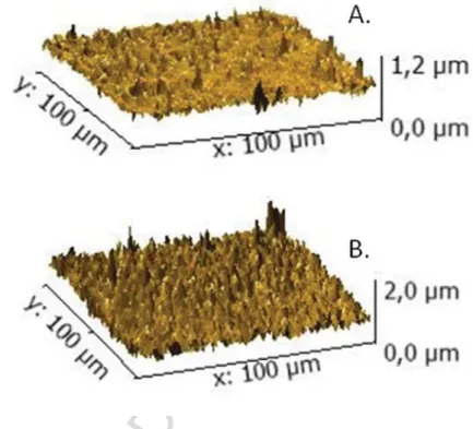
The roughness on the NiTi surfaces (untreated and nitrided) was evaluated by AFM. For
that, tapping mode was used in air with a silicon tip. To evaluate the roughness of both surfaces,
10 measurements were obtained from 100 × 100 μm AFM scans taken in different zone of each
surface. Examples of AFM maps for untreated and nitrided NiTi surfaces are presented in Fig.
3A and Fig. 3B, respectively. From the AFM maps, uniform roughness was obtained from both
surfaces. The roughness value, Ra, was measured to be 57 nm ± 6 nm for the untreated NiTi
alloy and 163 ± 9 nm for the nitrided NiTi alloy, respectively. Thus, the nitriding treatment
carried out within the framework of this study induces a roughness three times higher in
comparison with the untreated surface.
3.2. Endothelial cell response
Previous studies have shown that surface nitriding could enhance the attachment and spreading
of different types of cells on a variety of materials [20-27]. The improvement of cell affinity was
ascribed to the change in surface topography and increased adsorbtion of serum proteins
maintaining their native configuration on the coated surfaces. However, there are also published
ACCEPTED MANUSCRIPT
10
(nNiTi) as compared to un-treated surface were studied by SEM (Fig. 4A).
Although cell density was similar on both samples, the spreading area appeared to be
higher on the nNiTi. Indeed, quantification of cell spreading area by ImageJ software (Fig. 4B) at
2 h post-seeding showed that HUVECs spreading on nNiTi covered a median area of 988 μm2,
whereas HUVEC cells spreading on NiTi covered a median area of 691 μm2. Thus, these
experiments demonstrate that surface nitriding is competent to promote cell spreading. It is
known that strong adhesion of cells to substrate develops over time as the cells spread and more
focal adhesions are formed. Furthermore, focal adhesions activate signaling pathways which
control cell function and architecture [31]. Better cell adhesion and spreading result in higher
activation of intracellular signaling cascades through integrin coupled to the actin cytoskeleton.
A major protein of focal adhesions and a key player in the regulation of cell adhesion is vinculin
[32]. Therefore, vinculin provides a valuable detection system for focal adhesion sites by means
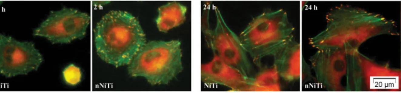
of specific antibody detection. In the present study, focal adhesion formation was analyzed by
immunocytochemistry using a monoclonal antibody against vinculin, whereas the organization
of actin cytoskeleton was visualized by incubation with Alexa Fluor 488 Phalloidin (Fig. 5).
Obvious focal adhesions marked by dot-like vinculin were detected in the HUVECs cultured on
both alloys at 2 h post-seeding. Endothelial cells maintained on NiTi revealed good cell
adhesion. However, increased cell spreading and more focal contacts have been observed on the
nitrided sample. At this time point, high resolution fluorescence images showed a circumferential
localization of actin filaments near the cell membrane and also some parallel oriented actin
filaments. After 24 h of culture, fluorescence analysis revealed typical appearance of endothelial
ACCEPTED MANUSCRIPT
11
the cell. Staining for vinculin revealed that HUVECs formed many focal adhesions, which were
present in peripheral region of the cells. These results demonstrate that nitriding is able to
stimulate both endothelial cell spreading and focal adhesion formation on NiTi substrates.
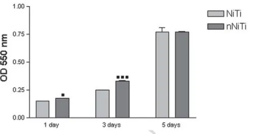
The effect of nitride coating on endothelial cells proliferation was further investigated by
MTT viability assay after incubation for 1-, 3- and 5- days. Fig. 6 shows the proliferation of
HUVECs on the surface of NiTi and nNiTi. As it can be seen, the number of viable
metabolically active endothelial cells continuously increased on both substrates. At the end of
incubation time, the level of cell proliferation on nNiTi alloy and control surface was
significantly higher than those found after 1 and 3 days of culture. More importantly, HUVECs
on nNiTi surfaces exhibited significantly increased proliferation compared with those on control
NiTi after 1 and 3 days of culture. By day 5, nNiTi still had a higher number of metabolically
active cells, but the differences were no longer statistically significant. Our data point to the
positive role that nitride coating can play in promoting endothelial cell coverage of vascular
stents.
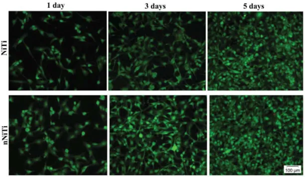
Furthermore, cells on the NiTi substrates were also stained with a solution containing
calcein AM and ethidium homodimer and imaged using fluorescence microscopy. Live cells
fluoresce green due to ester hydrolysis of calcein AM to calcein, while dead cells fluoresce red
due to DNA binding of ethidium homodimer in cells with compromised nuclear membranes.
After staining, highly viable cells were observed on both substrates (Fig. 7). Moreover,
microscopic observations confirmed the results of MTT assay showing higher cell density on
nitrided samples.
A possible explanation for the observed differences in cell spreading and growth between
ACCEPTED MANUSCRIPT
12
nanometer surface roughness was measured for nitrided compared with the conventional nitinol.
The growth of HUVECs was previously shown to increase on nano-scale rough surfaces [33].
Thus, increased surface roughness of biomaterial surfaces even at 10-10(2) nm scale has been
demonstrated to enhance the adhesion and growth of HUVECs.
The differences in the endothelial cell proliferation on the nitrided and control nitinol
samples might also result from the differences in corrosion resistance. Previous cell culture
evaluations indicated that nickel–based alloys did not affect cellular viability or morphology
[34]. However, nickel-based alloys released corrosion products which decreased cellular
proliferation. It was hypothesized that this decrease was due to an interference of the released
metal ions with the cellular energy metabolism. Given the fact that nitride coating has been
demonstrated to effectively prevent release of Ni ions [2], application of TiN coating could
explain the improvement of the proliferation rate of endothelial cells on nitinol substrate.
A clear beneficial effect of nitriding treatment on cell behavior is already acknowledged
in literature. Nitrogen plasma-implanted NiTi alloy has been shown to exhibit the highest amount
of cell proliferation in long term cultures of osteoblasts derived from calvarial bones of mice
when compared to untreated NiTi, medical grade stainless steel and Ti-6Al-4V [35]. Moreover,
the release of nickel ions was found to be significantly reduced compared with the untreated
NiTi. Biocompatibility enhancement of the porous nitinolTM was also reported after in-situ
nitriding [36], higher densities of bone marrow stromal cells being achieved on in-situ nitrided
samples than on unmodified nitinolTM. Furthermore, the porous NiTi samples were implanted
into rabbits’ bones to investigate their osseointegration capability and the results revealed that
ACCEPTED MANUSCRIPT
13
porous NiTi without surface modifications. More recently, Yang et al. [30] used vacuum filtered
arc plasma deposition technique to generate a nitride coating on nitinol and investigated
endothelial cell adhesion, morphology and viability on both alloys. Although initial cell
adhesion as well as proliferation were higher on nitrided samples, the differences were not
statistically significant. In addition, the serum protein layer adsorbed on bare NiTi alloy was
found to be qualitatively and quantitatively almost the same as the one on TiN-coated NiTi alloy.
Further on, the same authors [2] showed that, overtime, released nickel ions inhibited endothelial
cell function at molecular level, whereas TiN coating improved endothelial cell function.
Specifically, TiN coating promoted actin cytoskeleton and focal adhesion formation, increased
energy metabolism, enhanced regulation of inflammation, and promoted amino acid metabolism.
The long-term success of implanted cardiovascular NiTi devices is dependent upon the
ability of the material to resist corrosion, to support the growth, migration and function of
endothelial cells. Von Willebrand factor and eNOS are often used as vascular endothelial cell
markers to examine endothelial cell function.
The expression and cellular localization of vWf and eNOS were investigated by
immunofluorescence at 1-, 3- and 5- days post-seeding. As shown in Fig. 8, the cells grown on
nitrided samples had similar vWf and eNOS expression to the cells grown on untreated NiTi.
Taken together, these results suggest that surface modified NiTi promotes endothelial cell
adhesion and proliferation with functional vWf and eNOS marker expression.
ACCEPTED MANUSCRIPT
14
of NiTi shape memory alloy. Our results indicate that this approach can be used to enhance the
adhesion and proliferation of HUVECs on NiTi surface. Moreover, we report similar expression
of vWf and eNOS functional markers on both substrates. Although the presented results clearly
demonstrate an increase in NiTi biocompatibility by surface nitriding, the benefits of this
modification for in vivo condition remain to be determined.
Acknowledgment
This study was financially supported by research grant from the Romanian Ministry of National
Education, CNCS-UEFISCDI (project PN-II-ID-20-RO-FR-2014) and by the French ANR
ACCEPTED MANUSCRIPT
15 References
[1] S.A. Shabalovskaya, On the nature of the biocompatibility and on medical applications of
NiTi shape memory and superelastic alloys, Biomed. Mater. Eng. 6 (1996) 267–289.
[2] D. Yang, X. Lü, Y. Hong, T. Xi, D. Zhang, The molecular mechanism for effects of TiN
coating on NiTi alloy on endothelial cell function, Biomaterials 35 (2014) 6195–6205.
[3] M. Shayan, Y. Chun, An overview of thin film nitinol endovascular devices, Acta Biomater.
21 (2015) 20–34.
[4] S.D. Plant, D.M. Grant, L. Leach, Behaviour of human endothelial cells on surface modified
NiTi alloy, Biomaterials 26 (2005) 5359–5367.
[5] J.M. Schmehl, C. Harder, H.P. Wendel, C.D. Claussen, G. Tepe, Silicon carbide coating of
nitinol stents to increase antithrombogenic properties and reduce nickel release, Cardiovasc.
Revasc. Med. 9 (2008) 255–262.
[6] Y. Shen, G. Wang, L. Chen, H. Li, P. Yu, M. Bai, Q. Zhang, J. Lee, Q. Yu, Investigation of
surface endothelialization on biomedical nitinol (NiTi) alloy: Effects of surface micropatterning
combined with plasma nanocoatings, Acta Biomater. 5 (2009) 3593–3604.
[7] W. Shen, K. Cai, Z. Yang, Y. Yan, W. Yang, P. Liu, Improved endothelialization of NiTi
alloy by VEGF functionalized nanocoating, Colloids Surf. B Biointerfaces 94 (2012) 347– 353.
[8] M. Annunziata, L. Guida, L. Perillo, R. Aversa, I. Passaro, A. Oliva, Biological response of
human bone marrow stromal cells to sandblasted titanium nitride-coated implant surfaces, J.
ACCEPTED MANUSCRIPT
16
Guida, The effects of titanium nitride-coating on the topographic and biological features of TPS
implant surfaces, J. Dent. 39 (2011) 720–728.
[10] S. Durual, F. Pernet, P. Rieder, M. Mekki, M. Cattani-Lorente, H.W.A. Wiskott, Titanium
nitride oxide coating on rough titanium stimulates the proliferation of human primary
osteoblasts, Clin. Oral Implants Res. 22 (2011) 552–559.
[11] V.H. Pham, S.H. Jun, H.E. Kim, Y.H. Koh, Deposition of titanium nitride (TiN) on Co–Cr
and their potential application as vascular stent, Appl. Surf. Sci. 258 (2012) 2864–2868.
[12] D.M. Gordin, T. Gloriant, V. Chane-Pane, D. Busardo, V. Mitran, D. Hoche, C. Vasilescu,
S.I. Drob, A. Cimpean, Surface characterization and biocompatibility of titanium alloys
implanted with nitrogen by Hardion+ technology, J. Mater. Sci. Mater. Med. 23 (2012) 2953–
2966.
[13] D.M. Gordin, D. Busardo, A. Cimpean, C. Vasilescu, D. Höche, S.I. Drob, V. Mitran, M.
Cornen, T. Gloriant, Design of a nitrogen-implanted titanium-based superelastic alloy with
optimized properties for biomedical applications, Mater. Sci. Eng. C Mater. Biol. Appl. 33
(2013) 4173-4182.
[14] R. Ion, C. Vasilescu, P. Drob, E. Vasilescu, A. Cimpean, I. Drob, D.M. Gordin, T. Gloriant,
Long-term corrosion performances and cytocompatibility of nitrided Ti and Ti–6Al–4V alloy in
severe functional conditions, Mater. Corros. 65 (2014) 593–604.
[15] R. Ion, S. Vizireanu, C.E. Stancu, C. Luculescu, A. Cimpean, Gh. Dinescu, Surface plasma
functionalization influences macrophage behavior on carbon nanowalls, Mater. Sci. Eng. C
ACCEPTED MANUSCRIPT
17
[16] S.K. Wu, H.C. Lin, C.Y. Lee, Gas nitriding of an equiatomic TiNi shape memory alloy. Part
I: Nitriding parameters and microstructure characterization, Surf. Coat. Technol. 113 (1999)
17-24.
[17] Z.D. Cui, H.C. Man, X.J. Yang, Characterization of the laser gas nitrided surface of NiTi
shape memory alloy, Appl. Surf. Sci. 208-209 (2003) 388-393.
[18] G. Zorn, R. Adadi, R. Brener, V.A. Yakovlev, I. Gotman, E.Y. Gutmanas, C.N. Sukeni,
Tailoring the surface of NiTi alloy using PIRAC nitriding followed by anodization and
phosphonate monolayer deposition, Chem. Mater. 20 (2008) 5368-5374.
[19] H. Li, Z. Cui, Z. Li, S. Zhu, X. Yang,Surface modification by gas nitriding for improving
cavitation erosion resistance of CP-Ti, Appl. Surf. Sci. 298 (2014) 164–170.
[20] B. Groessner-Schreiber, A. Neubert, W.D. Müller, M. Hopp, M. Griepentrog, K.P. Lange,
Fibroblast growth on surface-modified dental implants: An in vitro study, J. Biomed. Mater. Res.
A 64 (2003) 591–599.
[21] H.H. Huang, C.H. Hsu, S.J. Pan, J.L. He, C.C. Chen, T.L. Lee, Corrosion and cell adhesion
behavior of TiN-coated and ion-nitrided titanium for dental applications, Appl. Surf. Sci. 244
(2005) 252–256.
[22] B. Grössner-Schreiber, M. Herzog, J. Hedderich, A. Dück, M. Hannig, M. Griepentrog,
Focal adhesion contact formation by fibroblasts cultured on surface-modified dental implants:
An in vitro study, Clin. Oral Implants Res. 17 (2006) 736–745.
[23] C.C. Chien, K.T. Liu, J.G. Duh, K.W. Chang, K.H. Chung, Effect of nitride film coatings on
cell compatibility, Dent. Mater. 24 (2008) 986–993.
[24] H.W. Jang, H.L. Lee, J.Y. Ha, K.H. Kim, T.Y. Kwon, Surface characteristics and osteoblast
ACCEPTED MANUSCRIPT
18
plasma nitriding enhances cell attachment to polymer surfaces, Appl. Surf. Sci. 273 (2013) 787–
798.
[26] G. Kaklamani, N. Mehrban, J. Bowen, H. Dong, L. Grover, A. Stamboulis, Nitrogen plasma
surface modification enhances cellular compatibility of aluminosilicate glass, Mater. Lett. 111
(2013) 225–229.
[27] E.P. Ferraz, J.C. Sa, P.T. de Oliveira, C. Jr. Alves, M.M. Beloti, A.L. Rosa, The effect of
plasma-nitrided titanium surfaces on osteoblastic cell adhesion, proliferation, and differentiation,
J. Biomed. Mater. Res. A 102 (2014) 991–998.
[28] W.C. Clem, V.V. Konovalov, S. Chowdhury, Y.K. Vohra, S.A. Catledge, S.L. Bellis,
Mesenchymal stem cell adhesion and spreading on microwave plasma-nitrided titanium alloy, J.
Biomed. Mater. Res. A 76 (2006) 279–287.
[29] C.Y. Li, S.Y. Gao, T. Terashita, T. Shimokawa, H. Kawahara, S. Matsuda, N. Kobayashi, In
vitro assays for adhesion and migration of osteoblastic cells (Saos-2) on titanium surfaces, Cell
Tissue Res. 324 (2006) 369–375.
[30] D. Yang, X. Lü, Y. Hong, T. Xi, D. Zhang, The molecular mechanism of mediation of
adsorbed serum proteins to endothelial cells adhesion and growth on biomaterials, Biomaterials
34 (2013) 5747–5758.
[31] M. Bigerelle, K. Anselme. Bootstrap analysis of the relation between initial adhesive events
and long-term cellular functions of human osteoblasts cultured on biocompatible metallic
substrates, Acta Biomater. 1 (2005) 499–510.
ACCEPTED MANUSCRIPT
19
[33] T.W. Chung, D.Z. Liu, S.Y. Wang, S.S. Wang, Enhancement of the growth of human
endothelial cells by surface roughness at nanometer scale, Biomaterials 24 (2003) 4655–4661.
[34] J.D. Bumgardner, J. Doeller, L.C. Lucas, Effect of nickel-based dental casting alloys on
fibroblast metabolism and ultrastructural organization, J. Biomed. Mater. Res. 29 (1995) 611–
617.
[35] K.W.K. Yeung, R.W.Y. Poon, P.K. Chu, C.Y. Chung, X.Y. Liu, W.W. Lu, D. Chan, S.C.W.
Chan, K.D.K. Luk, K.M.C. Cheung, Surface mechanical properties, corrosion resistance, and
cytocompatibility of nitrogen plasma-implanted nickel-titanium alloys: a comparative study with
commonly used medical grade materials, J. Biomed. Mater. Res. A 82A (2007) 403–414.
[36] H. Li, B. Yuan, Y. Gao, C.Y. Chung, M. Zhu, Remarkable biocompatibility enhancement of
porous NiTi alloys by a new surface modification approach: in-situ nitriding and in vitro and in
ACCEPTED MANUSCRIPT
20
ACCEPTED MANUSCRIPT
21
Fig. 2. XRD profile of the nitrided NiTi alloy.
35
45
55
65
75
2Theta, deg.
In
te
n
si
ty,
c
p
s.
TiN
Ti
2Ni
NiTi
0 300 1800 900 1500 1200 600ACCEPTED MANUSCRIPT
22
ACCEPTED MANUSCRIPT
23
Fig. 4. A. SEM images of HUVECs adhered to NiTi and nNiTi for 2 h. Scale bar represents 50
ACCEPTED MANUSCRIPT
24
Fig. 5. Merged fluorescence images of actin filaments (green) and vinculin (red) in HUVEC cells
ACCEPTED MANUSCRIPT
25
Fig. 6. Viability/proliferation of HUVEC cells cultured in direct contact with analyzed samples
for 1, 3 and 5 days as determined by MTT assay. Data analysis was based on mean ± SD (n = 3).
▪p ᦪ 0.05 versus control at 1 day post-seeding; ▪▪▪p ᦪ 0.001 versus corresponding control at 3 days post-seeding.
ACCEPTED MANUSCRIPT
26
Fig. 7. Live/Dead images of HUVEC cells at 1, 3 and 5 days post-seeding. Green = live cells;
ACCEPTED MANUSCRIPT
27
Fig. 8. Fluorescence images showing distribution of vWf (red) and eNOS (green) in HUVEC
ACCEPTED MANUSCRIPT
ACCEPTED MANUSCRIPT
29 Highlights
Gas nitriding process of NiTi is competent to promote cell spreading.
Surface nitriding of NiTi is able to stimulate focal adhesion formation and cell
proliferation.
Similar expression pattern of vWf and eNOS was exhibited by bare and nitrided NiTi. Gas nitriding treatment of NiTi shows promise for a better in vivo endothelialization.