HAL Id: tel-01058149
https://tel.archives-ouvertes.fr/tel-01058149
Submitted on 26 Aug 2014HAL is a multi-disciplinary open access archive for the deposit and dissemination of sci-entific research documents, whether they are pub-lished or not. The documents may come from teaching and research institutions in France or abroad, or from public or private research centers.
L’archive ouverte pluridisciplinaire HAL, est destinée au dépôt et à la diffusion de documents scientifiques de niveau recherche, publiés ou non, émanant des établissements d’enseignement et de recherche français ou étrangers, des laboratoires publics ou privés.
Autoantibody signatures defined by serological proteome
analysis in sera of patients with cholangiocarcinoma
Mohammad Zahid Mustafa
To cite this version:
Mohammad Zahid Mustafa. Autoantibody signatures defined by serological proteome analysis in sera of patients with cholangiocarcinoma. Human health and pathology. Université Paris Sud - Paris XI, 2014. English. �NNT : 2014PA11T025�. �tel-01058149�
UNIVERSITE PARIS-SUD
ÉCOLE DOCTORALE :
CANCEROLOGIE, BIOLOGIE, MEDECINE, SANTE
Laboratoire INSERM UMR-785
DISCIPLINE: IMMUNOLOGIE
THÈSE DE DOCTORAT
Soutenue le25/06/2014
Par
Mohammad Zahid MUSTAFA
Autoantibody signatures defined
by serological proteome analysis
in sera of patients with cholangiocarcinoma
Directeur de thèse :
Jean-Charles DUCLOS-VALLEE PU-PH, UMR 785 INSERM-Univ. Paris 11
Co- Directeur de thèse :
Eric BALLOT PH, UMR 785 INSERM
& Hôpital Saint-Antoine, Paris Composition du jury
Président du jury :
Malcolm BUCKLE DR, LBPA, UMR 8113 CNRS, ENS, Cachan
Rapporteurs :
Anne-Marie CASSARD-DOULCIER CR1, INSERM U 996-Univ. Paris 11
Sylvaine YOU CR1, INSERM U1151-CNRS UMR 8253
Univ. Paris 5 Examinateur :
UNIVERSITY PARIS-SOUTH
DOTORATE SCHOOL:
CANCÉROLOGY, BIOLOGY, MÉDECINE, SANTÉ
Laboratory INSERM UMR-785
DISCIPLINE: IMMUNOLOGY
Ph.D Thesis
Defense on25/06/2014
By
Mohammad Zahid MUSTAFA
Autoantibody signatures defined
by serological proteome analysis
in sera of patients with cholangiocarcinoma
Thesis director:
Jean-Charles DUCLOS-VALLEE PU-PH, UMR 785 INSERM-Univ. Paris 11 Thesis co-director:
Eric BALLOT PH, UMR 785 INSERM
& Hôpital Saint-Antoine, Paris Jury membres
President:
Malcolm BUCKLE DR, LBPA, UMR 8113 CNRS, ENS, Cachan
Reporters:
Anne-Marie CASSARD-DOULCIER CR1, INSERM U 996-Univ. Paris 11
Sylvaine YOU CR1, INSERM U1151-CNRS UMR 8253
Univ. Paris 5 Examiner:
Dedications
This thesis is dedicated to my parents. My father, the late Mohammad Mustafa, had a dream to see me at higher level of education but incidentally he met his demise during my childhood. My mother, who has always loved me unconditionally and whose good examples have taught me to work hard for the things that I aspire to achieve. This work is also dedicated to my wife, who has been a constant source of motivation and streanght during the moments of despair and discouragement. I am truly thankful for having you in my life.
Acknowledgement
I am deeply grateful to the jury members of my thesis, Malcolm Buckle, the president of jury, Sylvaine You and Anne-Marie Cassard-Doulcier for being the reporters and François Le Naour for evaluating the thesis as examiner.
I would particularly like to thank Prof. Didier Samuel, lab director of Inserm U 785 for giving me a home in his lab and support over the years.
I would like to express my gratitude to thesis director Prof. Jean-Charles Duclos-Vallee and co-director Dr. Eric Ballot. I am grateful for their guidance and the opportunities they had afforded me. They are incredibly organized and great problem solvers, both of these qualities were immensely helpful in moving my project forward. Under their mentorship I have learned the basics and advanced methodology of scientific research which is an invaluable tool to have as my career moves forward. I will remember my time in the lab very fondly.
I extend a warm thanks to Catherine Johanet for her hospitality at the laboratory of immunology at the Hospital Saint Antoine, her kindness and friendliness.
I would like to offer my special thanks to the students of autoimmunity group for their kindness and all shared moments: Elvire, Fouad, Ahmed, Simon, Setareh, Chayma, Sara, Karine and Eleonora.
I owe my deepest gratitude to the members of the laboratory Inserm U785: Jamila Faivre, Ama Gassama, Marion Bourgeade, Mylène Sebagh for their guidance as well as Nicolas, Nassima, Claire Lacoste, Marion, Franck, Nazha, Slavka, Juan, Alexander, Sokavuth, Myriam, Aline, Delphine, Viet, Mohyeddine, Chengyan, Sandrine, Alice, Ola and Guillaume for encouragement and exchange scientific views. I do not forget Claire Mony, Marina, Laurence and Vincent for their valuable assistance.
I would like to express the deepest gratitude to my family. My mother, my sisters, my uncle Dr.Tanveer Ahmad, my aunt Prof. Shehnaz Akhter, my brother in laws; Muhammad Rafi and Shahid Mahmood you have all provided support,
encouragement and interest in my thesis work. Thanks for listening to my problems and providing perspective. I would not be who am I today without you all. Finally, I would like to thank my wife, Zunera for standing beside me throughout my career. You have been continually supportive of my graduate education. Thank you for all the things you’ve done when I worked away from home. You have been patient with me when I’m frustrated, you celebrate with me when even the littlest things go right, and you are there whenever I need you to just listen. I also thank my wonderful children: Ahmad, Mahad and our new addition Duraid, who are just about the best children a dad could hope for: happy, loving, and fun to be with. Fundamentally what I love to do is create, so it’s wonderful watching you grow for always making me smile.
I feel a deep sense of gratitude for my late father and sister who formed part of my vision and taught me the good things that really matter in life. Their happy memories still provide a persistent inspiration for my journey in my life.
Finally, I would like to thank my friends for their continued support and encouragement. The individuals I have met during my graduate studies that I consider friends are too numerous to name. There are a few, however, that cannot go unmentioned, and I would specifically like to recognize Shoaib Ahmad, Abdul Malik, Rana Iftikhar, Hafiz Ali, Afaaq, Abdul Qadir, Abdullah Aqil, Junaid Ali, Adnan Arshad and Moazzam Azeem. These friends have been there for me when the challenges of graduate school seemed too difficult to overcome. Although many have moved away, I will never forget the experiences we’ve shared and hope to stay in touch.
I would like to acknowledge the support from Higher Education Commission of Pakistan for funding my Ph.D studies and University of Balochistan, Quetta, Pakistan for the grant of study leave.
As Always it is impossible to mention everybody who had an impact to this work. I would like to thank all whose direct and indirect support helped me completing my thesis.
SUMMARY
Cholangiocarcinoma is a rare but fatal primary liver cancer and accounts for an estimated 15% of primary liver cancer worldwide. It is associated with high mortality due to the lack of established diagnostic approaches. Autoantibodies can be used clinically as diagnostic markers for early cancer detection of cholangiocarcinoma (CC). Studies, indicating the presence of auto-antibodies (AAbs) in CC have not been reported yet. No immunological biomarker, correlated to the disease, has been identified. The objective of our study was to identify cellular proteins from liver tissues (tumoral and non tumoral) and cholangiocarcinoma cell lines which could be recognized by antibody of CC patients. We used serological proteome analysis (SERPA) technique which leads us to suggest some molecules as potential biomarkers for the early diagnosis of CC. Proteins from different origins were 2DE separated: CCSW1 and CCLP1 tumor cell lines, five different samples of hepatectomies for CC with respect to their tumoral and non-tumoral counterparts and a normal liver from amyloid neuropathy. Sera from 13 CC patients and a pool of 10 healthy subjects were probed on immunoblot performed with these different separations. Comparison of immunoblotting patterns given by patient’s sera compared to patterns given by controls allowed to define immunoreactive spots of interest and those reacting with more than one-third of sera were identified by orbitrap type mass spectrometry. In this way we identified 10, 11, 9, 14 and 16 proteins from CCSW1, CCLP1, tumor part, non-tumor counterpart and normal liver antigenic extracts respectively. Different patterns of reactivity were observed according to sera on the same antigenic extract, and for a same serum, according to the antigenic extract, even though few common patterns were also observed. This widespread of reactivity is not unusual and reported earlier in several studies of this sort. It is indicated that a single AAb have an ability to identify only a small proportion of patient. For this reason, several antibodies in combination must be used to ensure sensitivity and specificity of assays used in the daily clinic.
Identified proteins were then categorized by gene ontology analysis by which they fall into three main groups; biological process and molecular functions, protein class and molecular pathway and cellular component, according to the Panther classification. By Gene Ontology classification, two different patterns of targeted antigens were observed. The vast majority of targeted-proteins with catalytic activity were found in normal liver or non-tumor specimens. The second pattern was mainly represented by targeted proteins categorized as structural proteins extracted from CC cell lines and tumor tissues. Proteins identified with catalytic activity were: alpha-enolase, fructose biphosphate aldolase B and glyceraldedyde 3-phosphate dehydrogenase; which were reactive with more than 50% of CC sera. Proteins identified with structural activity, and detected with high rates by using cell lines and tumor tissues, were: vimentin, prelamine A/C, annexin A2 and actin; reactivity of each protein was higher than 62% with CC sera. Serotranferrin, identified under the category of transfer/carrier proteins, recognized by 100% of CC sera by using tumor tissues.
High sensitivity and specificity is a prime requisite of AAbs that might be used as CC biomarkers for CC diagnosis. Most of the AAbs detected in this study had previously been reported in other cancers and auto-immune disorders. Hence it is essential to prove the specificity of antigenic proteins, a combination of various antigens therefore needs to be tested to enable the development of new biomarkers for the diagnosis and prognosis of CC.
In conclusion, the proposed potential biomarkers need to be tested in a variety of different combinations with a panel of significant number of patients and using the most appropriate substrate defined during this study.
Key words: Autoantigens, autoantibodies, cholangiocarcinoma, tumor associated
RESUME
Le cholangiocarcinome (CC) est un cancer des voies biliaires qui représente environ 15% des cancers primitifs du foie, mais de pronostic redoutable en raison d'un diagnostic tardif faute de marqueurs spécifiques. La présence d'auto anticorps (Ac) est rapportée comme marqueurs diagnostiques précoce de certains cancers. La présence d'auto-Ac dans le CC n'a pas été signalée, et aucun biomarqueur immunologique de cette maladie n'a été identifié. L'objectif de notre étude était d'identifier des auto-Ac potentiellement utilisables comme biomarqueur de CC, par analyse sérologique du protéome.
Des immunoblots ont été réalisés à partir de la séparation par électrophorèse 2D de protéines de lignées tumorales de CC, CCSW1 et CCLP1, de 5 pièces d'hépatectomie avec leur partie tumorale et non tumorale, ainsi que de foie normal de neuropathie amyloïde. Les sérums de 13 patients atteints de CC et un pool de 10 sujets sains ont été testés sur ces immunoblot. La comparaison informatique des profils des protéines immunomarquées par les sérums des patients comparés aux profils des contrôles a permis de définir des spots immunoréactifs d'intérêt. Ces spots d'intérêt marqués par plus d'un tiers de sérums ont été ensuite identifiés par spectrométrie de masse de type Orbitrap®. Ainsi, nous avons identifié 10 protéines d'intérêt de CCSW1, 11 protéines de CCLP1, 9 de la partie tumorale des foies, 14 des parties non-tumoral et 16 protéines appartenant au foie normal. Une extrême variabilité était observée selon les sérums pour un même Ag. Différents profils de réactivité étaient observés sur le même extrait antigénique en fonction des sérums testés, et pour un même sérum selon l'extrait antigénique utilisé. Quelques spots communs ont également été observés. Cette diversité n'est pas rare et a été rapportée dans plusieurs études antérieures. Il en résulte qu'un AC d'intérêt donné ne peut être considéré comme biomarqueur de CC que pour une faible proportion de patients. Pour cette raison, il faut envisager la combinaison de plusieurs anticorps pour avoir un test avec une sensibilité et une spécificité utilisable en clinique.
Les protéines identifiées ont été classées par bio-informatique (logiciel Panther®) selon la description des gènes et de leurs produits selon une ontologie commune à toutes les espèces : fonctions moléculaires effectuées, processus biologiques assurés et localisation subcellulaire.
Dans cette classification, deux profils d'immunoréactivité se distinguent. La grande majorité des protéines cibles d'intérêt avec une fonction catalytique étaient présentes dans le foie normal ou dans les parties non tumorales des exérèses. L'autre profil était celui des protéines-cibles avec une fonction de protéines structurale et étaient présentes dans les lignées cellulaires tumorales ainsi que des parties tumorales des hépatectomies.
Les protéines identifiées avec une activité catalytique étaient : l'alpha-énolase, le fructose biphosphate aldolase B et la glyceraldedyde 3-phosphate déshydrogénase, toutes troisréactives avec plus de 50% des sérums de CC.
Les protéines de structure identifiées par plus de 60% des sérums de CC provenaient des lignées cellulaires et des tissus tumoraux. Il s'agissait de la vimentine, des prélamines A / C, de l'annexine A2 et de l'actine. Enfin, la sérotranferrine, protéines de transport, est reconnues par 100% des sérums CC en utilisant comme antigène des tissus tumoraux. Une sensibilité importante et une spécificité élevée sont des caractéristiques princeps d'un Ac pour pouvoir l'utiliser omme biomarqueur. La plupart des auto-Ac détectés dans cette étude avaient déjà été rapportées dans d'autres cancers et maladies auto-immunes. Pour trouver des protéines antigéniques spécifiques du CC, une combinaison de plusieurs semble nécessaire afin de permettre le développement de nouveaux biomarqueurs pour le diagnostic et le pronostic des CC. En conclusion, les biomarqueurs potentiels proposés dans cette étude doivent être testés en différentes combinaisons avec un panel en nombre significatif de patients et en utilisant le substrat antigénique le plus approprié comme défini au cours de cette étude.
Mots clés: auto-antigènes, auto-anticorps, cholangiocarcinome, antigènes associés à une tumeur, spectrométrie de masse, protéomique.
TABLE OF CONTENTS
General introduction ...10
PART I - INTRODUCTION CHAPTER A: Cholangiocarcinoma - generalities 1. Introduction...13
2. Incidence and prevalence of cholangiocarcinoma ...15
3. Classification of cholangiocarcinoma...20
4. Risk factors of cholangiocarcinoma ...21
5. Cholangiocarcinogenesis...24
6. Clinical features ...25
7. Diagnosis of cholangiocarcinoma ...25
8. Therapies of cholangiocarcinoma...28
9. Prognosis of cholangiocarcinoma...28
CHAPTER B: Cancer and immunity I. Introduction...31
II. Tumor associated antigens (TAAs) and tumor specific antigens (TSAs) ...32
1. Tumor associated antigens (TAAs)...32
1.1. Cancer testis antigens (CT) ...35
1.2. Differentiation antigens ...35
1.3. Mutational antigens ...35
1.4. Over-expressed antigens by tumoral cells ...36
1.5. Oncogene antigen products...36
2. Tumor specific antigens (TSAs)...36
III. Cancer Immunocontrolling...37
1. Elimination phase ...37
2. Equilibrium phase ...37
3. Escape phase ...39
IV. An overview of components of the immune system implicated in tumor process...42
1. Innate immunity and cancer...43
1.1. Humoral components implicated in cancer ...43
1.1.1 Complement system ...43
1.1.2. Natural autoantibodies (NAbs)...44
1.2 Cellular components of innate immunity in cancer...44
1.2.1. NK cells...44
a. Generalities...44
b. Receptors of NK cells ...45
c. Implication of NK cells in cancer ...46
1.2.2. NKT Cells...47
a. Receptors and different sorts of NKT cells...47
b. Implication of NKT cells in cancer ...48
1.2.3. γδ-T cells ...49
a. Receptors of γδ T-cells...49
c. Implication of γδ T-cells in cancer ...51 1.2.4. Other leukocytes ...52 a. Neutrophils ...52 b. Macrophages ...53 2. Adaptive immunity ...53 2.1. T Cells ...54 2.1.1. CD4 T cells ...54 a. TH1 cells...55 b. TH2 cells...55 c. TH17 cells...55 2.1.2. CD8 T cells ...56
2.1.3. CD4+CD25+Treg (Regulatory T cells) ...57
2.2. B Cells ...57
V. Autoantibodies in cancer ...59
1. Origin and regulation of autoantibodies ...59
1.1. B-1 lymphocytes and Nabs ...59
1.2. B-2 cells...62
1.3. Marginal zone B cells...63
2. Autoantibodies...63
2.1. Generalities ...63
2.2. Two types of antibodies ...64
a. Natural auto antibodies ...64
b. Antibodies due to self-tolerance breaking ...64
2.3. Autoantibodies in autoimmune diseases ...64
2.4. Autoantibodies in cancer ...66
2.4.1. Generalities...66
2.4.2. Auto antibodies and cancer destruction ...66
a. By ADCC mechanism ...66
b. By complement activation ...68
c. By opsonization of phagocytosis ...68
d. By antigenic modulation...68
e. By inhibition of cellular function...68
VI. Application: Immunologic tool in cancer detection...69
CHAPTER C: Methods of identifying autoantibodies in cancer patients I. Different techniques for identification of TAAs ...72
II. Methods of antigen recognition using proteins from cell lysates ...73
1. Serological proteome analysis ...73
1.1. Proteomics and proteome...73
1.2. Protein identification and proteomics ...75
1.3. Serological proteomic analysis (SERPA)...76
1.3.1. Basics of serological proteomic analysis. ...76
1.3.2. Advantages and limitations of SERPA ...77
3. Protein arrays ...80
3.1. Principle ...80
3.2. Advantages and limitations ...82
4. Two dimensional liquid chromatography (2D LC ) ...83
5. Reverse-capture microarray ...83
5.1. Principle ...83
5.2. Advantages and inconvenients ...85
III. Methods of antigen recognition using proteins from cDNA libraries ...86
1. Serological analysis of tumor antigens by recombinant cDNA expression cloning (SEREX)...86
1.1. Description...87
1.2. Advantages and limitations of SEREX technique ...87
2. Construction of cDNA libraries by phage display method ...89
2.1. Description...89
2.2. Limitations ...89
CHAPTER D: Mass spectrometry I. Definition of mass spectrometry...92
II. Ionic sources...92
1. Matrix-assisted laser desoeption/ionization (MALDI) source ...92
2. Electron spray ionization (ESI) source...94
III. Mass analyzers ...95
1. General principles of mass analyzers ...95
2. LTQ -Orbitrap®...96
2.1. Ion trap qudrupole...96
2.2. Orbitrap® ...98
3. Analyzers performance...98
3.1. LTQ ion trap/Orbitrap® ...102
V. Protein identification ...103
1. Protein mass map...103
2. Identification after MS/MS analysis...104
PART Il – EXPERIMENTAL WORK – Autoantibody signatures defined by serological proteome analysis in sera from patients with cholangiocarcinoma I. Introduction ...110
II. Article...114
III. Discussion ...148
1. Variability of immune response...148
2. Autoantibodies as cholangiocarcinoma biomarkers...149
3. AAbs as driving an effective response against cholangiocarcinoma ...150
PART IIl – General conclusion General conclusion ...151
Bibliography...156 Supplement articles with author contribution ...177
LIST OF PUBLICAIONS
Scientific article included in the thesis
Mohammad Zahid MUSTAFA, Viet Hung NGUYEN, François LE NAOUR, Elvire
BELEOKEN, Catherine GUETTIER, Catherine JOHANE, Didier SAMUEL, Jean-Charles DUCLOS-VALLEE and Eric BALLOT. Autoantibody signature defined by serological proteome analysis in sera from patients with cholangiocarcinoma. Submitted to the journal of Molecular and cellular proteomics (ISSN: 1535-9476). Submission ID: MCP/2014/039461.
Scientific articles not included in the thesis
Beleoken E, Sobesky R, Le Caer JP, Le Naour F, Sebagh M, Moniaux N Roche B,
Mustafa MZ, Guettier C, Johanet C, Samuel D, Bouhris JH, Duclos-Vallee JC, Ballot
E. Immunoproteomic analysis of potentially severe non-graft-versus-host disease hepatitis after allogenic bone marrow transplantation. Hepatology. 2013; 57(2):689-99.
Beleoken E, Leh H, Arnoux A, Ducot B, Nogues C, De Martin E, Johanet C, Samuel D, Mustafa MZ, Duclos-Vallée JC, Buckle M, Ballot E.. SPRi-based strategy to identify specific biomarkers in systemic lupus erythematosus, rhumatoid arthritis and autoimmune hepatitis. PLoS One. 2013 Dec 20;8(12):e84600.
Ballot E, Beleoken E, Mustafa MZ, Johanet C, Duclos-Vallée JC. Relations entre foie et immunité. EMC Hépatologie .2012 ; Doi : 10.1016/S1155-1976(12)54243-9.
Abstract publications and poster resentation
Mustafa MZ. Autoantibodies as diagnostic tools in the sera from patients with
Hepatocellular Carcinoma. Published in European Journal of Immunology (ISSN 0014-2980. EJIMAF 39 (S1) S1-S808. (2009). Vol. 39. No. S1. September 2009). Beleoken E, Mustafa MZ. Demonstration of an Hapatocyte canalicular localization of CYP2D6, the target of liver kidney microsome type 1 autoantibodies, markers of type 2 Autoimmune Hepatitis (ISSN 0014-2980. EJIMAF 39 (S1) S1-S808. (2009). Vol. 39. No. S1. September 2009).
Mustafa MZ. Autoantibodies as diagnostic tools in the sera from patients with
Hepatocellular Carcinoma and Cholangiocarcinoma (IAL very young science meeting, October 14, 2009).
LIST OF FIGURES ... Page
Figure 1. Cholangiocarcinoma, gross and glandular appearance...16
Figure 2. Classification of Cancers of the human biliary tract……….. 17
Figure 3. Primary liver cancer incidence rates in European Union and World regions .17 Figure 4. Worldwide incidence (cases/100,000) of CC………. 19
Figure 5. Incidence (case/100,00) IH-CC vs. EH-CC.Geographical variability in incidence of IH- and EH-CC among world areas in the period 1977 to 2007 ...19
Figure 6. Classification of CC………. 21
Figure 7. Molecular mechanism in cholangiocarcinoma development……… 25
Figure 8. Different processes leading to immunogenicity of self-proteins in tumoral cell ...33
Figure 9. Three phases of cancer immunoediting...38
Figure 10. Tumor escape mechanism ...41
Figure 11. Comparative characteristics of innate and adaptive immunity ...42
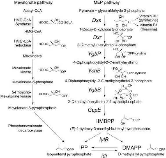
Figure 12. MEP and mavelonate pathways for isoprenoid biosynthesis ...50
Figure 13. Presentation of peptides to cytotoxic CD8 and CD4 T cells by a tumor cell ..56
Figure 14. An overview of different mechanisms leading to anti-tumor immunity ...58
Figure 15. Ontogeny and different types of B lymphocytes ...60
Figure 16. Different ways of actions of autoantibodies against tumor antigens ...67
Figure 17. Flow diagram of different techniques used for identification of TAAS ...72
Figure 18. Study areas of genomics, transcriptomics, proteomics, metabolomics and interactomics...74
Figure 19. Same genome, several proteomes...75
Figure 20. Main steps of serological proteome analysis (SERPA)...78
Figure 21. Identification of autoantibodies by MAPPing technique ...81
Figure 22. Different steps of protein microarray technique. ...84
Figure 23. Overviews of reverse capture microarray technique...85
Figure 24. Different steps of identification of TAAs by SEREX technology (immunoscreening of cDNA library)...88
Figure 25. Step vise overview of phage-display method ...90
Figure 26. A mass spectrometer is divided into three main parts ...93
Figure 27. MALDI, matrix-assisted laser desorption/ionization ...94
Figure 28. ESI, electrospray ionization ...95
Figure 29. The different instrumental configurations of mass analysers ...97
Figure 30. Linear ionic trap ...98
Figure 31. Orbitrap® apparatus ...99
Figure 32. Measurement curves of resolving power of mass analyzer ...100
Figure 33. Peptide bonds fragmentation after collision with gas molecules...101
Figure 34. A tandem MS/MS spectrometry analysis ...102
Figure 35. Different components of LTQ-Orbitrap® apparatus ...103
Figure 36. Bioinformatic analysis of MS spectra by mass homology search. ...106
Figure 37. Different strategies of bioinformatics analysis of a MS/MS spectrum ...107
LIST OF TABLES ... Page
Table 1. Risk factors of cholangiocarcinoma ...22 Table 2. Potential serum biomarkers for cholangiocarcinoma ...27
LIST OF ABBREVIATIONS
2D-PAGE Two-dimensional gel electrophoresis
A1BG/AFM α1β-Glycoprotein/afamin
AAG Auto-antigen
ADCC Antibody-dependent cell-mediated cytotoxicity
AFP Aalpha-fetoprotein
ANA Antinuclear antibodies
APRIL A proliferation-inducing ligand
BAFF B-cell activating factor
BLAST Basic local alignment search tool
BTCs Biliary tract cancers
CA 125 Carbohydrate antigen 125
CC Cholangiocarcinoma
CDC Complement-dependent cytotoxicity
CEA Carcinoembryonic antigen
CSF Colony-stimulating factor
DTT Dithiothreitol
DTE Dithioerythreitol
EBRT External-beam radiation
EBV Epstein barr virus
ECD Electron capture dissociation
EDD Electron detachment dissociation
ELISA Enzyme linked immunosorbent assay
ESI Electrospray ionization
ETD Electron transfer dissociation
F1ATPase F1-adenosine triphosphatase
FWHM Full width at half maximum
HBV Hepatitis B virus
HCC Hepatocellular carcinoma
HCD Higher-energy collisional dissociation
HCV Hepatitis C virus
HPLC High performance liquid chromatography
HPV Human papilloma virus
IEF Isoelectric focusing
IIF Indirect immunofluorescence
ILBT Iintraluminal brachytherapy
IPG Immobiline poly acrylamide gel dry strips
LC Liquid chromatography
LC1 Liver cytosol antigen type 1
LRG1 Leucine-rich α-2-glycoprotein
MAGE Melanoma antigen gene
MALDI-TOF Matrix assisted laser desorption ionisation-time of flight
MHC Major-histocompatibility-complex
MMP-7 Metalloproteinase 7
MMP-9 Metalloproteinase 9
mRNA Messenger ribo nuclic acid
NAbs Naturally occurring autoantibodies
PDGF Platelet-derived growth factor
PSA Prostate-specific antigen
PSC Primary sclerosing cholangitis
RCAS1 Receptor-binding cancer antigen expressed on SiSo cells
RPLC Rreversed phase liquid chromatography
SDS-PAGE Sodium dodecyl sulfate polyacrylamide gel electrophoresis
SEREX Serological cDNA expression libraries
SERPA Serological proteome analysis
SLA / LP Soluble liver / pancreas antigen
SLE Systemic lupus erythematosus
TAA Tumor associated antigen
TAMs Tumor-associated macrophages
TBP Tributyl phosphine
TSA Tumor specific antigen
VEGF Vascular endothelial growth factor
GENERAL
INTRODUCTION
Cancer is known to be related with genes mutations. Some alterations of oncogenes or tumor suppressor genes were largely documented in the cancer genesis. In the past 30 years, it was established that the transformation of a normal cell into a tumoral cell may be considered as a multistep process, with multiple mutations of cells that allow to surmount cellular controls that normally restraint the diffusion of these mutations consequences. Yet, it is also well documented that cancer development depends upon changes in the interactions between tumoral cells and normal cells in their vicinity (Hanahan and Weinberg 2000). It seems that all types of tumors, including their metastasis, form complex mixture of several cell types that collaborate. Several extrinsic tumor suppressor mechanisms have been reported to screen the presence of abnormal cells. There is trophic signal in the microenvironment which implicates interactions with the extra cellular matrix. There is also a control of cellular junctions and proliferations through genes implicated in the control of cell polarity, in order to avoid cell cycle progression in front of deregulated junctional complexes. There also exists tumor-suppressor mechanism involving the immune system. However, the immune system acts also as a promoter of tumor progression (Vesely et al. 2011).
This continual process where the immune system both protects against tumor development and promotes their outgrowth is named immunoediting. During this process, the instability of the genome of tumoral cells leads to the synthesis of abnormal proteins, changes in protein expression, and changes in tumor microenvironment. The shelf-modifications may be recognized by the immune system as external agents, and both cellular and humoral immunity may be activated. The targets of the immune response are known under the word tumor-associated antigens and tumor-tumor-associated antibodies are now well reported as cancer biomarkers. Easy to detect in the blood, with a half-life of 21 days for IgG1, superior to many biochemical molecules potentially also cancer biomarkers, synthetized in response to very small quantities of antigens, there appear to be very useful as cancer biomarkers. But their role in the carcinogenesis is not well understood.
The improvement of the proteomics technologies with development of bio-informatics enables the discovery of many associated antibodies for a particular
tumor. Many antibody profiles in different cancers are now well documented, with better diagnosis value than a single marker. Nevertheless, concerning a cancer of the biliary tract, the cholangiocarcinoma, there are no reports, contrasting with numerous reports about another liver cancer, the hepatocellular carcinoma. Cholangiocarcinoma account for 15% of primary liver cancer and its incidence is increasing in western countries.
In this study, we used the proteomics tool into a method for identifying autoantibodies in patients with cholangiocarcinoma, named the serological proteome analysis or SERPA.
After a chapter about the cholangiocarcinoma, we envisage the immunological general mechanisms implicated in cancer immunoediting, with a special attention to the genesis of autoantibodies. By which technologies these autoantibodies may be detected for the profiling of tumor-associated autoantibodies is the third chapter of this review. At last, we envisage the mass spectrometry in its technology dimension, especially the LTQ-orbitrap we have used. As results we obtained, we propose a relevant combination of autoantibodies as potential biomarkers and we discussed about the results under the light of the technology, with the inconvenient of the SERPA, but also with the advantages to the other technologies. The variability of the immune response we observed and the implication of the autoantibody we reported in the immunoediting are also a part of the discussion.
PART l - INTRODUCTION
CHAPTER A:
1. CHOLANGIOCARCINOMA – GENERALITIES
Cholangiocarcinoma (CC) was first reported by Durand Fardel in 1840 (Olnes and Erlich 2004). Although this is a rare cancer but primarily it is a highly lethal liver cancer, which can be difficult to diagnose and to treat and is associated with a high mortality because it is usually detected at the advanced stage of the disease; therapeutic treatment options are often limited and of least utility. Its incidence is increasing worldwide, since last few decades, especially intrahepatic cholangiocarcinoma and its pathogenesis remains unclear (Khan et al. 2005). Usually it occurs less frequently rather than hepatocellular carcinoma. In the progression of cancer metastasis is an important event. CC metastasizes to several organs, including brain, bones, lungs, and adrenal glands (Hyun et al. 2011). Skin metastasis, interestingly, is uncommon for internal organ cancers, has been reported in CC patients (Hyun et al. 2011; Yanagi et al. 2007). Though, the molecular mechanisms underlying the metastasis process in this malignancy remain unclear.
Cholangiocarcinoma, also termed as bile duct cancer, arises from the bile duct tissues (from the epithelial cells of the intrahepatic and extra hepatic bile ducts). Bile duct is a 4 to 5 inch tube that connects the liver and gallbladder to the small intestine. Bile is synthesised in the apical face of hepatocytes in liver and stored in the gallbladder and bile duct allows it to flow into the small intestine. Bile is a fluid that helps to break fats present in foods for digestion and helps the body to get rid of waste material filtered out of the bloodstream by the liver.
The bile duct originates in the liver. Inside the liver, capillaries like smaller tubes drain bile from the cells in the liver into larger and larger branches, ending in a tube called the common bile duct. The bile duct opens into the small intestine, outside of the liver. The gallbladder acts as a reservoir and stores bile until the food reaches the intestines. That is attached to the common bile ducts by a cystic duct about one-third of the way down the bile ducts from the liver. The end of the bile duct opens into the small intestine. If gall bladder is being removed, then bile flows directly from liver to the small intestine.
Cholangiocarcinoma is an adenocarcinoma of bile ducts, type of cancer that arises in glandular cells, which is a common form of cancer and begins in bile duct
lining which accounts for up to 90% of all cholangiocarcinomas (Ishak et al. 1994). This adenocarcinoma arises from the mucus glands lining the inside of the bile duct (Fig 1). Cancer can develop in any area of the bile duct. The part of the duct that presents outside of the liver is called extrahepatic. Cancer usually arises in this portion of the bile duct. 60-70% CC is perihilar cancer, also called a Klatskin tumor, grows where many small channels join into the bile duct at the point where it leaves the liver (Fig 2), about two-thirds of all cholangiocarcinomas occur at this point (Nakeeb et al. 1996). Distal cholangiocarcinoma occurs at the opposite end of the bile duct from perihilar cancer, near where the bile duct drains into the small intestine. About one-fourth of all cholangiocarcinomas are distal cholangiocarcinomas. About 5% to 10% of cholangiocarcinomas are intrahepatic, or inside the liver (Nakeeb et al. 1996). In most of the cases, intrahepatic cholangiocarcinoma presents as a large mass because in early stages of the tumor it does not show clinical symptoms. On the other hand, extrahepatic cholangiocarcinoma is generally small at the time of presentation because the bile ducts are occluded in its early stage and patients present with jaundice. The pathologic and radiologic appearance of cholangiocarcinoma can be describe in a variety of ways, different terminology and classifications have been adapted to define this malignancy and each explains a specific aspect of the tumor (Lim 2003).
2. INCIDENCE AND PREVALENCE OF CHOLANGIOCARCINOMA
Primary cholangiocarcinoma is not a common disease; frequency of CC incidence is highly variable in different areas of the world. It is the second commonest primary
malignant hepatic neoplasm cancer (Khan et al. 2002b) and accounted for an
estimated 15% of primary liver cancer worldwide (Parkin et al. 1993). Worldly and European Incidence rate of primary liver cancer per 100,000 has been illustrated in
Fig 3.
Approximately more than 3500 new cases are diagnosed in the United States while in northeast Thailand, an extremely high incidence rate (85/100,000) has been reported, where CC represents approximately 85% of total primitive liver cancers (Poomphakwaen et al. 2009). Shin HR et al. reported that occurrence of CC varies
widely by region from 5% in Japan and 20% in Pusan (Busan) Korea to 90% in Khon Kaen in Thailand (Shin et al. 2010).
Figure 1. Macroscopic and histological aspect of cholangiocarcinoma a. Gross: solitary, 7-10 cm, multinodular or
diffuse small nodules < 1 cm; gray-white and firm; often hepatomegaly and satellite nodules; rarely cirrhosis; rarely bile stained; may invade portal vein.
b. The carcinoma has a glandular appearance that is most consistent with cholangiocarcinoma.
Figure 2. Classification of Cancers of the Human Biliary Tract. Panel A shows the overall classification of biliary tract cancers. Panel B shows the Bismuth classification of perihilar cholangiocarcinomas. Yellow areas represent tumor, and green areas normal bile duct (de Groen et al. 1999).
Figure 3. Incidence of primary liver cancer
a) Primary liver cancer incidence rates per 100,000 populations, world regions (Ferlay et al. 2010).
b) Primary liver cancer incidence rates per 100,000, European union-27 countries (Ferlay et al. 2010).
Few reports indicated that the incidence of CC has increased in Western countries (Shin et al. 2010). Though, the mortality rate due to the incidence of cholangiocarcinoma is increasing, indeed high mortality rate is due to the lack of
tools for early diagnosis and treatment (Patel 2002), one study indicates that the rising rates of intrahepatic cholangiocarcinoma in Western Europe, Australia and Japan from 1979-1998 is a reason of high mortality rate (Khan et al. 2002b). Whereas gradually increase incidence of both intra and extra-cholangiocarcinoma has been observed between 1992 and 2000 in Crete (Mouzas et al. 2002).
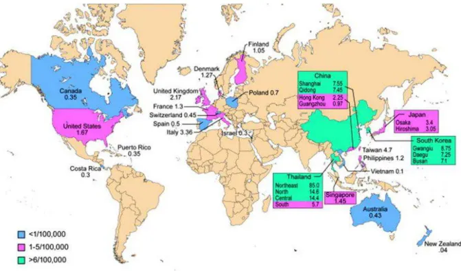
The reason for this increase is unknown. It may be due to have better tests and ability to diagnose even smaller tumors more accurately, although in many areas around the Globe, the increases have predated the advent of advanced technologies such as endoscopy retrograde cholangiography and cholangio-magnetic resonance imaging (Taylor-Robinson et al. 2001). Earlier, they may have been considered to be a different sort of cancer. In some regions of the world, Thailand, China, Korea, Japan, Malaysia, Vietnam, Laos, and Cambodia (de Groen et al. 1999; Shin et al. 1996; Shin et al. 2010; Watanapa 1996; Watanapa and Watanapa 2002), a parasite called liver flukes can infect the bile duct and cause interahepatic cholangiocarcinoma to form (Shin et al. 2010) because the parasitic infection of biliary track is endemic in these regions of Globe which is a strong risk factor of CC together with chronic liver inflammation (Patel 2002; Shin et al. 2010). So, the incidence of CC is high (>6/100,000 cases) in some of these regions (Fig 4
and 5). Report of WHO indicates that Opisthorchis Viverrini is a Group I human
carcinogenic specie and prolonged infection may leads to CC (Sripa et al. 2007). Liver flukes are very common in Asia and the Middle East, and consequently cholangiocarcinoma incidence is more frequent in these areas. Gall stones and gastrointestinal (GI) tract chronic inflammatory conditions, such as ulcerative colitis or an associated condition called sclerosing cholangitis, increase the risk of cholangiocarcinoma (Patel 2002).
Figure 4. Worldwide incidence (cases/100,000) of CC. With pink color are represented areas with rare incidence (1-5/100,000 cases), in green color are indicated countries in which CC is a non-rare cancer (>6/100,000 cases), while in blue color indicted very lower incidence (< 1/100,000) (Bridgewater et al. 2014).
Figure 5. Incidence (case/100,00) CCA vs. EHCCA.Geographical variability in incidence of IH-and EH-CC among world areas in the period 1977 to 200 (Bragazzi MC et al. 2012).
It is necessary to interpret statistics of cancer survival prudently. This data cannot be applied on a single person because it is based on the estimates on the data obtained from large number of CC cases in US. It is not possible to predict a person how long he may live with this cancer. For the reason that these survival statistics are regularly measured in five-year (or sometimes one-year) intervals, they may not correspond to advances made in the treatment or diagnosis of this cancer.
3. CLASSIFICATION OF CHOLANGIOCARCINOMA
Anatomically cholangiocarcinomas are broadly classified into intrahepatic or extrahepatic tumors (Fig 6). Intrahepatic cholangiocarcinomas arise from small intrahepatic ductules (termed peripheral cholangiocarcinomas) or large intrahepatic ducts proximal to the bifurcation of the right and left hepatic ducts. The extrahepatic bile ducts are further divided into proximal, middle, and distal segments. The proximal extrahepatic bile duct extends from the confluence of the right and left hepatic bile ducts to the level of the cystic duct. The middle portion of the extrahepatic bile ducts extends from the cystic duct to the level of the duodenum (Fig
2) (de Groen et al. 1999). The distal ducts are made up of the bile duct that extends
to the level of the ampulla. A detailed classification of hilar tumors is provided by the Bismouth-Corlette classification. This classification is based on tumors that are within 1 cm of the common hepatic duct (Klatskin tumors). These are divided into five types of tumors: the tumors that do not extend to the bifurcation of the right and left extrahepatic bile ducts (Type I), tumors that extend to the bifurcation (Type II), tumors that extend to either the right (Type IIIa) or the left (Type IIIb) intrahepatic bile ducts, and tumors that extend to both the right and left (Type IV) intrahepatic bile duct tumors (Ganeshan et al. 2012).
Figure 6. Classification of CC. Cholangiocarcinoma is broadly classified into intrahepatic (also known as peripheral) or extrahepatic tumors. Each one is morphologically categorized into mass-forming, periductal-infiltrating or intraductal-growing. This classification also provides a parallels description of extrahepatic tumors as nodular, sclerosing or papillary (Malhi and Gores 2006).
4. RISK FACTORS OF CHOLANGIOCARCINOMA
The cause of most cholangiocarcinomas is unknown. However, there are a number of risk factors that can increase the risk of developing this cancer (Table 1). These include, inflammatory conditions, abnormal bile ducts with (congenital abnormalities), hepatitis B & C virus infection, increasing age, hepatolithiasis (intrahepatic stones), Caroli’s disease, chronic typhoid carrier state, thorotrast (thorium dioxide, a chemical was used in medical imaging as radiologic contrast medium) exposure, chronic bile duct adenoma, biliary papillomatosis, smoking, parasitic biliary infestation as underlined before, and choledocholithiasis (presence of gallstones), alcoholic liver disease, etc… (Khan et al. 2002a; Shaib et al. 2005). HIV infection was also identified as a potential risk factor for CC, although it was unclear
whether HIV itself or other confounding factors (e.g. hepatitis C infection) were responsible for the association (Shaib et al. 2005).
People having chronic inflammatory bowel condition, known as ulcerative colitis, have an increased risk of developing cholangiocarcinoma. Similarly, people who have primary sclerosing cholangitis (PSC), an inflammatory condition that affects the bile ducts, are also at an increased risk of developing this type of cancer but the prevalence of cholangiocarcinoma in these patients ranges from 5 to 15% (Scofield 2004). The mechanism by which PSC increases the risk of CC is not well understood.
Risk Factors Type Reference
1.Established risk factors
Cystic disorders
(Caroli’s disease,Choledochal cysts)
IH-CCA, EH-CCA (de Groen et al. 1999; Lee et al. 2008; Patel 2011; Tyson and El-Serag 2011; Welzel et al. 2007)
Hepatobiliary flukes C. sinesis
O. viverrini
IH-CCA, EH-CCA (Kaewpitoon et al. 2008; Shin et al.
1996; Sripa et al. 2007; Sripa and Pairojkul 2008; Tyson and El-Serag 2011; Watanapa 1996; Watanapa and Watanapa 2002),
Primary Sclerosing Cholangitis IH-CCA, EH-CCA (Broome et al. 1995; de Groen et
al. 1999; Patel 2011)
Toxins (Thorotrast exposure) IH-CCA, EH-CCA (de Groen et al. 1999)
Hepatolithiasis IH-CCA, EH-CCA (Patel 2011; Su et al. 1997)
2. Potential risk factors
ulcerative colitis IH-CCA, EH-CCA (Broome et al. 1995; Chapman
1999; de Groen et al. 1999)
Cirrhosis IH-CCA (Patel 2011; Sorensen et al. 1998)
Hepatitis B viral infection IH-CCA, EH-CCA (Patel 2011; Shin et al. 1996)
Hepatitis C viral infection IH-CCA, EH-CCA (Patel 2011; Shin et al. 1996)
HIV IH-CCA, EH-CCA (Patel 2011)
Gallstones IH-CCA, EH-CCA (Nordenstedt et al. 2013)
Obesity IH-CCA, EH-CCA (Oh et al. 2005; Parsi 2013; Welzel
et al. 2007)
Diabetes IH-CCA (Chaiteerakij et al. 2013)
Smoking IH-CCA, EH-CCA (Chaiteerakij et al. 2013; de Groen
et al. 1999; Sorensen et al. 1998)
Limited data are available to validate diabetes, obesity, alcohol consumption, and smoking exposure as potential risk factors, similarly a large number of genetic polymorphisms also have been reported to increase risk of CC, but require further investigation (Tyson and El-Serag 2011).
People who are born with (congenital) abnormalities of the bile ducts, such as choledochal cysts (fibropolycystic malformations of the biliary tree), have a higher risk of developing this cancer which estimates about 1% per year cumulative increase in cancer risk in this population (Sahin et al. 1995). In Africa and Asia, cholangiocarcinoma is thought to be caused by infection with a parasite known as liver fluke “Opisthorchis Viverrini” and “Clonorchis sinensis” due to the consumption of uncooked cyprinoid fish which is endemic in certain areas (Green et al. 1991; Klatskin 1965).
The parasite persists and progressively accumulates in biliary system for years and contributes to biliary damage in the human host resulting in tissue damage even early in infection which leads to a chronic inflammatory response and increased risk of cholangiocarcinoma. In an endemic area the odds ratio adjusted prevalence for CC was 14.1% (Haswell-Elkins et al. 1994).
In Eastern countries chronic hepatitis C virus (HCV) infection was initially reported as a risk factor for intrahepatic cholangiocarcinoma then it was also reported in United States as a risk factor of CC (Yamamoto et al. 2004). The presence of chronic biliary inflammation is the only common feature of all these risk factors for cholangiocarcinoma. In recent times, considerable progress is achieved in the understanding of the role of cellular and molecular mechanisms in developing biliary cancer including tumor suppressor genes and oncogenes (Fig 7) (Zhang et al. 2008). Although CC can occur in younger people but more than two out of three occur in people over 65 years of age. Bile duct cancer can block the bile’s flow from the liver to the intestine, which causes bile to flow back into the blood and body tissues. Skin and whites of the eyes turn into yellow or jaundice. Urine becomes a dark yellow color and stools (bowel motions) look pale. The skin may become itchy. Other possible symptoms may include discomfort in the abdominal area, loss of appetite, high temperature and weight loss.
Many other diseases can also show these types of symptoms beside bile duct cancer, but it needs a thorough investigation by an experienced physician.
5. CHOLANGIO-CARCINOGENESIS
A number of successive genomic mutations similar to the sequence of events usually observed in gastrointestinal cancers are probably required for conversion from normal to malignant bile duct tissue. Mutations in oncogenes and tumor-suppressor genes have been defined in specimens of biliary tract tumors. Oncogenes mutations include K-ras, c-myc, c-neu, c-erb-b2, and c-met and the tumor-suppressor genes p53 and bcl-2 (de Groen et al. 1999). As a result of these mutations detectable phenotypic changes might be observed; for example, biliary epithelial cells change from expressing MUC-1 apomucin before birth to MUC-3 after birth (Sasaki et al. 1995). This process can be reversed by malignant transformation, many cholangiocarcinomas show staining with antibody to MUC-1 (de Groen et al. 1999). Similarly, core mucin carbohydrate Tn and sialyl-Tn antigens were also expressed in many intrahepatic bile-duct cancers (Sasaki et al. 1995; Yamashita et al. 1993). Nevertheless, mutations and phenotypic changes are also observed under nonmalignant conditions with other tissue types, precluding their routine use in clinical practice. Factors involved in the induction of various mutations that cause cholangiocarcinoma are not known although there is much speculation regarding the factors such as chronic inflammation, ethnic background, diet, and exposure to carcinogens.
Bcl-2 is an apoptosis regulatory gene and altered expression of Bcl-2 gene involves in malfunctioning of apoptosis regulatory function and thus it is closely related with carcinogenesis (Fig 7) (Jeon and Yoon 2012). Role of anti-apoptotic involvement of Bal-2 in human is unknown (Jeon and Yoon 2012) however a study indicates an over expression of Bcl-2 in 72% patients (n=08) and suggested Bcl-2 as a potential biomarker for CC (Charlotte et al. 1994). Another study indicates a limited role of Bcl-2 in cholangiocarcinogenesis (Terada and Nakanuma 1996).
Figure 7. Molecular mechanism in cholangiocarcinoma development. In PSC related cholangiocarcinoma, apoptosis is probably important for its development. If apoptosis is impaired, the genetic damage may become fixed. Effective apoptosis removes cells with serious genetic damage-beyond repair (Celli and Que 1998).
6. CLINICAL FEATURES
The presentation of cholangiocarcinoma is mainly governed by anatomic location. Rarely, an asymptomatic cholangiocarcinoma is found during the assessment of abnormal liver tests. Intrahepatic cholangiocarcinoma presents as mass lesion; obstructive symptoms are rare. Fever, night sweats and weight loss may also occur in addition to right upper quadrant abdominal pain. In contrast; cholangitis, biliary obstruction, and right upper quadrant pain are symptoms of extrahepatic bile duct and hilar type cancers. Other symptoms may coexist too, related to hepatitis C infection, cirrhosis, or systemic metastases.
7. DIAGNOSIS OF CHOLANGIOCARCINOMA
Cholangiocarcinoma usually presents at advanced stage of the disease.
However, some cases are diagnosed incidentally by deranged LFTs (liver function
available for cholangiocarcinoma. Liver function tests often show an obstructive picture with raised alkaline phosphatase, bilirubin, gamma glutamyl transpeptidase etc.
There are no specific serum tumor markers for CC diagnosis. Generally, the sensitivity and specificity of tumor marker measurements are low but might be advantageous in conjunction with other diagnostic modalities where diagnostic doubt subsists. At the present, most widely used serum tumor markers are CA 19-9, carcinoembryonic antigen (CEA) (Patel et al. 2000; Ramage et al. 1995), and CA-125.
Elevation level of CA 19-9 is up to 85% of patients with cholangiocarcinoma. The value of CA 19-9 in patients which are suspected to cholangiocarcinoma is unclear.
Elevation of CA 19-9 can be observed in obstructive jaundice with no malignancy but continues increase of CA 19-9 levels after biliary decompression propose malignancy. CA 19-9 does not has an ability to distinguish between cholangiocarcinoma, gastric or pancreatic malignancy and it may also be raised in severe hepatic injury of any cause; the value of CA 19-9 for cholangiocarcinoma detection in patients without PSC is not known but CA 19-9 may be beneficial for the differential diagnosis of cholangiocarcinoma but more studies are still needed (Ramage et al. 1995).
Carcinoembryonic antigen (CEA) is raised in approximately 30% of patients with cholangiocarcinoma, so we cannot consider it as specific marker for CC diagnosis. CEA may also be raised in biliary obstruction, inflammatory bowel disease, other cancers, and severe liver injury.
The elevated level of CA-125 is 40±50% of cholangiocarcinoma patients may suggest the presence of peritoneal involvement but further investigations are necessary. A number of potential serum biomarkers have been associated to
cholangiocarcinoma diagnosis such as CA-S27, CA-242, CA-195, IL-6, DU-PAN-2
and trypsinogen-2 (Juntermanns et al. 2010; Silsirivanit et al. 2013) . Their clinical role is currently unclear. The sensitivity and specificity of different potential biomarkers of CC are listed in Table 2.
Molecular diagnosis of CC is still not possible and a lot of research is further
required, but in future, molecular diagnosis might be possible because PSC
associated CC is often occurred due to the aberrant expression of tumor suppressor genes, p53 mutations and p16 inactivation (Reeves and DeMatteo 2000). However inactivation of tumor suppressor genes, for example, p53, APC, Smad-4, bcl-2, and p16 is often associated with CC. Oncogenes mutations have also been described, for example, K-ras (Boberg et al. 2000; Sturm et al. 1999), myc, erbB-2 met, and c-neu (de Groen et al. 1999). Chromosomal ac-neuploidy has been reported in up to 25% of periampullary tumours. The diagnostic or prognostic usefulness of these mutations is unclear and molecular profiling is not yet established clinically in patients with cholangiocarcinoma.
Biomarker Sensitivity Specificity Reference
Prediction accuracy
CA19-9 53-92 %*, 89%** 50-98%*, 86%** (Alvaro 2009)*,
(Nichols et al. 1993)**
CAE 33-68% 79-100% (Alvaro 2009)
Bilirubin (Briggs et al. 2009)
CRP (Briggs et al. 2009)
Sialic acid (Briggs et al. 2009)
IL-6 73% 92% (Alvaro 2009)
MUC5AC 71.01% 90% (Alvaro 2009)
CYFRA 21-1 74.7% 92.2% (Alvaro 2009)
TTR+CA19-9 98.2% 100% (Alvaro 2009)
AP1+CA19-9 88.9% (Sriwanitchrak et al. 2011)
CA 19-9+ CAE 86% Accuracy (Ramage et al. 1995)
CYFRA21-1 87% 95% (Uenishi et al. 2003)
MUC5AC 62.6% (Wongkham et al. 2003)
A1BG/AFM 87.5% 84.4% (Tolek et al. 2012)
MMP-7 (Leelawat et al. 2009)
MMP-9 (Leelawat et al. 2009)
4204 Da peptide 75.8% (Kikkawa et al. 2012)
LRG1 (Sandanayake et al. 2011)
LRG1+CA19-9+IL-6 (Sandanayake et al. 2011)
RCAS1 74.4% (Enjoji et al. 2004)
PDGFA 84.6 %*, 80%** (Boonjaraspinyo et al. 2012a)*,
(Boonjaraspinyo et al. 2012b)**
CA-S27 87.5% (Silsirivanit et al. 2013)
8. THERAPIES OF CHOLANGIOCARCINOMA
Surgery for cholangiocarcinoma can be difficult due to the sensitivity and location of the bile duct area while progression of the disease is usually lethal in absence of curative surgery. Resectable patients with CC are less than 30%. About 5% to 10% of patients cannot survive after this complicated operation, 25% to 45% patients experience serious complications, such as infection, bleeding, or leaking of bile or pancreatic juices. Unresectable cholangiocarcinoma has been treated by entire removal of the liver and bile ducts followed by transplantation of a donor liver.
Biliary stenting is a widely-accepted palliative procedure practiced to treat patients with unresectable hilar cholangiocarcinoma and obstructive jaundice. Patient prognosis is poor, even with the absence of metastasis (Isayama et al. 2012).
External beam radiotherapy (EBRT), with or without intraluminal brachytherapy (ILBT), is broadly used to treat patients suffering from hilar cholangiocarcinoma. A trail has been conducted in order to know the significance of radiotherapy plus biliary stenting and stenting alone, which revealed that both procedures pointedly prolonged survival of patient and stent patency. Hence, the special effects of radiotherapy alone, and the advantages of ILBT, are unknown. The prognosis of patients with unresectable intrahepatic cholangiocarcinoma seems to improve by EBRT (Zeng et al. 2006).
However chemotherapy can be used in an effort to control recurrent, irresectable and metastastic cholangiocarcinoma. Neoadjuvant chemotherapy might be used before surgery to reduce the primary tumor or when surgery is not an option. Few cases shows that, the tumor can be reduced by chemotherapy, but still it is not proved that it prolongs life or quality of life improves by this (Sripa et al. 2007). The ever best chemotherapy for CC still remains to be determined (Thongprasert 2005).
9. PROGNOSIS OF CHOLANGIOCARCINOMA
The overall prognosis of PSC related cholangiocarcinoma is worst due to the late diagnosis (Kaya et al. 2001; Rosen et al. 1991). Potential chances of cure can be achieved by surgical resection in cholangiocarcinoma. The 5-year survival rate is 0%
for non-resectable cancer because of the metastases of distal lymph nodes (Yamamoto et al. 1999), and in general less than 5%.
CHAPTER B:
I. INTRODUCTION
In 1909, Paul Ehrlich proposed that the cancer incidence would be much higher were it not for the vigilance of our immune system in identifying and eradicating emerging tumor cells. This indicated the generally accepted concept that the immune system has a vital role in the recognition, identification and elimination of altered or transformed cells. After 50 years, Frank MacFarlane Burnet and Lewis Thomas have taken the original idea of Paul Ehrlich’s, and step further to propose that T cell was the crucial (key) sentinel in the immune response against cancer. This elaboration led to the denomination of the term “immunosurveillance or immune surveillance” to define the concept whereby the immune system is on high alert against transformed cells or tumor cells. Shankaran et al. 2001 stated that the immune system can prevent cancers from developing, and therefore plays a strong protective function against cancer (Shankaran et al. 2001). Shankaran and colleagues further uncovered important new insights regarding the immune system and tumor development that they dubbed “immunoediting” or equilibrium phase which occurs if elimination is not fully successful and in which tumor cells undergo changes and mutations that aid their survival as a result of selection pressure imposed by the immune system.
In order to know how the immune system acts against the tumors, we must review few basic roles:
First, why tumoral autoantigen can activate the immune system? Immune system can protect the body from tumors induced by viruses either by suppressing viral infections or eliminating them. Eliminate the favorable inflammatory environment to tumorigenesis before its establishment by quickly removal of other pathogens and inflammation. And finally, by the identification and elimination of specific tumor cells in certain body tissues by recognizing the tumor specific antigens (TSAs) expression.
Second, the phases and mechanisms of interaction between immune system and cancer have been termed as cancer immunosurveillance, which activates when transformed cells are recognized by the immune system after the failure of cell-intrinsic tumor-suppressor functions and eradicates these abnormal cells before establishment of malignancy. To facilitate this elimination the effector immune cells work in different ways by adopting the different pathways, such as interaction
between FAS-FAS ligand, TNF-TNF receptors, and TRAIL-TRAIL receptors and by mitochondrial caspase to lead apoptosis, to reduce or inhibit tumor growth. These interrelated diverse intrinsic and extrinsic tumor-suppressor functions are highly specific and effective.
Third, to have an overview of the different effector functions of the immune system implicated in the tumor process.
II. TUMOR ASSOCIATED ANTIGENS (TAAS) AND TUMOR SPECIFIC
ANTIGENS (TSAS)
Classically tumor antigens are grouped into two categories, according to their pattern of expression, i.e. tumor associated antigens (TAAs) and tumor specific antigens (TSAs).
1. Tumor associated antigens (TAAs)
TAAs are the antigens expressed by tumor cells and normal cells. When compare to the normal cells TAAs may be expressed at increased levels on tumor cells. In other words, they may be expressed only during cell development and absent during adult life but re-appeared in tumors.
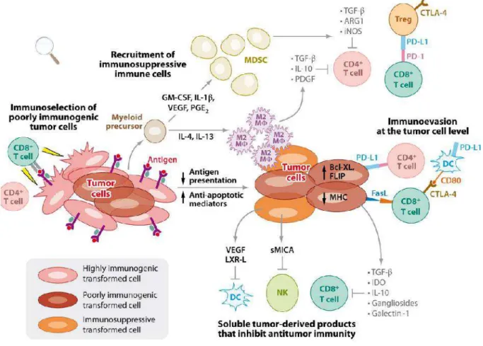
It is well known evidence that cancer sera possess antibodies which react with cellular autologous antigens called tumor-associated antigens (TAAs) (Tan 2001). There are many studies indicating that the immune system has an ability to recognize the antigenic changes in cancer cells, and further develop autoantibodies against these antigens which have been termed tumor-associated antigens (Houghton 1994; Old and Chen 1998). TAAs are actually the cellular proteins and they trigger the production of autoantibodies, different factors (Fig 8) are involved in this process which are not completely established (Zhang et al. 2009).
Figure 8. Different ways for antigens to become tumor antigens. Peptides from normal self-proteins (red, grey, and green) are presented on the cell surface as normal self-peptides (red, grey and green) in major-histocompatibility-complex (MHC) molecules. In cases of mutation, the tumor cell fails to repair DNA damage which results in a mutation (red) of normal protein and, consequently, presentation of mutated peptides (red) on the surface of tumor cells. Due to a mutation or factors that regulate its expression, a normal protein (grey) might be over-expressed in a tumor cell and its peptides presented on surface of the cell at extremely abnormal levels. In cases of post-translational modification, a normal protein can be abnormally processed (spliced, glycosylated, phosphorylated, or lipidated) post-translationally (green), resulting in an abnormal repertoire of peptides on the surface of the tumor cell. (Finn et al, 2008).
1) On tumor cells, TAAs can be different qualitatively in structure, because of post-translational modifications, misfolding, altered gene expression etc (Desmetz
2011), from those antigens that are present on normal cells. In this situation there is modification of the self-proteins which is able to be recognizing by immune system.
2) They are present quantitatively in higher significant amounts. Due to their great quantity, they are often released into the bloodstream. Increased amount of these antigens in serum can be used as tumor markers as reporters of a tumor.
3) These factors might include the mutation of oncogenes and tumor suppressor genes, products of other mutated genes, overexpression or aberrant expression of cellular proteins, tumor antigens produced by oncogenic viruses, oncofetal antigens, altered cell surface glycolipids and glycoproteins and cell type-specific differentiation antigens. In other words these situations are characterized by inappropriate expression of self-proteins (Zhang et al. 2009). In certain cancer patients the immune system appears to have the ability of recognizing these abnormalities or antigenic changes in cellular proteins and further responds by generating autoantibodies against these cellular antigens generally called TAAs (Liu et al. 2011). Particularly, the immune system of patient has the ability to recognize abnormalities in proteins involved in carcinogenesis before clinical diagnostic measures can (Imai et al. 1993).
Many studies indicated that the immune system itself interacts with cancer and promotes tumor development; the interaction between immune system, pre-cancerous and cancer cells becomes expected part of carcinogenesis (Heo et al. 2012).
List of tumoral antigens recognized by the cytotoxic T lymphocytes is very long and still growing. Antigens derived from the melanoma, and were characterized very well, were the beginning of human antigen recognition (van der Bruggen et al. 1991), after that many tumor antigens have been cloned. On the basis of tissue distribution, molecular structure and source the tumoral antigens can be classified into five categories:
• Cancer testis (CT) antigens • Differentiation antigens • Mutational antigens
• Antigen expressed by the normal cells and overexpressed by tumoral cells • Oncogene antigen products
1.1. Cancer testis (CT) antigens
Cancer testis antigens are of particular interest because of their unique characteristics. Prototypes of this category are the antigens encoded by the gene MAGE (melanoma antigen gene). Most of these antigens were initially characterized by the melanoma cells. Placental trophoblasts and germinal cells are the only normal cells that express the gene MAGE, while a variable quantity of wide range of CT antigens is expressed by different tumor types in cancer patients. Antigen NY-ESO-1 (Chen et al. 1997) of CT class is frequently expressed by melanoma, bladder, kidney and lung cancer. Furthermore, with the advancement of serological analysis of recombinant cDNA expression libraries (SEREX) technique, more than 20 cancer testis antigens have been recognized in human cancer (Scanlan et al. 2002). More recent information about new entries of antigens can be obtained from the site: (http:/www.cancerimmunity.org/peptidedetabase/mutation.htm).
1.2. Differentiation antigens
Differentiation antigens are the specific antigens of normal cells of a tissue and identified in the corresponding tumoral cells. A typical example of this type of antigen is tyrosinase which is expressed by normal melanocytes and also by the cells of melanoma (Renkvist et al. 2001; Wolfel et al. 1994). Thus, CD4+ and CD8+ cells specific for the tyrosinase peptide exist in the patients of melanoma (Wolfel et al. 1994). Though, the in vivo antitumor role of specific cytotoxic lymphocytes of differentiation antigens of melanocytes is not clear.
1.3. Mutational antigens
Mutational antigens; the genes encoding these peptides are the genes of ubiquitous proteins undergoing somatic mutations in tumor cells. Different mutation antigens are found in human tumoral cells including the mutation affecting the gene CDK4 (Wolfel et al. 1995) cyclin dependent kinase that promotes uncontrolled cell proliferation, mutation of β-catenin (Robbins et al. 1996; Rubinfeld et al. 1997) gene stimulates cell proliferation and /or inhibits apoptosis (Chen et al. 2001), mutation of caspase 8