4D Mapping of Network-Specific Pathological Propagation in Alzheimer's Disease by
Rebecca Gail Canter
B.S. The Johns Hopkins University (2009)
Submitted to the Department of Brain and Cognitive Sciences in Partial Fulfillment of the Requirements for the Degree of
Doctor of Philosophy at the
MASSACHUSETTS INSTITUTE OF TECHNOLOGY
September 2016
2016 Massachusetts Institute of Technology. All rights reserved.
Signature of Author
Certified By
Accepted By
Signature redacted
Department of Brain and Cognitive SciencesJune, 2013
Signature redacted
Li-Huei Tsai, Ph.D., D.V.M. Director, The Picower Institute for Learning and Memory Picower Professor of Neuroscience Thesis Supervisor
Signature redacted
Matthew A. Wilson, Ph.D. Sherman Fairchild Professor of Neuroscience Direc of Graduate Education for Brain and Cognitive Sciences
MASSACHUSETTS INSTITUTE
OF TECHNOLOGY
DEC 20 2016
LIBRARIES
MITLibraries
77 Massachusetts Avenue
Cambridge, MA 02139 http://Iibraries.mit.edu/ask
DISCLAIMER NOTICE
Due to the condition of the original material, there are
flaws in this reproduction. We have made every effort
provide you with the best copy available.
unavoidable
possible to
Thank you.
The images contained in this document are of the
4D Mapping of Network-Specific Pathological Propagation in Alzheimer's Disease By
Rebecca Gail Canter
Submitted to the Department of Brain and Cognitive Sciences on June 13, 2016 in Partial Fulfillment of the Requirements for the Degree of
Doctor of Philosophy in Neuroscience Abstract
Alzheimer's disease (AD) causes a devastating loss of memory and cognition for which there is no cure. Without effective treatments that slow or reverse the course of the disease, the rapidly aging population will require astronomical investment from society to care for the increasing numbers of AD patients. Additionally, the financial and emotional burden on families of affected individuals will be profound. Traditional approaches to the study of AD use either biochemical assays to probe cellular pathophysiology or non-invasive imaging platforms to investigate brain-wide network alterations. Though decades of research using these tools have advanced the field significantly, our increased understanding of AD has not led to successful interventions. One reason for this impediment may be that the tools used in neither approach can achieve the spatial and temporal precision necessary to study the consequences of molecular insults across the brain over time. In this thesis, I capitalize on recent advances in tissue processing technologies to gain a network-level perspective on the molecular and cellular progression of AD. First, I present optimized methods for in situ proteomic phenotyping of large-volume tissue specimens. Then, I use the techniques to map amyloid-beta (AP) aggregates at the whole-brain scale across disease stages in a mouse model of AD. The spatially-unbiased, temporally-precise map demonstrates hierarchical susceptibility of increasingly large, memory-related brain networks to Ap deposition. Importantly, the 4D nature of the map reveals that subcortical nodes and white matter tracts of the Papez memory circuit exhibit unique, early vulnerability to AP aggregates. Finally, using large-volume labeling approaches, I confirm the molecular findings by showing disease-specific AP aggregation in human samples from the early hub regions. Together, this data unites desperate observations of network-level deficits and identifies critical locations of early AP deposition in the brain. By linking molecular and network observations, I begin to provide biological explanations for the clinical manifestation of AD. This perspective can guide earlier patient identification and refine experimental approaches to developing cognitively efficacious treatments. These discoveries emphasize the necessity of multi-level investigations in neuroscience research and highlight the potential impacts of tools that enable researchers to bridge the gap.
Thesis supervisor: Li-Huei Tsai, Ph.D., D.V.M.
Title: Director, The Picower Institute for Learning and Memory Picower Professor of Neuroscience
"Then leaf subsides to leaf.
-Robert Frost
TABLE OF CONTENTS
ACK NO W LEDG EM ENTS ... 7
PREFACE ... 9
THE COSTS OF M ENTAL ILLNESS ... 9
TRADITIONAL APPROACHES TO AD RESEARCH ... 10
A CIRCUIT AND NETW ORK PERSPECTIVE OF AD...
11
ADDRESSING THE M ULTIFACETED ETIOLOGY OF AD ... 12
THESIS OVERVIEW ... 13
CHAPTER 1: INTRODUCTION ... 15
PREFACE ... 15
INTRODUCTION... 16
AD THROUGH THE AMYLOID HYPOTHESIS LENS... 17
M ULTIPLE FACTORS INDUCE NETWORK DYSFUNCTION IN AD ... 22
PROSPECTIVE ... 33
CHAPTER 2: ISWITCH LABELING FOR IN SITU PROTEOMIC INVESTIGATION OF LARGE VO LUM E TISSUE SPECIM ENS ... 39
ABSTRACT ... 39
INTRODUCTION... 40
RESULTS... 42
DISCUSSION... 49
M ETHODS ... 52
ACKNOW LEDGEMENTS AND CONTRIBUTIONS:... 55
CHAPTER 3: 4D MAPPING OF CIRCUIT-SPECIFIC ALZHEIMER'S PATHOLOGY REVEALS EARLY SUBCORTICAL VULNERABILITY TO AB DEPOSITION...63
ABSTRACT ... 63
INTRODUCTION... 64
RESULTS...65
DISCUSSION... 73
M ETHODS ... 76
ACKNOW LEDGEMENTS AND CONTRIBUTIONS:... 80
CO NCLUSIO NS AND FUTURE DIRECTIONS... 93
APPENDIX A: PROTOCOL FOR ISWITCH LABELING THICK TISSUE SECTIONS...97
APPENDIX B: PROTOCOL FOR ISWITCH LABELING INTACT TISSUE SAMPLES...101
APPENDIX C: PROTOCOL FOR ISWITCH LABELING HUMAN AUTOPSY SPECIMENS.. 103
APPENDIX D: RECOMMENDATIONS TO THE NIA-AA 2015 AD SUMMIT... 107
Acknowledgements
This thesis culminates years of work, not only my own and my colleagues within the laboratories at MIT, but also on behalf of my teachers, mentors, friends, and family who have supported me in getting to this point. Without them, I would not be the researcher, thinker, or person that I am today, and I am eternally grateful for their contributions to this work.
First and foremost I must acknowledge Dr. Li-Huei Tsai, my advisor and mentor and without whom I would not have been able to write this thesis on multiple levels. I wish I could acknowledge each way in which Li-Huei has significantly impacted my life, but it would take too many pages to list each of the things I have learned from her mentorship, advice, and example. To Li-Huei - thank you for taking me in when I thought there was nothing left for me in science, and helping to rekindle the love of exploration and investigation that drove me to research. Thank you for your enthusiasm for science, your visionary questions, and your courage to pursue the most challenging projects. Working with you has changed my life. I cannot express how profound and important your mentorship has been throughout the journey to this thesis and how much I will cherish your influence as I move through my scientific career.
Next, I must acknowledge my family for their continual support of my exploration and wacky endeavors. They encouraged my earliest scientific inquiries (if you can call them that) and have endured the stress of my personal adventures as I've grown. Mom, Dad, and Matt - you are the best. No words written here can acknowledge what you have done for me. Thank you for always reminding me who I am and where I came from. To my 'extended' family who have always been a huge part of my life and championed my nerdiness since I was young - Nanny, Aunt Marsha, Uncle Michael, Uncle Mark, Aunt Nancy, Aunt Gail, Uncle Rob, Uncle Jimmy, and Aunt Sheila, and all of my cousins - it just doesn't get any better than this. And to Poppy who is always listening in - thank you for providing the motivation to keep going when things got hard.
Throughout my life I have the privilege of learning from some amazing teachers. In my earliest years Elaine Russeniello and Deborah V.H. Cook helped foster the sensitivity, inquisitiveness, and creativity that have influenced my character and love of science in immeasurable ways. In high school, two teachers in particular - Jennifer Mockensturm and Zachary Marks - took me under their wings and encouraged my curiosity. They let me explore areas outside our coursework and encouraged me to work beyond the books. Thank you for your continued confidence that I could accomplish anything I tried.
My undergraduate mentors set me off on the path of science. Dr. Gregory Ball, Dr. Peter Holland, and Dr. Alexander Johnson - thank you for providing me the opportunity, tools, and vocabulary to ask questions I never thought tractable and introducing me to neuroscience and behavioral research. The unique resources, ideas and information you taught me have truly shaped my intellectual interests and instilled the questions that have driven my pursuit of neuroscience.
Acknowledgements
while also observing the personal and professional fortitude it takes to be successful has been invaluable. Thank you to Dr. Matthew Wilson, Dr. Kay Tye, Dr. Ivana Delalle, and Dr. Dava Newman for these lessons. Also, I must add a special acknowledgement to Dr. Kwanghun Chung whose mentorship contributed to my scientific and personal growth at MIT, and who taught me that positivity, optimism, and perseverance pay off.
In addition to working with Li-Huei, I have been fortunate to work in her laboratory filled with some of the most brilliant scientists on the planet. The Tsai lab is full of dedicated researchers who are also fun, thoughtful, and engaging colleagues. Without the technical advice, moral support, and insightful discussions from the entire lab, this thesis would have been an unconquerable beast. Ram, Jinsoo, Elizabeth, Ashley, Nina, Alison, Ying, Susan, Matt, Fatema, Jay, Chinna, Marco, and Hiruy deserve a special acknowledgement for their friendship and help. As do the incredible undergraduate women who worked with me - Christine Yao, Stephanie Bousleiman, and Kamilla Tekiela - your dedication, company, and insight have helped bring the findings in this thesis from ideas to fruition.
My graduate student friends and colleagues have been there every step of the way and made MIT a home away from home. I am lucky to have lived, played, and worked with you all. Meg, Joe, Stetner, Eitan, and Kean - it was a great run together. To Evan and Naveed, thank you for working long nights alongside me so I was rarely alone. To Corey and Zach - thank you for letting me word-vomit my day when I came home stressed and for expanding my culinary horizons. To Amy, Jo, Larissa, Shuyu, Dale, John, Glenn and the rest of the MIT Rowing Club -the camaraderie of -the sport got me through -the most difficult times in my life. I am lucky to have been a part of the team. To Leyla and Dan -- as fellow Blue Jays, Baker House GRTs, and friends, I may be most thankful to you for introducing me to CrossFit. To Susana, Tyler, Dom, and Tracy - although you haven't been graduate students for most (all) of the time I have known you, your support and our adventures and celebrations are some of my best memories from my time at MIT.
I am lucky to have made many great friends outside of MIT. Sarah, Andrea, and Katie - thank you for accepting my grad student lifestyle, sending constant encouragement and support, and providing a respite to normal life. You have kept me sane throughout. Amrit and Eddie - thank you for your wisdom, fun times, and good conversations that kept me grounded.
Living in Boston away from home I have also been lucky enough to stumble on to a family-away-from-home. Retsina and Reuben- thank you for everything. I could not have gotten here without you. Thank you for your support, encouragement, advice, adventures, and everything else I've missed.
Last, but certainly not least, Andrea. You are everything. Thank you for your optimism, for keeping it silly, and for your tactical and emotional support. Your love encourages me to be stronger, smarter, and more capable than I ever thought possible. Your inspiration helps me strive to make the world a better place each day.
Preface
The Costs of Mental Illness
In 2011, the World Economic Forum commissioned a report that indicated non-communicable diseases will cost the world nearly $47 trillion by 2030. Of this economic loss, the largest portion -- nearly 35%
--will be due to mental illnesses.' These diseases can occur in any stage of life and are often chronic, impacting affected individuals' abilities to function in society for long periods of time. Fortunately, the development of treatments for depression, anxiety, and schizophrenia2
that can ameliorate disease symptoms has brought relief to many patients.3 Though more work must be done to find drugs with better efficacy and specificity, especially for patients that do not respond to the currently available treatments, the success has affirmed that many mental illnesses are biologically tractable diseases for which there may be cures. However beyond the diseases for which drugs exist, there remain other mental illnesses that have no treatments or cures, and with mounting failures to develop efficacious drugs, the potential grows increasingly less certain.4
Dementias are a group of diseases that cause cognitive impairment and memory loss and have become the most costly mental illness for which there are no treatments or cures."5
In 2010, nearly 15% of the US population had dementia at a cost of over $55,000 per affected individual. By 2040, conservative predictions estimate that dementias alone will cost the US nearly $400 billion6, and although there are several types of dementia, including frontotemporal dementia, vascular dementia, Creutzfeldt-Jakob disease, and dementia with Lewy bodies, Alzheimer's disease (AD) is the most prevalent and will account for most of that cost.7
Though the calculated economic costs of AD include care and account for the time spent by non-compensated caregivers (e.g. family members), they often do not reflect the emotional and physical toll that these diseases wreak on individuals throughout society.7 These unquantifiable consequences of AD not only impact families, but also can lead to or exacerbate mental illnesses amongst the caregiving population. With few drug prospects and an aging population, the burden of AD will rise, and it is likely society has yet to fully realize the costs of rising AD prevalence. For neuroscientists, the wide-reaching impact of AD, alongside failed drug trails and mounting individual and societal costs, presents an opportunity to review our approach dementia research and potentially revolutionize the way
Traditional Approaches to AD Research
Since the discovery of AD-causative genes, research has focused on finding heritable mutations that predispose individuals to AD and the cellular consequences of those mutations.8'9 Though this approach has provided insight to the molecular climates that drive neurodegeneration in AD, they have largely highlighted the role of observable hallmark pathologies and failed to explain cognitive deficits.'0 Furthermore, these studies have yet to account for variable correlations between pathology and memory loss, especially at the earliest clinical stages." With increasing evidence that observable AD pathology is not informative for cognitive decline," and that therapeutically rescuing burden does not improve cognition, research has shifted to examine the cellular changes that underlie the disease.'4
Indeed,
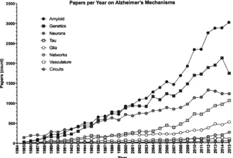
3500. Papers per Year on Alzheimer's Mechanisms
-- Amyloid 3000 -- Genetics G Neurons -a- Tau 2500 -0- Glia o Networks C 2000 4 Vascula Ire -0 Circuits 1500 1000 Year
Figure 1. Papers published per year on putative mechanisms of Alzheimer's disease pathogenesis. Numbers were taken from 'Results by year' graph that accompanies Pubmed.gov search results of "Alzheimer's [xx]". [xx] is the search term indicated in the legend. If the 'Results by year' data were not available, numbers were tabulated from the search data. Papers including multiple search terms will be represented in each category.
while investigation of AD genetics and the hallmark AD pathologies - amyloid beta (AP) aggregates and hyperphosphorylated neurofibrillary tau tangles (NFT) - by number of papers published per year remains
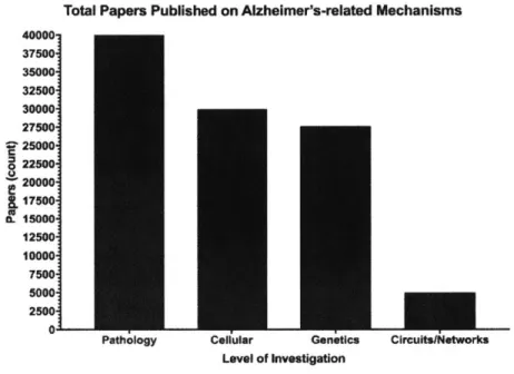
high (Fig. 1), studies looking at the cellular contributions to disease progression are increasing (Fig. 1), largely due to renewed interest in glia over the last decade. Studies of cellular mechanisms together surpass examinations of AD genetics (Fig. 2). Though this is a promising shift towards a functional
understanding of AD-related pathophysiological processes, an understanding of how cellular-level alterations impact cognition is still untenable. Instead, circuit- and network-based studies are beginning to
I
Total Papers Published on Alzheimer's-related Mechanisms
40000-37500: 350001 32500' 300001 27500-25000 0 22500 20000 * 17500 o. 15000 12500 10000 7500 50007 25001 0-Circuits[Networks
Figure 2. Total papers published on AD-related mechanisms at each level of basic neuroscience investigation. Pathology: tau, am yloid; Cellular neurons, glia, vasculature; Genetics: genetics; Circuits/Networks: circuits, networks, white matter.
illuminate how brain activity drives memory and behavior. Unfortunately, network- and cellular-level studies are lacking in the AD field, with nearly 5-fold fewer papers published on the topic (Fig. 2).
A Circuit and Network Perspective of AD
At its core, AD is episodic memory impairment.1 5 This impairment starts as an anterograde amnesia for specific events, and progresses to a loss of long-term memory recall. The loss of cognitive function and progression are core criteria for diagnosis,' and importantly, the loss of episodic recall (remembering details of the event) seems to be specific for AD, whereas recognition memory (familiarity with an event or person) impairments are typical of multiple amnesic dementias." This is important because in humans, identifying which brain regions is possible with high resolution magnetic resonance imaging (MRI), and studies of structural connectivity in patients with mild cognitive impairment (MCI), a
Cellular Genetics
Level of Investigation Pathology
prodromal stage of AD, suggest that neural connections shifts to compensate when recall circuits have damage.'" Thus, unlike pathological burden which occurs non-specifically across dementias and poorly correlates to memory function,19 network-level investigations are beginning to reveal disease-specific alterations that do correspond to observed patient deficits.
There is a lot of evidence for circuit and network disruption in AD, and increasing studies suggest that network deficits are consistent between human patient observations and preclinical murine models. For example, in AD-like mouse models of amyloidosis, a core region of learning and memory, the hippocampus (HPC), demonstrates marked hyperexcitability.20 This effect recapitulates seizure-like activity seen in AD patients21, and rescue of this hyperexcitability in both mice and humans improved
22,2
cognition. Although these experiments lack the molecular specificity of targeted cellular interventions, it is likely that the physiological changes in AD result from complex and widespread alterations that are not amenable to such specific treatments. This is likely to be particularly true at the later stages of
24
memory loss, before which pathological burden had plateaued for many years. Moving towards a network-understanding of AD may provide avenues for interventions that bypass the need for molecular specificity, and with emerging technologies it is possible that these can be targeted directly to the brain with high chances for cognitive efficacy in the absence of off-target systemic side effects. Furthermore, because network dysfunction results from and drives cellular and molecular changes, optimizing this framework will inevitably provide new information on specific, druggable molecular targets, bringing the field closer to treatments and cures.
Addressing the Multifaceted Etiology of AD
In addition to long-range networks within the brain, understanding how peripheral biological influences and lifestyle factors contribute to AD is critical. Lifetime experience factors like education, diet, and socioeconomic status greatly impact cognition with aging, and may increase risk for AD.6,25'26
Additionally, studies demonstrating links between peripheral inflammation and AD suggest that despite its relative immune privilege from the blood brain barrier, factors outside of the brain influence its function. This is not surprising, as research into neuroendocrinology and metabolism demonstrate clear roles for peripheral signals in controlling brain function, however how these influences related to AD remain uncertain. Some evidence for the role of these factors in AD susceptibility comes from insulin and obesity studies, which suggest that diabetes and high adiposity increase AD risk2 7, however causal links between these factors and cognitive decline are lacking.
Thesis Overview
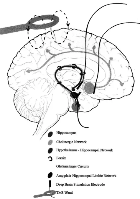
This thesis seeks to establish the importance of network-specific investigations in AD by developing tools for their unbiased characterization and using those tools to identify specific circuits that are vulnerable to novel pathologies. The first chapter presents a perspective that elaborates on the role of pathological observations and circuit and network research to progress AD therapeutic goals. Then, the second chapter outlines the technical specifications for large tissue specimen proteomic analyses techniques we worked to develop and optimize including: sample preparation, imaging, and quantification techniques. The concluding work highlights the utility of the tools for both preclinical and patient research, and uses the protocols to create an unbiased map of AD pathological progression. The data demonstrates network-specific spread of AD pathology and identifies novel diencephalic regions of the Papez long-term memory circuit as particularly vulnerable to early pathological burden, and that this susceptibility results in physiological impairment. Furthermore, the 3D nature of the data show, for the first time, specific white matter affliction of a particular long-range tract, the fornix, which connects the major nodes of the Papez circuit. This data is confirmed in human postmortem specimens, which demonstrate that the regions identified in the murine models also show disease-specific pathology throughout the AD progression spectrum, indicating this may be a useful biomarker of early AD in the patient population. The thesis concludes with a summary of the potential impact and a hypothesis that future work to understand the contributions of the Papez circuit hubs (particularly the mammillary bodies and fornix) to memory and mechanisms of pathological AD propagation in this system will shed light on the earliest stages of the disease and may provide a unifying framework onto which researchers can map the increasingly complex pathophysiology of AD.
Chapter 1: Introduction
The complex road to restoring circuits for the treatment of Alzheimer's disease.a
Preface
Alzheimer's disease (AD) is a progressive loss of memory and cognition for which there is no cure. While genetic studies initially suggested a primary role for amyloid-beta (AP) in AD, Ap-targeted treatment strategies have failed to ameliorate cognitive symptoms. These clinical results suggest cognitive decline results from complex pathophysiology, and AP treatment alone may not be sufficient to treat the disease. This review discusses evidence that pathophysiological mechanisms outside the amyloidogenic pathway contribute to systemic dysfunction, cognitive decline, and AP accumulation and proposes that a broad outlook on circuit-damaging pathologies in AD will yield new insights to therapeutic strategies.
Contributions:
R.G.C & L-H.T. came up with the idea and curated the content.
R.G.C wrote the initial draft of the document and generated the figures.
J.P. helped edit the manuscript and wrote the initial draft of the HDAC2 paragraph.
a This chapter was accepted as a review article in the journal Nature on August 3, 2016. The citation for this publication is: Canter, RG, Penney, J, Tsai, L-H. The complex road to restoring circuits for the treatment of Alzheimer's disease. Nature. In Press.
Introduction
Age related dementias will affect almost 10% of the population, and place a tremendous burden on affected individuals and their families.6 The most prevalent type of dementia, Alzheimer's disease (AD), is a devastating, progressive loss of cognition for which there is no treatment or cure. Analyses of AD patients' brains suggest that extracellular aggregates of amyloid beta peptides (AP), intracellular inclusions of tau-rich neurofibrillary tangles (NFT), and the appearance of neuritic plaques (NP) are pathological hallmarks of the disease, yet there is no conclusive link between these observations and the
cognitive symptoms28
. The inability to definitively connect progressive memory loss to pathological
biomarkers is a huge impediment in the quest for effective AD therapeutics, but enhanced efforts to understand mechanisms of cognitive decline are beginning to reveal new avenues for intervention.
The insidious onset of AD-related memory loss has hampered etiological studies because cognitive symptoms emerge late in disease progression when there is already rampant AP deposition, NFT formation, and cellular death; each of which might contribute to memory loss.'0 Early genetic studies implicated AP as the primary causative factor because mutations in or duplications of amyloid precursor
protein (APP)29-32 and its processing pathway components presenilins 1 & 2 (PSi, PS2)3 3-31 lead to
inherited, early-onset AD. These breakthroughs laid the foundation for the amyloid cascade hypothesis, which postulates that the accumulation of AP peptides is the proximal event in the development of AD, that in turn leads to NFT pathology, neurodegeneration, and memory loss.8 Corollary to this hypothesis, increased levels of AP in the brain-- due to excessive peptide generation or an inability to clear it--underlie cognitive dysfunction, thus treatments to reduce AP load should improve cognition.9 Surprisingly, despite extensive evidence for AP-driven neurodegeneration, all AP-targeted therapeutics have failed to reverse memory deficits or halt cognitive decline, even alongside significant AP reduction.'2 As the AP-modifying strategies for treating AD shift to preventing accumulation, refining drug targets, and improving compound design,36'37 conceptual advances in functional AD pathophysiology, coupled with data from -omics technologies and brain imaging platforms, are highlighting other promising avenues.
Beyond the genes in the AP pathway, additional newly identified human genomic loci significantly increase the risk of AD. While studies link some of the genes to amyloid metabolism, many appear to function in cellular signaling pathways and the brain's immune system,38 indicating the importance of atypical cellular responses to changing brain states in AD. Emerging functional data also shifts etiological focus from a neuron-centric view of the disease to an integrated outlook that acknowledges the synergistic
functions of the different cell types that make up the brain. Consistent wich this holistic view, multiple pathophysiological processes in AD are interconnected and feedback and feed-forward to provoke a cycle dysfunction as the disease progresses.'4 The array of dysfunctional cellular processes ultimately affects neural circuit function and network connectivity,39 and the aberrant circuit activity not only triggers additional cellular coping mechanisms, but may also propagate pathological spread to connected brain regions.
The complexity of the various interactions makes disentangling AD etiology a daunting task, however systematic examination of the genetic and cellular changes can provide mechanistic insight into the cascade of pathological events that occur during AD.14
Importantly, however, only an understanding of how these alterations contribute to progressive network and circuit dysfunction will connect observable pathophysiology to biologically-abstract cognitive impairment. Additionally, identifying vulnerable networks and susceptibility nodes within them may hold clues to the disease origins and progression. This review will briefly explore the role of AP driven pathogenesis in AD. Then, rooted in the newly identified genetic risk factors, will examine growing evidence for genetic, cellular, and circuit dysregulation as pathophysiological mechanisms that not only result from, but can also lead to, Ab accumulation, tau aggregation, and cognitive impairment. With this perspective, the field can move past unsuccessful Ap-targeted clinical trials towards implementing a multipronged approach to treatment including cellular- and circuit-level therapeutic strategies.
AD through the amyloid hypothesis lens.
Familial genetics implicate Af in AD pathogenesis.
The most compelling evidence for the central role of AP in AD comes from studies of familial Alzheimer's disease (FAD). FAD cases account for 5%40 of the patient population and results from pathogenic, autosomal dominant mutations or duplications in APP, or mutations in genes for y-secretase
components presenilin-1 (PSI) and presenilin-2 (PS2)30'331'4. APP, PSI, and PS2 mutations shift APP
processing (see Box 1) to the amyloidogenic pathway and bias cleavage towards the longer, toxic AP species4
1,
42. The mechanism of APP duplication toxicity is less clear, however FAD-related APP duplications exist43'44, and individuals with Down syndrome (DS), who have 3 copies of the APP gene,also develop early onset AD45
'46. Thus, despite differences in the specific location, mutation, or gene, heritable changes that either qualitatively or quantitatively increase longer AP species cause AD. Alternatively, mutations in the MAPT gene encoding tau, the other major pathological feature of AD, usually result in frontotemporal dementia, a similar but ultimately different disease.47
protection from AD. One recently identified APP variant in the Icelandic population reduces amyloidogenic processing and protects against AD and normal age-related cognitive decline5. This
suggests the relationship between AP levels and cognitive impairment is bidirectional, and the genetic data strongly support the idea that accumulation of AP, especially the longer forms, plays an important etiological role in AD-related cognitive decline.
A/i impairs synapses and destabilizes circuits.
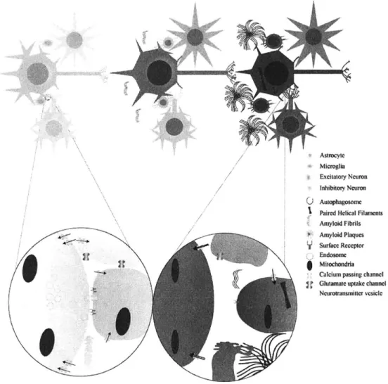
AP peptides can exert multiple detrimental effects on neurons as well as the other cell types that make up the brain (Figure 1)4. Though neurons primarily transmit information throughout the brain, glia also make significant contributions to signal propagation and modulation, especially at the synapse; thus any discussion of circuit changes in AD must incorporate activities of these cells too . In addition to
modifying communication, many non-neural cells support neuronal health and thus participate in AP clearance to protect neuronal function during disease progression. For example, astrocytes and microglia are the major source of apolipoprotein E (ApoE)5
3-5 5, a protein for which allelic variation confers risk of
developing AD. ApoE isoforms differentially facilitate AP clearance into astrocytes or through the vasculature, and the &4 variant impairs this ability and reduces AP clearance, which augments its
parenchymal levels5 6'57. Similarly, microglia are brain-resident myeloid-lineage cells that have
phagocytotic capability58. Though it remains controversial whether microglia or blood-derived
macrophages degrade AP in AD (but see ref 59), microglial responses seem to affect aggregate levels and
the cells themselves are capable of uptaking AP, suggesting they play a role60'61. Though oligodendrocyte fate in AD is less certain, the main component of the myelin they produce to ensheathe axons, myelin basic protein (MBP), may aid AP clearance.62
Ultimately a complete characterization of non-neural A-induced AD pathophysiology is lacking, however it is likely that disruptions in the normal functions of these cells impact neuronal health and circuit dynamics.
As an acutely toxic species, oligomeric AP (oAP) directly incites neuronal apoptosis via interactions with cell surface receptors, and AP accumulation indirectly leads to oxidative damage of DNA and proteins, injured organelles, and dysregulated internal calcium (Ca 2
) levels, which can also provoke cellular death.
In vitro, AP exposure induces neuronal dysfunction and can cause death within hours6 3. Although in vivo these pathogenic processes take years to have detectable consequences, widespread neuronal loss in the brain accompanies the onset of severe cognitive decline24
. Though neuronal death has profound impacts on memory performance, deficits in synaptic plasticity, circuit function, and cognition all develop well before cellular loss occurs9. Indeed, synapse loss occurs before overt pathological aggregates
A large body of evidence indicates that amyloid plays an important physiological role at the synapse and that synaptic activity is a major source of parenchymal Ap67,68. Excitatory activity promotes APP proteolysis and release into the extracellular space.677 Once there, the effects of synaptic AP on excitatory activity vary with its extracellular concentration: low AP levels promote excitatory activity and higher levels depress it. Small elevations in AP promote activity via presynaptic acetylcholine receptors, which elevate internal Ca concentrations to increase glutamate release probability.7273
Though the resulting postsynaptic excitation could result in positive feedback whereby additional AP release increases synaptic excitability, increasingly high levels actually depress activity through a number of synaptic-strength modifying mechanisms, including glutamate receptor internalization74
'75 (Figure 1). While acute elevations in synaptic AP can impair potentiation of synaptic strength (LTP) and promote
depression (LTD)76-79, long-term elevations can weaken connectivity, alter dendritic spine dynamics,
69
promote synapse loss, and impair the processes that shape circuits and underlie learning and memory The loss of spines can lead to neuron-intrinsic hyperexcitability66, and A$ also alters excitatory tone by impairing inhibitory interneurons' abilities to affect excitatory cells. Loss of inhibition occurs through multiple pathways, including down-regulation of cell-surface voltage-gated sodium channel subunits. Indeed, lower Nav1.1 sodium channel subunit expression in AD-model mice hinders action potential propagation through inhibitory parvalbumin interneurons, resulting in reduced release of the primary inhibitory neurotransmitter, GABA, and the loss of inhibition on excitatory neurons2 2
With long-term pathological accumulation of AP, disinhibition of excitatory cells and synaptic loss result in hyperactivity, which occurs in some pre-symptomatic patients in brain regions associated with learning and memory21
. Over time, this may lead to epileptiform activity and compensatory inhibitory sprouting, which can impair learning processes20. AD patients that have seizures exhibit worse cognitive outcomes80. Treating patients with inhibitory receptor agonists can reduce the abnormal activity and restore cognition, suggesting aberrant spiking is directly related to memory
loss23.
Interestingly, restoring inhibition in mouse models not only rescues local circuit dynamics, but also reinstates the observed loss of long-range network coherence to enhance learning81.Network susceptibility is key to A/3 pathogenesis.
Amyloid deposition is a critical event in AD pathogenesis therefore identifying the brain networks that are susceptible to AP-induced dysfunction should reveal how the disease propagates through the brain, and may explain how spreading AP accumulation contributes to cognitive symptoms. Initial
cross-led to the observation that AP deposition initiated within the neocortical regions involved in cognition, spread to neural hubs underlying learning and memory, and finally progressed to motor and sensory structures, and also provided a framework for understanding the successive deterioration of cognitive and sensory impairments2 .Because they suggested the serial involvement of interconnected brain regions, but could not functionally demonstrate propagation through networks or provide critical insight to mechanisms of AP propagation, these observations paved the way for longitudinal studies of AP
accumulation in patients.
The development of AP binding tracers, like ["C]-Pittsburgh Compound B (PiB) and ["F]-florbetapir, for positron emission tomography (PET) has enabled researchers to assess the pattern of AP deposition across the course of the disease83,84 (Figure 2). Cross-sectional PET studies of AD patients confirm the neuropathological findings of significant AP throughout the neocortex, and importantly demonstrate the
amount of cortical retention is predictive of cognitive decline24
,85. This indicates that AP levels may be a good biomarker for AD.86 The consistent locations of tracer retention across studies suggest that an AD-specific pattern of AP accumulation is a fundamental and necessary change to effect cognitive symptoms, and correlational and functional studies suggest that memory decline may relate to AP within regions of the Default Mode Network (DMN).87 The DMN is a group of functionally connected brain regions that co-activate during passive thinking, remembering, and planning, and early investigations with PiB-PET demonstrated overlap between areas of high AP and DMN regions, suggesting the network may be vulnerable in AD.8 Studies using functional connectivity magnetic resonance imaging (MRI) demonstrated DMN susceptibility by showing reduced connectivity within the network7. Interestingly, ~25% of cognitively healthy individuals highly retain PiB, and this correlates with reductions in DMN connectivity, worse episodic memory, and predicts dementia in follow up studies8
9-
91. Together this evidence suggests that DMN alterations may be a proximal network dysfunction underlying cognitive impairment. Surprisingly, despite cognitive deterioration, further increases in AP levels over the conversion period from aMCI to AD are relatively modest, which is consistent with observations that AD patients show little retention differences throughout disease progression92-95. This data suggests that amyloid deposition may plateau at pathological levels long before the onset of cognitive symptoms.
Though human imaging studies suggest AP pathology precedes cortical network dysfunction, the techniques have yet to reveal precipitating patterns of amyloid accumulation or give insight to mechanisms of propagation. Some preclinical studies suggest AP may propagate in a prion-like manner and undergo cell-cell transmission 96,97, yet other studies suggest that Ap-induced circuit dysfunction
affects network connectivity8 1
, and local aberrant activity may result in AP accumulation at projection target structures, creating the appearance of sequential spread through these networks. Whichever mechanism contributes to AP progression, the realization that As pathology begins many years before overt memory loss and spreads through the brain increases the urgency of identifying the key nodes for early intervention.
A/-based treatments have yet to restore cognition.
Despite the complexities, it is clear that As contributes to AD pathogenesis and its probable contributions to AD progression make it a prime target for therapeutic intervention (Figure 2). Because elevated AP levels likely underlie its pathogenicity, treatment strategies have emerged targeting the two determinants of AP load: aberrant generation and faulty clearance mechanisms.
Drugs to modulate 0- or y- secretase activity are primary therapeutic strategies to reduce Ap generation by limiting amyloidogenic proteolysis, and phase II trials have identified a few safe first-generation pharmacologics for each enzyme.9'"99 However, these trials have mostly failed due to problems with target specificity, brain permeability, or study design without testing cognitive or AP outcomes 36,100. The few trials that have reported primary outcomes show reductions in CSF markers of AP, but have conflicting cognitive results with some showing hastened decline'0 1. Given the pharmacological setbacks and uninformative memory outcomes, it is difficult to conclude the feasibility of AP proteolytic targets as AD treatments. However, with redesigned clinical trials that utilize updated dosing and outcome design, and increased understanding of the complexity of the enzymatic functions of the targets, hope remains that the next generation of compounds will be safe and effective.
Enhancing AP clearance from the parenchyma is an alternative approach to modifying AP levels. The primary avenue to affect these processes has been immunization, employing both active and passive strategies. Early clinical trials investigating active immunization successfully reduced amyloid burden, but also caused severe side effects like brain swelling and hemorrhage10 2
. The promising effects on AP levels has led to new generations of immunotherapies, which have advanced quickly through clinical trials. Patients generally tolerate the newer immunotherapies well and phase
I/II
clinical trials indicate successful Ap reductions0 3. These significant AP clearance results are unfortunately tempered by cognitive performance outcomes that suggest these treatments may only successfully slow decline in prodromal or mild AD, and across different therapeutics, the efficacy even within this population is mixed12,0 4
may be necessary for testing treatments with different mechanisms of action'0 5
. As clinical trials advance through stages III/IV, more insight into the long-term cognitive outcomes may provide a better understanding of the efficacy of these strategies for long-term prognosis. Additionally, preventative trials to hinder early AP accumulation are underway, and should be informative of the primacy of AP toxicity to AD-related cognitive impairment.
Together, the results from AP-modifying clinical trials herald significant progress towards reducing AP burden. Though variable and slight, the cognitive benefits of these therapeutics suggest that decreasing AP levels slows AD progression. However, because the effects are seen early in the disease, and many patients seek treatment only once they experience memory loss, preventing further deterioration is only the first step in an effective treatment regimen. The identification of additional interventions that can restore cognitive function and treat cellular pathophysiology late in the disease will be critical to improve quality of life for AD patients and to avert the looming public health crisis.
Multiple factors induce network dysfunction in AD.
Genetic risk and dysregulation prime AD pathology.
AD cases that have no identifiable genetic cause account for >90% of patients and they usually develop symptoms late in life40
. 'Sporadic' or late-onset AD (LOAD) patients experience amnesic memory loss and develop AP aggregates and NFTs similarly to FAD; however the levels of pathology in LOAD patients vary considerably'9,28
. Because individual variation is high, and multiple complex and interrelated processes can lead to AP accumulation and tau aggregation, understanding the mechanisms behind LOAD pathology has proven even more difficult than making sense of FAD dysfunction'4. While functional studies of the AP protein in FAD models have yielded insight into the pathologic mechanisms of peptide accumulation, the description of additional intracellular functions for APP and PS, alongside the identification of increasing numbers of genetic risk alleles and regulatory mechanisms, has shed light on additional pathophysiological processes that may precipitate AP accumulation and NFT formation, and contribute to cognitive decline in AD.
Though FAD-related protein changes directly increase AP levels, they may also have consequences outside of altered APP proteolysis. Mutations in APP, PSI, and PS2 may affect the proteins' other roles in cholesterol binding, cellular adhesion, cytoskeletal dynamics, ion homeostasis, endocytosis, and synaptic plasticity (for review: 106,107) - all processes which can affect circuit properties. Mice lacking APP exhibit deficits in LTP induction, and also show age dependent cognitive decline' 8"0 9. Similarly, deletion of presenilins suggests they play critical roles in neural development, synaptic plasticity, and
memory performance, while FAD mutations affect their function as calcium leak channels in the endoplasmic reticulum"o'"'. Thus, though FAD-linked mutations in these proteins do clearly affect AP production, it is possible that disruption of their additional physiological functions also contributes to circuit dysfunction and memory impairment in AD.
The early identification of FAD-causative mutations intensified the search for additional genetic factors. Though no additional causative genes arose, multiple genetic loci do contribute to disease susceptibility. The most significant among these, and the earliest identified, was the E4 allelic variant of apolipoprotein E (ApoE), which leads to at least ~3-4 fold increase in the likelihood of developing AD" ". ApoE4
carriers also show more profound amyloid pathology than non-carrier LOAD patients, clearly demonstrating that ApoE participates in AP regulation'
"4.
The initial association of ApoE and AP drew focus away from investigations into the contribution of variable ApoE lipid functions to cellular health and cognitive decline; however changes in membrane cholesterol content and lipid rafts in AD patient brains suggest alterations in these processes may be a major pathogenic mechanism"5. The identification of additional genetic risk factors in recent years has underscored the potential relevance of these processes to cognitive decline.
The establishment of ApoE as a heritable, but not causative, genetic factor paved the way for large-scale patient screens to identify additional risk loci that increase individual. susceptibility to AD. These studies strongly implicate genes involved in lipid metabolism and cholesterol homeostasis, cellular adhesion, cytoskeletal dynamics, ion regulation, vascularization and maintenance, transcriptional regulation, inflammation, and endocytosis38,1'6-122. To date, confirmation of a subset of the hits in functional experiments demonstrates that lipid processing, endocytosis, and inflammation may contribute substantially to AD pathogenesis123125 Unlike ApoE, APP, and PS, the exact pathogenic gene variants and cellular consequences of alterations in these loci are largely unknown38; however the processes the genes support are important for maintenance of normal cellular function126128. Though many of the implicated proteins appear to play roles in AP processing or clearance8, they may also be important for molecular homeostasis, cellular function, and synaptic dynamics, all of which influence circuit activity and network connectivity '.
In addition to encoded genetic risk factors, epigenetic mechanisms such as DNA methylation and histone acetylation can also alter gene expression, and can respond to both environmental influences and intrinsic factors that affect cellular metabolism or health. Indeed, LOAD patients show altered DNA methylation,
affecting multiple different AD risk loci.29,"30. The further association of methylation status at some of these loci with disease risk and cellular pathology confirms the functional significance of such alterations'3 . Studies in AD model mice suggest that disease progression is also associated with widespread shifts in epigenetic histone modifications in cells from the hippocampus, which is correlated with transcriptional repression of synaptic genes and an induction of immune genes.132 Importantly, evolutionarily conserved immune gene regulatory regions with altered histone modification profiles are enriched for AD patient-identified genetic variants, supporting the genetic links between immune processes and AD risk3 2. Further insight into the basis of epigenetic changes associated with neuronal
genes in AD comes from the observation of increased levels of histone deacetylase 2 (HDAC2), a critical negative regulator of synaptic plasticity, in both AD model mice and human patients.133,134
Beyond epigenetic regulation, other experiential factors like circuit activity and peripheral signaling can modulate genetic risk by acting on transcriptional components, which may also lead to fundamental changes in cellular homeostasis. One example of a protective transcription factor is repressor element 1-silencing transcription factor (REST, aka NRSF), which upregulates cellular protection pathways in aging135. Higher REST expression levels correlate with increased hippocampal volume, improved cognitive outcomes with age, and protection against AP toxicity; but levels decline with age. Though the reason for age-related decline of REST remains unclear, its time-dependent expression kinetics may provide clues to the influence of circuits in healthy aging because the protein itself is regulated by non-cell autonomous signaling, indicating an important role for neuronal communication and circuit function
in neuroprotection against AD35136.
Intracellular pathophysiology impairs circuits.
Recently identified genetic loci pinpoint cellular processes that may be involved in AD pathogenesis, and highlight the importance of not only neurons, but also non-neuronal cells, in disease progression'4. Considerable evidence supports the idea that loss of cellular homeostasis across brain-resident cell types can result in circuit dysfunction and cognitive impairment. The 'cellular phase' of AD refers to the time during which processes that participate in feedback and feed-forward loops result in cellular pathology, and aligns with the observation that AP deposition plateaus before the onset of memory symptoms.14
Though cognition remains largely intact at the initiation of pathological deposition, it is likely that progressive changes during and following the cellular phase alter network connections and circuit activity to produce cognitive decline39. Because the events leading to AP aggregation in the absence of causative mutations remain vague, understanding the convergent cellular factors that affect circuit activity and
contribute to cognitive impairment, and which may also initiate AP accumulation, is crucial to restoring memory capacity in advanced AD.
Metabolic and mitochondrial functions are major determinants of brain health that play central roles in multiple neurodegenerative disorders, including AD' . Glucose-metabolizing mitochondria accumulate age-related damage that can reduce metabolic efficiency, increase reactive oxygen species (ROS) production, and induce cellular dysfunction. ROS oxidize proteins, induce lipid peroxidation, and increase levels of BACE1, which hinder gene expression, alter membrane content, and increase AP generation, respectively137-140. Another major effect of ROS is the induction of oxidative DNA damage, which can directly perturb gene expression and induce broad changes in chromatin structure41. Indeed, aging is associated with reduced expression and increased oxidative damage to the promoters of a subset of genes 142 in the human brain, effects that are mimicked by direct induction of oxidative stress in vitro 4
Furthermore, ROS can cause widespread relaxation of heterochromatin, a tightly packaged form of DNA that functions to repress certain portions of the genome.143 A reduction in heterochromatin-associated marks was observed in AD patient brains, as well as in animals overexpressing tau protein, and correlated with expression of genes normally repressed in healthy individuals43. In addition to oxidative damage, DNA strand breaks can also be detected at very early stages of disease progression in AD mouse models and patients44
' .Interestingly, recent evidence suggests physiological breaks in DNA arise in response to neuronal activity and AP can exacerbate such damage.146 Breaks that form during a learning event initiate transcription of genes important for learning and memory, suggesting the possibility that as DNA repair mechanisms fail with age, mutations and breaks may accumulate at plasticity-related loci to stunt cognition.142,147 This suggests a direct link between neural circuit activity, DNA integrity, and memory. In addition to effects on DNA, mitochondria affect cellular energetics and damage to mitochondrial membranes can dysregulate cytosolic calcium levels148. These alterations perturb the intracellular milieu and can induce deficits in synaptic plasticity in neurons, and reactivity and excitability across all cell types.
Calcium (Ca2
+) critically mediates many intracellular events and its levels regulate synaptic plasticity and
signal propagation12 7
. Brain cells tightly regulate cytosolic Ca2+ levels via influx through cell surface Ca2+ channels and certain neurotransmitter receptors, and from Ca stores in the endoplasmic reticulum (ER) 7. In AD, multiple processes ranging from mitochondrial or plasma membrane damage to circuit hyperactivity and Ap-induced Ca2 influx across the plasma membrane can increase cytosolic Ca2+, which
ER Ca2+ release"'. Furthermore, the AD risk gene Slc24A4 encodes a potassium-dependent, Na+/Ca2+ exchanger. While its impacts on cellular function remains unknown, Slc24A4 mutations could alter cellular Ca ' handling, thereby increasing LOAD risk38. Ca2+ can also induce pathologic signaling
cascades; for example, Ca 2 mediated calpain cleavage of Cdk5 activator p35 to p25 regulates physiologic synaptic depression in neurons'49. However, under pathologic conditions, enhanced p25-Cdk5 signaling
results in HDAC2 increases, synaptic depression, spine loss, neurodegeneration, inflammation, and aberrant tau phosphorylation, which in mouse models also correlates with NFT-like pathology.132,149,5 0 Elevated Ca2+ abundance in the cell can also produce ionic imbalance, facilitate aberrant presynaptic neurotransmitter release, and dysregulate postsynaptic signal transduction that ultimately alters gene
regulation, cellular dynamics, and activity13,127,149
In the brain, cells span large distances and distal processes require transport of signaling molecules, nutrients, and organelles to and from the soma for proper functioning 26. Dystrophic neurites and aggregated cytoskeletal proteins are hallmarks of AD and indicate that abnormalities arise in the maintenance of cellular structure28. One possible cause is the molecular insults that destabilize assemblies responsible for structural organization and subcellular traffic. In neurons, microtubules are the primary cytoskeletal protein, and the tubulin filaments serve as tracks on which molecular motors run. While the pathophysiological processes underlying neurite collapse remain uncertain, numerous studies demonstrate axonal transport deficits in AD 26. These may result from destabilization of either the motor-vesicle
associations or microtubule assemblies; it is likely both play a role in deficits and dystrophy1"5 2
The stability of microtubule assemblies depends on microtubule-associated proteins (MAP), and the tau protein found in NFT pathology in AD is an important MAP that dynamically binds within the microtubule network'5 3154. NFT pathology is requisite for AD diagnosis and the levels of NFT correlate well with AD progression and cognitive symptoms28. NFT result from assembly of hyperphosphorylated tau into paired helical filaments, which further combine inside neurites and soma to form mature pathology. tau binds microtubules in its unphosphorylated state, and site-specific phosphorylation weakens its microtubule binding capacity, making tau cytosolic and less soluble3154. Dephosphorylation by PP2A phosphatases maintains appropriate tau dynamics, allowing it to actively bind to and release from the cytoskeletal network55'1 56. However, in AD, pathological processes like circuit hyperactivity and Ca 2 influx trigger aberrant tau phosphorylation by kinases like GSK3p, Cdk5, and (cAMP dependent
protein kinase A (PKA), and induce concomitant dysfunction of PP2A that leads to tau
y50,152,157
activity and direction, and loss of tau may disrupt transport kinetics'58. The demise of axonal transport impairs bidirectional signaling, reduces mitochondrial number at the synapse, and destabilizes actin--another cytoskeletal protein-- in distal, highly dynamic processes 158,159. For many years, the assumed
pathogenicity of tau related to disassembly of the microtubule network, induction of axonal transport deficits, and mislocalization to dendrites' 54'160161 , however recent studies suggest tau plays an important
role at the synapse, where it coordinates postsynaptic density proteins and may be released in an activity-dependent manner162
,163. Additional observations of aberrant acetylation of tau and accumulation of tau-rich autophagosomes in AD brains indicate tau acts in complex cellular systems, the dysfunction of which is only beginning to be understood64'67.
Endocytosis and endocytic/lysosomal sorting and degradation are also critical processes that regulate
168
intra- and extracellular milieus to affect cellular and circuit homeostasis . Intracellular proteostasis requires that large cytosolic protein assemblies and aged organelles undergo lysosomal degradation via macroautophagic processes that utilize clathrin-mediated endocytosis (CME) of donor membrane to engulf particles into autophagosomes164. These organelles mediate protein degradation, and alterations in risk loci associated with endocytosis (e.g. BIN]) may impair this ability' .. A recent study shows that bidirectional modulation of levels of another AD risk product, PICALM, slows autophagosome formation and hinders tau engulfment, leading to increased cytosolic presence, aggregation, and NFT formation. The same manipulations also impaired autolysosome formation64. Cumulatively, the effects of arrested autophagosome progression and slowed intracellular tau clearance potentially reconcile the presence of tau pathology despite an apparent increase in degradative autophagosome number in AD brain64"69.
Synaptic activity and plasticity also critically depend on CME in both pre- and postsynaptic terminals. At the presynapse, it regulates recycling of neurotransmitter vesicles, which is essential to maintain the readily releasable synaptic vesicle pool that enables fast signaling'. At the postsynapse, CME mediates plasticity via receptor trafficking and signal transduction cascades79. Importantly, synaptic APP and BACE I undergo internalization from the plasma membrane via CME, and one recent study suggests that despite separate secretory pathways, rapid fusion of APP/BACE1 containing vesicles into recycling endosomes regulates activity dependent cleavage to ADM . There is also evidence that activity-dependent
67
trafficking of late-endosomes mediates
Y-secretase
proximity to APP. Interestingly, landmark studies in yeast looking at AD-induced dysfunction identified several genetic toxicity modifiers in the endocytic pathway.128Thus, in addition to genetic risk in the CME pathway that can confer deficits that impair synaptic plasticity and enhance As production, As itself can also disrupt CME. The yeast studies suggest
AP may alter the pattern of clathrin aggregation at the plasma membrane, exemplifying the complex feedback processes that regulate this cellular function28
As with intracellular degradation and clearance pathways, maintenance of the extracellular milieu also requires the removal of debris to protect connectivity and signaling capacities. The primary cells responsible for this elimination in the brain are microglia, which survey their environments for unwanted
and potentially pathogenic debris5 ',6 1. Microglia can remove such waste from the parenchyma via
specialized endocytic processes called phagocytosis and pinocytosis. Phagocytosis of AP seems to be a primary means of AP aggregate degradation, and recent studies showed that loss of TREM2, a LOAD risk
gene, reduced phagocytic cell migration toward plaques and exacerbated AP pathology5 9'60. Another
recent study showed down-regulation of phagocytic genes in microglia following AP accumulation7 1
.
Thus there may be pathogenic feed-forward processes at play, where genetic- or AP-induced deficits in phagocytosis lead to increased AP, and further dysfunction. Microglia have also been shown to uptake AP oligomers via pinocytosis, a related endocytic process6 1. Because these oligomers induce LTD and have other deleterious effects on the synapse79, loss of phagocytic functioning may contribute to inefficient communication and circuit dysfunction even before overt plaque pathology.
Interestingly, emerging functions of microglia at synapses suggest they also directly participate in synaptic remodelling.8"2s While considerable data implicate microglia in AD pathogenesis, their exact role in disease progression remains contentious. Two ground-breaking studies suggest that activated microglia can be both protective and damaging to circuits, further complicating the understanding of their role in AD12172. In one study, Chen and colleagues demonstrate that lipopolysaccharide-activated microglia shear inhibitory presynaptic terminals from the soma of excitatory neurons to enhance coherent firing and up-regulate protective pathways.72
However, a more recent study in an AD-specific model shows that in response to oAP, microglia pathologically phagocytose synapses via a complement-mediated pathway. 12 Though both studies indicate that microglia play a significant role in shaping synaptic communication and circuit firing properties, whether they initiate neurodegeneration, confer protection, or affect both outcomes remains unclear. It is possible these contrasting findings may reflect distinct responses of microglia during different phases of disease progression. Although microglia may augment circuit dysfunction through aberrant synaptic pruning, how their responses to increasing pathological burden impact cellular and circuit dysfunction as AD progresses remains unknown. More generally, microglial-mediated immune responses and inflammation are also likely to impact AD progression, however these responses also remain poorly understood in AD. Future studies utilizing