1
The effect of gender on Helicobacter pylori and gastric cancer
by Alexander Sheh
B.S., Biological Engineering (2003) Cornell University
Submitted to the Department of Biological Engineering in Partial Fulfillment of the Requirements for the Degree of
Doctor of Philosophy Degree in Biological Engineering at the
Massachusetts Institute of Technology June 2011
© 2011 Massachusetts Institute of Technology All rights reserved
Signature of Author _____________________________________________________________ Department of Biological Engineering
Division of Comparative Medicine April 11, 2011
Certified by ___________________________________________________________________ James G. Fox Professor, Department of Biological Engineering Director, Division of Comparative Medicine Thesis Co-supervisor
Accepted by ___________________________________________________________________ Forest M. White Co-chairman, Department Committee on Graduate Students Associate Professor of Biological Engineering
2 A thesis advisory committee consisting of the following members has examined this doctoral
thesis: David B. Schauer
Professor, Department of Biological Engineering and Division of Comparative Medicine Thesis Co-supervisor
Leona D. Samson
Director, Center for Environmental Health Sciences Professor of Toxicology, and Biological Engineering
American Cancer Society Research Professor Committee Chair
Andrew Camilli
Investigator, Howard Hughes Medical Institute
Professor of Molecular Biology and Microbiology at Tufts University School of Medicine
Jacquin C. Niles
3 The effect of gender on Helicobacter pylori and gastric cancer
By Alexander Sheh
Submitted to the Department of Biological Engineering on
April 11, 2011, in Partial Fulfillment of the Requirements for the Degree of Doctor of Philosophy Degree in Biological Engineering
ABSTRACT
Gastric cancer is the 2nd leading cause of cancer death worldwide and the 4th most commonly diagnosed cancer worldwide. Helicobacter pylori infection is the major risk factor of gastric cancer, and as such, this bacterium has been classified as a type 1, or definite, carcinogen by the International Agency for Research on Cancer. H. pylori infects the gastric mucosa of more than half of the world's population and promotes gastric carcinogenesis by inducing chronic inflammation. Over decades of persistent H. pylori infection and chronic inflammation, the stomach goes through a well characterized pathological progression involving chronic gastritis, atrophy, intestinal metaplasia, dysplasia, and ultimately cancer. Interestingly, there are strong gender differences in the development of gastric cancer, as men are twice as likely to develop the disease than women. Given the importance of H. pylori and chronic inflammation in gastric carcinogenesis, this thesis investigated the role of gender in modulating host immune responses to H. pylori. The aims of this thesis explored 1) the effect of gender on H. pylori’s ability to induce mutations and 2) the effect of estrogen and the anti-estrogen, Tamoxifen, on H. pylori-induced gastric cancer. For the first aim, the gpt delta mouse model, a murine mutational analysis model, was used to study chronic infection with H. pylori. Increased frequency of point mutations was observed in infected female mice at 12 months post infection. These mutations
4 were not observed in infected male mice. Further analysis revealed that H. pylori induced a greater immune response in female mice in this model, as measured by increased severity of gastric lesions, decreased bacterial counts and the higher levels of Th1 antibodies for H. pylori. The spectra of mutations pointed towards oxidative damage as the underlying cause of induction. This study revealed that gender differences in mutagenesis were mediated by the severity and duration of the immune response.
In the second aim, 17β-estradiol prevented the formation of gastric cancer in the INS-GAS mouse model, which develops gastric cancer in a male-predominant manner. Unexpectedly, this study led to the discovery that Tamoxifen may act as an agonist in this model of gastric cancer, as it was able to prevent gastric cancer using mechanisms similar to 17β-estradiol. Both compounds downregulated pathways associated with cellular movement and cancer. CXCL1, a murine homolog of IL-8, was downregulated by treatment at both local and systemic levels, which led to a decreased neutrophilic infiltrate. 17β-estradiol and Tamoxifen mediated the disruption of a positive feedback loop coupling CXCL1 secretion with neutrophil recruitment, which dampened the activation of proinflammatory and oncogenic pathways, leading to protection against gastric cancer. In conclusion, these studies provide further insight into the role of gender modulation of host immune response in H. pylori-induced mutagenesis and carcinogenesis.
Thesis Co-supervisor: James G. Fox
Title: Professor, Department of Biological Engineering Director, Division of Comparative Medicine
5 Thesis Co-supervisor: David B. Schauer
6
Acknowledgements
This thesis is dedicated in loving memory of David Schauer, a mentor and friend.
First and foremost, I'd like to thank God for bringing me to MIT and for His providence and sustenance during this PhD. He has also provided me with great colleagues and friends for whom I am deeply grateful. To my two wonderful advisors, David Schauer and James Fox, I am thankful for the ways that you complemented each other in mentoring an inexperienced graduate student. I will always recall your patience and investment of time in my work, and your meaningful contributions and insights that made it all possible. Thank you for allowing me to sample a wide swath of the research related to H. pylori pathogenesis but also for teaching me to focus and distill the relevant questions when necessary. I am appreciative of my many colleagues in DCM and the Schauer lab who were always attentive for discussions on research, life and family.
I'd also like to thank a lot of good friends that I've made throughout my time at MIT. To Abhinav Arneja, Bonnie Huang, Brandon Kwong, Carol Koh, James Mutamba, Megan McBee, Mike Murrell, Nidhi Shrivastav, and Ta-Chun Hang, thanks for lots of good times at lunch, coffee and otherwise discussing intricacies of science, movies and every other topic. You've all helped me become a better scientist and friend. Finally, to my loving wife, Karen, our son, Nicholas, and our child-to-be, I love you all so much. Your love helped me through this. I am truly blessed.
7
Table of Contents
Abstract 3 Chapter 1: Introduction 11 1.1 Gastric cancer 11 1.2 Helicobacter pylori 16 1.3 Host factors 21 1.4 Environmental factors 281.5 Mouse models of Helicobacter pylori infection 29
1.6 Transgenic mouse mutation systems 30
1.7 References 34
1.8 Tables and Figures 43
Chapter 2: Mutagenic potency of Helicobacter pylori in the gastric mucosa of mice is determined by sex and duration of infection
50
2.1 Introduction 52
2.2 Results and Discussion 54
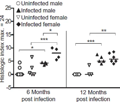
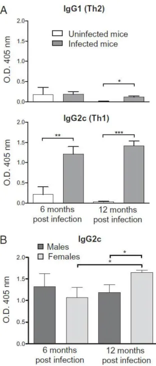
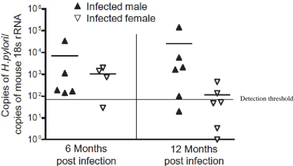
2.2.1 Pathology, Cytokine and iNOS Expression, and Serologic Responses to H. pylori Infection and H. pylori Levels
54
2.2.2 Frequency and Nature of Mutations 56
2.3 Materials and Methods 62
2.4 References 66
2.5 Tables and Figures 69
2.6 Supplemental Information 75
Chapter 3: 17β-estradiol and Tamoxifen prevent gastric cancer by modulating leukocyte recruitment and oncogenic pathways in Helicobacter pylori-infected INS-GAS male mice
86
3.1 Introduction 88
3.2 Materials and Methods 90
3.3 Results 96
3.3.1 E2 and Tamoxifen reduce reproductive tissue size and serum E2 concentrations through different mechanisms
96 3.3.2 E2, Tamoxifen and dual treatment prevent gastric cancer in
infected males
97 3.3.3 E2 and Tamoxifen decreased MPO+ neutrophils and F4/80+
macrophages in the stomach
99 3.3.4 E2 and Tamoxifen modulate cellular movement and immune
responses responsible for cancer and chronic inflammatory diseases in infected mice
100
3.3.5 E2 modulates inflammatory serum cytokines involved in neutrophil and macrophage chemotaxis
102
3.4 Discussion 103
3.5 References 111
8 3.7 Supplemental Tables 125 Chapter 4: Summary 150 4.1 Summary 150 4.2 References 155 4.3 Table 4.1 157
Appendix A: Functional classification of Helicobacter pylori isolates by gene expression analysis during coculture with gastric epithelial cells
158
A.1 Introduction 159
A.2 Materials and Methods 162
A.3 Results 165
A.3.1 ATCC43504 elicits greater IL-8 secretion and elongation of AGS cells
165 A.3.2 ATCC43504 injects more CagA and induces greater phosphorylation of Erk and Akt
166
A.3.3 Microarray Validation 166
A.3.4 Classification of Colombian strains 166
A.4 Discussion 168
A.5 References 171
A.6 Figures 175
Appendix B: Published version of Chapter 2: Sheh et al. "Mutagenic potency of Helicobacter pylori in the gastric mucosa of mice is determined by sex and duration of infection." PNAS August 24, 2010 vol. 107 no. 34, 15217-15222.
9
List of Figures and Tables
Figure 1.1 Mortality rates of gastric cancer in 2008 for men and women 43 Figure 1.2 Age-standardized incidence rates of gastric cancer 44 Figure 1.3 Histological progression of Helicobacter-induced gastric cancer 45
Figure 1.4 Divergent responses to H. pylori infection 46
Figure 1.5 Effects of CagA translocation on host epithelial cells 47
Figure 1.6 Host responses to H. pylori infection 48
Table 1.1 Polymorphisms in genes studied for association with gastric cancer 49 Figure 2.1 H. pylori infection elicits more gastric pathology in female mice 69 Figure 2.2 The effect of H. pylori infection on H. pylori-specific IgG1 and IgG2c 70
Figure 2.3 H. pylori levels in the stomach 71
Figure 2.4 H. pylori infection increases the frequency of point mutations 72 Table 2.1 H. pylori infection increases A:TG:C and G:CT:A mutations 73 Figure 2.5 Mutant frequency of deletions was unchanged by H. pylori infection 74 Figure 2.S1 Gastric histomorphological alterations caused by H. pylori 82 Figure 2.S2 H. pylori infection increases IFNg, TNFa, iNOS and IL-17 83 Figure 2.S3 H.pylori infection targets specific hot spots in the gpt gene 84 Table 2.S1 Comparison of mutations across treatment groups 85
Figure 3.1 Ratios of reproductive tissues/body weight 118
Figure 3.2 Serum E2 levels 119
Figure 3.3 Corpus pathology after 28 weeks of H. pylori infection 120 Figure 3.4 Individual histological parameters of corpus pathology 121 Figure 3.5 Immune cell infiltration of neutrophils and macrophages 122 Figure 3.6 Significant molecular networks affected by hormone treatment 123
Figure 3.7 Serum levels of cytokines and chemokines 124
Table 3.S1 Genes differentially expressed by treatment 126
Table 3.S2 Networks associated to differentially expressed genes 140 Table 3.S3 Biological functions and pathways associated with E2 treatment 142 Table 3.S4 Biological functions and pathways associated with TAM treatment 145
10 Table 3.S5 Biological functions and pathways associated with both E2 & TAM treatment 147 Table 3.S6 Individual histological parameters in the corpus and antrum 148 Table 4.1 Comparison of H. pylori pathogenesis in mice and humans 157 Figure A.1 IL-8 secretion in AGS cells cocultured with H. pylori 175 Figure A.2 Comparison of cell deformation in AGS cells cocultured with H. pylori 176
Figure A.3 Effects of H. pylori on cell signaling 177
Figure A.4 Hybridization of eukaryotic & prokaryotic RNA to H. pylori microarray 178
Figure A.5 Clustering analysis of laboratory strains 179
Figure A.6 Clustering analysis of clinical isolates 180
Figure A.7 Clustering analysis of clinical isolates using differentially expressed genes between
one low risk strain and one high risk strain 181
Figure A.8 Clustering analysis of clinical isolates using differentially expressed genes between
11
Chapter 1: Introduction
1.1 Gastric cancer
Overview. Cancer is the leading cause of deaths worldwide, accounting for 7.6 million deaths
worldwide (13% of all deaths) in 20081. In 2008, there were 989,000 cases of gastric cancer diagnosed and 738,000 gastric cancer associated deaths, making gastric cancer the fourth most common cancer and the second leading cause of cancer death1. The high mortality to incidence ratio (0.75) is due in part to the lack of clinical symptoms in most cases of early gastric cancer, which makes screening and treatment difficult. At diagnosis, gastric cancer is usually at an advanced stage, making prognosis poor and the overall 5-year survival rate less than 25%2.
Up until 1985, gastric cancer was the number one cause of cancer deaths worldwide3. At the time, the causes and pathogenesis of stomach cancer were not well understood, but for reasons unknown, a gradual decline in the incidence of gastric cancer had been observed over the last several decades. Improvements in sanitation and nutrition are believed to have contributed by increasing the amount of fresh fruits and vegetables available and by decreasing dietary salt intake. In the field of medicine, the development of endoscopies allowed for better management of precancerous lesions. However, it was the discovery in 1982 of a curved bacillus in the stomachs of patients with gastritis and peptic ulcers that revolutionized our understanding of the etiology and pathogenesis of gastric cancers4. This bacillus named, Helicobacter pylori, was recognized in 1994 by the International Agency for Research on Cancer (IARC) as a class I, or definite, carcinogen, for its role in the development of gastric cancer5. H. pylori was one of the first infectious agents recognized as a potential carcinogen. For their seminal discovery, Robin Warren and Barry Marshall were awarded the Nobel Prize in Medicine
12 in 2005. The decline in H. pylori infection, resulting from increased sanitation, improved nutrition, the common usage of antibiotics and changes in family structures due to modern life, is thought to be one of the main reasons for the decreased incidence of gastric cancer6. Nevertheless, despite falling incidence rates, the total number of new gastric cancer cases and deaths continues to increase worldwide due to overall population growth and aging in high-risk areas.
Epidemiology. Marked differences in gastric cancer incidence are observed based on
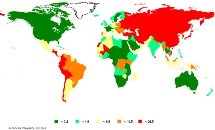
geographical location, ethnicity and gender. Over 70% of cases occur in developing nations. 50% of cases worldwide occur in Eastern Asia, with a majority of them in China, Japan and South Korea. Western industrialized nations like the United States have relatively low gastric cancer incidence rates. In the United States, gastric cancer-related incidence and mortality have been reduced approximately 50% since 19751,7. Not surprisingly, in 2008, Eastern Asia, including Japan, China, Korea and Mongolia, has the highest mortality rates (28.1 per 100,000 men and 13.0 per 100,000 women), while North America, including Canada and the United States, has the lowest (2.8 and 1.5 per 100,000 men and women, respectively)1. Other gastric cancer "hot spots" include Central and Eastern Europe, as well as Central and South America. In contrast, Australia, Africa, Southern Asia, Western Europe and North America are areas of low risk (Figure 1.1). Interestingly, country of birth is a better predictor of gastric cancer risk than country of current residence. Immigrants from regions with high gastric cancer risk to regions with lower gastric cancer risk have an intermediate risk of gastric cancer but have higher risk compared to second-generation immigrants8-9.
13 Despite the aforementioned influence of location, gastric cancer incidence and mortality differed significantly among different ethnic groups. In the United States between 2003-2007, the highest annual incidence rates were observed among Asian/Pacific Islanders (17.5 per 100,000 men and 10.0 per 100,000 women), followed by blacks(16.7 per 100,000 men and 8.6 per 100,000 women), Hispanics (14.8 per 100,000 men and 9.1 per 100,000 women), American Indians and Alaska Natives (15.5 per 100,000 men and 7.3 per 100,000 women), and whites (9.6 per 100,000 men and 4.7 per 100,000 women)10. Gastric cancer mortality rates, in the United States during the same period, mostly paralleled the trends observed for gastric cancer incidence with rates being highest among black Americans (10.7 per 100,000 men and 5.0 per 100,000 women), followed by Asian/Pacific Islanders (9.4 per 100,000 men and 5.6 per 100,000 women), American Indian/Alaska native (9.2 per 100,000 men and 4.2 per 100,000 women), Hispanic (8.0 per 100,000 men and 4.6 per 100,000 women), and white Americans (4.6 per 100,000 men and 2.4 per 100,000 women)10.
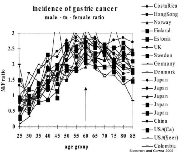
However, irrespective of location and ethnicity, men are twice as likely as women to develop gastric cancer with age-standardized incidence rates ranging from 3.9 in Northern Africa to 42.4 in Eastern Asia for men and from 2.2 in Southern Africa to 18.3 in Eastern Asia for women10-11. The pattern of the M/F incidence of gastric cancer is a global phenomenon, equally seen in populations with high and low risk for gastric cancer (Figure 1.2). This remains one of the unresolved epidemiological questions as this sexual dimorphism has not been explained by putative risk factors such as smoking, alcohol and obesity12. However, epidemiological evidence points to the protective role of female hormones13 and we are just starting to study this using in vivo models14-15. The question of gender is central to the work of this thesis and will be
14 examined in the context of mutagenesis in Chapter 2 and carcinogenesis in Chapter 3.
Pathology. Approximately 90% of gastric cancers are adenocarcinomas, malignant epithelial
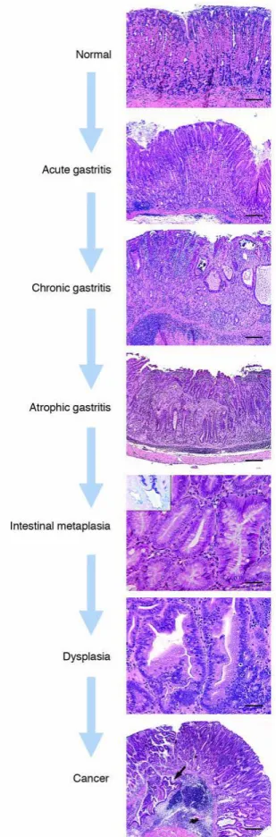
tumors that arise from the gastric glandular epithelium16. Adenocarcinomas, referred to as gastric cancer in this work, can be further subdivided by anatomical site and histologic type. Anatomically, gastric cancers are categorized as cardia and non-cardia. Cardia adenocarcinomas are more similar pathologically and epidemiologically to esophageal adenocarcinomas, and are not the focus of this work, as their development may be associated with the absence of H. pylori6. Most non-cardia gastric cancers originate in the antrum, exhibit a male-predominant pattern and are associated with chronic H. pylori infections. Histologically, gastric cancers are classified as either diffuse- or intestinal-type adenocarcinomas17. Diffuse-type gastric cancer is present in younger populations and is prevalent in both men and women, while the intestinal-type gastric cancer is most commonly associated with high risk populations and elderly men. Diffuse-type adenocarcinomas are characterized by poorly-differentiated cells without glandular structures and a distinct progression of precancerous lesions leading to cancer has not been identified17-18. Furthermore, diffuse-type gastric cancers can be familial in distribution as germline mutations leading to reduced E-cadherin (CDH1) have been associated with this histologic type19. In contrast, intestinal-type adenocarcinomas have well-differentiated neoplastic cells connected by tubules and glands similar to those observed in normal intestinal mucosa. This intestinal phenotype preserve cell polarity, partially due to the action of E-cadherin18. No germline mutations have been associated with this form of gastric cancer. The intestinal-type gastric cancer is associated with tissues with chronic inflammation, and follow a clear disease progression described by Correa's model of gastric carcinogenesis20.
15 Briefly, Correa's model characterized the series of sequential lesions that lead to gastric cancer. The stages of the precancerous cascade are normal stomach, chronic active nonatrophic gastritis, multifocal atrophic gastritis, intestinal metaplasia (complete, followed by incomplete), dysplasia and ultimately invasive carcinoma (Figure 1.3)20. This work focuses on non-cardia, intestinal-type gastric adenocarcinomas, which represent the most common form of gastric cancer worldwide.
Mutations associated with gastric cancer. Data from literature examining mutations in gastric
cancer as well as the sequencing efforts of the Cancer Genome Project is compiled in the Catalogue of Somatic Mutations in Cancer (COSMIC). COSMIC lists the genes most frequently mutated as well as the type of mutations seen in these mutations21-23. According to COSMIC data, the top ten mutated genes found in gastric cancer samples are TP53, KRAS, CTNNB1, APC,
PIK3CA, CDH1, CDKN2A, PTEN, MSH6 and FBXW7
(http://www.sanger.ac.uk/genetics/CGP/cosmic/ accessed on 2/22/2011). Analysis of the mutational spectra of gastric cancer shows that G:C>A:T (mostly at CpG sites) transitions are the most common mutation observed, followed by A:T>G:C transitions and G:C>T:A transversions22. These mutations can be induced by reactive oxygen and nitrogen species (RONS)24, such as nitric oxide25. RONS are constantly released by inflammatory cells in response to H. pylori infection.
Etiology. The role of H. pylori infection in gastric carcinogenesis has been established through
epidemiological26-28 and experimental evidence29-30. H. pylori promotes precancerous lesions and the development of gastric cancer by inducing a chronic inflammatory state. The association of cancer with inflammation has been recognized since 1863 when Virchow
16 detected leukocytes in neoplastic tissues31. The causal relationship between RONS produced by inflammatory cells and DNA damage that leads to cancer is now widely accepted32. The combination of DNA damage, increased cell proliferation, deregulation of apoptosis and tissue remodeling that occurs during chronic inflammation increases the risk of neoplasia33. These processes are largely regulated by proinflammatory cytokines, chemokines and growth factors that are part of the inflammatory environment. This model of persistent H. pylori infection leading to chronic inflammation and cancer is supported by chronic viral (hepatitis B-induced liver cancer) and parasitic (Schistosomiasis-induced bladder cancer) infections34-35.
Fifty percent of the world population is infected with H. pylori, but only a small fraction (1-2%) develops multifocal atrophic gastritis which leads to gastric cancer. The majority of infected individuals present no clinical symptoms of infection while 5-15% of infected individuals develop peptic duodenal ulcers (Figure 1.4)36-37. Duodenal ulcers are characterized by antral predominant nonatrophic gastritis which may reduce the risk of gastric cancer38. While H. pylori infection drives gastric carcinogenesis, the outcome of infection is modulated by the virulence of the bacterial strain, the genetic susceptibility of the host and the external environment.
1.2 Helicobacter pylori
Overview. The first well-documented report of spiral-shaped bacteria in human stomachs was
by Bizzozero in 189339. Early reports linking these microorganisms to gastritis, and even carcinomas, were dismissed as the bacteria were not found by others and were considered contaminants, as bacteria were not expected to survive in the acidic gastric environment40-42.
17 The field stagnated until 1982 when Marshall and Warren "rediscovered" unidentified curved bacilli in gastric biopsies associated with active, chronic gastritis4. Later, Marshall fulfilled Koch's postulates by developing gastritis after swallowing a pure culture of H. pylori43. H. pylori's ability to persist for years in the human gastric mucosa was demonstrated in another experiment involving self-induced infection, which required antibiotics to eradicate the bacterium44. Within the last several decades, investigators around the world have proven that H. pylori is the causative agent of gastritis, ulcers, gastric mucosa-associated lymphoid tissue lymphoma and gastric cancer in humans and laboratory animal models45.
Epidemiology. While 50% of the world's population is infected with H. pylori, prevalence differs
dramatically between developing (>80%) and developed (<40%) nations46. H. pylori is the most common bacterial infection worldwide. Bacterial phylogenetic studies demonstrate that H. pylori has infected humans for at least 58,000 years, before humans migrated out of Africa47. Currently, H. pylori incidence has been estimated at 1-3% in developed nations and 3-10% in developing nations48-49. H. pylori infection is usually acquired in childhood and normally persists for life within the gastric mucosa6. In recent years, H. pylori prevalence worldwide has been decreasing coinciding with better hygiene and improved socioeconomic status, particularly in the industrialized world46.
The mechanism of H. pylori transmission remains largely unknown. The bacterium is almost exclusively found in humans and some nonhuman primates with no known reservoirs of H. pylori in the environment, which makes direct person-to-person transmission the most likely method of infection. The fecal-oral (e.g. fecal contamination in institutions), oral-oral (e.g. premastication of food) and gastric-oral (e.g. iatrogenic infection or vomitus) routes have been
18 proposed but conclusive evidence is not yet available46. There is a strong association of H. pylori infection with family size, as intra-familial transmission is facilitated by increased number of siblings, possibly due to reinfection among children6.
Virulence factors. H. pylori is a microaerophilic, gram-negative, curved rod with multiple
flagella that is capable of persistently infecting the harsh gastric environment. The bacterium's success is due largely to an array of virulence factors that have adapted to the stomach for millennia. The first obstacle the bacteria have to overcome is navigating the acidic lumen of the stomach and finding its niche in the gastric mucosa. To survive the acid pH, H. pylori utilizes urease to convert gastric urea into ammonia and CO2 which may form a protective layer of ammonia to neutralize the acid adjacent to the bacteria50. A second mechanism which utilizes an α-carbonic anhydrase takes the CO2 produced and converts it to HCO32- which helps maintain the periplasmic pH of the bacteria around 6.151. In order to escape the lumen, H. pylori swims following gradients of carbonate and urea secreted by gastric epithelia to chemotactically find the mucin layer. Mutants deficient in chemotaxis (cheW mutant) or sensing gradients (TlpB mutant), as well as mutants deficient in motility (nonflagellated mutants and flagellated but nonmotile mutants), do not infect the gastric mucosa at all or are outcompeted by wild-type counterparts52-54. Once in the mucin layer, H. pylori's adhesins mediate persistent infection by binding sulfated mucin sugars or epithelial cells to prevent the sloughing of the bacterium. The best characterized adhesin is BabA which binds Lewis B antigens on epithelial cells. An allele of BabA, babA2, has been strongly associated with worse clinical outcomes55. Other known H. pylori adhesins include SabA, SabB, OipA, AlpA and AlpB46.
19 The final set of virulence factors have perhaps been the most studied as they are the genes associated with H. pylori pathogenicity and epithelial cell damage.
Damaging epithelial cells or destabilizing tight junctions is believed to release nutrients and substrates required by the bacteria to survive56. The most important virulence factors in this set are CagA, VacA, HP-NAP, and LPS. Recently, a prospective study conducted in Spain demonstrated that infection with H. pylori strains harboring CagA and VacA s1/m1 was associated with the development of gastric preneoplastic lesions (Odds ratio of 4.80, 95% CI 1.71–13.5) compared to H. pylori-infected individuals without these virulence factors57. The cytotoxin-associated gene A (CagA) is part of the H. pylori Cag pathogenicity island (CagPAI) which contains 31 genes required for the assembly and function of a type IV secretion system. CagA is a bacterial effector protein injected into the cytoplasm of gastric epithelial cells that can be phosphorylated by human kinases and directly interferes with signaling networks inducing changes in morphology and IL-8 secretion (Figure 1.5)58. H. pylori strains possessing CagA, specifically CagA variants common in East Asia, are believed to increase the risk of gastric cancer58. VacA, or vacuolating cytotoxin A, produces large acidified vacuoles in epithelial cells in vitro, which leads to cell death. Epithelial cell vacuoles are derived from late endosomes and lysosomes, affecting endocytosis. VacA also forms membrane channels through the epithelial layer, increasing its permeability46. The activity of the s1/m1 type of VacA is strongly correlated with increased H. pylori pathogenicity in Western populations46. HP-NAP is the H. pylori neutrophil activation protein that specifically targets and activates neutrophils, presumably to increase epithelial damage59. H. pylori's LPS, or lipopolysaccharide, is interesting as it poorly elicits an inflammatory response, as H. pylori's LPS moeities mimic the Lewis antigens x and y on
20 gastric epithelial cells. This immune mimicry might have two distinct objectives: 1) limit specific immune responses to H. pylori, or 2) elicit immune responses against gastric epithelial cells to further destabilize epithelial polarity46. All together, H. pylori has evolved to infect the gastric niche and utilizes the appropriate combination of virulence factors to transform the environment for its survival. Determining the role of virulence factors in different strains of H. pylori is the purpose of experiments using H. pylori microarrays that are introduced in Appendix A.
Diagnosis and Treatment. The methods for diagnosing H. pylori infection include urea breath
testing, serology, fecal antigen tests, histology and culture. Urea breath testing is the least invasive and works by detecting the activity of H. pylori's urease. Patients swallow urea labeled with a carbon isotope and breathe into a tube that detects the presence of labeled carbon dioxide, which would be produced by the decomposition of urea. Serological tests detect specific antibodies against H. pylori, but cannot definitively determine active infection as antibodies can be produced for years after clearance. Fecal antigen tests use H. pylori-specific antibodies to detect H. pylori antigen. Both histology and culture are used as the gold standard of detection but are the most invasive as they require an endoscopy46.
The current treatment of H. pylori in symptomatic carriers is eradication of the bacteria. Antimicrobial therapy to eradicate H. pylori consists of triple or quadruple therapy as individual antibiotics are not as effective in the gastric niche. Triple therapy incorporates two antibiotics, usually clarithromycin and amoxicillin, with a bismuth compound or a proton pump inhibitor, while quadruple therapy has both the bismuth compound and the proton pump inhibitor46. However, these therapies have shown suboptimal results in some studies leading to concerns
21 of antibiotic resistant strains60. Due to H. pylori's association with gastric cancer, wide-scale eradication and vaccines have been proposed to prophylactically prevent gastric cancer. Early stage eradication of H. pylori prevents gastric cancer61, and reduces atrophy and physiological alterations to the stomach that allow colonization by other microbiota, which may be involved in gastric carcinogenesis62-66. The possibility of developing antibiotic resistant strains and the possible relationship between H. pylori eradication and development of proximal gastric cancer and esophageal cancer have quelled discussions advocating H. pylori eradication in asymptomatic carriers worldwide. Efforts to develop a H. pylori vaccine have produced mixed results60. However, a viable solution is aggressive screening by X-ray or endoscopy and eradication of the bacteria in symptomatic carriers in high-risk regions. These methods have proven effective in Japan, a nation with one of the highest incidence rates of gastric cancer, as they have also experienced the worldwide decrease in gastric cancer incidence and increased their overall 5 year survival rate67. From 1962 to 1992, Japan doubled the overall 5 year survival of gastric cancer patients from 20% to 40% by detecting stomach cancer at the localized stage and surgically removing cancerous tissues60.
1.3 Host factors
Although H. pylori is the major causative agent of gastric cancer, the host plays an important role in the progression of gastric cancer through its response to the bacterium68. There is a two- to three-fold increase in gastric cancer risk in individuals with blood relatives with gastric cancer, and 10% of gastric cancer cases show familial clustering69. While H. pylori infection is likely to be transmitted throughout a family, controlling for H. pylori infection indicated that a family
22 history of gastric cancer is still a risk factor for disease. These families have been shown to have increased susceptibility to H. pylori and its associated precancerous lesions which suggest a strong host component in disease development70. As H. pylori pathogenesis is mediated through chronic inflammatory processes, the modulation of the host's immune response may affect disease development and will be discussed below.
The adaptive immune response. The current model of persistent H. pylori infection leading to a
chronic inflammatory state that causes gastric cancer (through DNA damage, errors in replication due to hyperproliferative conditions or recruitment of oncogenic bone-marrow derived cells) is largely dependent on the host's specific immune response to the bacterium68. In response to initial H. pylori infection, circulating phagocytic cells including neutrophils, macrophages and monocytes are recruited to the site of infection by the cytokine/chemokine system and the complement system (Figure 1.6). However, failure to eradicate the bacteria by the innate immune response and its associated acute inflammation lead to the recruitment of T and B lymphocytes and more macrophages, signaling the involvement of the adaptive immune system and the establishment of chronic inflammation. Following their recruitment, immature helper T cells (Th0) are activated by H. pylori antigens expressed on antigen presenting cells (APCs) and differentiate into several subtypes, including Th1, Th2 and the more recently discovered Th17 cells, which secrete different sets of cytokines that modulate the type of immune response (Figure 1.6). Broadly, Th1 cells secrete IL-2 and IFN-γ, increase proinflammatory cues and are associated with macrophage activation. Th2 cells secrete 4, IL-5, and IL-10, increase antiinflammatory cues and stimulate B cell responses. Th17 cells secrete IL-17 and IL-22, also increase proinflammatory cues and are associated with neutrophil
23 recruitment and inflammatory disorders. By mechanisms incompletely understood, H. pylori infection promotes a strong Th1 and Th17 proinflammatory response71. Historically, Th1 responses have been shown to mediate H. pylori-associated gastritis71-75, but there is increasing recognition of the role of Th17 cells in H. pylori pathogenesis76-77. However, due to the recent discovery of Th17, its contributions have not been as thoroughly studied. Based on the Th1/Th2 model of H. pylori pathogenesis, the proinflammatory Th1, and possibly now Th17, cytokines promote gastritis, while Th2 cytokines, and possibly T regulatory cells that counteract Th17 actions, protect against gastric inflammation. The importance of T cells, particularly Th1 cells, in the development of Helicobacter-induced gastric disease has been demonstrated in mouse models lacking functional lymphocytes and utilizing adoptive transfer to investigate the effect of specific T cell subsets74,78-79. Furthermore, different mouse models of Helicobacter spp. infection demonstrate the importance of Th1/Th2 responses, as C57BL/6 mice which are susceptible to gastric atrophy caused by H. pylori have stronger Th1 responses while BALB/c mice which are less susceptible to gastric atrophy mount Th2 responses68. These models will be further discussed in the section on "Mouse models of H. pylori infection" as the effects of the immune response in the context of gender of great importance to this thesis.
As differences in the host's T cell response affect the outcome of H. pylori infection, it has been hypothesized that modulating the host's response might exacerbate or ameliorate gastric disease. Coinfection with Heligmosomoides polygyrus, a murine parasite, and Helicobacter felis, a gastric Helicobacter, decreased Th1-associated cytokines and antibodies to H. felis leading to attenuated gastritis and less severe premalignant lesions80. Coinfection with Helicobacter bilis, an enterohepatic helicobacter that subclinically infects numerous strains of
24 mice, also attenuated H. pylori induced gastritis in C57BL/6 mice81. The opposite effect was observed by modulating the host's response using a parasite that induces a strong Th1 response, such as Toxoplasma gondii. Coinfection with T. gondii exacerbated H. felis infection leading to increased morbidity82. These results support the "African enigma," the hypothesis that the low incidence of gastric cancer observed in some developing nations is due to the immunomodulating effect of coinfections leading to Th2 responses83. This effect has been observed in a study involving a coastal population and an Andean population in Colombia with vastly different incidences in gastric cancer84. While H. pylori prevalence (>90%) was similar, the coastal population had a lower incidence of gastric cancer which was associated with coinfection with enteric helminths and a systemic Th2 response to H. pylori, unlike the Andean population that experience a higher incidence of gastric cancer, had fewer parasites and a systemic Th1 response84.
Inflammation, oxidative stress and DNA repair. T helper cells create an environment that
recruits and activates the appropriate inflammatory cells to deal with specific antigens. T helper cells direct the immune response through the secretion of specific cytokines. H. pylori-infected gastric epithelium has increased levels of IL-1β, IL-2, IL-6, IL-8 and TNFα, which is a potent proinflammatory combination characterized by the predominance of phagocytes85. IL-1β promotes a strong proinflammatory response and inhibits gastric acid secretion70. IL-8, a neutrophil activating chemokine, is strongly induced by H. pylori85-86. After following cytokine and chemokine gradients to the site of infection, phagocytes have two functions: a) eradicate H. pylori and b) repair the tissue. To eradicate H. pylori, phagocytes utilize lysosomal proteins, such as proteases, lysozymes, and myeloperoxidases. Myeloperoxidases produce superoxide
25 (O2-), a precursor of peroxide (H2O2) and hypochlorous acid. Monocytes and macrophages release nitric oxide (NO) which also forms peroxynitrite (OONO-). Lysosomal proteins and RONS released during H. pylori infection damage adjacent tissues and are the main source of epithelial damage. To accomplish the goal of repairing the tissue, phagocytes secrete cytokines and growth factors to recruit more inflammatory cells and initiate wound healing responses, such as proliferation, ECM degradation, angiogenesis, etc. required to return the tissue to homeostasis33. As H. pylori persistently infects the stomach, the proinflammatory and wound healing responses are not shut down and over time can promote cancer.
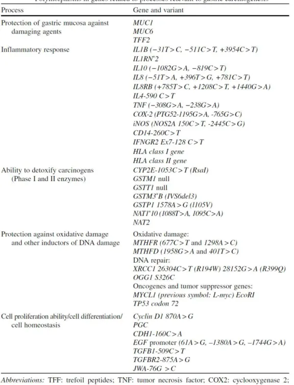
Another way in which the host can modulate the inflammatory response is through host genetics. The association of single nucleotide polymorphisms (SNPs) of inflammation-related genes with increased risk of gastric cancer highlights the importance of inflammation and oxidative stress in H. pylori driven carcinogenesis87-88. SNPs have been associated with increased susceptibility to H. pylori infection, H. pylori-induced gastric atrophy and gastric cancer. SNPs in many inflammatory mediators, such as IL-1β, IL-1RN2, IL-10, IL-8, CXCR2 (IL-8RB) , IL-4, COX2, TNF, iNOS, and IFNGR2, are relevant in gastric carcinogenesis as they further alter the balance of both Th cells and phagocytes (Table 1.1). The associations of IL1β-31*C and -511*T with increased risk of gastric atrophy and cancer in Western populations were among the first discovered70. Studies in East Asian populations have associated SNPs in the IL-1 family with increased gastric cancer risk89-91. Polymorphisms in the promoter region of IL-8 (IL-8 -251 T to A) have been associated with increased gastric cancer risk92-93. The role of CXCL1, a murine homolog of IL-8, in H. pylori induced carcinogenesis will be discussed in Chapter 3.
26 carcinogenesis by decreasing oxidative DNA damage. H. pylori-induced chronic inflammation increases DNA damage, double-strand breaks and DNA fragmentation in the gastric mucosa through the effect of oxidative stress and RONS94-97. RONS cause DNA damage via multiple mechanisms such as direct base oxidation, nucleoside deamination, and the formation of etheno adducts by lipid peroxidation24,32,98-99. H. pylori gastritis selectively increases the activity of DNA damage and repair proteins, such as Ku, poly (ADP-ribosyl) polymerase, 8-hydroxyguanine glycosylase (OGG1), and MSH2, in human gastric mucosa96. Studies of SNPs in genes related to DNA repair, such as MGMT, and OGG1, have not always demonstrated clear links with gastric cancer87-88,100. MGMT, also known as O6-methylguanine-DNA methyltransferase, repairs DNA adducts such as O6-methylguanine and O4-methylthymine101-102, and its inactivation by promoter hypermethylation has been associated with increased gastric cancer risk103-104. MGMT Ile143Val polymorphism was associated with increased gastric cancer risk, but only in patients with low intake of fruits and vegetables100. Polymorphisms of OGG1, or 8-oxoguanine DNA glycosylase, cause the accumulation of 8-oxo-dG during H. pylori gastritis, but their association with increased gastric cancer risk is still controversial105-107. Given their role in repairing DNA damage induced by H. pylori, DNA repair proteins may greatly influence H. pylori carcinogenesis.
Gender. Men experience higher rates of infection and its associated mortality than women.
Normal immune responses are sexually dimorphic with a heightened inflammatory response in women compared to men. This greater inflammatory response in women provides advantages during infection, sepsis and trauma but makes women more susceptible to autoimmune diseases108. Epidemiological and immunological evidence suggests that the menstrual cycle,
27 pregnancy and menopausal status influence the etiology and progression of chronic inflammatory diseases, such as gastric cancer and rheumatoid arthritis, which suggests the importance of female sex hormones109-110. Hormone therapies in men and women are also associated with decreased gastric cancer risk13. Postmenopausal hormone replacement therapy (HRT) may lower the rate of gastrointestinal cancer, particularly colonic cancer111-113. Prostate cancer patients treated with estrogen had a decreased risk of gastric cancer110. Additionally, the usage of anti-estrogen therapy, particularly the breast cancer drug Tamoxifen, is linked with increased incidence rates of gastrointestinal malignancies. Considerable evidence exists that Tamoxifen, a mixed agonist/antagonist of estrogen signaling, is associated with protection against breast cancer, but also increases the risk of endometrial cancers114-115. However, Tamoxifen treatment and its association with gastrointestinal cancers, and gastric cancer in particular, is not clear; one study reports no effect115 while others demonstrate Tamoxifen treatment with increased risk116-118.
A review of the effects of estrogens on inflammation indicate that estrogens, of which 17β-estradiol or E2 is the most important, decrease the apoptosis of immune cells, inhibit reactive oxygen species formation, and inhibit nitric oxide production in the presence of inflammatory stimuli108. Another mechanism by which estrogens may affect chronic inflammatory diseases is by directly affecting the secretion of cytokines by T cells, B cells and macrophages. Generally E2 in premenopausal women favors the downregulation of T cell-dependent immunity. Periovulatory to pregnancy levels of E2 promotes IL-4, IL-10 and IFN-γ secretion from CD4+ T cells while inhibiting the production of TNFα. Furthermore, mouse studies demonstrate that high concentrations of E2 increase Th2 and T regulatory transcription
28 factors, thus increasing the antiinflammatory response. In contrast, B cell antibody secretion is normally stimulated by E2 in pre- and post-menopausal women. Furthermore, very high levels of E2 inhibit the secretion of IL-1β and TNFα, while lower levels stimulated both cytokines demonstrating the importance of E2 levels in shaping the inflammatory response108. As estrogens can strongly decrease T cell-dependent immunity and the proinflammatory cytokine environment, the role of estrogens in gastric cancer is further explored in Chapter 3.
1.4 Environmental factors
The sharp decrease in the incidence and mortality of gastric cancer in developed countries suggest that the environment is a third factor to consider in the etiology of gastric cancer. As mentioned previously, second-generation immigrants had a reduction in gastric cancer risk compared to first-generation immigrants indicating that the environment can be more important that host genetics and bacterial factors (assuming intra-familial transmission of H. pylori)9. Diet and lifestyle have been associated with increased gastric cancer risk. High salt consumption and low intake of fresh fruits and vegetables are independent dietary factors that increase gastric cancer risk119. Of these dietary factors, the intake of high levels of dietary salt has been studied most extensively due to the association of high incidences of gastric cancer to countries with high salt intake, which is thought to promote further injury to the stomach120-121. Animal models assessing the role of high salt in gastric pathology have yielded mixed results66,122 Smoking also increases gastric cancer risk123. Associations between meat consumption or alcohol and gastric cancer are not as clear119,124. Additionally, the impact of
29 long-term proton pump inhibitors in gastric carcinogenesis is now being probed, as proton pump inhibitors have been linked to precancerous lesions and cancer in rodents125-126
1.5 Mouse models of H. pylori infection
Many animal models, such as neonatal gnotobiotic piglets, Macaca species and Mongolian gerbils, have been utilized to model H. pylori infection in humans127-129. However, the mouse is the most widely used animal model in H. pylori studies due to its myriad advantages, such as well-documented immune responses, the sequencing of its genome, availability of reagents and transgenic mice and low cost relative to other models. Early H. pylori experiments with mice did not lead to persistent colonization in wild-type strains leading to the use of athymic, or nude mice130 or other Helicobacter spp., most notably H. felis131. H. felis infection of C57BL/6 mice causes oxyntic atrophy and gastric cancer by 15 months post-infection (MPI)132-133. The first reports of successful adaptation of H. pylori to the mouse model came from Italy in the mid-1990's134-136, but they were superseded by the adaptation of the cagPAI+ Sydney strain, or SS1, which has become the standard for H. pylori mouse studies due to its ability to persistently infect many inbred mouse strains, such as C57BL/6, BALB/c, DBA/2 and C3H/He137. As mentioned previously, SS1 elicits a robust chronic active gastritis in C57BL/6 mice accompanied by a Th1 immune response while BALB/c mice develop less pathology accompanied by a Th2 response due to host strain differences (e.g. differences in phospholipase A2 secretion)132,138-139. Each system has particular differences that must be considered in order to address the right biological questions. For example, in C57BL/6 mice, females have a stronger inflammatory
30 response compared to males leading to increased gastritis and cell proliferation, which is an important factor to consider when determining the interpretation of data such as histopathological scores140. These differences in the models are useful in determining how changes in the immune response and gender affect the formation of gastric lesions. Nevertheless, H. pylori infection alone does not reliably induce gastric cancer in either of these strains138. Limitations associated with the murine model are differences in gastric architecture, the lack of H. pylori-associated ulcers and gastric cancer in some models, and the presence of Lactobacillus spp. in the proximal murine stomach, which are not found in the human stomach46. However, new murine models that more closely recreate conditions in humans continue to progress. For example, the development of H. pylori-induced gastric cancer has been documented in male hypergastrinemic INS-GAS FVB mice at 7 MPI141 and C57BL/6 × 129S6/SvEv (B6129) mice at 15 MPI122. Using two of these models, this thesis probes the interactions between gender and immune response in gastric mutagenesis using C57BL/6 mice (Chapter 2) and the role estrogens and innate immunity in gastric carcinogenesis in INS-GAS mice (Chapter 3).
1.6 Transgenic mouse mutation systems
Murine mutational analysis systems are mouse models used to detect somatic mutations and assess the mutagenicity of chemical, biological or physical agents. These in vivo models use reporter genes to detect mutations under the assumption that damage to reporter genes are indicative of mutations to the host genome. Based on the origin of their reporter genes, murine mutational analysis systems are classified as endogenous or exogenous systems.
31 Endogenous systems include the Dlb-1 mouse assay, the Aprt mouse assay, the pink-eyed unstable (pun) mouse assay and the mammalian spot assay142. Exogenous systems include the Big Blue® assay, Muta ™Mouse assay and the gpt delta assay143. The main advantage of the endogenous systems is that the reporter genes are host genes that are expressed in their native chromosomal conformation142. This allows the induction of mutations in active genes which can often be assessed in vivo. In the Dlb-1, pun, and mammalian spot models, assessment of the induction of mutations can be done visually by examining changes on the fur or staining in tissue samples. An exception is the Aprt system which requires in vitro culture but also allows sequencing of mutations144-145. However, a limitation of endogenous systems is that they can only detect mutations in certain cell types. In contrast, exogenous, or transgenic, systems express the reporter genes in every cell of the body which permits the detection of mutations in any organ. Transgenic systems insert multiple tandem copies of lambda phage carrying a reporter gene like lacI, lacZ or gpt to detect mutations ex vivo143. After extracting the DNA, lambda phage is repackaged, and used to infect Escherichia coli. E. coli expresses the transgene providing a readout indicating mutations in the transgene. The genes can be isolated and sequenced to determine the exact mutational spectra. The drawbacks of this method are that the reporter genes are 1) not actively expressed in the mouse and 2) are prokaryotic with differences in methylation and nucleotide composition. Furthermore, the ex vivo expression makes the reporter genes susceptible to further mutations outside the mouse.
Different transgenic systems have been used to measure the induction of mutations caused by chronic inflammation, including H. pylori, in the gastrointestinal tract146-149. The Big Blue® assay, Muta ™Mouse assay and the gpt delta assay are attractive candidates to study H.
32 pylori pathogenesis as they are available on the C57BL/6 background which develops
precancerous lesions in response to H. pylori infection. As these three transgenic systems utilize similar mechanisms to report mutations, the gpt delta assay will be utilized to further describe the methods. The gpt delta transgenic mouse was developed to allow the detection of point mutations by the inactivation of gpt, or guanine phosphoribosyltransferase, activity under 6-thioguanine (6-TG) selection150. Importantly, in contrast to other transgenic systems, the gpt delta mouse can also measure deletions less than 10kb in size using Spi- selection150.
The 456 bp gpt gene encodes the Gpt enzyme which adds activated ribose-5-phosphate to guanine bases creating guanine monophosphate in the purine salvage pathway. Gpt can also salvage 6-TG but the product is toxic to the cell143. Using this principle, E. coli infected with plasmids expressing a functional gpt cannot grow on minimal media plates with 6-TG, whereas plasmids with inactivated, or mutant, gpt will form colonies. The gpt gene was inserted into a plasmid construct flanked by lox sites. After murine genomic DNA is isolated, the phage DNA is rescued and packaged into phage particles that transduce E. coli expressing Cre recombinase. Cre acts upon the lox sites allowing the propagation of the plasmid. The plasmid also carries the CAT gene which confers resistance to chloramphenicol (Cm). Using Cm alone or Cm and 6-TG selection, the total number of E. coli expressing the plasmid and the total number of E. coli expressing the plasmid with a mutated gpt gene can be calculated. 6-TG resistant colonies are then cultured and a 739 bp DNA product containing the gpt gene is amplified by PCR. This product can then be sequenced to determine the mutational spectra. Mutations are classified as transitions, transversions, deletions, insertions or complex (multiple changes).
33 The Spi- assay selects for lambda phage that are not sensitive to interference from prophage P2. Induction of wild-type lambda phage on E. coli carrying prophage P2 results in the host's death and inhibition of phage replication, which makes lambda phage sensitive to P2 interference, or Spi+151. However, deficiency in both red and gam gene function, along with chiC expression, confer phages with resistance to P2's interference, or a Spi- phenotype, and importantly do not immediately kill E. coli allowing phage propagation. The lambda phage in the gpt delta mouse incorporates the red, gam and chiC elements. When gpt delta mice are exposed to agents that cause deletions capable of inactivating both red and gam, the recovered lambda phage are Spi -which allows them to form plaques on a lawn of E. coli carrying P2. Using strains of E. coli with and without P2, the total number of lambda phage with red and gam inactivation and the total number of lambda phage recovered can be determined. Mutated phage can be recovered and passaged to determine the location of the deletion by a combination of PCR and sequencing. Due to its ability to determine the mutagenic potency of infectious agents in the stomach, I utilized the gpt delta mouse model on a C57BL/6 background to determine the role of H. pylori in the induction of gastric mutations in Chapter 2.
34
1.7 References
1. Ferlay, J., et al. Estimates of worldwide burden of cancer in 2008: GLOBOCAN 2008. Int J Cancer (2010).
2. Chan, A.O., Wong, B.C. & Lam, S.K. Gastric cancer: past, present and future. Can J Gastroenterol 15, 469-474 (2001).
3. Pisani, P., Parkin, D.M. & Ferlay, J. Estimates of the worldwide mortality from eighteen major cancers in 1985. Implications for prevention and projections of future burden. Int J Cancer 55, 891-903 (1993).
4. Marshall, B.J. & Warren, J.R. Unidentified curved bacilli in the stomach of patients with gastritis and peptic ulceration. Lancet 1, 1311-1315 (1984).
5. Schistosomes, liver flukes and Helicobacter pylori. IARC Working Group on the Evaluation of Carcinogenic Risks to Humans. Lyon, 7-14 June 1994. IARC Monogr Eval Carcinog Risks Hum 61, 1-241 (1994).
6. Blaser, M.J. Hypothesis: the changing relationships of Helicobacter pylori and humans: implications for health and disease. J Infect Dis 179, 1523-1530 (1999).
7. Parkin, D.M., Stjernsward, J. & Muir, C.S. Estimates of the worldwide frequency of twelve major cancers. Bull World Health Organ 62, 163-182 (1984).
8. Coggon, D., Osmond, C. & Barker, D.J. Stomach cancer and migration within England and Wales. Br J Cancer 61, 573-574 (1990).
9. Kolonel, L.N., Nomura, A.M., Hirohata, T., Hankin, J.H. & Hinds, M.W. Association of diet and place of birth with stomach cancer incidence in Hawaii Japanese and Caucasians. Am J Clin Nutr 34, 2478-2485 (1981).
10. Altekruse, S.F., et al. SEER Cancer Statistics Review, 1975-2007. (National Cancer Institute, Bethesda, MD, 2010).
11. Sipponen, P. & Correa, P. Delayed rise in incidence of gastric cancer in females results in unique sex ratio (M/F) pattern: etiologic hypothesis. Gastric Cancer 5, 213-219 (2002). 12. Lindblad, M., Rodriguez, L.A. & Lagergren, J. Body mass, tobacco and alcohol and risk of
esophageal, gastric cardia, and gastric non-cardia adenocarcinoma among men and women in a nested case-control study. Cancer Causes Control 16, 285-294 (2005).
13. Chandanos, E. & Lagergren, J. Oestrogen and the enigmatic male predominance of gastric cancer. Eur J Cancer 44, 2397-2403 (2008).
14. Ohtani, M., et al. Protective role of 17 beta -estradiol against the development of Helicobacter pylori-induced gastric cancer in INS-GAS mice. Carcinogenesis 28, 2597-2604 (2007).
15. Ohtani, M., et al. 17β-estradiol suppresses Helicobacter pylori-induced gastric pathology in male hypergastrinemic INS-GAS mice. Carcinogenesis (Submitted 2010).
16. Kumar, V., Abbas, A.K., Fausto, N. & Aster, J. Robbins and Cotran Pathologic Basis of Disease, (Saunders Elsevier, Philadelphia, PA, 2010).
17. Lauren, P. The two histological main types of gastric carcinoma: diffuse and so-called intestinal-type carcinoma. An attempt at a histo-clinical classification. Acta Pathol Microbiol Scand. 64, 31-49 (1965).
18. Correa, P. Human gastric carcinogenesis: a multistep and multifactorial process--First American Cancer Society Award Lecture on Cancer Epidemiology and Prevention. Cancer Res 52, 6735-6740 (1992).
35 19. Guilford, P., et al. E-cadherin germline mutations in familial gastric cancer. Nature 392,
402-405 (1998).
20. Correa, P., Haenszel, W., Cuello, C., Tannenbaum, S. & Archer, M. A model for gastric cancer epidemiology. Lancet 2, 58-60 (1975).
21. Futreal, P.A., et al. A census of human cancer genes. Nat Rev Cancer 4, 177-183 (2004). 22. Greenman, C., et al. Patterns of somatic mutation in human cancer genomes. Nature
446, 153-158 (2007).
23. Bamford, S., et al. The COSMIC (Catalogue of Somatic Mutations in Cancer) database and website. Br J Cancer 91, 355-358 (2004).
24. De Bont, R. & van Larebeke, N. Endogenous DNA damage in humans: a review of quantitative data. Mutagenesis 19, 169-185 (2004).
25. Wink, D.A., et al. DNA deaminating ability and genotoxicity of nitric oxide and its progenitors. Science 254, 1001-1003 (1991).
26. Nomura, A., et al. Helicobacter pylori infection and gastric carcinoma among Japanese Americans in Hawaii. N Engl J Med 325, 1132-1136 (1991).
27. Parsonnet, J., Friedman, G.D., Orentreich, N. & Vogelman, H. Risk for gastric cancer in people with CagA positive or CagA negative Helicobacter pylori infection. Gut 40, 297-301 (1997).
28. Forman, D., et al. Association between infection with Helicobacter pylori and risk of gastric cancer: evidence from a prospective investigation. BMJ 302, 1302-1305 (1991). 29. Watanabe, T., Tada, M., Nagai, H., Sasaki, S. & Nakao, M. Helicobacter pylori infection
induces gastric cancer in mongolian gerbils. Gastroenterology 115, 642-648 (1998). 30. Wang, T.C., et al. Synergistic interaction between hypergastrinemia and Helicobacter
infection in a mouse model of gastric cancer. Gastroenterology 118, 36-47 (2000). 31. Balkwill, F. & Mantovani, A. Inflammation and cancer: back to Virchow? Lancet 357,
539-545 (2001).
32. Dedon, P.C. & Tannenbaum, S.R. Reactive nitrogen species in the chemical biology of inflammation. Arch Biochem Biophys 423, 12-22 (2004).
33. Coussens, L.M. & Werb, Z. Inflammation and cancer. Nature 420, 860-867 (2002).
34. IARC. Schistosomes, liver flukes and Helicobacter pylori. IARC Working Group on the Evaluation of Carcinogenic Risks to Humans. Lyon, 7-14 June 1994. IARC Monogr Eval Carcinog Risks Hum 61, 1-241 (1994).
35. Chen, C.J. & Chen, D.S. Interaction of hepatitis B virus, chemical carcinogen, and genetic susceptibility: multistage hepatocarcinogenesis with multifactorial etiology. Hepatology (Baltimore, Md 36, 1046-1049 (2002).
36. Kuipers, E.J., et al. Long-term sequelae of Helicobacter pylori gastritis. Lancet 345, 1525-1528 (1995).
37. de Vries, A.C. & Kuipers, E.J. Helicobacter pylori infection and nonmalignant diseases. Helicobacter 15 Suppl 1, 29-33 (2010).
38. Hansson, L.E., et al. The risk of stomach cancer in patients with gastric or duodenal ulcer disease. N Engl J Med 335, 242-249 (1996).
39. Bizzozero, G. Ueber die schlauchformigen drusen des magendarmkanals und die beziehungen ihres epithels zu dem oberflacheepithel der schleimhaut. Arch. Mikr. Anat.
36 40. Doenges, J.L. Spirochetes in the gastric glands of macacus rhesus and of man without
related diseases. Arch. Pathol. 27, 469-477 (1939).
41. Kreinitz, W. Ueber das Auftreten von Spirochaeten verschiedener Form im Mageninhalt bei Carcinoma ventriculi. Dtsch Med Wochenschr 32, 872 (1906).
42. Palmer, E.D. Investigation of the gastric mucosa spirochetes of the human. Gastroenterology 27, 218-220 (1954).
43. Marshall, B.J., Armstrong, J.A., McGechie, D.B. & Glancy, R.J. Attempt to fulfil Koch's postulates for pyloric Campylobacter. Med J Aust 142, 436-439 (1985).
44. Morris, A. & Nicholson, G. Ingestion of Campylobacter pyloridis causes gastritis and raised fasting gastric pH. Am J Gastroenterol 82, 192-199 (1987).
45. Houghton, J., Fox, J.G. & Wang, T.C. Gastric cancer: laboratory bench to clinic. J Gastroenterol Hepatol 17, 495-502 (2002).
46. Kusters, J.G., van Vliet, A.H. & Kuipers, E.J. Pathogenesis of Helicobacter pylori infection. Clin Microbiol Rev 19, 449-490 (2006).
47. Linz, B., et al. An African origin for the intimate association between humans and Helicobacter pylori. Nature 445, 915-918 (2007).
48. Graham, D.Y., et al. Epidemiology of Helicobacter pylori in an asymptomatic population in the United States. Effect of age, race, and socioeconomic status. Gastroenterology
100, 1495-1501 (1991).
49. Parsonnet, J. The incidence of Helicobacter pylori infection. Aliment Pharmacol Ther 9
Suppl 2, 45-51 (1995).
50. Bauerfeind, P., Garner, R., Dunn, B.E. & Mobley, H.L. Synthesis and activity of Helicobacter pylori urease and catalase at low pH. Gut 40, 25-30 (1997).
51. Wen, Y., Feng, J., Scott, D.R., Marcus, E.A. & Sachs, G. The HP0165-HP0166 two-component system (ArsRS) regulates acid-induced expression of HP1186 alpha-carbonic anhydrase in Helicobacter pylori by activating the pH-dependent promoter. J Bacteriol
189, 2426-2434 (2007).
52. Williams, S.M., et al. Helicobacter pylori chemotaxis modulates inflammation and bacterium-gastric epithelium interactions in infected mice. Infect Immun 75, 3747-3757 (2007).
53. Croxen, M.A., Sisson, G., Melano, R. & Hoffman, P.S. The Helicobacter pylori chemotaxis receptor TlpB (HP0103) is required for pH taxis and for colonization of the gastric mucosa. J Bacteriol 188, 2656-2665 (2006).
54. Ottemann, K.M. & Lowenthal, A.C. Helicobacter pylori uses motility for initial colonization and to attain robust infection. Infect Immun 70, 1984-1990 (2002).
55. Gerhard, M., et al. Clinical relevance of the Helicobacter pylori gene for blood-group antigen-binding adhesin. Proc Natl Acad Sci U S A 96, 12778-12783 (1999).
56. Tan, S., Tompkins, L.S. & Amieva, M.R. Helicobacter pylori usurps cell polarity to turn the cell surface into a replicative niche. PLoS Pathog 5, e1000407 (2009).
57. Gonzalez, C.A., et al. Helicobacter pylori cagA and vacA Genotypes as Predictors of Progression of Gastric Preneoplastic Lesions: A Long-Term Follow-Up in a High-Risk Area in Spain. Am J Gastroenterol (2011).
58. Hatakeyama, M. Helicobacter pylori CagA -- a bacterial intruder conspiring gastric carcinogenesis. Int J Cancer 119, 1217-1223 (2006).