Report of Two Cases of Gastric Cancer after Bariatric Surgery:
Lymphoma of the Bypassed Stomach after Roux-en-Y Gastric Bypass and
Gastrointestinal Stromal Tumor (GIST) after Vertical Banded
Gastroplasty
Arnaud De Roover, MD, PhD1; Olivier Detry, MD, PhD1; Laurence de Levai, MD, PhD2; Carla Coimbra, MD1; Claude Desaive, MD, PhD1; Pierre Honoré, MD, PhD1; Michel Meurisse, MD, PhD1
1 Department of Abdominal Surgery and Transplantation,
2Department of Pathology, CHU, ULG, Domaine du Sart Tilman, Liège, Belgium
Abstract
We report two new cases of gastric cancer diagnosed after a bariatric operation. The first case is a 66-year-old male who 3 years after gastric bypass suffered from a perforation of the fundus that was found to be secondary to a diffuse large B-cell lymphoma of the distal stomach. The second case is a 47-year-old woman who presented 12 years after a vertical banded gastroplasty with a gastric pouch outlet obstruction caused by a gastrointestinal stromal tumor (GIST). Based on the few reports of cancer in the literature, analysis of these cases suggests that the main risk of gastric cancer after bariatric surgery comes from the delayed diagnosis of malignancy.
Key words: Lymphoma, gastrointestinal stromal tumor, GIST, gastric bypass, vertical banded gastroplasty,
morbid obesity, cancer
Introduction
Malignancy of the stomach in patients who had undergone a bariatric operation may initially be overlooked. The symptoms may be attributed to dietary indiscretion with hard foods, excess intake, or with a gastroenterostomy, as dumping. Furthermore, the patient may be happy with the start of further weight loss. We present two cases of gastric malignancies, whose diagnosis was difficult after bariatric surgery.
Case Report 1
A 66-year-old male was admitted through the emergency-room with fever and left shoulder pain. In past history, at the age of 63 he had undergone a Roux-en-Y gastric bypass (RYGB) for morbid obesity (BMI 44) associated with type 2 diabetes, hypertension, and sleep apnea syndrome.
Six months before the present admission, he was explored at another hospital for severe anemia (Hb 4.7 g/dl) associated with melena, in the context of non-steroidal anti-inflammatory drug (NSAID) use for left scapulalgia. Work-up at time included upper and lower GI endoscopies, and small bowel barium study, and was negative. The patient was then transfused, supplemented with iron and followed in the clinic without recurrence of acute bleeding. While the origin of the anemia was not determined, it was considered to be secondary to NSAIDs, facilitated by a possible iron deficiency due to the RYGBR.
The distal excluded stomach was not suggested or explored as the source of bleeding.
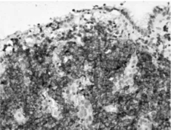
On admission to our hospital, laboratory tests showed anemia with hemoglobin 7.6 g/dl, elevated white count (12,890/ml), fibrinogen 9.92 g/l and C reactive protein 329.4 mg/l. Chest X-ray demonstrated a left pleural effusion. CT-scan showed a left subphrenic abscess. Preoperative diagnosis was perforated gastric ulcer. An exploratory laparotomy confirmed a gastric perforation at the level of the fundus, with a 5 cm left subphrenic abscess. A gastrectomy of the distal stomach was performed. Pathology disclosed a diffuse large B-cell
lymphoma (Figure 1). Helicobacter pylori (HP) was not identified in the specimen. The patient was treated with chemotherapy after convalescence from this operation.
A 47-year-old lady presented in our clinic 12 years after a vertical banded gastroplasty (VBG) with a history of several months of progressive dysphagia and vomiting, These symptoms had been attributed initially to
inadequate respect for dietetic rules after VBG. The symptoms had also been minimized by the patient who was initially happy with the ensuing weight loss. Endoscopy and barium studies demonstrated a subtotal stenosis at the level of the gastric pouch outlet with normal mucosa.
Figure 1. Diffuse large B-cell lymphoma. Destructive infiltration of the gastric mucosa by large lymphoid cells
(hematoxylin and eosin, x100) stained positively with an anti-CD20 antibody (immunoperoxidase, x200).
At laparotomy, a 9-cm intramural gastric mass was discovered in the antrum, just below the gastric pouch outlet, and was treated by a total gastrectomy. Pathology diagnosis was a CD117-positive stromal tumor compatible with a gastrointestinal stomal tumor (GIST) (Figure 2). Resections margins were free of tumor invasion. Mitosis count per 50 high power fields was 14, classifying the lesion as high risk together with its size. Additional work-up by abdominal and thoracic CT-scan and PET-scan was negative.
She underwent regular clinical and radiological follow-up every 4 months after this operation. Systemic recurrence was diagnosed by CT-scan in the lungs and the liver 3 years later. She was placed on imatinib treatment and is currently asymptomatic 12 months after recurrence. The liver and lung metastases have shown a 50% decrease in size since initiation of therapy, with a suppression of metabolic activity on PET-scan.
Discussion
The risk of cancer in the excluded distal stomach after RYGBP is unknown. The distal gastric segment is excluded from the alimentary channel and thus from contact with exogenous carcinogens, but could theoretically suffer from prolonged contact with stagnant bile, shown to promote carcinogenesis experimentally.1,2 The distal stomach after gastric bypass appears to be exposed to pancreaticobiliary reflux with pooled bile found in the excluded stomach.2,3 Superficial gastritis of this segment is common. While Sinar et al2 reported that biopsies from the distal stomach in 4 of 33 patients showed intestinal dysplasia at 3-24 months after RYGBP, this was not observed in a later study with a 2 years follow-up by the same group.3
It is uncertain if local conditions in the bypassed stomach played a role in the development of lymphoma. The fact that this is the first report of lymphoma at that location and the relatively short time between RYGBP and malignancy diagnosis precludes a definite answer. Gastric lymphoma is a rare event, representing 3% of all gastric cancers.4 Lymphomas have been described in the proximal gastric stump 5 to 43 years after partial gastrectomy, with 15 cases reported in the western literature and 27 cases in Japan.5,6 The gastric stump and the distal gastric segment may share in common an increased exposure to duodenal reflux with accompanying mucosal inflammation. It has been suggested that lymphocyte infiltration associated with chronic inflammation of the mucosa could develop into lymphoma.7 H. pylori has been shown to play an intermediary step in the development of gastric lymphoma, inducing genetic changes within the B-cells of the MALT8-10 H. pylori was not detected in our patient, and previous gastric stump lymphoma reports have shown a low correlation with H. pylori.6
Figure 2. GIST. Spindle cell tumor with focal collagen deposition; note the presence of atypical mitosis
(hematoxylin and eosin, x100).
After gastric restrictive operations, a potential mechanism leading to cancer could be prolonged contact with the food and potential exogenous carcinogens in the gastric pouch. Increased pressure with chronic irritation in the pouch and inflammation caused by the banding material could also be evoked as potential carcinogenic factors. However, these hypotheses remain speculations because of the few cases of cancer reported in the literature. Four cases of adenocarcinoma of the stomach have been reported after VBG. Two occurred in the gastric pouch, at 2 and 15 years after surgery11,12 A third lesion appeared in the distal stomach 6 years after VBG,13 while the fourth patient was diagnosed with a linitis plastica of the entire stomach 13 years after VBG14 A fifth case of gastric pouch cancer was described 10 years after adjustable gastric banding in a 62-year-old lady15 Most of the cases were diagnosed with advanced disease, while a non-specific history of epigastric pain, nausea and vomiting had already been present for considerable time before the diagnosis was made.
This delay in the diagnosis of cancer is a concern after bariatric surgery. The negligence of upper GI symptoms is illustrated in the second case report of this paper. Nausea and vomiting are often considered as normal symptoms after bariatric surgery in the general and medical community. This is particu-lary true in the first weeks after operation when the patient needs to adjust dietary behavior to the dietetic rules imposed by the procedure. However, these symptoms occurring de novo in a stable patient need adequate investigation, because this can be the only chance to make an early diagnosis.
A second difficulty for early diagnosis of gastric cancer concerns lesions arising in the excluded distal stomach, which become difficult to explore by retrograde endoscopy, with long Roux and biliopan-creatic limbs. Four cases of adenocarcinoma of the distal stomach have been reported after gastric bypass.16-19 Cancer was diagnosed at an advanced stage in 3 of the cases at 5, 8 and 22 years after the gastric bypass, when symptoms were now secondary to occlusion of the pylorus by the tumor. A fourth patient presented with anemia that was investigated for a year before an endoscopy evaluated the distal stomach through the loop gastroentrostomy and diagnosed two polypoid early gastric cancers.
This delay is illustrated in our first case report above where the problem was attributed 6 months earlier to the RYGBP operation. In addition to the difficulty to investigate the distal stomach, this case emphasizes the need to diffuse information about the type of bariatric operations to the medical community.
The intramural location of the GIST also represents a challenge for early diagnosis, because the tumor can reach a significant size before producing symptoms. GISTs are the most common mesenchymal tumors of the GI tract, 40-70% occurring in the stomach. The estimated annual incidence of GISTs is at least 10 to 20 cases per million. This is underestimated because most patients with GISTs are asymptomatic. Ten to 30% are discovered
incidentally,20 as illustrated in a paper by Sanchez et al21 who reported a 0.8% incidence of GISTs discovered incidentally during laparoscopic RYGBP on inspection of the stomach. Their 4 GISTs were <l cm, and had no histologic evidence of malignancy. DeMatteo et al22 found that in two-thirds of 200 patients found to have GISTs, the tumor size was >5 cm. All GISTs are currently considered to have some degree of malignant
treatment of localized GIST still rests on surgical resection, the prognosis of advanced disease had been transformed over the last years by imatinib, shown to induce sustained tumor response or halt disease progression.24
In view of the malignant potential of any such lesion, the difficulty to define the risk without histology, and the complexity of follow-up, it is recommended to remove any such lesion discovered incidentally during a bariatric operation.
An increased incidence of different types of cancer has been reported in the obese population.25 At this point in time, bariatric surgery does not appear to be associated with an increased risk for gastric cancer in view of the few cases of adenocarcinoma reported after many thousands of operations. Our paper adds to this short list the first cases of lymphoma and GIST. While the low incidence could reflect under-reporting or the effect of pre-surgical screening, the main risk associated with cancer after bariatric surgery appears to be related to late diagnosis of malignancy. Investigation of new or modified upper GI symptoms may allow earlier diagnosis of malignancy. This, however, requires an adequate knowledge of bariatric operations and complications in the medical community.
References
1. Safatle-Ribeiro AV, Ribeiro U Jr, Reynolds JC. Gastric stump cancer: what is the risk? Dig Dis 1998; 16: 159-68.
2. Sinar DR, Flickinger EG, Park HK et al. Retrograde endoscopy of the bypassed stomach segment after gastric bypass surgery: unexpected lesions. South Med J 1985; 78: 255-8.
3. Park HK, Sinar DR, Sloss RR et al. Histologic and endoscopic studies before and after gastric bypass surgery. Arch Pathol Lab Med 1986; 110: 1164-7.
4. Ruskoné-Fourmestraux A. MALT gastric lymphomas. Rev Med Interne 2004; 25:573-81.
5. Di Cosimo S, Ferretti G, Partenzi A et al. Gastric stump lymphoma five years after distal gastrectomy. Leuk Lymphoma 2003; 44: 365-7.
6. Oshita H, Tanemura H, Kanno A et al. Malignant lymphoma occurring in the residual stomach following gastrectomy: plus discussion based on the literature in Japan. Gastric Cancer 2003; 6: 60-3.
7. Sigal SH, Saul SH, Auerbach HE et al. Gastric small lymphocytic proliferation with immunoglobulin gene rearrangement in pseudolymphoma versus lymphoma. Gastroenterology 1989 ; 97: 195-201.
8. Wotherspoon AC, Ortiz-Hidalgo C, Falzon MR et al. Helicobacter pylori associated gastritis and primary B-cell gastric lymphoma. Lancet 1991; 338:1175-6.
9. Isaacson PG Update on MALT lymphomas. Best Pract Res Clin Haematol. 2005; 18: 57-68.
10. Alsolaiman MM, Bakis G, Nazeer T et al. Five years of complete remission of gastric diffuse large B cell lymphoma after eradication of Helicobacter pylori infection. Gut 2003; 53: 507-9.
11. Jain PK, Ray B, Royston CM. Carcinoma in the gastric pouch years after vertical banded gastroplasty. Obes Surg 2003; 13: 136-7.
12. Zirak C, Lemaitre J, Lebrun E et al. Adenocarcinoma of the pouch after silastic ring vertical gastroplasty. Obes Surg 2002; 12: 693-4.
13. Papakonstantinou A, Moustafellos P, Terzis I et al. Gastric cancer occurring after vertical banded gastroplasty. Obes Surg 2002; 12: 118-20.
14. Sweet WA. Linitis plastica presenting as pouch outlet stenosis 13 years after vertical banded gastroplasty. Obes Surg 1996; 6: 66-70.
15. Hackert T, Dietz M, Tjaden C et al. Band erosion with gastric cancer. Obes Surg 2004; 14: 559-61. 16. Escalona A, Guzman S, Ibanez L et al. Gastric cancer after Roux-en-Y gastric bypass. Obes Surg 2005; 15: 423-7.
17. Lord RV, Edwards PD, Coleman MJ. Gastric cancer in the bypassed segment after operation for morbid obesity. Aust NZ J Surg 1997; 67: 580-2.
18. Khitin L, Roses RE, Birkett DH. Cancer in the gastric remnant after gastric bypass: a case report. Curr Surg 2003; 60: 521-3.
19. Raijman I, Strother SV, Donegan WL. Gastric cancer after gastric bypass for obesity. Case report. J Clin Gastroenterol 1991; 13: 191-4.
20. Saund MS, Demetri GD, Ashley SW. Gastrointestinal stromal tumors (GISTs). Curr Opin Gastroenterol 2004; 20: 89-94.
21. Sanchez BR, Morton JM, Curet MJ et al. Incidental finding of gastrointestinal stromal tumors (GISTs) during laparoscopic gastric bypass. Obes Surg 2005; 15:1384-8.
22. Fletcher CD, Berman JJ, Corless C et al. Diagnosis of gastrointestinal stromal tumors: A consensus approach. Hum Pathol 2002; 33: 459-65.
23. De Matteo RP, Lewis JJ, Leung D et al. Two hundred gastrointestinal stromal tumors. Recurrence patterns and prognostic factors for survival. Ann Surg 2000; 231: 51-8.
24. Benjamin RS, Blanke CD, Blay J-Y et al. Management of gastrointestinal stromal tumors in the imatinib era: selected case studies. Oncologist 2006; 11: 9-20.
25. Calle EE, Rodriguez C, Walker-Thurmond K et al. Overweight, obesity, and mortality from cancer in a prospectively studied cohort of U.S. adults. N Engl J Med 2003; 348: 1625-38.