Effect of Non-Steroidal Anti-Inflammatory Drugs on
Amyloid-
 Formation and Macrophage Activation after
Platelet Phagocytosis
Dominique M. Jans, PharmD,* Wim Martinet, PhD,* Marianne Fillet, PharmD, PhD,†
Mark M. Kockx, MD, PhD,*‡ Marie-Paule Merville, PhD,† Hidde Bult, PhD,*
Arnold G. Herman, MD, PhD,* and Guido R.Y. De Meyer, PharmD, PhD*
Abstract:Recently, we showed that platelet phagocytosis occurs in human atherosclerotic plaques and leads to foam cell formation. Platelet phagocytosis, resulting in macrophage activation and iNOS induction, was associated with the formation of amyloid- peptide (A) via proteolytic cleavage of platelet-derived amyloid precursor protein (APP), possibly by secretases. To test the involvement of ␥-secretase in this process, we used indomethacin, ibuprofen, and su-lindac sulfide, non-steroidal anti-inflammatory drugs (NSAIDs) known to alter the␥-secretase cleaving site of APP, on their ability to inhibit macrophage activation evoked by platelet phagocytosis. J774 macrophages were incubated with human platelets or lipopolysaccha-ride (LPS) with or without NSAIDs. Nitrite was quantified as a mea-sure for inducible nitric oxide synthase (iNOS) activity. Indometha-cin, ibuprofen, sulindac sulfide, and meloxicam concentration-dependently reduced nitrite production by macrophages incubated with platelets, but did not alter LPS-induced iNOS activity or platelet uptake. However, acetylsalicylic acid and naproxen, two NSAIDs without effect on the␥-secretase cleaving site of APP, did not affect nitrite production in either platelet- or LPS-stimulated macrophages. Surface-enhanced laser desorption/ionization time-of-flight mass-spectrometry demonstrated time-dependent formation of A -containing peptides after platelet phagocytosis, which could be inhib-ited by indomethacin. In conclusion, these results point to the
involvement of␥-secretase in macrophage activation following plate-let phagocytosis.
Key Words: amyloid precursor protein,-amyloid peptide, induc-ible nitric oxide synthase, macrophage activation, NSAID
(J Cardiovasc Pharmacol™ 2004;43:462–470)
R
ecently, we demonstrated the presence of amyloid- (A), which until then had only been studied in Alzheimer’s dis-ease, in macrophages around microvessels in advanced human atherosclerotic plaques.1In these macrophages, the formation of A was associated with the expression of inducible nitric oxide synthase (iNOS). A source for A in atherosclerotic plaques is blood platelets, which contain amyloid precursor protein (APP) in the␣-granules. The platelets can enter ath-erosclerotic plaques via leaky microvessels and are then phagocytosed by macrophages,2which subsequently trans-form to foam cells.1We proposed that during this process A -like peptides are formed, which then activate the macrophage as indicated by iNOS expression.1This novel mechanism of macrophage activation in atherosclerotic plaques may favor plaque expansion and/or rupture. Indeed, the massive nitric ox-ide (NO) release produced by iNOS has been reported to in-duce apoptotic cell death of smooth muscle cells by enhancing Fas-L/Fas interactions.3In the atherosclerotic plaque, smooth muscle cells are the only cells that are able to produce collagen isoforms that contribute to the strength of the plaque.4 More-over, activated macrophages can trigger the activation of ma-trix metalloproteinases in the vascular interstitium,5resulting in degradation of the interstitial collagen fibers and decreased strength of the fibrous cap of an atherosclerotic plaque.The proteolytic enzymes responsible for A formation in macrophages following platelet phagocytosis are not known yet. In Alzheimer’s disease the insoluble A1-42peptide is gen-erated by the cleavage of its precursor APP, by two enzymes designated- and ␥-secretase. Recently, it was shown that in-domethacin, ibuprofen, and sulindac sulfide directly affect the
Received for publication August 18, 2003; accepted December 17, 2003. From the *Division of Pharmacology, University of Antwerp, Antwerp,
Bel-gium; †Department of Medicinal Chemistry, University of Liège, Liège, Belgium; and ‡Department of Pathology, General Hospital Middelheim, Antwerp, Belgium.
Supported in part by the Fund for Scientific Research Belgium (FWO, FNRS) (Nr: 1.5.206.00 and G.0180.01), The Bekales Foundation, University of Antwerp (NOI-BOF), and the Flemish Institute for Improvement of Sci-entific and Technological Research in Industry (IWT). Dr. M. Kockx is a holder of a fund for fundamental clinical research of the Fund for Scientific Research–Flanders. D. Jans was supported by the Flemish Institute for Improvement of Scientific and Technological Research in Industry (IWT). Reprints: Dominique Jans, Division of Pharmacology, University of Ant-werp (UA), Universiteitsplein 1, B-2610 AntAnt-werp, Belgium (e-mail: dominique.jans@ua.ac.be).
␥-secretase cleaving site of APP, thereby altering the amyloid pathology in the brain.6The latter effect results in a reduction of the highly amyloidogenic A1-42levels and an increase in the less harmful A1-38isoform. The interference with the ␥-secretase cleaving site is not seen with all NSAIDs and was not dependent on cyclooxygenase (COX) inhibition. Indeed, acetylsalicylic acid and naproxen do not affect the␥-secretase cleaving site of APP. Since specific␥-secretase inhibitors are not available, we used a series of NSAIDs to investigate the participation of␥-secretase in macrophage activation and the formation of A-like peptides following platelet phagocytosis.
METHODS
Cell Culture
The murine macrophage cell line J774A.1 (American Type Culture Collection, Rockville, MD) was grown in RPMI 1640 medium supplemented with 10% fetal calf serum, 100 U/ml penicillin, 100 µg/ml streptomycin, 150 U/ml polymyxin B, and 50 µg/ml gentamycin in a humidified 5% carbon dioxide/95% air incubator at 37°C. All cell culture media and supplements were from Invitrogen (Paisley, UK). For cell cul-ture experiments 48 well plates (Costar, Cambridge, MA) or culture slides (Beckton Dickinson Labware, Sunnyvale, CA) were used. Cells (0.5 × 10⁶/800 µL) were allowed to adhere at 37°C for 2 hours. Thereafter, cells were washed with warm medium and the medium was replaced by Dulbecco Modified Eagles Medium (DMEM) supplemented with 100 U/ml peni-cillin, 100 µg/ml streptomycin, and 50 µg/ml gentamycin.
Human blood platelet concentrates were kindly pro-vided by the blood transfusion center of the University Hospi-tal of Antwerp (Belgium). These platelet concentrates had been deleukocytized by filtration and contained only 1 to 2 white blood cells per 300,000 platelets. Platelets were washed in warm PBS and resuspended in serum free Dulbecco Modi-fied Eagles Medium (DMEM) supplemented with 100 U/ml penicillin, 100 µg/ml streptomycin, and 50 µg/ml gentamycin. J774 macrophages, primed with recombinant mouse IFN␥ (100 U/ml, Sigma), were incubated with washed human platelets (108per 0.5 × 10⁶macrophages)1in the presence of an NSAID or its solvent. After an incubation period of 18 hours, the culture medium was removed from the macrophages, cen-trifuged, and stored at −20°C for nitrite determination. To study the specificity of NSAID treatment on the nitrite produc-tion evoked by platelet phagocytosis, experiments using lipo-polysaccharide (LPS) from Salmonella typhosa as an alterna-tive stimulus for iNOS induction were performed as well.
Drugs
The following NSAIDs were examined (highest final solvent concentration is indicated): indomethacin (Federa, Brussels, Belgium, 0.5 mM TRIS pH 8.3), ibuprofen (Federa, Brussels, Belgium, 0.5% ethanol), acetylsalicylic acid
(Fe-dera, Brussels, Belgium, 0.5% ethanol), naproxen (Fe(Fe-dera, Brussels, Belgium, 1% ethanol), meloxicam (Calbiochem, San Diego, CA, 0.2% DMSO), and sulindac sulfide (Sigma, St Louis, MO, 0.2% ethanol), the active metabolite of the prodrug sulindac. They were dissolved at the start of every experiment and the solutions were sterilized by filtration.
Nitrite Measurement
Nitric oxide synthase activity was assessed by measur-ing nitrite, a stable NO metabolite, usmeasur-ing the Griess reaction.7 Briefly, 200 µL culture medium was transferred to a 96-well microtiter plate and mixed with 40 µL of 6.5 M HCl and 37.5 mM sulphanilic acid (1:1). After 10 minutes, 20 µL 12.5 mM N-(1-naphtyl)-ethylene diamine dihydrochloride was added. After 30 minutes at 37°C, the absorbance was read at 540 nm using a Titertek Multiscan MCC/340 plate reader (Lab-systems). Nitrite concentration was calculated from a standard curve of sodium nitrite in culture medium.
Oil Red O Stain
To examine whether platelet engulfment by phages was altered by NSAIDs, co-incubations of macro-phages and platelets were stained with oil red O8after fixation of the cells with 4% formaldehyde (10 minutes). Nuclei were stained with hematoxylin.
Flow Cytometry
Phagocytosis of platelets by macrophages was also evaluated by flow cytometry. J774 macrophages and washed human platelets were incubated for 45 minutes with DMEM supplemented with 5 µM of 5- and 6-([(4-chloromethyl) ben-zoyl] amino) tetramethylrhodamine (Cell Tracker Orange, Molecular probes) or 2.5 µM 5- chloromethylfluorescein diac-etate (Cell Tracker Green, Molecular Probes), respectively.9 Thereafter, the media were refreshed, and the macrophages were incubated with the platelets for 3.5 hours in the presence or absence of the highest concentrations of these NSAIDs that inhibited macrophage activation. This time point was based on previous experiments on the evaluation of platelet phagocyto-sis using confocal and electron microscopy.1Flow cytometry was performed using a FACSort (BD Biosciences, San Diego, CA). Forward and side scatters were set to include cells but to exclude debris. Green fluorescent-free platelets were moni-tored in the FL 1 channel, whereas orange fluorescent mac-rophages were measured in the FL 3 channel. Data were analyzed using CellQuestPro software (BD Biosciences). Phagocytosis (per cent) was calculated as the (number of cells containing cell tracker green and orange / total number of cells containing cell tracker orange) × 100.
Cyclooxygenase Inhibition and Determination
of Prostaglandin E
2To document COX inhibition, we tested all NSAIDs on their ability to inhibit arachidonic acid-induced blood platelet
aggregation. Blood samples (9 mL) were obtained from a cannulated carotid artery of pentobarbitone (30 mg/kg)-anesthetized New Zealand White rabbits and collected on tri-sodiumcitrate (1 mL; 3.8%). Platelet-rich plasma was prepared by centrifugation (10 minutes, 200 g). The samples were ex-posed to 330 µM arachidonic acid after 3 minutes incubation in the presence or absence of the NSAIDs in a Chronolog dual channel aggregometer (37°C, 900 rpm).9The maximum in-crease in light transmission after the addition of arachidonic acid was measured.
PGE2was measured in the culture medium by radio-immunoassay. Dose interpolation was done with a four-parameter logistic function using the IBM-PC RIA data reduction package provided by M.L. Jaffe (Silver Spring, MD).10
Surface-Enhanced Laser Desorption/Ionization
Time-of-Flight (SELDI-TOF) Mass Spectrometry
Analysis of APP fragments in cell lysates of macro-phages, platelets, and macrophages co-incubated with platelets for 3, 6, or 18 hours with or without indomethacin was per-formed by SELDI-TOF mass spectrometry using the Protein-Chip system (PS20, protein chip array, Ciphergen Biosystems Inc., Fremont, CA). Cells were lysed for 30 minutes in 50 mM TRIS buffer containing 250 mM NaCl, 5 mM EDTA, 0.1% (vol/vol) Triton X 100, 1 mM dithiothreitol, 1 mM NaF, 10 mM-glycerophosphate, 0.1 mM Na3VO4, and 0.1 mM para-methylsulphonylfluoride.
The chip arrays were incubated overnight at 4°C with the antibody (6E10 or IgG) to allow coupling. 6E10 is an antibody specific for the 17 N-terminal amino acids of-amyloid pep-tides (672–688 of the full-length APP sequence). IgG antibody coating was used as a control to discriminate between specific and aspecific peptide binding to the protein chip. The samples were diluted and 20 µg total protein was spotted on the 6E10 or IgG antibody-coated chips. After 4 hours, non-bound peptides were removed by extensive washing with PBS and PBS con-taining 0.5% Triton X 100. The arrays were rinsed several times with a 1.0-mM HEPES buffer, and allowed to air dry. Thereafter, 1 µL of a 20% saturated solution of ␣-cyano-4-hydroxycinnamic acid in 0.5% (vol/vol) trifluoroacetic acid: 50% (vol/vol) acetonitrile was applied to each spot. Mass spec-trometric analysis was performed by averaging a minimum of 100 laser shots in a Ciphergen SELDI Protein Biology system II, with a linear time-of-flight mass analyzer (mass accuracy: 0.1%).11–14
Statistical Analysis
Nitrite values were compared by using analysis of vari-ance (ANOVA). A P value less than 0.05 was considered to be significant. All data are represented as mean ± SEM.
RESULTS
Effect of NSAIDs on Nitrite Production
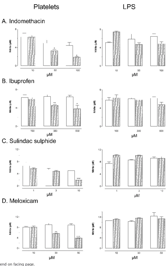
Incubation of IFN␥-primed J774 macrophages with washed human platelets or LPS resulted in nitrite production by the macrophages. The extent of the nitrite production was comparable for both stimuli. Addition of indomethacin, ibu-profen, meloxicam, or sulindac sulfide to the culture medium concentration-dependently reduced nitrite production by mac-rophages evoked by platelet phagocytosis (Fig. 1, A–D), whereas acetylsalicylic acid or naproxen were without such an inhibitory effect (Fig. 1, E and F). As compared with indo-methacin, sulindac sulfide, and meloxicam, the effect of ibu-profen was less pronounced. When macrophages were stimu-lated with LPS, nitrite production was not affected by any NSAID (Fig. 1, A–F).
Effect of NSAIDs on Platelet Aggregation and
PGE
2Production
The production of PGE2by macrophages stimulated with either platelets or LPS was significantly reduced in the presence of all NSAIDs (results not shown). Moreover, all NSAIDs inhibited arachidonic acid-induced platelet aggrega-tion. After 3 minutes of exposure the percentage inhibition was: indomethacin (10, 50, 100 µM), 97% for all concentra-tions, ibuprofen (100, 200, 300 µM), 9%, 92%, and 96%, meloxicam (10, 30, 50 µM), 50%, 97%, and 97%, acetylsali-cylic acid (50, 250, 500 µM), 13%, 96%, and 96% and naproxen (75, 150 µM), 12% and 97%. Sulindac sulfide was less effective as platelet inhibitor: 1 µM reduced the platelet aggregation with 3% and 10 µM with 18%.
Platelet Phagocytosis and Foam Cell Formation
Flow cytometry revealed that after 3.5 hours platelet phagocytosis was not altered in the presence of 100 µM indo-methacin, 500 µM ibuprofen, 10 µM sulindac sulfide, and 50 µM meloxicam (Fig. 2).
Lipid accumulation in the macrophages as a result of platelet phagocytosis was not changed in the presence of 100 µM indomethacin, indicating that platelet phagocytosis was not altered by this NSAID (Fig. 2). Similarly, the other NSAIDs did not alter platelet phagocytosis (not shown).
Detection of APP-Fragments by SELDI-TOF
Mass Spectrometry
The mass spectrum of the lysate of macrophages incu-bated for 3 hours with human blood platelets did not show peaks corresponding to A1-40or A1-42fragments (Fig. 3). However, in the lysate of macrophages incubated with blood platelets for 6 or 18 hours, two peaks at m/z 6277.5 and m/z 6541.5 were detected, which correspond to the [M + H]+peaks of proteins with a molecular mass of 6276.5 Da and 6540.5 Da, respectively. The intensity of these peaks increased with an
increasing incubation time of platelets and macrophages (Fig. 3), which paralleled nitrite production (0.49 ± 0.03 µM after 3 hours, 0.80 ± 0.07 µM after 6 hours and 6.8 ± 0.2 µM after 18 hours). For the 6276.5 Da protein, six different APP-derived peptides are theoretically possible within a mass range of ± 5 Da (Table 1). The peak at m/z 6541.5 was not detected in every experiment and will not be considered in the present study.
When macrophages were incubated with platelets in the presence of indomethacin for 18 hours, [M + H]+peaks at m/z 6277.5 and 6541.5 disappeared completely. This was accom-panied by peaks in the mass ranges m/z 5000–6000 and m/z 7000–8000 (Fig. 4). In all cases, a standard mixture gave the expected profile with [M + H]+peaks at m/z 1953.5 (A1-16, not shown), 4343.5 (A1-40), and 4527.6 (A1-42).
DISCUSSION
Recently, it was demonstrated that in neuronal cells a subset of NSAIDs including indomethacin, ibuprofen, and su-lindac sulfide alter the␥-secretase cleaving site of APP.6As a result, the formation of A1-42, the most toxic form of A, was reduced in favor for the less toxic A1-38.6Indeed, it has been suggested6that this interference with the␥-secretase cleaving site of APP may explain the benefit of some NSAIDs seen in epidemiological15–17and clinical studies18–20of Alzheimer’s disease. Since specific inhibitors of␥-secretase are currently not available or cytotoxic (unpublished observations), we in-vestigated a series of NSAIDs to test whether␥-secretase is involved in platelet-induced macrophage activation. These NSAIDs were merely selected as pharmacological tools to
un-FIGURE 1. Effect of indomethacin (A, Platelets: n = 6, LPS: n = 2–4), ibuprofen (B, n = 4), sulindac sulfide (C, n = 3), meloxicam (D, n = 3), acetylsalicylic acid (E, PLT: n = 6, LPS: n = 2), and naproxen (F, PLT: n = 9, LPS: n = 3) on nitrite production by macrophages after incubation with platelets or LPS for 18 hours. Indomethacin, ibuprofen, sulindac sulfide, and meloxicam reduced platelet-induced nitrite production by macrophages concentration-dependently but did not affect LPS-induced nitrite production. Acetylsalicylic acid and naproxen did not alter nitrite production by macrophages after incubation with either blood platelets or LPS. Open bars: solvent control, hatched bars: NSAID-treated; *P < 0.05, **P < 0.01, ***P < 0.001 versus solvent control.
ravel the mechanisms of macrophage activation after platelet phagocytosis.
The present data confirm that platelets are phagocytosed by macrophages (flow cytometry), that the macrophages be-come activated (nitrite production) and transform to foam cells (oil red O). Foam cell formation can result from other mecha-nisms than platelet phagocytosis. Since we used serum-free medium, endocytosis of lipid droplets can be excluded. En-gulfment of LDL or modified LDL bound to the platelets is possible but seems to be less important because we used washed human platelets. With flow cytometry, we could dem-onstrate that platelets are indeed phagocytosed by macro-phages, confirming previous results.1
Subsequently, our results clearly showed a reduced ni-trite production when macrophages were incubated with plate-lets in the presence of indomethacin, ibuprofen, sulindac sul-fide, and meloxicam as compared with the controls. Importantly, the concentrations of the NSAIDs used in the present study did not affect LPS-induced nitrite production, confirming previous reports.21–23The latter finding indicates that the inhibitory activity of NSAIDs on macrophage activa-tion following platelet phagocytosis was not due to
interfer-ence with the cell signaling pathways essential for iNOS in-duction or with the activity of iNOS. Indeed, Aeberhard et al22 showed that only extremely high concentrations of NSAIDs, exceeding those of the present study, inhibit iNOS activity in LPS-stimulated macrophages. Moreover, the inhibitory activ-ity of indomethacin, ibuprofen, sulindac sulfide, and meloxi-cam on macrophage activation evoked by platelet ingestion was not due to reduced platelet phagocytosis or processing. The different compounds did not inhibit the early platelet up-take as studied by flow cytometry, nor did they suppress the accumulation of platelet-derived lipid droplets inasmuch as oil red O staining of NSAID-treated macrophages did not differ from that of untreated macrophages. Furthermore, these ex-periments revealed no differences in platelet uptake between NSAID-treated and control macrophages.
Both PGE2measurements in the supernatant and arachi-donic acid-induced blood platelet aggregation assays showed that all NSAIDs inhibited COX, confirming their activity and excluding possible degradation of the drugs. However, inhibi-tion of macrophage activainhibi-tion by the NSAIDs was not due to COX-inhibition. Except for ibuprofen, the concentrations of the NSAIDs required to affect the macrophage activation after
FIGURE 2. Effect of NSAIDs on platelet phagocytosis. Flow cytometric evaluation of platelet phagocytosis by macrophages with or without indomethacin 100 µM (Indo, A), ibuprofen 500 µM (Ibu, B), sulindac sulfide 10 µM (Sul, C), and meloxicam 50 µM (Mel, D). Results are presented as percentage of the population that has engulfed platelets. Oil red O stain of control macrophages (E) and macrophages incubated with human platelets in the presence (F) or absence (G) of 100 µM indomethacin. Lipid accumulation in the macrophages (arrowheads) was not changed in the presence of indomethacin further indicating that platelet phagocytosis was not altered by this NSAID. Bar = 10 µm.
platelet ingestion were higher than those necessary for COX inhibition. Secondly, sulindac sulfide, the least effective COX inhibitor, was even the most active inhibitor of macrophage activation following platelet phagocytosis. More importantly, although acetylsalicylic acid and naproxen inhibited COX, they did not alter nitrite production of macrophages induced by platelet phagocytosis. Indeed, the latter NSAIDs do not affect the ␥-secretase cleaving site of APP.6Taken together, these findings suggest that the effect of some NSAIDs on nitrite for-mation could be due to interference with the␥-secretase cleav-ing site of APP, thereby reduccleav-ing the formation of A-like peptides.
Previously, Ogawa et al24 reported that A1-40 could stimulate nitrite production in IFN␥-primed J774 macro-phages. They further showed that indomethacin and ibuprofen inhibited the A-effect and proposed that the inhibitory effects of these NSAIDs on iNOS expression were mediated via per-oxisome proliferator-activated receptor-␥ (PPAR-␥). Unfortu-nately, we were unable to reproduce these experiments. We encountered a great batch-to-batch variability in the response to A1-40as well as A1-42and the nitrite production of IFN
␥-TABLE 1. Different Theoretically Possible APP-Derived Peptides With a Molecular Mass of 6276.5Ⳳ 5 Da That Contain the Epitope of the 6E10 Antibody
Measured Mass (Da) Theoretical Mass (Da) ⌬ Mass (Da) Number of Amino Acids Amino Acids in APP Sequence 6276.5 6278.9 2.4 56 635–690 6271.9 −4.6 56 640–695 6273.9 −2.6 57 644–700 6273.9 −2.6 57 648–704 6275.1 −1.4 58 656–713 6273.3 −3.2 57 672–728
All peptides contain the complete epitope of the 6E10 antibody (17 N-terminal amino acids of A (ie amino-acids 672–688 of full length APP)). Peptides are ranked according to their sequence in full-length APP, with their theoretical mass, the difference between the theoretical mass and the measured mass and the number of amino acids. Cleaving sites of- or ␥-secretase are marked in bold.
FIGURE 3. SELDI-TOF mass spectra of the lysate of co-incubations of macrophages and blood platelets (M + PLT) (3 hours, 6 hours, and 18 hours). When macrophages were incubated with blood platelets, an A-like fragment was time-dependently formed with a molecular mass of 6276.5 Da. A standard mixture of A1-16, A1-40, and A1-42gave the expected profile with [M + H]+peaks
primed macrophages after stimulation with either peptide was too low to study the effect of inhibitors.1Since Ogawa et al24 did not investigate whether the inhibition by the NSAIDs was specific for A1-40by including an alternative stimulus, such as LPS, their experiments cannot be compared with the present study in which we used platelets as a stimulus. Furthermore, the active concentrations of indomethacin and ibuprofen were even higher than in the present study and could have interfered directly with the activity of iNOS, as reported by Aeberhard et al.22Since NSAIDs used in the present study did not interfere with the LPS-induced expression or activity of iNOS, it seems less likely that the concentrations used in the present study in-terfered with PPAR-␥ nuclear receptors as proposed by Ogawa et al24. However, it cannot be excluded that some of their in-hibitory effects were due to interference with iNOS- inducing pathways distal from A-production.
Subsequently, SELDI-TOF mass spectrometry was used to substantiate the ␥-secretase involvement. This technique demonstrated that incubation of macrophages with platelets re-sulted in a time-dependent formation of a peptide with a mo-lecular mass of 6276.5 Da, containing an A-like domain. The
properties and the importance of this peptide are still unclear. Only its molecular mass is known and the fact that it contains the epitope recognized by the 6E10 antibody. Six theoretically possible A-like peptides with a molecular mass of 6276.5 ± 5 Da remained after excluding peptides without the epitope of the 6E10 antibody on the SELDI chip. Two of these candidates deserve further consideration. The first one, which has a mass of 6275.1 Da, is 58 amino acids long and contains the complete A1-42sequence (ie, amino acids 672–713 of full-length APP) at its C-terminal site, ending just at the ␥-secretase cleaving site (Table 1). The difference between the theoretical mass and the measured mass was the lowest for this peptide as compared with the other five candidates. Another interesting candidate has a mass of 6273.3 Da, is 57 amino acids long and contains the complete A1-42sequence at its N-terminal site, starting with the-secretase cleaving site (Table 1). The exact identity of the A-like peptide with a mass of 6276.5 ± 5 Da formed during platelet phagocytosis by macrophages remains to be es-tablished. Also the importance of the presence of the complete A1-42sequence in this A-like peptide remains to be deter-mined, since A1-42itself had only a limited effect on the
ac-FIGURE 4. SELDI-TOF mass spectra of the lysate of co-incubations of J774 macrophages and blood platelets (M + PLT) for 18 hours in the presence (M + PLT + Indo 100 µM) or absence (M + PLT + TRIS) of 100 µM indomethacin. Incubation of macrophages and platelets for 18 hours in the presence of indomethacin inhibited the formation of the [M + H]+peak at m/z 6277.5. The standard,
a mixture of A1-16, A1-40, and A1-42, gave the expected profile with [M + H]
+peaks at m/z 1953.5 (not shown), 4343.5, and
tivation of both J774 macrophages, as discussed before, and microglia cells.25Nevertheless, the present study showed that addition of indomethacin to the co-incubations of platelets and macrophages inhibited the formation of this peptide, as well as nitrite production. This strongly suggests that the A-like pep-tide with a mass 6276.5 ± 5 Da participates in macrophage activation following platelet phagocytosis.
In summary, our results demonstrate that NSAIDs known to alter the␥-secretase cleaving site of APP, as well as meloxicam, reduced macrophage activation following platelet phagocytosis, but not after LPS stimulation. Furthermore, NSAIDs like acetylsalicylic acid and naproxen, which do not affect␥-secretase cleavage were also without effect on macro-phage activation after platelet ingestion. This indicates that the reduced macrophage activation is independent of COX-inhibition and points to an effect on␥-secretase. The inhibition of the formation of A-related peptides by indomethacin, mea-sured by SELDI-TOF, gives further support to the idea that NSAIDs act via interference with a␥-secretase-related path-way, although further research is necessary to identify the pep-tide(s). Finally, our findings indicate that the novel platelet-induced pathway of macrophage activation can be treated in a pharmacological way. This may open new perspectives for the development of specific drugs for intervention in the complex matter of plaque physiology.5
ACKNOWLEDGMENTS
The authors thank Prof. M. Claeys for critical comments on the manuscript. M. Fillet is a Senior Research Assistant at the National Fund for Scientific Research (FNRS, Belgium) and M.-P. Merville is a Research Associate (FNRS, Belgium). The use of SELDI-TOF mass spectrometry technology was made possible by cooperation with the Laboratory of Medici-nal Chemistry (Prof. J. Gielen), University of Liège, Belgium. Human blood platelets were kindly provided by the Blood Transfusion Center, University Hospital of Antwerp, Ant-werp, Belgium.
REFERENCES
1. De Meyer GRY, De Cleen DMM, Cooper S, et al. Platelet phagocytosis and processing of beta-amyloid precursor protein as a mechanism of mac-rophage activation in atherosclerosis. Circ Res. 2002;90:1197–1204. 2. Kockx MM, Cromheeke KM, Knaapen MWM, et al. Phagocytosis and
macrophage activation associated with hemorrhagic microvessels in hu-man atherosclerosis. Arterioscler Thromb Vasc Biol. 2003;23:440–446. 3. Boyle JJ, Weissberg PL, Bennett MR. Human macrophage-induced
vas-cular smooth muscle cell apoptosis requires NO enhancement of Fas/Fas-L interactions. Arterioscler Thromb Vasc Biol. 2002;22:1624– 1630.
4. Newby AC, Zaltsman AB. Fibrous cap formation or destruction—the
critical importance of vascular smooth muscle cell proliferation, migra-tion and matrix formamigra-tion. Cardiovasc Res. 1999;41:345–360. 5. Rajagopalan S, Meng XP, Ramasamy S, et al. Reactive oxygen species
produced by macrophage-derived foam cells regulate the activity of vas-cular matrix metalloproteinases in vitro. Implications for atherosclerotic plaque stability. J Clin Invest. 1996;98:2572–2579.
6. Weggen S, Eriksen JL, Das P, et al. A subset of NSAIDs lower amyloido-genic Abeta42 independently of cyclooxygenase activity. Nature. 2001; 414:212–216.
7. Schmidt HH, Nau H, Wittfoht W, et al. Arginine is a physiological pre-cursor of endothelium-derived nitric oxide. Eur J Pharmacol. 1988;154: 213–216.
8. Luna LG. Manual of histologic staining methods of the armed forces in-stitute of pathology. L.G.Luna, ed. Armed Forces Inin-stitute of Pathology: Washington, DC; 1968:140–142.
9. Baker GR, Sullam PM, Levin J. A simple, fluorescent method to inter-nally label platelets suitable for physiological measurements. Am J He-matol. 1997;56:17–25.
10. Zamora R, Bult H, Herman AG. The role of prostaglandin E2 and nitric oxide in cell death in J774 murine macrophages. Eur J Pharmacol. 1998; 349:307–315.
11. Merchant M, Weinberger SR. Recent advancements in surface-enhanced laser desorption/ionisation-time of flight-mass spectrometry. Electropho-resis. 2000;21:1164–1167.
12. Austen BM, Frears ER, Davies H. The use of seldi proteinchip arrays to monitor production of Alzheimer’s beta-amyloid in transfected cells. J Pept Sci. 2000;6:459–469.
13. Vehmas AK, Borchelt DR, Price DL, et al. beta-Amyloid peptide vacci-nation results in marked changes in serum and brain Abeta levels in APPswe/PS1DeltaE9 mice, as detected by SELDI-TOF- based Protein-Chip technology. DNA Cell Biol. 2001;20:713–721.
14. Beher D, Wrigley JD, Owens AP, et al. Generation of C-terminally trun-cated amyloid-beta peptides is dependent on gamma-secretase activity. J Neurochem. 2002;82:563–575.
15. Stewart WF, Kawas C, Corrada M, et al. Risk of Alzheimer’s disease and duration of NSAID use. Neurology. 1997;48:626–632.
16. McGeer PL, Schulzer M, McGeer EG. Arthritis and anti-inflammatory agents as possible protective factors for Alzheimer’s disease. Neurology. 1996;47:425–432.
17. Anthony JC, Breitner JCS, Zandi PP, et al. Reduced prevalence of AD in users of NSAID’s and H2 receptor antagonists. Neurology. 2000;54: 2066–2071.
18. Rogers J, Kirby LC, Hempelman SR, et al. Clinical trial of indomethacin in Alzheimer’s disease. Neurology. 1993;43:1609–1611.
19. Aisen PS, Schmeidler J, Pasinetti GM. Randomized pilot study of nime-sulide treatment in Alzheimer’s disease. Neurology. 2002;58:1050–1054. 20. Aisen PS, Schafer KA, Grundman M, et al. Effects of rofecoxib or naproxen vs placebo on Alzheimer disease progression: a randomized controlled trial. JAMA. 2003;289:2819–2826.
21. Amin AR, Vyas P, Attur M, et al. The mode of action of aspirin-like drugs: effect on inducible nitric oxide synthase. Proc Natl Acad Sci U S A. 1995; 92:7926–7930.
22. Aeberhard EE, Henderson SA, Arabolos NS, et al. Nonsteroidal anti-inflammatory drugs inhibit expression of the inducible nitric oxide syn-thase gene. Biochem Biophys Res Commun. 1995;208:1053–1059. 23. Salvemini D, Misko TP, Masferrer JL, et al. Nitric oxide activates
cyclo-oxygenase enzymes. Proc Natl Acad Sci U S A. 1993;90:7240–7244. 24. Ogawa O, Umegaki H, Sumi D, et al. Inhibition of inducible nitric oxide
synthase gene expression by indomethacin or ibuprofen in beta-amyloid protein-stimulated J774 cells. Eur J Pharmacol. 2000;408:137–141. 25. Meda L, Cassatella MA, Szendrei GI, et al. Activation of microglial cells
by beta-amyloid protein and interferon- gamma. Nature. 1995;374:647– 650.